Review

Stem Cell Niche Concept: Search for Current Expert Consensus

1
Department of Morphology and General Pathology, Siberian State Medical University, 634050 Tomsk, Russia
2
Center for Immunology and Cellular Biotechnology, Immanuel Kant Baltic Federal University, 636016 Kaliningrad, Russia
3
Medical Research and Educational Institute, Lomonosov Moscow State University, 119234 Moscow, Russia
*
Author to whom correspondence should be addressed.
Int. J. Mol. Sci. 2025, 26(17), 8422; https://doi.org/10.3390/ijms26178422
Submission received: 20 June 2025 / Revised: 20 August 2025 / Accepted: 26 August 2025 / Published: 29 August 2025

Abstract

Postnatal stem cells are crucial for tissue homeostasis and repair and are regulated by specialized microenvironmental microterritories known as “stem cell niches”. Proposed by R. Schofield in 1978 for hematopoietic stem cells, niches maintain self-renewal, guide differentiation and maturation, and can even revert progenitor cells to an undifferentiated state. Niches respond to injury, oxygen levels, mechanical cues, and signaling molecules. While the niche concept has advanced regenerative medicine, bioengineering, and 3D bioprinting, further progress is hindered by inconsistent interpretations of its core principles. To address this, we proposed a consensus-building initiative among experts in regenerative medicine and bioengineering. We have developed a questionnaire covering the niche topography, hierarchy, dimension, geometry, composition, regulatory mechanisms, and specifically the mesenchymal stem cell niches. This pilot survey, being conducted under the auspices of the National Society for Regenerative Medicine in the Russian Federation, aims to establish a standardized framework on the eve of the 50th anniversary of Schofield’s hypothesis. The resulting consensus will guide future research and innovation in this pivotal field.

1. Introduction

Accumulating data indicates that postnatal stem cells play a central role in tissue homeostasis, repair, and regeneration. Stem cells require a favorable and instructive microenvironment in order to be well controlled. The concept of the stem cell niche was proposed almost 50 years ago by R. Schofield (1978) for hematopoietic stem cells (HSCs) and later further developed by others who have claimed that a niche can maintain the self-renewal of stem cells and stimulate their differentiation as well as the return of progenitor cells to an undifferentiated state. As part of the regulation of stem cell fate, tissue-specific niches should respond to injury and sense microenvironmental changes such as oxygenation, position, mechanotransduction, and multiple secreted factors that mediate cell-to-cell communication. The theory of stem cell niches has not only made a significant contribution to basic research in the field of regeneration but also provides the technological basis for the practical implementation of innovative approaches in regenerative medicine, tissue bioengineering, 3D bioprinting, microfluidic technologies, etc. However, although several more or less comprehensive reviews and experimental studies have been published in recent years demonstrating the progress made in stem cell niche research, a certain stagnation can be observed in this field, mainly due to the different and broad interpretations of the fundamental issues of the “niche” concept. We propose that this problem can be partially overcome by building a consensus among experts in regenerative medicine, developmental biology, and bioengineering. To this end, we have developed a questionnaire with six sections addressing the topography, hierarchy, and size of the stem cell niche; the contribution of different niche components to the regulatory function; the control of cell behavior within the niche; and the characteristics of stem cell niches of mesenchymal origin.
In this focused review, we discuss the key problems and current achievements in the development of the niche concept, present the questionnaire, and justify the wording of the statements to be interpreted and approved by the selected experts.

2. General Concept (Definition) of the Stem Cell Niche

A pivotal role of tissue-specific stem cells in tissue renewal and regeneration has been actively investigated for several decades [1,2,3,4]. The discussion about the inner mechanisms of stem cell regulation has been ongoing for the past 60 years [5]; the theory of hemopoietic-inductive microenvironment (HIM) [6] and the stochastic model (hemopoiesis engendered randomly, HER) [7,8] are among the main speculations of bone marrow (BM) research.
Within the HIM theory, an intriguing section is the HSC niche in adult mammals, formulated almost 50 years ago by Robert Schofield [9] and later further developed by other researchers who have claimed that a niche can maintain stem cell self-renewal and stimulate their differentiation as well as the return of progenitor cells to an undifferentiated state. According to Schofield’s own opinion, the “stem cell niche” hypothesis was introduced to explain the dependence of stem cells on their microenvironment [10].
Although many intrinsic and extrinsic factors (niche components) of HIM have been identified in the regulation of HSC origination, expansion, migration, and localization, the underlying mechanisms remain largely unknown [11]. In the context of stem cell fate control, tissue-specific niches should respond to injury and sense microenvironmental changes such as oxygenation, position, mechanotransduction, and a variety of secreted factors that mediate cell-to-cell communication. The specific microenvironment, or the stem cell niche, demands the presence of certain niche components that can maintain the stem cell pool and restore the microenvironment in injured tissues for their subsequent appropriate functioning. Sagaradze et al. (2020) and others consider that this role belongs to mesenchymal stromal/stem cells (MSCs) [12].
The evolving theory of stem cell niches has not only made a significant contribution to basic research in the field of regeneration but also provides the technological basis for the practical implementation of innovative approaches in regenerative medicine, tissue bioengineering, 3D bioprinting, microfluidic technologies, etc. In this regard, a query ‘cell niche’ showed 33,385 results generated by the text retrieval engine PubMed (https://pubmed.ncbi.nlm.nih.gov/; accessed on 7 May 2025) since 1956. In turn, the key phrase ‘stem cell niche’ was found in 14,546 publications from 1978 onwards. Finally, the search query ‘hematopoietic stem cell niche’ determines more than 4000 papers since Schofield’s paper in 1978; 62% of these (2564 papers) were published in the years 2012–2021. It is noteworthy that, according to the PubMed search engine (https://pubmed.ncbi.nlm.nih.gov/; accessed on 7 May 2025), the maximum number of annual publications in this field was reached in 2021 (342 references). By 2024, the number of published papers decreased to 208.
Similarly, the word combination ‘MSC niche’ showed a total of 1037 results in the PubMed database from 2004 onwards. The peak (105 publications) was observed in 2020, with the number of articles published annually decreasing to 65 (by 38%) by 2024. Thus, although several more or less comprehensive reviews and experimental studies have been published in recent years demonstrating the progress in stem cell niche research, certain stagnation can be observed in this field, possibly due to different opinions, broad interpretations, and a lack of solutions to the fundamental and applied issues of the “niche” concept. In particular, there is still no answer to the key question: how do the components of a niche as a specialized microterritory differ from the same well-known components of the entire HIM?
In this review, we discuss recent findings about the major milestones of the niche concept, focusing on BM-derived HSCs and MSCs as the most extensively studied yet more complex systems as we have understood them so far [13,14], and highlight the problematic questions that remain to be explored at the threshold of 50 years of the stem cell niche concept. We propose that these problems can be partially overcome by establishing a consensus among experts in regenerative medicine, developmental biology, and bioengineering using the niche questionnaire we have developed (Table S1).
The term “niche” was used by Calvo et al. (1976) to describe the osteal sites in the trabecular bones where groups of cells (colony-forming units, CFU) demonstrated either erythropoiesis, myelopoiesis, or megakaryocytopoiesis during a recovery after lethal irradiation [15]. In other words, distinct BM microenvironments have been identified in which cellular components contribute to the niche, many of which regulate differentiation scenarios.
In 1978, Schofield proposed a theoretical basis in the form of a “niche” hypothesized as a specialized site of HIM to maintain the HSC self-renewal and stemness [9,16].
Despite the obvious progress of Schofield’s hypothesis, the spatial organization of BM hematopoiesis remains poorly understood. Hence, since the birth of the term “stem cell niche”, there is no certainty in its interpretation so far. The understanding of the niche for cells of mesenchymal origin by different authors contradicts each other in key aspects. These opinions can be divided into two large directions: the orthodox variant versus the alternative point of view (Table 1 [13,14,15,16,17,18,19,20,21,22,23,24,25,26,27,28,29,30,31,32,33,34,35,36,37,38,39,40,41,42,43,44,45,46,47,48,49,50,51,52,53,54,55,56,57,58,59]).
Some orthodox variants of niche definition are as follows:
  • The fundamental property of a stem cell is self-renewal, which depends on the microenvironment in which the stem cell is seen in association with other cells, determining its behavior [10].
  • The niche is a highly specialized, complex, and instructive microenvironment that physically localizes the stem cells and maintains their fate [17,18].
  • The stem cell niche is a specialized microenvironment in which stem cells reside primarily in a quiescent state by providing anti-proliferation signals [19].
  • The stem cell niche is a specific microenvironment that directly controls the dual capacity for self-renewal and multilineage differentiation of stem cells, maintaining the HSC in a quiescent state [20].
  • The niche is a restricted site in an organ that supports the stem cell self-renewal [21].
  • Stem cells reside in specialized microenvironments, also called niches, which maintain them in an undifferentiated and self-renewing state [22].
  • Niches are the anatomical regions within the microenvironment [60].
Forty years after Schofield’s article, Purton and Scadden (2008) have formulated several questions that are largely fundamental to the development of the HSC niche hypothesis, particularly: are all niches equal; do one or numerous niches exist; are the niches in a quiescent or activated state; and does niche hierarchy correspond to stem cell hierarchy [16]? As a continuation of these questions, Lucas (2021) considers distinct niches for HSCs and their progenitors in the BM [61]. Based on new proposals and accumulated experimental data, alternative definitions of the HSC niche have emerged, some of which we present below.
An alternative point of view reflecting the hematopoiesis dynamicity is presented by certain descriptions:
  • The stem cell niche is a group of cells in a special tissue location that are intended for the maintenance of stem cells. It has a variable structure, and different cell types can provide the niche environment. There is a niche hierarchy of HSC due to HIM heterogeneity at diverse regions of the BM [23].
  • There are the hierarchical HSC osteoblastic (and MSC) niches in terms of their localization, composition, function, and multi-layer regulation systems [24].
  • The stem cell niches are the distinct, structurally functional, energetically favorable microterritories in a contiguous space of HIM where quantitative parameters of a microenvironment promote the qualitative control of stem cell fate [13].
  • According to Pinho et al. (2018), the HSC pool is functionally and molecularly heterogeneous; consequently, there are distinct ‘specialized’ niches for distinct subpopulations of HSCs [25].
  • The HSC niche is considered to be a complex multicellular network that provides molecular signals and physical interactions that are essential for HSC localization, maintenance, and differentiation [26].
  • The niche provides a microenvironment that supports the self-renewal and multi-lineage differentiation of stem cells [27].
  • The niche represents a sophisticated and dynamic system of cellular and molecular components coupled with heterogeneous signaling mechanisms. It serves as an interface between stem cells and the organism, orchestrating their adaptive responses to tissue damage [62].
  • The BM niche is a complex environment composed of heterogeneous cell populations that regulate the hematopoietic stem and progenitor cells (HSPCs) function and activity through the secretion of a wide array of cytokines and growth factors [63].
  • The stem cell niche is a dynamic and specialized microenvironment with a specific architecture that regulates self-renewal of stem cells, the balance between their quiescent and proliferative status, as well as their choice of fate and differentiation of their progenitor cells [28].
The mentioned definitions could also be applied to the niches of other tissue-specific stem cells. However, it should be noted that differences in structure and functional features are observed for distinct types of stem cell niches belonging to epithelial or stromal niches (reviewed in detail in [62]).
Briefly, epithelial niches (such as niches for intestinal stem cells, epithelial stem cells, hair follicle stem cells, limbal stem cells, spermatogonial stem cells, etc.) are generally determined as a specific microterritory in the tissue, and their components are usually clearly distinguishable by histological analysis. On the contrary, stromal niches (HSPC and MSC niches, etc.) have an elusive microanatomical structure and are characterized by a pronounced dynamicity. Below, we will discuss the key components of all stem cell niches and the critical regulatory mechanisms involved in the interactions between a stem cell and its niche, focusing mainly on BM niches.

2.1. Core Components and Regulatory Mechanisms in Stem Cell Niches

2.1.1. Cellular and Non-Cellular Components of Stem Cell Niches in Mammalian Tissues
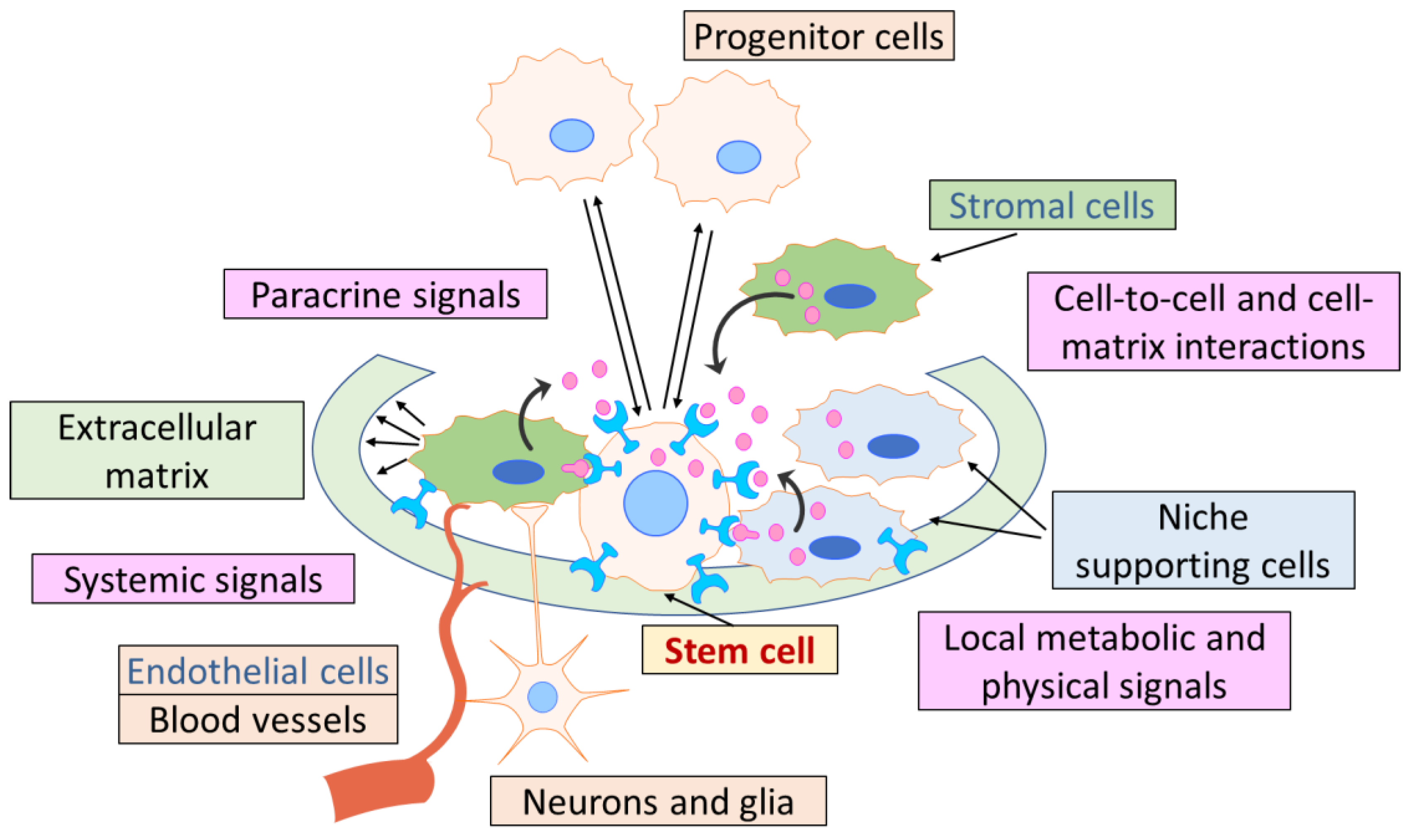
The stem cell niche in postnatal tissues, being a sophisticated, heterotypic, and dynamically regulated microenvironment, was shown to be composed of multiple interacting components of different natures (Figure 1). The cellular components consist of adjacent niche-supporting cells, MSCs, immune cells, and parenchymal cells; the noncellular components include the extracellular matrix (ECM), signaling molecules (e.g., growth factors and cytokines, extracellular vesicles, etc.), physical cues (e.g., shear stress, substrate rigidity, and topographical features), and extrinsic environmental stimuli (e.g., glucose and metabolic byproducts, oxygen tension, small ions, and inflammatory mediators) (Figure 1). Importantly, stem cell niches require extensive vascularization and neural integration, making them susceptible to both direct and indirect modulation by circulatory and neuronal inputs as systemic signals [62,64,65,66,67,68].
Figure 1. Key components of a stem cell niche. The stem cell niche provides a structural and regulative microenvironment for stem cells, supporting their maintenance and adequate response to the differentiation stimuli. The existing opinion is schematically emphasized: a niche is a small part of the microenvironment, which nevertheless has the same structural and functional organization as the broader HIM space. Please see the text for additional details.
Ijms 26 08422 g001
The stem cell niche comprises multiple cell types from distinct lineages that collectively regulate stem cell maintenance and their response to activating stimuli. Thus, in the HSC niche within the BM, stromal cells such as osteoblasts and MSCs, endothelial and immune cells, together with distinctive signaling factors, regulate the balance between HSC quiescence, activation, proliferation, and differentiation to maintain the blood cell system. HSCs reside in osteoblastic niches near the endosteal bone surface and in perivascular niches (including periarterial, perisinusoidal, and perivascular in the transition zone), where they communicate locally with their cellular microenvironment through cell–cell interactions and secreted factors or extracellular vesicles released into the BM [69]. The osteoblastic niche may provide a quiescent microenvironment for HSC maintenance, whereas the vascular niche facilitates HSC transendothelial migration during mobilization or homing and may favor HSC proliferation and further differentiation. Quiescent HSCs are adjacent to the endosteal lining, where they are in close contact with N-cadherin+ osteoblasts, which regulate HSC quiescence through secretion of osteopontin and angiopoietin. Activated HSCs are attracted to the central marrow region in the perivascular niche, close to the sinusoid, where they can give rise to multipotent HSPCs and subsequently new blood cells, which are then released into the circulation [70]. Leptin receptor-expressing (LepR+) perivascular MSCs and endothelial cells that surround sinusoids and potentially other blood vessels throughout the BM are required for HSC maintenance [71]. It was found that CD150+CD48+CD41 lineage-negative HSCs localize in the perivascular zone near sinusoidal blood vessels in close contact with not only vascular and perivascular cells but also megakaryocytes and other hematopoietic cells [30].
Recent advances in biomedicine have established the crucial role of MSCs in the maintenance of many stem cell niches in various tissues [12,72,73,74]. One of the most studied niches in this context is the niche of HSCs. In the hematopoietic niche, separate subpopulations of MSCs have been described, and they are closely connected with HSCs due to intercellular contacts and also produce various factors supporting hematopoiesis [73,74]. Deletion of the population of MSCs expressing the marker nestin resulted in impaired homing of HSCs to the BM in transplantation after complete irradiation of the recipient BM. In addition, MSCs are known to support hematopoiesis ex vivo when HSCs are isolated in culture and can also form an HSC niche-like microenvironment in heterotopic transplantation in vivo [75]. Human skeletal stem cells (SSCs), often referred to as MSCs in skeletal tissues, were shown to have a capacity to maintain human HSCs in serum-free culture conditions ex vivo [76].
Accumulating evidence demonstrates that the specific population of MSCs, termed CXC chemokine ligand 12 (CXCL12)-abundant reticular cells (CAR), which overlap with LepR+ cells, represents the crucial cellular component within the perivascular HSC niche [68]. CAR cells, as well as endothelial and perivascular stromal cells, produce CXCL12 and stem cell factor (SCF), regulating HSC maintenance and activation. Macrophages in close proximity to nestin-expressing MSCs can modulate the secretion of CXCL12 by MSCs [77].
MSCs could also support the multipotent stem cell subpopulation within their heterogeneous population, which is involved in the stromal tissue renewal and regeneration. Thus, ECM components produced by MSCs could promote the differentiation of multipotent stem cells into the adipogenic, osteogenic, and chondrogenic lineages [78].
In the epithelial stem cell niche, i.e., the intestinal stem cell niche, the population of Gli1-positive MSCs, located in the depth of the intestinal crypt, is one of the key participants of niche regulation through the production of ligands of the Wnt signaling pathway, even when the function of the main sources of these factors—Paneth cells—is impaired. MSCs also negatively regulate intestinal stem cells via bone morphogenetic protein 2 (BMP2) and BMP4 secretion [77]. In the hair follicle niche, MSCs participate in the activation of stem cells by secreting Noggin and Wnt factors as well as Shh [79]. In the spermatogonial niche, MSCs regulate the functioning of Leydig cells and Sertoli cells, thus stimulating the restoration of the niche after damage [80]. MSCs may also be involved in the regulation of stem cell niches in other tissues. However, there is little experimental data on this, and the specific mechanisms by which MSCs exert their regulatory function in the niche remain poorly understood.
Other cell types, such as endothelial and immune cells, have recently attracted increasing attention from researchers as regular contributors to stem cell regulation within a niche. Thus, in the BM, endothelial cells interact with osteolineage cells in a paracrine manner (so-called angiogenic-osteogenic coupling) and, together with perivascular stromal cells, also directly form the perivascular niche for HSCs. Recent studies highlight the heterogeneity of vascular niches at different bone regions, suggesting that HSCs are regulated by locally distinct mechanisms [81]. Immune cells, classical hematopoietic components derived from HSPCs, such as neutrophils, macrophages, dendritic cells, B cells, and others, mature predominantly in the BM and are considered important cellular regulators of HSC quiescence, self-renewal, and multi-lineage differentiation through either direct cell-to-cell interactions or the secretion of various immunological factors. They regulate not only HSCs but also other cells within the niche, such as MSCs, endothelial cells, osteoblasts, and osteoclasts, to promote their survival and modulate the supportive microenvironment for HSPCs [82].
Adipocytes derived from CAR/LepR+ cells could act as negative or positive regulators of HSCs during functional hematopoiesis and the myelosuppressive condition (discussed in detail in [68]). Within the complex BM microenvironment, HSCs constantly cooperate with the SSC niche to maintain homeostasis and regulate skeletal formation as well as immune system functioning [83]. There is also a constant neural-immune crosstalk regulating niche activity by the contact with sympathetic neurons [84].
Recent advances in the development of novel genetic and histology tools, including single-cell RNA sequencing and spatial multi-omics, have made it possible to elucidate cellular heterogeneity and specific cell populations within the HSC niche and other stem cell niches and generate single-cell atlases to decipher the microenvironment of stem cells [69]. These approaches provide novel data regarding the cellular components of stem cell niches in BM and other tissues, expanding our understanding of how the niche is regulated.
Among the non-cellular components of the niches involved in the regulation of stem cell functioning, the ECM is considered to be one of the most important. The ECM represents a dynamic and intricate microenvironment distinguished by tissue-specific biophysical, mechanical, and biochemical properties that play a crucial role in modulating stem cell behavior [74]. The multiple components of the ECM exert direct and indirect control over stem cell maintenance, proliferation, self-renewal, and differentiation [85]. Various ECM molecules exhibit regulatory functions across different stem cell types, and the ECM’s molecular composition can be precisely assembled and adjusted to establish an optimal niche for stem cells in specific tissues (reviewed in detail in [36]). Integrins, expressed on the surface of stem cells, are often involved in the interaction with ECM components within a niche. Thus, it was demonstrated that the αv integrin subunit could regulate HSC proliferation via interaction with periostin [86].
Intercellular communication within stem cell niches occurs through multiple modalities, including direct cell–cell contact, secreted factors, and extracellular vesicles, which enable both local and long-range coordination of cellular activities [74]. Cell adhesion molecules play particularly important roles in maintaining proper spatial organization and facilitating direct signaling between adjacent cells. Among the signaling pathways, Wnt, Notch, BMP, and transforming growth factor (TGFb) can be highlighted as critical regulators of stem cell self-renewal and cell fate. These evolutionarily conserved pathways enable precise control over cell fate decisions while maintaining the flexibility required for appropriate responses to physiologic demands. Extracellular vesicle-mediated communication also represents an emerging area of research that may provide new insights into how niche cells coordinate their activities over longer distances.
The molecular regulation of stem cell niches relies heavily on sophisticated chemokine and cytokine signaling networks that coordinate cellular behavior and maintain tissue homeostasis. CXCL12 and SCF represent two of the most critical regulatory molecules within the BM niche and serve as essential regulators that manage HSC behavior, including quiescence, proliferation, migration, and differentiation [74]. SCF binds to c-Kit on HSCs and is essential for their repopulating activity. It exists in both membrane-bound (mSCF) and soluble (sSCF) forms, with the former being more potent in retaining HSCs within their niche [69].
CXCL12, also known as stromal cell-derived factor 1 (SDF-1), is a chemokine that plays a crucial role in stem cell niches. In the BM, it is essential for the survival, maintenance, and trafficking of HSCs [87]. CXCL12 acts through its receptor CXCR4, which is expressed on HSCs, to promote their quiescence and self-renewal. In response to changes in CXCL12 levels, a portion of HSC progenies leave the niche and begin to mobilize and circulate. Targeted deletion of CXCL12 from MSCs in the BM was shown to reduce the normal HSC numbers [88].
The process of HSPC recruitment into the vascular niche may also depend on endothelium-derived fibroblast growth factor 4 (FGF-4) and differences in oxygen level. Higher FGF-4 and oxygen concentration gradients during the transitions of cells from the osteoblastic niche to the vascular niche regulate the recruitment, proliferation, and differentiation of HSPCs. Under stress such as thrombocytopenia, SDF-1 and vascular endothelial growth factor (VEGF) activate metalloproteinases (MMP) like MMP-9, which convert membrane-associated Kit ligand to soluble Kit ligand (sKitL), thus promoting HSC entry into the cell cycle, mobilization to the vascular niche, and differentiation [47].
While most studies of the niche focus on those residing in the BM, HSC niches in extramedullary tissues are also actively studied. Thus, HSCs were found to reside in adult mouse meninges [89]. Locally expressed CXCL12 and SCF maintain meningeal HSCs and regulate their adaptation to the central nervous system immunosurveillance. These cells could be replenished from blood upon niche depletion. Gao et al. demonstrated that epitranscriptomic control resulting in RNA modifications (e.g., m6A) dictates niche-specific HSC fates [90]. Tissue-specific HSC niche was also described in spleen [91], where it functions under the megakaryocyte-driven regulation and neural signals, adipose tissue and lungs [92], as well as placenta and fetal liver [93]. However, extramedullary HSC niches, especially in human tissues, remain understudied.
In other stem cell niches, the mediators of intercellular communication may vary depending on the rate of cell turnover and the specificity of niche-supporting cells, but the crucial principles of the regulation are similar to those in the HSC niche—a balance between stem cell maintenance and activation on demand. Thus, in the intestinal crypt Leucine-rich repeat-containing G protein coupled receptor 5 (LGR5)+ intestinal stem cells are located at the base of the crypt, embedded between their specialized daughter cells, the so-called Paneth cells, which supply Wnt, epidermal growth factor (EGF), Delta-like protein 4 (DLL4) and Noggin ligands necessary for the self-renewal and maintenance of stem cells [77]. In the spermatogonial stem cell niche, the population of stem cells strongly depends on the supply of fibroblast growth factors (FGFs) secreted by lymphatic endothelial cells, which provide the competitive microenvironment regulating stem cell homeostasis under the conditions of limited mitogen availability [94,95].
Considering the high complexity and heterotypic structure of the stem cell niches, it is still controversial which components and mechanisms lead to the regulation of stem cells within their niches. These questions are included in the questionnaire developed to consolidate expert opinions on this issue (Table S1).

2.1.2. Comparison of HSPC Niche Structure and HIM Morphologic Components in the BM

This morphofunctional section of the niche concept can be regarded as the most advanced since the formulation of Schofield’s hypothesis and is constantly evolving. This is not surprising, since the niches are the specialized HIM territories (surrounding the HSPCs); the HIM concept has been evolving since the 1960s [6,96].
At the present time, the intravital microscopy technique allows topographical visualization in the BM and in situ detection of the function of numerous cell types, including bone cells, megakaryocytes, macrophages, perivascular cells, and Schwann cells, as niche regulators of HSC quiescence, mobilization, and responsibility [26,52]. Mature hematopoietic and innate immune cells (macrophages, dendritic cells, B cells, Breg cells, CD4+ T cells, CD8+ T cells, Treg cells, and neutrophils) are proposed to be the regulators of the HSPC niches through feedback mechanisms [82,97].
HIM morphologic components were previously defined (prior to Scofield’s publication) as follows [98]:
  • Tissue-resident and migrating cells.
  • ECM with fibers and soluble molecules.
  • Blood vessels.
  • Nerve terminals.
The main components of the HSPC niche, in turn, are described in multiple publications (e.g., [13,26,29,30,31,32,33,34,35,36,99]) and consist of the following:
  • Various hematopoietic and stromal support cells, including cell–cell adhesion molecules and secreted soluble factors, are located in close proximity to stem cells.
  • ECM, which serves as an “anchor” for stem cells and forms a mechanical scaffold for the transmission of stem cell signaling.
  • Blood vessels that supply the niche with nutrients and systemic signals from other organs and are involved in the recruitment of circulating stem cells from and into the niche.
  • Nerve terminals and Schwann cells.
Comparing the core components of the HIM space and the HSPC niches, their strong similarity can be easily noticed. Onward, briefly examining general regulatory mechanisms, Tavassoli (1975) has described that HIM components control the HSC fate via direct (cell–cell and cell–matrix) and indirect (soluble molecules) cues [98]. The functioning of the stem cell niche, in turn, is based on both the physical contacts and diffusible factors (e.g., [26,100]). It appears that the control mechanisms of HSPC behavior in the niche and in HIM are largely similar.
Of course, significant progress has been made recently in deciphering the intimate processes of both cellular components (MSCs, inflammatory and immune cells, megakaryocytes, etc.) [75,97,101,102] and molecular signaling (exosomes, integrins and other anchor molecules, chemokines, hypoxia, etc.) [103,104,105,106,107] within the hematopoietic microenvironment.
However, virtually all cells, molecules, and cues in the BM can be considered to be components of one or another stem and/or progenitor cell niche (Table 1). This gives rise to the following controversial assumptions:
  • The entire HIM space of the BM consists of niches for HSCs, MSCs, and their progenitors, which are in close contact with each other.
  • Between the niches, there is a non-specialized HIM space, which has extrinsic effects on its functioning.
  • It is unclear whether there are significant (qualitative or quantitative) differences between the local specialized microterritories (referred to as the niches) and the known components of the entire HIM space. What are the external borders of stem cell niches to distinguish their structural and functional features?
These and other crucial issues, also summarized in Table 1, have largely shaped the further argumentation and conclusions in the present review. As it is crucial to understand the mechanisms of stem cell niche regulation, we dedicated the selected part of the developed questionnaire to the issues related to the control of stem cells within a niche (Table S1).

3. Hierarchy of Potential Niche-Candidates for Stem and Progenitor Cells
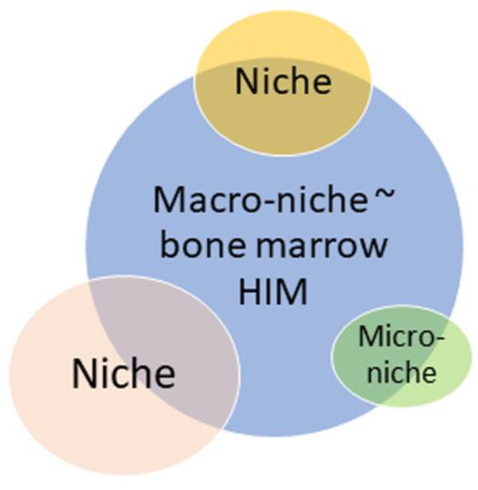
The niche supporting HSPCs in the BM is a highly heterogeneous and dynamic structure that responds to changing physiologic or pathologic signals [52,97]. Therefore, Yu and Scadden (2016) [52] viewed the BM environment (HIM; [6]) as composed of multiple micro-niches, each consisting of a unique pairing of distinct supportive stromal cells with different hematopoietic subtypes to regulate a particular branch of hematopoietic cell process.
In particular, Baccin et al. (2020) described perivascular micro-niches established by CAR cell subsets (Adipo-CAR and Osteo-CAR) that localize differently on sinusoidal and arteriolar surfaces [53]. Conversely, Kandarakov et al. (2022) considered mammalian BM as a macro-niche for HSCs [40]. These hypothetical speculations about Schofield’s niche, as well as micro- and macro-niches, blur the lines of niches from single cell associations to the entire hematopoietic organ. In this case, the term ‘niche’ equated with the term HIM (Figure 2).
Since the size and composition of the HSC microterritories are still unclear, even the classical Schofield’s niche can vary significantly in value (Figure 2). For example, macrophages and megakaryocytes vary significantly in their dimensions; thereafter, megakaryocytic candidate niche [44] and erythroblastic islands [42] appear to be somewhat different BM microterritories.
Figure 2. Bubble map of understanding size variability for distinct HSC candidate niches. HIM—hematopoietic-inductive microenvironment. The areas of bubbles reflect schematically the variations in the unitless value of the niches described by different authors. The figure begs the question about certain borderlines and dimensions of the niche; otherwise, it can be considered ‘cloud’ (functional) subtypes only.
Ijms 26 08422 g002
Regarding BM as a macro-niche, Kokkaliaris et al. (2020) [108] have shown interesting results. It was found that the distribution of HSCs relative to the candidate niches was stochastic and did not differ from a mapping of computationally generated points randomly placed throughout the entire BM volume. Finally, Kandarakov et al. (2022) [40] supported the idea of [52] that HSC niches are anatomically abundant and are not a limiting factor in vivo.
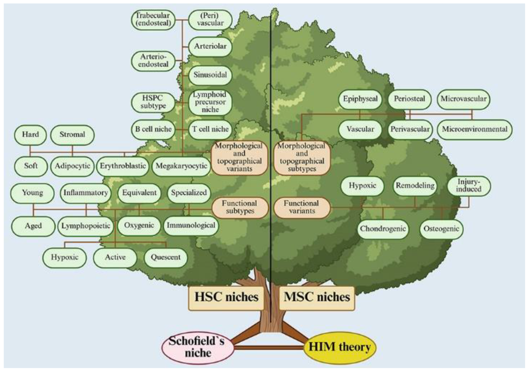
In the current literature, various authors have described a wide range of cells and structural-functional units that are assumed to be candidate niches for HSPCs, as well as for stromal cells themselves, including MSCs. Since our previous review [54], the list of candidates for HSPC and MSC niches has expanded considerably. It can be conditionally divided into structural-functional, topographical, and functional variants (Figure 3, Table S2).
Of particular interest among candidate HSC niches are hematopoietic (erythroblastic) islands and megakaryocytes as native BM cells and structural-functional units, whose size is easy to determine. It can serve as a dimension scale for separating other cell associations that are not always correctly considered as stem cell niches [109].
Figure 3. Candidate niche tree for bone marrow HSCs and MSCs described in the references. Drawn from data presented in Table S2 with corresponded references [11,13,26,28,40,41,42,43,44,45,46,47,48,49,50,52,54,56,61,110,111,112,113,114,115,116,117,118,119,120,121,122,123,124,125,126,127,128,129,130,131,132,133,134,135,136].
Ijms 26 08422 g003

3.1. Erythroblastic Islands as Candidate Niche for HSPCs
Almost 70 years ago, the erythroblastic islands (EIs) were primarily defined by Bessis as structural-functional units of the BM [116]. The EIs have been currently updated to be renamed as ‘erythroblastic island niche’ [117,118,137], supported by a subset of macrophages that provide erythroblast maturation near sinusoid vessels [117].
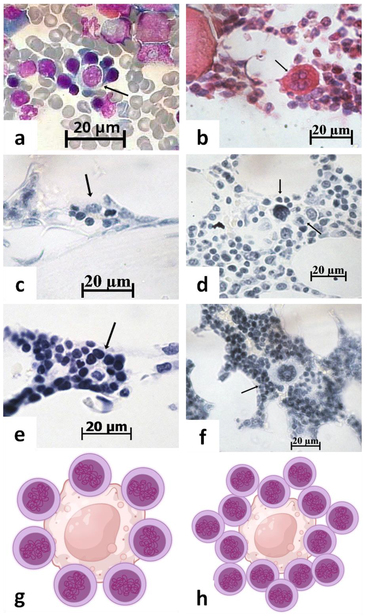
Ex vivo EI reconstitution is used to mimic an interaction of HSCs with macrophages in the natural BM environment under different stress conditions [138]. In all this, Han et al. (2023) [138] consider the unclear relation of EIs reconstructed in vitro to their in vivo features. Hematopoietic islands (HIs) can be easily identified in medullary tissue by standard cytologic [56] and histologic techniques (Figure 4). HIs consist of immature hematologic cells that actively synthesize the nucleic acids (Figure 4c,e) and incorporate 3H-thymidine [56]. It is proposed to consider HIs as active hematologic niches.
Moreover, HIs have an apparent size (Figure 4a,c,e) that may be useful for the comparative evaluation with the dimensions of other niche candidates. In particular, such an estimation was recently conducted by [109].
Interestingly, Sathyanarayana et al. (2007) hypothesized the existence of a stromal niche for early-stage erythropoiesis [139], while EIs consisting of about 5–30 erythroid cells surrounding a central resident macrophage have been proposed as niches for the proliferation, differentiation, and enucleation of late-stage erythroid precursors [42,117,140].
Indeed, the central stromal elements within the EIs are surrounded by erythroid cells (Figure 4a), with varying concentrations of nucleic acids in their nucleus and cytoplasm (Figure 4c,e), stained using the well-known Einarson technique [141].
Figure 4. Composition of hematopoietic islands (a,c,e) and megakaryocytic niche-candidate (b,d,f) on cytological smear (a) and histological sections (bf) of mouse (a), rabbit (bd,f), and human (e) bone marrow. Black arrows specify cell associations. (a) Erythroblastic island. Azure II and eosin staining; (b) megakaryocyte rounded by hematopoietic cells. Hematoxylin and eosin staining; (c,e) hematopoietic islands with different nucleic acid (DNA and RNA) contents in hematopoietic cells; (d,f) megakaryocyte surrounded by single (d) or multicellular layers (f). The Einarson staining; dividing cell (black arrow) near megakaryocyte (d). Scale bar 20 µm; (g,h) schematic 2D reconstruction of hematopoietic niche-candidate: central stromal cell (macrophage, fibroblast-like cell, or megakaryocyte) surrounded by single (a,c,d) or multilayered (b,e,f) hematopoietic cells. Own microscopic images and cartoons are shown.
Ijms 26 08422 g004

3.2. Megakaryocytic Candidate for HSPC Niches

Bruns et al. (2014) showed that about 30% of HSCs are localized near megakaryocytes (MKs), which promote stem cell quiescence via CXCL4 production [44]. Later, Pinho et al. (2018) found ~60% of von Willebrand factor-positive HSCs within 5 μm distance of MKs [25]. In summary, they suggested that MKs maintain HSC quiescence and may be specialized hematopoietic niches [26,118].
However, there are serious contradictions that MKs are primarily the niches and secondarily promote the HSC quiescent state. Thus, Lucas (2021) proposed the existence of a megakaryocyte/sinusoidal niche that maintains HSCs biased towards megakaryocyte fates and an arteriolar niche that maintains more quiescent HSCs [61].
In turn, Kandarakov et al. (2022) suggested the possibility of direct contact between MKs and HSCs due to the cell–cell interaction through chemokines (CXCL4) and growth factors (TGF-β1; thrombopoietin; FGF1) secreted by MKs [40]. If the authors’ hypothesis is correct, MKs are mainly part of the mutual HIM but not a specialized microterritory for the HSCs. Moreover, light microscopy of BM histology clearly confirms a close contact of MKs with hematopoietic cells (Figure 4b), which actively synthesize DNA and RNA molecules (Figure 4d,f). There are some cells with a mitotic figure (Figure 4d, black arrow). Therefore, hematopoietic quiescence near MKs has not been confirmed morphologically, and the role of MKs as potential niches for HSCs remains controversial.

3.3. MSC Niche

SSCs play a crucial role in shaping the medullary cavity by providing both the structural framework and the hematopoietic microenvironment necessary for BM function [142]. These cells establish specialized niches, including SSC-derived HSC microenvironments tailored to specific physiologic needs [76,143].
However, SSCs exhibit significant heterogeneity, with distinct populations residing in articular cartilage, the growth plate, periosteum, and perivascular regions. Remarkable progress has been made in characterizing various SSCs and skeletal stem and progenitor cell (SSPC) populations in postnatal bone. Their isolation, detection, and functional characterization remain debatable, which is exacerbated by inconsistencies in nomenclature—such as the interchangeable use of “SSCs” and “mesenchymal stem cells (MSCs)” [143]. Consequently, the existence of SSPC niches in the BM remains under discussion [144]. SSPCs were localized in the periosteum and within the BM stroma, including subsets localizing around arteriolar and sinusoidal blood vessels; they can display osteogenic, chondrogenic, adipogenic, and/or fibroblastic potential, and exert critical hematopoiesis-supportive functions (for a detailed review, see [145]).
Despite these uncertainties, recent studies employing advanced in situ techniques—including laser-capture microdissection, confocal fluorescence microscopy, and immunohistochemistry—suggest the presence of an SSPC niche in the postnatal epiphyseal growth plate in mice. These niches appear to support sustained chondrogenesis over prolonged periods [50]. Other potential SSPC niches have been proposed in other anatomical regions, such as articular cartilage and periosteum [28,146].
The interpretation of these findings is complicated by conflicting data. For instance, a combination of micro-CT imaging, molecular profiling, and clonal cell tracing failed to identify stem cell niches in salamander cartilage during development or regeneration [147]. Nevertheless, many studies highlight the spatial association of MSC niches with bone, cartilage, and the microvasculature (Figure 3, Table S2), as pericytes—often considered as MSCs—can differentiate into osteoblasts, chondrocytes, and adipocytes [148]. The precise regulatory role of these niches in determining MSC fate, HSC behavior, or both remains unclear. However, emerging evidence suggests the existence of functionally distinct MSC niches (e.g., osteogenic, chondrogenic) (Figure 3, Table S2).
Despite these challenges, ex vivo modeling offers a promising approach to predict the structural and dimensional characteristics of functional MSC niches, facilitating their identification in native bone [43,54]. In combination with in vivo imaging, such models could provide critical insights into the quantitative and spatial organization of the stem cell microenvironments across different tissues.

3.4. Live Imaging of Niches

Intravital microscopy is a rapidly developing field for niche detection within the mammalian BM [149]. For example, many studies have attempted to determine which of the two cellular elements in the BM, osteoblasts or endothelial cells, could serve as a niche for long-term HSCs [30]. In this context, pioneering live imaging work that has captured labeled HSCs within their normal 3-dimensional tissue environment has helped to address this issue by suggesting that HSC niches may indeed contain both osteoblast and vascular components that are closely apposed [150]. Multiphoton laser microscopy (MLM) can penetrate the outer bone thickness (~50 µm in the murine cranium) and then image in the marrow region at an additional depth of 250 to 300 µm [151] with high (subcellular) resolution (0.5 µm in the lateral direction and 1–2 µm in the axial direction) [152].
At the same time, MLM is unable to penetrate into the deepest regions of the BM; therefore, it is complicated to observe the niches for short-lived HSCs and multipotent progenitors maintaining day-to-day hematopoiesis in adult animals [153]. In addition, according to Pinho et al. (2019), the location of a stem cell within tissue is not sufficient to define its niche, as the niche must have both anatomical and functional features [26]. In particular, determining the site of a labeled stem cell in the BM does not allow us to outline the borders of its specialized maintaining area in the general space of the HIM. This can only be assumed, since the currently known cellular and molecular components of the niche and the entire HIM are, on a large scale, the same.

3.5. Niche Aging as One of the Functional Manifestations of Specialized Microterritories

The stem cell niche is the most important intrinsic factor regulating cellular aging in adult stem cells [20]. In particular, Pinho et al. (2019) consider aging-related changes in the BM niche that influence HSC aging as the alterations in the vasculature and mesenchymal stem and progenitor cells, increased adipogenesis and reduced osteogenesis, altered secretion of niche factors, and a reduced number of adrenergic nerves [26]. According to a review by Kandarakov et al. (2022), aging-related alterations occur in HSCs and niche-supporting cells (MSCs, endothelial cells, osteoblasts, adipocytes, MKs, macrophages, and lymphocytes) and components (nerve fibers, molecule secretion) due to the increased proinflammatory molecules and cells [40].
Thus, Young et al. (2024) showed a reduction in cues from MSCs to HSCs in old mice, resulting in HSC impaired lymphoid differentiation in aged niches versus balanced lymphoid/myeloid signaling in young HSC microterritories [45]. Aged HSCs exhibit high proliferation with increased myeloid-biased differentiation and reduced regenerative capacity, which promotes myeloid malignancies [26].
In addition, neutrophils, as contributors to the HSPC niche [97] and proinflammatory cells, may support hematopoietic niche aging. Therefore, Yang and de Haan (2021) [154] highlighted the emerging role of inflammation in HSC aging, leading to low functionality of aged HSCs, including a declined repopulation capacity and myeloid and platelet-restricted differentiation. Taken together, both cell-intrinsic and microenvironmental extrinsic cues contribute to HSC aging.

4. Challenges of the Dimension and Geometry of Stem and Progenitor Cell Niches
Lovegrove et al. (2025) [155] have recently shown an instructive influence of pre-mitotic cell morphology and shape (spherical or spindle-like) on its symmetric (via mitotic rounding) or asymmetric (isomorphic) division. The isomorphic mode triggered heterogeneous morphology, unequal distribution of signaling pathways, determinant polarization, and, finally, variable differentiation of mesenchymal-derived daughter cells. Accordingly, the dimensions and geometric features of specialized HIM sites (niches) can also influence the morphogenetic events in stem cells due to the precise control of gene expression during organogenesis [156].
The specific niches for the distinct developing hematopoietic lineages are poorly understood; still less is known about the changes in niche size and function in these distinct BM anatomical sites under stress conditions and aging [157]. Niche features are not only important for the control of malignancy processes of stem and precursor cells [17,21,158]. The bioengineering phase of the niche concept development [13] requires precise characteristics of the stem cell microenvironment to mimic biological niches by designing artificial microterritories that have structural, functional, and biomechanical parameters similar to the natural conditions. However, there are some crucial quantitative properties of niches [54] that need to be replicated ex vivo. The lack of consensus on the generally accepted dimensions and geometry of the specific microenvironment for stem cells makes their biomimetic reconstruction, for example, using modern additive technologies, for the needs of regenerative medicine, impossible.
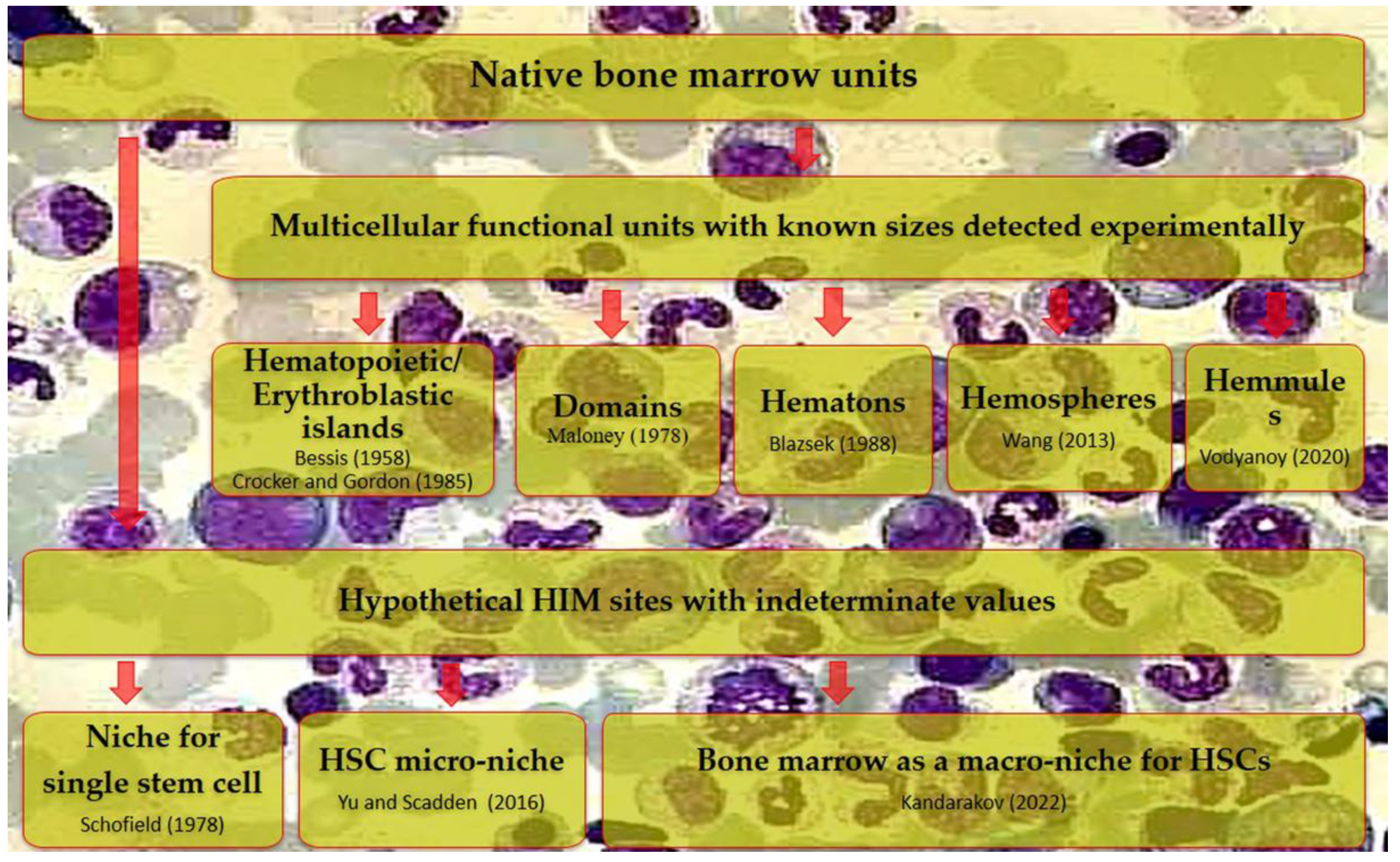
Recently, we have attempted to compile the available quantitative information on the natural microterritories for mesenchymal cells (HSCs, HSPCs, and MSCs) [109]. It turned out that the size of niches as associations of a few cells is poorly represented in the available literature. Much more is known about the scales of other, larger cell associations in the BM (in particular, “domains”, Figure 5), which are often referred to as “niches” in compliance with the existing trend.
BM domains were described by Maloney et al. (1978) in the same year as Schofield’s niche [55]. The domain represents a large (up to 0.1 mm3; cube of 50 cell diameters) multicellular association (~90,000 cells) of hematopoietic and stromal elements. The results were further developed experimentally by other authors, establishing various cellular formations (hematons, hemospheres, hemulles, Figure 5) corresponding to Maloney’s domains; these data were compiled in the review [109].
Figure 5. Some native sites of the BM hematopoietic inductive microenvironment (HIM) have been found or proposed in vivo. BM—bone marrow, HSCs—hematopoietic stem cells. The figure highlights the authors’ view about the necessity for transition from theoretical discussions about the existence of hypothetical stem cell niches to the definition and expert approval of their composition, dimension, and bounds, as has been described for larger cell associations detected in the BM. Following references are cited here: [9] Schofield (1978); [24] Wang (2013); [40] Kandarakov (2022); [52] Yu and Scadden (2016); [55] Maloney (1978); [56] Crocker and Gordon (1985); [57] Blazsek (1988); [59] Vodyanoy (2020); and [116] Bessis (1958).
Ijms 26 08422 g005
From the perspective of the continuous hierarchy of biological tissues, niches should be structural and functional elements in a puzzle of larger domains. For example, His in BM have different sizes. Approximately 40% of HIs are 20–25 µm in diameter (~5–10 cells); in turn, 60% of cluster sizes are 60–80 μm on average (up to 100 cells) [56]. These dimensions may be, respectively, classified as HSPC niches or (micro)domains composed of single-cell niches [109].
Such experiments and fundamental considerations are not only of theoretical importance for the development of the niche concept at the present stage. Domain-like structures can be designed ex vivo using state-of-the-art solutions such as spheroid techniques (e.g., [159]) and various additive methods, primarily lithographic processing [160].
Regarding the need for bioengineering to mimic natural niches, there are only sporadic articles in the Scopus, PubMed, Web of Science, and Science Direct databases. For instance, Santos Rosalem et al. (2022) found only 21 research papers published between 2009 and 2020 [161,162]. Secondly, the publications share a major drawback: the authors attempt to design ‘niches’ in the broadest sense of the word, i.e., as a “dimensionless” or stochastic in its size stem cell microenvironment.
Thus, HIs and megakaryocytic niche candidates for HSPCs (Figure 5), to a certain extent also hemosheres with small sizes (~30–100 µm) [58], as well as MSC ‘osteogenic’ niches (Figure 3, Table S2), have a specific range of quite measurable parameters (size, geometry, composition) for their real reproduction in the synthetic version of the natural microenvironment.
While the morphofunctional and topographical intricacies of stem cell niches in BM and other tissues could be studied indefinitely, advancing the niche concept to quantitative and 3D bioengineering stages [13] requires a strong consensus among researchers. A clear agreement on the key challenges and well-defined milestones (Table 1) is essential to guide future progress in this field.
To gradually bridge the gap between researchers in the complex niche problematics, we developed and proposed an expert questionnaire (Table S1) covering, to some initial extent, the definition, topography, hierarchy, dimension, geometry, composition, and regulatory mechanisms of the stem cell niche. This pilot survey, being conducted under the auspices of the National Society for Regenerative Medicine in the Russian Federation, aims to establish a unified framework of niche understanding on the eve of the 50th anniversary of Schofield’s hypothesis.

5. Prospects for Further Research

High expectations for the determination of spatial distribution of HSC, MSC and cancer cell niches in tissues, as well as their functioning mechanisms, are laying on the latest approaches including ex vivo modeling (e.g., see [54,109,163,164,165,166], live imaging (see Section 3.4), single-cell omics (genomics, transcriptomics, proteomics, and metabolomics) techniques, and their combination. A query ‘stem cell niches and omics’ showed 72 papers in the Pubmed database since 2012, reflecting the importance of these approaches in the field of spatial molecular and cellular processes at a single-cell and multicellular levels. Single-cell RNA-sequencing, spatial transcriptomics, epigenomics, and multi-omics combined with in vivo lineage-tracing estimation within highly complex and plastic BM structure have identified multicellular diversity, growth, and regeneration of hematopoietic and non-hematopoietic cells [166,167].
Mapping the hematopoietic, stromal, and tumor cells allows designing the tissue atlases and big data-based profiles revealing the localization, fate heterogeneity, and intercellular communication of HSCs, MSCs, as well as leukemia cells in human BM [112]. There was an in situ transformation of HIM stromal elements contacted with leukemic blast cells in leukemia patients [40,112]. Certainly, the combination of live imaging, single-cell omics, and bioinformatics provides novel clinical tools to localize the pathological BM microenvironment [168,169] for improving early diagnostics, predicting and curing malignancies relapse. Age-related and pathological changes in BM niches can contribute to HSPC functional aging [40,124] or even its leukemic transformation and progression [40,170]. Therefore, anti-leukemic reprogramming or remodeling of the specific HIM [171], as well as a transplantation of young niche components [172] seems at first sight to be a fantastic clinical perspective for revitalizing aged or compromised hematopoiesis. However, HIs are real candidate niches for HSPCs (see Section 3.1) if expert consensus is reached. HIs are obviously capable of ex vivo self-assembly and subsequent transplantation; moreover, their stromal and hematopoietic elements may be precisely reprogrammed using omics technologies.
Thus, these advanced single-cell tools and techniques allow for the localization and differentiation of the micro-sites of health and pathological hematopoiesis in the HIM volume. The current evidence suggests these formations should be more accurately characterized as niche-like structures rather than definitive niches, given the persistent lack of consensus within the expert community regarding their defining characteristics. Fundamental parameters, including geometric organization, dimensional boundaries, metabolic profiles, and biomechanical properties, remain subjects of ongoing debate. Establishing expert consensus on stem cell niches represents a particularly challenging and protracted scholarly endeavor that will require coordinated, multi-stage investigation. The present review, along with the accompanying developed questionnaire (Table S1), provides a foundational framework to initiate this essential dialog. We propose that this systematic approach will enable gradual but meaningful progress toward resolving these fundamental definitional challenges that currently limit conceptual clarity in the field and restrict the clinical translation of the promising experimental results.
Importantly, a stem cell niche could be a potential therapeutic target for regenerative technologies [173]. Thus, we have recently developed a novel biopharmaceutical drug based on MSC secretome for the treatment of male infertility, building upon scientific research that elucidates the role of MSCs in the restoration of the damaged spermatogonial stem cell niche [80,173]. As another example, three-dimensional (3D) bioprinting is a promising additive (bio)technology that enables the reproducible and precise production of biological tissues and organs [174] based on living cells and natural or synthetic ECM components to mimic the hierarchical organization of multicellular populations [175]. Low-cost 3D printers allow the organization of cells and biological materials into complex scaffolds with reported resolution ~1–15 µm and average errors < 2% [176]. Current accuracy corresponds to a spatial dimension of HSC and MSC microterritories reviewed in [54,109].
Hence, niche printing may significantly improve the biomimetic properties of the scaffolds, promoting a new class of bioinspired bioengineered ECM for industrial production. Therefore, a wide-range expert consensus on the niche features can be a driver for large-scale development of a short-term strategy of 3D manufacturing for clinical applications in tissue engineering and regenerative biomedicine.

6. Primary Minimal Criteria for HSPC Niche

Two decades ago, the International Society for Cellular Therapy (ISCT) established some minimal criteria to define the morphofunctional properties of MSCs [177]. However, the complex properties of cells derived from the MSC pool have not yet been fully elucidated; therefore, it is probably not yet time to determine the features of their niches. Firstly, it is not always clear whether it is a niche or domain for MSCs themselves, or an MSC-derived niche for HSCs (e.g., the (peri)vascular microenvironment), or their shared specialized microspace. Secondly, even when it is obvious (i.e., chondrogenic or osteogenic niches for MSCs, Table S2), there are few data on their borders to generalize existing knowledge about the existential features of these microterritories.
At the same time, pilot experiments already need to be undertaken to discuss the quantitative parameters of HSPC niches if a common consensus is to be reached on HIs as candidates for stem and progenitor cell microterritories. It does not seem timely to summarize the properties of ‘megakaryocytic niches’ because MKs range in size (Figure 4b,d,f) from 10 µm to about 65 µm due to their significant polyploidization (up to 32N) [178,179].
Based on the above speculations, the following primary minimal quantitative and qualitative criteria for a single-cell niche candidate for HSPC can be suggested to initiate a constructive discussion in the scientific community:
-
Approximately round or oval shape (Figure 4);
-
Up to 5–10 mature and/or immature hematopoietic cells in close contact with stromal-derived cells [56,108];
-
Niche diameter up to 100 µm, approximately [109].

7. Conclusions

Since R. Schofield proposed the first conception of ‘stem cell niche’, it has been considerably refined and further developed by the work of subsequent researchers. In the meantime, tissue-specific niches for multiple types of postnatal stem cells have been discovered and characterized in detail. To date, dozens of cellular and molecular components involved in stem cell regulation within the niche have been identified. However, the accumulation of this knowledge has led to a certain conceptual ambiguity and a multiplication of definitions, demanding a critical reassessment of the existing data and potentially a reformulation of the core tenets of the niche concept.
In this review, we have discussed the main recent achievements and key problems in the development of the niche concept, focusing mainly on adult mammalian HSCs and MSCs. General definitions of the stem cell niche, core niche components and regulatory mechanisms, hierarchy of potential stem and progenitor cell niche candidates, and challenges of dimension and geometry of niches were described. Some properties of erythroblastic (hematopoietic) islands as a possible niche for HSPCs, megakaryocytic niche candidate for HSPCs, and MSC niche were recognized to formulate and propose primary minimal criteria for a niche of hematopoietic stem and progenitor cells.
Finally, we have proposed three primary minimal criteria for HSPC niches (Section 6). A state-of-the-art questionnaire with six sections (Table S1) was prepared on the basis of crucial issues highlighted in the review and described briefly in Table 1. Expert surveys address the niche definition, topography, hierarchy, dimension, geometry, contribution of different niche components to regulatory function, control of cell behavior within the niche, and specific features of stem cell niches of mesenchymal origin. We plan to conduct a pilot survey using the developed questionnaire under the auspices of the National Society for Regenerative Medicine in the Russian Federation. The qualified answers, reflecting the professional positions of the experts, will be used to prepare the National Expert Consensus on the stem cell niche concept, dedicated to the 50th anniversary of R. Schofield’s hypothesis.
For this purpose, the opinions of domestic experts on each question will be collected online, and the strength of consensus on each issue will be calculated as part of the responses (agree, disagree, or abstain) of all voting experts. Further, the strength of consensus will be clarified as strong (>75%), middle (50–75%), or poor (<50%) value to validate the conclusions according to statistical analysis. Finally, the results of the National Expert Consensus will be introduced to the scientific community, and open discussion will be welcomed and developed at scientific meetings in the field.
We hope that the establishment of an up-to-date consensus is expected to further facilitate meaningful and in-depth discussions regarding the challenges and future directions of one of the most compelling issues in regenerative medicine. We anticipate that the prospective implications of our efforts will include the following key aspects:
  • Fundamental and applied scientific solutions:
    -
    Enhanced international research collaboration by defining clear priorities and directions;
    -
    Systematic development of a unified classification system for diverse structural-functional units within the native hierarchy of hematopoietic tissues, building upon previous work outlined in [101];
    -
    Innovative digital solutions for three-dimensional prototyping and printing of synthetic stem cell niches and other microscale tissue constructs, enabling precise bioengineering applications.
  • Educational and terminological contributions:
    -
    Specialized training programs to equip early-career researchers with cutting-edge knowledge in the field;
    -
    Development and refinement of a standardized glossary to harmonize terminology related to stem cells and their niches.

Supplementary Materials

The following supporting information can be downloaded at https://www.mdpi.com/article/10.3390/ijms26178422/s1. Table S1 and Table S2 are supplied.

Author Contributions

Conceptualization, I.K. and A.E.; methodology, I.K. and A.E.; resources, A.E.; writing—original draft preparation, I.K. and A.E.; writing—review and editing, L.L.; visualization, I.K. and A.E.; supervision, I.K. and A.E.; project administration, I.K.; funding acquisition, A.E. All authors have read and agreed to the published version of the manuscript.

Funding

This research was supported by the Russian Science Foundation grant 19-75-30007, https://rscf.ru/project/19-75-30007/ (accessed on 27 August 2025).

Data Availability Statement

Table S1 is available on request.

Acknowledgments

The article has been prepared based on the results of the School for Young Scientists organized by the Russian Science Foundation, grant RSF 19-75-30007 “Fundamental Problems of Regenerative Medicine: Regulation of Renewal and Repair of Human Tissues,” held within the VI National Congress of Regenerative Medicine (13–15 November 2024), symposium “Interaction of Components of the Stem and Progenitor Cell Niche” chaired by Efimenko A.Yu., Buravkova L.B. (Institute of Biomedical Problems of the Russian Academy of Sciences, Russia), and Kalinina N.I. (Lomonosov Moscow State University, Russia). The authors thank the symposium co-chairs and the speakers of the RSF School for Young Scientists for presenting the results of their research, which contributed to the development of the topic of stem cell niches. Litvinova L.S. and Khlusov I.A. are leading scientists in this field and were invited speakers at the school. The authors gratefully acknowledge Buravkova L.B. and Andreeva E.R. (Institute of Biomedical Problems of the Russian Academy of Sciences, Russia) for the valuable discussion of the questionnaire. The authors thank Vladimir Malashchenko and Maria Bograya from Immanuel Kant Baltic Federal University (Kaliningrad, Russia), as well as Temur Nasibov and Anna Gorokhova from Siberian State Medical University (Tomsk, Russia) for technical support in figures, text style, and reference list corrections.

Conflicts of Interest

The authors declare no conflicts of interest.

Glossary

The following definitions are used in this manuscript:
HIMThe microenvironment of hematopoietic tissues (bone marrow, spleen, and liver in rodents); its morphological components (stromal cells, ECM, blood vessels, and nerve terminals) control the hematopoietic cell fate via direct (cell–cell and cell–matrix) and indirect (soluble molecules) signals.
NicheNo common definition exists. The simplest definition is that the niche is the specific microenvironment of a stem cell. Our description of the niche is as follows: the stem cell niches are the distinct, structural-functional, energetically favorable microterritories (structural-functional regions) in a contiguous spatial space of HIM where quantitative parameters of a microenvironment promote the qualitative control of stem cell fate.
DomainHSC regulatory volume in mouse bone marrow, with the value of ~0.1 mm3, consisted of ~90,000 cells (stem, progenitor, and mature hematopoietic cells + HIM components) [55,109].

Abbreviations

The following abbreviations are used in this manuscript:
HSCsHematopoietic stem cells
HIMHemopoietic-inductive microenvironment
HERHemopoiesis engendered randomly
MSCsMesenchymal stromal/stem cells
BMBone marrow
HSPCsHematopoietic stem and progenitor cells
CFUColony-forming units
ECMExtracellular matrix
LepR+leptin receptor-expressing (cells)
SSCsSkeletal stem cells
CARCXC chemokine ligand 12 (CXCL12)-abundant reticular cells
SCFStem cell factor
BMPBone morphogenetic protein
TGFTransforming growth factor
SDF-1Stromal cell-derived factor 1
FGF-4Fibroblast growth factor 4
VEGFVascular endothelial growth factor
MMPMetalloproteinase
KitLKit ligand
LGR5Leucine-rich repeat-containing G-protein-coupled receptor 5
EGFEpidermal growth factor
DLL4Delta-like protein 4
FGFsFibroblast growth factors
EIsErythroblastic islands
HIsHematopoietic islands
MKsMegakaryocytes
SSPCsSkeletal stem and progenitor cells
MLMMultiphoton laser microscopy
ISCTInternational Society for Cellular Therapy

References

  1. Shiba, Y. Pluripotent Stem Cells for Cardiac Regeneration—Current Status, Challenges, and Future Perspectives. Circ. J. 2020, 84, 2129–2135. [Google Scholar] [CrossRef] [PubMed]
  2. Berika, M.; El-Hashash, A.H.K. Stem Cells in Bone Repair and Regeneration. Curr. Stem Cell Res. Ther. 2023, 18, 460–469. [Google Scholar] [CrossRef] [PubMed]
  3. Trompet, D.; Melis, S.; Chagin, A.S.; Maes, C. Skeletal Stem and Progenitor Cells in Bone Development and Repair. J. Bone Miner. Res. 2024, 39, 633–654. [Google Scholar] [CrossRef]
  4. Augustine, R.; Gezek, M.; Nikolopoulos, V.K.; Buck, P.L.; Bostanci, N.S.; Camci-Unal, G. Stem Cells in Bone Tissue Engineering: Progress, Promises and Challenges. Stem Cell Rev. Rep. 2024, 20, 1692–1731. [Google Scholar] [CrossRef]
  5. Kent, D.G. Hematopoietic Stem Cells Depend on HIM and HER. Exp. Hematol. 2022, 116, 15–17. [Google Scholar] [CrossRef] [PubMed]
  6. Curry, J.L.; Trentin, J.J.; Wolf, N. Hemopoietic Spleen Colony Studies. II. Erythropoiesis. J. Exp. Med. 1967, 125, 703–720. [Google Scholar] [CrossRef]
  7. McCulloch, E.A.; Siminovitch, L.; Till, J.E.; Russell, E.S.; Bernstein, S.E. The Cellular Basis of the Genetically Determined Hemopoietic Defect in Anemic Mice of Genotype Sl/Sld. Blood 1965, 26, 399–410. [Google Scholar] [CrossRef]
  8. Till, J.E.; McCulloch, E.A. Hemopoietic Stem Cell Differentiation. Biochim. Biophys. Acta 1980, 605, 431–459. [Google Scholar] [CrossRef]
  9. Schofield, R. The Relationship between the Spleen Colony-Forming Cell and the Haemopoietic Stem Cell. Blood Cells 1978, 4, 7–25. [Google Scholar]
  10. Schofield, R. The stem cell system. Biomed. Pharmacother. 1983, 37, 375–380. [Google Scholar]
  11. Huang, D.; Chen, C.; Xie, L.; Yu, Z.; Zheng, J. Hematopoietic Stem Cell Metabolism and Stemness. Blood Sci. 2019, 1, 12–18. [Google Scholar] [CrossRef]
  12. Sagaradze, G.D.; Basalova, N.A.; Efimenko, A.Y.; Tkachuk, V.A. Mesenchymal Stromal Cells as Critical Contributors to Tissue Regeneration. Front. Cell Dev. Biol. 2020, 8, 576176. [Google Scholar] [CrossRef]
  13. Khlusov, I.A.; Litvinova, L.S.; Khlusova, M.Y.; Yurova, K.A. Concept of Hematopoietic and Stromal Niches for Cell-Based Diagnostics and Regenerative Medicine (a Review). Curr. Pharm. Des. 2018, 24, 3034–3054. [Google Scholar] [CrossRef]
  14. Yurova, K.A.; Khaziakhmatova, O.G.; Melashchenko, E.S.; Malashchenko, V.V.; Shunkin, E.O.; Shupletsova, V.V.; Ivanov, P.A.; Khlusov, I.A.; Litvinova, L.S. Cellular and Molecular Basis of Osteoblastic and Vascular Niches in the Processes of Hematopoiesis and Bone Remodeling (A Short Review of Modern Views). Curr. Pharm. Des. 2019, 25, 663–669. [Google Scholar] [CrossRef]
  15. Calvo, W.; Fliedner, T.M.; Herbst, E.; Hügl, E.; Bruch, C. Regeneration of Blood-Forming Organs after Autologous Leukocyte Transfusion in Lethally Irradiated Dogs. II. Distribution and Cellularity of the Marrow in Irradiated and Transfused Animals. Blood 1976, 47, 593–601. [Google Scholar] [CrossRef]
  16. Purton, L.E.; Scadden, D.T. The Hematopoietic Stem Cell Niche. In StemBook; Harvard Stem Cell Institute: Cambridge, MA, USA, 2008. [Google Scholar]
  17. Scadden, D.T. The Stem-Cell Niche as an Entity of Action. Nature 2006, 441, 1075–1079. [Google Scholar] [CrossRef] [PubMed]
  18. Scadden, D.T. The Stem Cell Niche in Health and Leukemic Disease. Best Pract. Res. Clin. Haematol. 2007, 20, 19–27. [Google Scholar] [CrossRef] [PubMed]
  19. Li, L.; Neaves, W.B. Normal Stem Cells and Cancer Stem Cells: The Niche Matters. Cancer Res. 2006, 66, 4553–4557. [Google Scholar] [CrossRef] [PubMed]
  20. Wagner, W.; Horn, P.; Bork, S.; Ho, A.D. Aging of Hematopoietic Stem Cells Is Regulated by the Stem Cell Niche. Exp. Gerontol. 2008, 43, 974–980. [Google Scholar] [CrossRef]
  21. Gentry, S.N.; Jackson, T.L. A Mathematical Model of Cancer Stem Cell Driven Tumor Initiation: Implications of Niche Size and Loss of Homeostatic Regulatory Mechanisms. PLoS ONE 2013, 8, e71128. [Google Scholar] [CrossRef]
  22. Birbrair, A. Stem Cell Microenvironments and Beyond. Adv. Exp. Med. Biol. 2017, 1041, 1–3. [Google Scholar] [CrossRef]
  23. Li, L.; Xie, T. Stem Cell Niche: Structure and Function. Annu. Rev. Cell Dev. Biol. 2005, 21, 605–631. [Google Scholar] [CrossRef] [PubMed]
  24. Wang, H.; Zhang, P.; Liu, L.; Zou, L. Hierarchical Organization and Regulation of the Hematopoietic Stem Cell Osteoblastic Niche. Crit. Rev. Oncol. Hematol. 2013, 85, 1–8. [Google Scholar] [CrossRef]
  25. Pinho, S.; Marchand, T.; Yang, E.; Wei, Q.; Nerlov, C.; Frenette, P.S. Lineage-Biased Hematopoietic Stem Cells Are Regulated by Distinct Niches. Dev. Cell 2018, 44, 634–641.e4. [Google Scholar] [CrossRef]
  26. Pinho, S.; Frenette, P.S. Haematopoietic Stem Cell Activity and Interactions with the Niche. Nat. Rev. Mol. Cell Biol. 2019, 20, 303–320. [Google Scholar] [CrossRef]
  27. Chen, S.; Jing, J.; Yuan, Y.; Feng, J.; Han, X.; Wen, Q.; Ho, T.-V.; Lee, C.; Chai, Y. Runx2+ Niche Cells Maintain Incisor Mesenchymal Tissue Homeostasis through IGF Signaling. Cell Rep. 2020, 32, 108007. [Google Scholar] [CrossRef] [PubMed]
  28. Kurenkova, A.D.; Medvedeva, E.V.; Newton, P.T.; Chagin, A.S. Niches for Skeletal Stem Cells of Mesenchymal Origin. Front. Cell Dev. Biol. 2020, 8, 592. [Google Scholar] [CrossRef] [PubMed]
  29. Ferraro, F.; Celso, C.L.; Scadden, D. Adult Stem Cels and Their Niches. Adv. Exp. Med. Biol. 2010, 695, 155–168. [Google Scholar] [CrossRef]
  30. Morrison, S.J.; Scadden, D.T. The Bone Marrow Niche for Haematopoietic Stem Cells. Nature 2014, 505, 327–334. [Google Scholar] [CrossRef]
  31. Calvi, L.M.; Link, D.C. Cellular Complexity of the Bone Marrow Hematopoietic Stem Cell Niche. Calcif. Tissue Int. 2014, 94, 112–124. [Google Scholar] [CrossRef]
  32. Gattazzo, F.; Urciuolo, A.; Bonaldo, P. Extracellular Matrix: A Dynamic Microenvironment for Stem Cell Niche. Biochim. Biophys. Acta 2014, 1840, 2506–2519. [Google Scholar] [CrossRef]
  33. Domingues, M.J.; Cao, H.; Heazlewood, S.Y.; Cao, B.; Nilsson, S.K. Niche Extracellular Matrix Components and Their Influence on HSC. J. Cell. Biochem. 2017, 118, 1984–1993. [Google Scholar] [CrossRef]
  34. Muncie, J.M.; Weaver, V.M. The Physical and Biochemical Properties of the Extracellular Matrix Regulate Cell Fate. Curr. Top. Dev. Biol. 2018, 130, 1–37. [Google Scholar] [CrossRef]
  35. Ghosh, J.; Koussa, R.E.; Mohamad, S.F.; Liu, J.; Kacena, M.A.; Srour, E.F. Cellular Components of the Hematopoietic Niche and Their Regulation of Hematopoietic Stem Cell Function. Curr. Opin. Hematol. 2021, 28, 243–250. [Google Scholar] [CrossRef]
  36. Lee-Thedieck, C.; Schertl, P.; Klein, G. The Extracellular Matrix of Hematopoietic Stem Cell Niches. Adv. Drug Deliv. Rev. 2022, 181, 114069. [Google Scholar] [CrossRef]
  37. Wu, Q.; Zhang, J.; Lucas, D. Anatomy of Hematopoiesis and Local Microenvironments in the Bone Marrow. Where to? Front. Immunol. 2021, 12, 768439. [Google Scholar] [CrossRef]
  38. Quesenberry, P.; Abedi, M.; Dooner, M.; Colvin, G.; Sanchez-Guijo, F.M.; Aliotta, J.; Pimentel, J.; Dooner, G.; Greer, D.; Demers, D.; et al. The Marrow Cell Continuum: Stochastic Determinism. Folia Histochem. Cytobiol. 2005, 43, 187–190. [Google Scholar] [PubMed]
  39. Shimoto, M.; Sugiyama, T.; Nagasawa, T. Numerous Niches for Hematopoietic Stem Cells Remain Empty during Homeostasis. Blood 2017, 129, 2124–2131. [Google Scholar] [CrossRef] [PubMed]
  40. Kandarakov, O.; Belyavsky, A.; Semenova, E. Bone Marrow Niches of Hematopoietic Stem and Progenitor Cells. Int. J. Mol. Sci. 2022, 23, 4462. [Google Scholar] [CrossRef] [PubMed]
  41. Drummond-Barbosa, D. Stem Cells, Their Niches and the Systemic Environment: An Aging Network. Genetics 2008, 180, 1787–1797. [Google Scholar] [CrossRef]
  42. Chasis, J.A. Erythroblastic Islands: Specialized Microenvironmental Niches for Erythropoiesis. Curr. Opin. Hematol. 2006, 13, 137–141. [Google Scholar] [CrossRef]
  43. Khlusov, I.A.; Shevtsova, N.M.; Khlusova, M.Y. Detection in Vitro and Quantitative Estimation of Artificial Microterritories Which Promote Osteogenic Differentiation and Maturation of Stromal Stem Cells. Methods Mol. Biol. 2013, 1035, 103–119. [Google Scholar] [CrossRef]
  44. Bruns, I.; Lucas, D.; Pinho, S.; Ahmed, J.; Lambert, M.P.; Kunisaki, Y.; Scheiermann, C.; Schiff, L.; Poncz, M.; Bergman, A.; et al. Megakaryocytes Regulate Hematopoietic Stem Cell Quiescence through CXCL4 Secretion. Nat. Med. 2014, 20, 1315–1320. [Google Scholar] [CrossRef]
  45. Young, K.A.; Telpoukhovskaia, M.A.; Hofmann, J.; Mistry, J.J.; Kokkaliaris, K.D.; Trowbridge, J.J. Variation in Mesenchymal KITL/SCF and IGF1 Expression in Middle Age Underlies Steady-State Hematopoietic Stem Cell Aging. Blood 2024, 144, 378–391. [Google Scholar] [CrossRef]
  46. Kopp, H.-G.; Avecilla, S.T.; Hooper, A.T.; Rafii, S. The Bone Marrow Vascular Niche: Home of HSC Differentiation and Mobilization. Physiology 2005, 20, 349–356. [Google Scholar] [CrossRef]
  47. Yin, T.; Li, L. The Stem Cell Niches in Bone. J. Clin. Investig. 2006, 116, 1195–1201. [Google Scholar] [CrossRef]
  48. Zhang, J.; Niu, C.; Ye, L.; Huang, H.; He, X.; Tong, W.-G.; Ross, J.; Haug, J.; Johnson, T.; Feng, J.Q.; et al. Identification of the Haematopoietic Stem Cell Niche and Control of the Niche Size. Nature 2003, 425, 836–841. [Google Scholar] [CrossRef] [PubMed]
  49. Calvi, L.M.; Adams, G.B.; Weibrecht, K.W.; Weber, J.M.; Olson, D.P.; Knight, M.C.; Martin, R.P.; Schipani, E.; Divieti, P.; Bringhurst, F.R.; et al. Osteoblastic Cells Regulate the Haematopoietic Stem Cell Niche. Nature 2003, 425, 841–846. [Google Scholar] [CrossRef]
  50. Newton, P.T.; Li, L.; Zhou, B.; Schweingruber, C.; Hovorakova, M.; Xie, M.; Sun, X.; Sandhow, L.; Artemov, A.V.; Ivashkin, E.; et al. A Radical Switch in Clonality Reveals a Stem Cell Niche in the Epiphyseal Growth Plate. Nature 2019, 567, 234–238. [Google Scholar] [CrossRef] [PubMed]
  51. Chan, C.K.; Chen, C.C.; Luppen, C.A.; Kim, J.B.; DeBoer, A.T.; Wei, K.; Helms, J.A.; Kuo, C.J.; Kraft, D.L.; Weissman, I.L. Endochondral ossification is required for haematopoietic stem-cell niche formation. Nature 2009, 457, 490–494. [Google Scholar] [CrossRef] [PubMed]
  52. Yu, V.W.C.; Scadden, D.T. Heterogeneity of the Bone Marrow Niche. Curr. Opin. Hematol. 2016, 23, 331–338. [Google Scholar] [CrossRef] [PubMed]
  53. Baccin, C.; Al-Sabah, J.; Velten, L.; Helbling, P.M.; Grünschläger, F.; Hernández-Malmierca, P.; Nombela-Arrieta, C.; Steinmetz, L.M.; Trumpp, A.; Haas, S. Combined Single-Cell and Spatial Transcriptomics Reveal the Molecular, Cellular and Spatial Bone Marrow Niche Organization. Nat. Cell Biol. 2020, 22, 38–48. [Google Scholar] [CrossRef]
  54. Khlusov, I.; Litvinova, L.; Yurova, K.; Khlusova, M. Precise Tissue Bioengineering and Niches of Mesenchymal Stem Cells: Their Size and Hierarchy Matter. BIOCELL 2022, 46, 1365–1373. [Google Scholar] [CrossRef]
  55. Maloney, M.A.; Dorie, M.J.; Lamela, R.A.; Rogers, Z.R.; Patt, H.M. Hematopoietic Stem Cell Regulatory Volumes as Revealed in Studies of the Bgj/Bgj:W/WV Chimera. J. Exp. Med. 1978, 147, 1189–1197. [Google Scholar] [CrossRef]
  56. Crocker, P.R.; Gordon, S. Isolation and Characterization of Resident Stromal Macrophages and Hematopoietic Cell Clusters from Mouse Bone Marrow. J. Exp. Med. 1985, 162, 993–1014. [Google Scholar] [CrossRef]
  57. Blazsek, I.; Misset, J.L.; Comisso, M.; Mathé, G. Hematon: A Multicellular Functional Unit in Primary Hematopoiesis. Biomed. Pharmacother. 1988, 42, 661–668. [Google Scholar]
  58. Wang, L.; Benedito, R.; Bixel, M.G.; Zeuschner, D.; Stehling, M.; Sävendahl, L.; Haigh, J.J.; Snippert, H.; Clevers, H.; Breier, G.; et al. Identification of a Clonally Expanding Haematopoietic Compartment in Bone Marrow. EMBO J. 2013, 32, 219–230. [Google Scholar] [CrossRef]
  59. Vodyanoy, V.; Pustovyy, O.; Globa, L.; Kulesza, R.J.; Sorokulova, I. Hemmule: A Novel Structure with the Properties of the Stem Cell Niche. Int. J. Mol. Sci. 2020, 21, 539. [Google Scholar] [CrossRef]
  60. Ju, F.; Atyah, M.M.; Horstmann, N.; Gul, S.; Vago, R.; Bruns, C.J.; Zhao, Y.; Dong, Q.-Z.; Ren, N. Characteristics of the Cancer Stem Cell Niche and Therapeutic Strategies. Stem Cell Res. Ther. 2022, 13, 233. [Google Scholar] [CrossRef] [PubMed]
  61. Lucas, D. Structural Organization of the Bone Marrow and Its Role in Hematopoiesis. Curr. Opin. Hematol. 2021, 28, 36–42. [Google Scholar] [CrossRef]
  62. Nimiritsky, P.; Sagaradze, G.D.; Efimenko, A.Y.; Makarevich, P.; Tkachuk, V.A. The Stem Cell Niche. Tsitologiya 2018, 60, 575–586. [Google Scholar] [CrossRef]
  63. Emmons, R.; Niemiro, G.M.; De Lisio, M. Hematopoiesis with Obesity and Exercise: Role of the Bone Marrow Niche. Exerc. Immunol. Rev. 2017, 23, 82–95. [Google Scholar] [PubMed]
  64. Morrison, S.J.; Spradling, A.C. Stem Cells and Niches: Mechanisms That Promote Stem Cell Maintenance throughout Life. Cell 2008, 132, 598–611. [Google Scholar] [CrossRef] [PubMed]
  65. Wagers, A.J. The Stem Cell Niche in Regenerative Medicine. Cell Stem Cell 2012, 10, 362–369. [Google Scholar] [CrossRef]
  66. Scadden, D.T. Nice Neighborhood: Emerging Concepts of the Stem Cell Niche. Cell 2014, 157, 41–50. [Google Scholar] [CrossRef]
  67. Hicks, M.R.; Pyle, A.D. The Emergence of the Stem Cell Niche. Trends Cell Biol. 2023, 33, 112–123. [Google Scholar] [CrossRef]
  68. Nakatani, T.; Nagasawa, T. Bone Marrow Niches for Hematopoietic Stem Cells in Homeostasis and Aging. Exp. Hematol. 2025, 144, 104749. [Google Scholar] [CrossRef]
  69. Yip, R.K.H.; Hawkins, E.D.; Bowden, R.; Rogers, K.L. Towards Deciphering the Bone Marrow Microenvironment with Spatial Multi-Omics. Semin. Cell Dev. Biol. 2025, 167, 10–21. [Google Scholar] [CrossRef]
  70. Boulais, P.E.; Frenette, P.S. Making sense of hematopoietic stem cell niches. Blood 2015, 125, 2621–2629. [Google Scholar] [CrossRef]
  71. Ding, L.; Saunders, T.L.; Enikolopov, G.; Morrison, S.J. Endothelial and Perivascular Cells Maintain Haematopoietic Stem Cells. Nature 2012, 481, 457–462. [Google Scholar] [CrossRef]
  72. Crippa, S.; Bernardo, M.E. Mesenchymal Stromal Cells: Role in the BM Niche and in the Support of Hematopoietic Stem Cell Transplantation. Hemasphere 2018, 2, e151. [Google Scholar] [CrossRef] [PubMed]
  73. Kfoury, Y.; Scadden, D.T. Mesenchymal Cell Contributions to the Stem Cell Niche. Cell Stem Cell 2015, 16, 239–253. [Google Scholar] [CrossRef] [PubMed]
  74. Huang, Z.; Iqbal, Z.; Zhao, Z.; Liu, J.; Alabsi, A.M.; Shabbir, M.; Mahmood, A.; Liang, Y.; Li, W.; Deng, Z. Cellular Crosstalk in the Bone Marrow Niche. J. Transl. Med. 2024, 22, 1096. [Google Scholar] [CrossRef] [PubMed]
  75. Méndez-Ferrer, S.; Michurina, T.V.; Ferraro, F.; Mazloom, A.R.; Macarthur, B.D.; Lira, S.A.; Scadden, D.T.; Ma’ayan, A.; Enikolopov, G.N.; Frenette, P.S. Mesenchymal and Haematopoietic Stem Cells Form a Unique Bone Marrow Niche. Nature 2010, 466, 829–834. [Google Scholar] [CrossRef]
  76. Chan, C.K.F.; Gulati, G.S.; Sinha, R.; Tompkins, J.V.; Lopez, M.; Carter, A.C.; Ransom, R.C.; Reinisch, A.; Wearda, T.; Murphy, M.; et al. Identification of the Human Skeletal Stem Cell. Cell 2018, 175, 43–56.e21. [Google Scholar] [CrossRef]
  77. Soteriou, D.; Fuchs, Y. A Matter of Life and Death: Stem Cell Survival in Tissue Regeneration and Tumour Formation. Nat. Rev. Cancer 2018, 18, 187–201. [Google Scholar] [CrossRef]
  78. Novoseletskaya, E.; Grigorieva, O.; Nimiritsky, P.; Basalova, N.; Eremichev, R.; Milovskaya, I.; Kulebyakin, K.; Kulebyakina, M.; Rodionov, S.; Omelyanenko, N.; et al. Mesenchymal Stromal Cell-Produced Components of Extracellular Matrix Potentiate Multipotent Stem Cell Response to Differentiation Stimuli. Front. Cell Dev. Biol. 2020, 8, 555378. [Google Scholar] [CrossRef]
  79. Avigad Laron, E.; Aamar, E.; Enshell-Seijffers, D. The Mesenchymal Niche of the Hair Follicle Induces Regeneration by Releasing Primed Progenitors from Inhibitory Effects of Quiescent Stem Cells. Cell Rep. 2018, 24, 909–921.e3. [Google Scholar] [CrossRef]
  80. Sagaradze, G.; Basalova, N.; Kirpatovsky, V.; Ohobotov, D.; Nimiritsky, P.; Grigorieva, O.; Popov, V.; Kamalov, A.; Tkachuk, V.; Efimenko, A. A Magic Kick for Regeneration: Role of Mesenchymal Stromal Cell Secretome in Spermatogonial Stem Cell Niche Recovery. Stem Cell Res. Ther. 2019, 10, 342. [Google Scholar] [CrossRef]
  81. Shimizu, S.; Kubota, Y. Intra-Bone Marrow Diversity of Endothelial Cells and Its Impact on Hematopoietic Stem Cell Development and Maintenance. Exp. Hematol. 2025, 149, 104817. [Google Scholar] [CrossRef]
  82. Xu, H.; Li, Y.; Gao, Y. The Role of Immune Cells Settled in the Bone Marrow on Adult Hematopoietic Stem Cells. Cell. Mol. Life Sci. 2024, 81, 420. [Google Scholar] [CrossRef] [PubMed]
  83. Quarato, E.R.; Salama, N.A.; Calvi, L.M. Interplay Between Skeletal and Hematopoietic Cells in the Bone Marrow Microenvironment in Homeostasis and Aging. Curr. Osteoporos. Rep. 2024, 22, 416–432. [Google Scholar] [CrossRef]
  84. Akinyemi, D.E.; Chevre, R.; Soehnlein, O. Neuro-immune crosstalk in hematopoiesis, inflammation, and repair. Trends Immunol. 2024, 45, 597–608. [Google Scholar] [CrossRef]
  85. Novoseletskaya, E.S.; Grigorieva, O.A.; Efimenko, A.Y.; Kalinina, N.I. Extracellular Matrix in the Regulation of Stem Cell Differentiation. Biochemistry 2019, 84, 232–240. [Google Scholar] [CrossRef]
  86. Khurana, S.; Schouteden, S.; Manesia, J.K.; Santamaria-Martínez, A.; Huelsken, J.; Lacy-Hulbert, A.; Verfaillie, C.M. Outside-in integrin signalling regulates haematopoietic stem cell function via Periostin-Itgav axis. Nat. Commun. 2016, 7, 13500. [Google Scholar] [CrossRef] [PubMed]
  87. Nakatani, T.; Sugiyama, T.; Omatsu, Y.; Watanabe, H.; Kondoh, G.; Nagasawa, T. Ebf3+ Niche-Derived CXCL12 Is Required for the Localization and Maintenance of Hematopoietic Stem Cells. Nat. Commun. 2023, 14, 6402. [Google Scholar] [CrossRef] [PubMed]
  88. Agarwal, P.; Isringhausen, S.; Li, H.; Paterson, A.J.; He, J.; Gomariz, Á.; Nagasawa, T.; Nombela-Arrieta, C.; Bhatia, R. Mesenchymal Niche-Specific Expression of Cxcl12 Controls Quiescence of Treatment-Resistant Leukemia Stem Cells. Cell Stem Cell 2019, 24, 769–784.e6. [Google Scholar] [CrossRef]
  89. Niu, C.; Yu, J.; Zou, T.; Lu, Y.; Deng, L.; Yun, H.; Si, C.-Y.; Wu, X.; Jiang, H.; Guo, T.; et al. Identification of Hematopoietic Stem Cells Residing in the Meninges of Adult Mice at Steady State. Cell Rep. 2022, 41, 111592. [Google Scholar] [CrossRef]
  90. Gao, L.; Lee, H.; Goodman, J.H.; Ding, L. Hematopoietic Stem Cell Niche Generation and Maintenance Are Distinguishable by an Epitranscriptomic Program. Cell 2024, 187, 2801–2816.E17. [Google Scholar] [CrossRef]
  91. Oda, A.; Tezuka, T.; Ueno, Y.; Hosoda, S.; Amemiya, Y.; Notsu, C.; Kasahara, T.; Nishiyama, C.; Goitsuka, R. Niche-induced extramedullary hematopoiesis in the spleen is regulated by the transcription factor Tlx1. Sci. Rep. 2018, 8, 8308. [Google Scholar] [CrossRef]
  92. Conrad, C.; Magnen, M.; Tsui, J.; Wismer, H.; Naser, M.; Venkataramani, U.; Samad, B.; Cleary, S.J.; Qiu, L.; Tian, J.J.; et al. Decoding Functional Hematopoietic Progenitor Cells in the Adult Human Lung. Blood 2025, 145, 1975–1986. [Google Scholar] [CrossRef]
  93. Lewis, K.; Yoshimoto, M.; Takebe, T. Fetal liver hematopoiesis: From development to delivery. Stem Cell Res. Ther. 2021, 12, 139. [Google Scholar] [CrossRef]
  94. Yoshida, S. Heterogeneous, Dynamic, and Stochastic Nature of Mammalian Spermatogenic Stem Cells. Curr. Top. Dev. Biol. 2019, 135, 245–285. [Google Scholar] [CrossRef]
  95. Kitadate, Y.; Jörg, D.J.; Tokue, M.; Maruyama, A.; Ichikawa, R.; Tsuchiya, S.; Segi-Nishida, E.; Nakagawa, T.; Uchida, A.; Kimura-Yoshida, C.; et al. Competition for Mitogens Regulates Spermatogenic Stem Cell Homeostasis in an Open Niche. Cell Stem Cell 2019, 24, 79–92.e6. [Google Scholar] [CrossRef] [PubMed]
  96. Trentin, J.J. Determination of Bone Marrow Stem Cell Differentiation by Stromal Hemopoietic Inductive Microenvironments (HIM). Am. J. Pathol. 1971, 65, 621–628. [Google Scholar]
  97. Cossío, I.; Lucas, D.; Hidalgo, A. Neutrophils as Regulators of the Hematopoietic Niche. Blood 2019, 133, 2140–2148. [Google Scholar] [CrossRef] [PubMed]
  98. Tavassoli, M. Studies on Hemopoietic Microenvironments. Report of a Workshop Held in La Jolla, California, August 8–9, 1974. Exp. Hematol. 1975, 3, 213–226. [Google Scholar] [PubMed]
  99. Crane, G.M.; Jeffery, E.; Morrison, S.J. Adult Haematopoietic Stem Cell Niches. Nat. Rev. Immunol. 2017, 17, 573–590. [Google Scholar] [CrossRef]
  100. Walker, M.R.; Patel, K.K.; Stappenbeck, T.S. The Stem Cell Niche. J. Pathol. 2009, 217, 169–180. [Google Scholar] [CrossRef]
  101. Norozi, F.; Shahrabi, S.; Hajizamani, S.; Saki, N. Regulatory Role of Megakaryocytes on Hematopoietic Stem Cells Quiescence by CXCL4/PF4 in Bone Marrow Niche. Leuk. Res. 2016, 48, 107–112. [Google Scholar] [CrossRef]
  102. Wuchter, P.; Diehlmann, A.; Klüter, H. Closer to Nature: The Role of MSCs in Recreating the Microenvironment of the Hematopoietic Stem Cell Niche in Vitro. Transfus. Med. Hemother. 2022, 49, 258–267. [Google Scholar] [CrossRef] [PubMed]
  103. Morikawa, T.; Takubo, K. Hypoxia Regulates the Hematopoietic Stem Cell Niche. Pflugers Arch. 2016, 468, 13–22. [Google Scholar] [CrossRef]
  104. Niazi, V.; Parseh, B.; Ahani, M.; Karami, F.; Gilanchi, S.; Atarodi, K.; Soufi, M.; Soleimani, M.; Ghafouri-Fard, S.; Taheri, M.; et al. Communication between Stromal and Hematopoietic Stem Cell by Exosomes in Normal and Malignant Bone Marrow Niche. Biomed. Pharmacother. 2020, 132, 110854. [Google Scholar] [CrossRef]
  105. Krenn, P.W.; Montanez, E.; Costell, M.; Fässler, R. Integrins, Anchors and Signal Transducers of Hematopoietic Stem Cells during Development and in Adulthood. Curr. Top. Dev. Biol. 2022, 149, 203–261. [Google Scholar] [CrossRef]
  106. Zhang, J.; Wu, X.; Ma, J.; Long, K.; Sun, J.; Li, M.; Ge, L. Hypoxia and Hypoxia-Inducible Factor Signals Regulate the Development, Metabolism, and Function of B Cells. Front. Immunol. 2022, 13, 967576. [Google Scholar] [CrossRef]
  107. Binder, V.; Li, W.; Faisal, M.; Oyman, K.; Calkins, D.L.; Shaffer, J.; Teets, E.M.; Sher, S.; Magnotte, A.; Belardo, A.; et al. Microenvironmental Control of Hematopoietic Stem Cell Fate via CXCL8 and Protein Kinase C. Cell Rep. 2023, 42, 112528. [Google Scholar] [CrossRef] [PubMed]
  108. Kokkaliaris, K.D.; Kunz, L.; Cabezas-Wallscheid, N.; Christodoulou, C.; Renders, S.; Camargo, F.; Trumpp, A.; Scadden, D.T.; Schroeder, T. Adult blood stem cell localization reflects the abundance of reported bone marrow niche cell types and their combinations. Blood 2020, 136, 2296–2307. [Google Scholar] [CrossRef]
  109. Khlusov, I.A.; Khlusova, M.Y.; Litvinova, L.S. Native cell domains as stem cell regulatory microterritories for precise tissue engineering. Next Mater. 2023, 1, 100021. [Google Scholar] [CrossRef]
  110. Garrett, R.W.; Emerson, S.G. Bone and Blood Vessels: The Hard and the Soft of Hematopoietic Stem Cell Niches. Cell Stem Cell 2009, 4, 503–506. [Google Scholar] [CrossRef]
  111. Kiel, M.J.; Yilmaz, O.H.; Iwashita, T.; Yilmaz, O.H.; Terhorst, C.; Morrison, S.J. SLAM family receptors distinguish hematopoietic stem and progenitor cells and reveal endothelial niches for stem cells. Cell 2005, 121, 1109–1121. [Google Scholar] [CrossRef] [PubMed]
  112. Bandyopadhyay, S.; Duffy, M.P.; Ahn, K.J.; Sussman, J.H.; Pang, M.; Smith, D.; Duncan, G.; Zhang, I.; Huang, J.; Lin, Y.; et al. Mapping the cellular biogeography of human bone marrow niches using single-cell transcriptomics and proteomic imaging. Cell 2024, 187, 3120–3140.e29. [Google Scholar] [CrossRef]
  113. Wei, Q.; Frenette, P.S. Niches for hematopoietic stem cells and their progeny. Immunity 2018, 48, 632–648. [Google Scholar] [CrossRef]
  114. Bello, A.B.; Park, H.; Lee, S.-H. Current Approaches in Biomaterial-Based Hematopoietic Stem Cell Niches. Acta Biomater. 2018, 72, 1–15. [Google Scholar] [CrossRef]
  115. Vercellino, J.; Małachowska, B.; Kulkarni, S.; Bell, B.I.; Shajahan, S.; Shinoda, K.; Eichenbaum, G.; Verma, A.K.; Ghosh, S.P.; Yang, W.-L.; et al. Thrombopoietin Mimetic Stimulates Bone Marrow Vascular and Stromal Niches to Mitigate Acute Radiation Syndrome. Stem Cell Res. Ther. 2024, 15, 123. [Google Scholar] [CrossRef]
  116. Bessis, M. Erythroblastic island, functional unity of bone marrow. Rev. Hematol. 1958, 13, 8–11. [Google Scholar] [PubMed]
  117. May, A.; Forrester, L.M. The Erythroblastic Island Niche: Modeling in Health, Stress, and Disease. Exp. Hematol. 2020, 91, 10–21. [Google Scholar] [CrossRef]
  118. Yahata, T.; Ibrahim, A.A.; Muguruma, Y.; Eren, M.; Shaffer, A.M.; Watanabe, N.; Kaneko, S.; Nakabayashi, T.; Dan, T.; Hirayama, N.; et al. TGF-β-induced intracellular PAI-1 is responsible for retaining hematopoietic stem cells in the niche. Blood 2017, 130, 2283–2294. [Google Scholar] [CrossRef] [PubMed]
  119. Ugarte, F.; Forsberg, E.C. Haematopoietic Stem Cell Niches: New Insights Inspire New Questions. EMBO J. 2013, 32, 2535–2547. [Google Scholar] [CrossRef]
  120. Sottoriva, K.; Pajcini, K.V. Notch Signaling in the Bone Marrow Lymphopoietic Niche. Front. Immunol. 2021, 12, 723055. [Google Scholar] [CrossRef] [PubMed]
  121. Chen, L.; Pronk, E.; van Dijk, C.; Bian, Y.; Feyen, J.; van Tienhoven, T.; Yildirim, M.; Pisterzi, P.; de Jong, M.M.E.; Bastidas, A.; et al. A Single-Cell Taxonomy Predicts Inflammatory Niche Remodeling to Drive Tissue Failure and Outcome in Human AML. Blood Cancer Discov. 2023, 4, 394–417. [Google Scholar] [CrossRef]
  122. Taylor, C.T.; Colgan, S.P. Regulation of Immunity and Inflammation by Hypoxia in Immunological Niches. Nat. Rev. Immunol. 2017, 17, 774–785. [Google Scholar] [CrossRef] [PubMed]
  123. Piccin, D.; Tufford, A.; Morshead, C.M. Neural Stem and Progenitor Cells in the Aged Subependyma Are Activated by the Young Niche. Neurobiol. Aging 2014, 35, 1669–1679. [Google Scholar] [CrossRef]
  124. Matteini, F.; Mulaw, M.A.; Florian, M.C. Aging of the Hematopoietic Stem Cell Niche: New Tools to Answer an Old Question. Front. Immunol. 2021, 12, 738204. [Google Scholar] [CrossRef]
  125. Bianco, P. Minireview: The stem cell next door: Skeletal and hematopoietic stem cell “niches” in bone. Endocrinology 2011, 152, 2957–2962. [Google Scholar] [CrossRef]
  126. Kolf, C.M.; Cho, E.; Tuan, R.S. Mesenchymal Stromal Cells. Biology of Adult Mesenchymal Stem Cells: Regulation of Niche, Self-Renewal and Differentiation. Arthritis Res. Ther. 2007, 9, 204. [Google Scholar] [CrossRef]
  127. Kuhn, N.Z.; Tuan, R.S. Regulation of Stemness and Stem Cell Niche of Mesenchymal Stem Cells: Implications in Tumorigenesis and Metastasis. J. Cell. Physiol. 2010, 222, 268–277. [Google Scholar] [CrossRef]
  128. Lin, H.; Sohn, J.; Shen, H.; Langhans, M.T.; Tuan, R.S. Bone Marrow Mesenchymal Stem Cells: Aging and Tissue Engineering Applications to Enhance Bone Healing. Biomaterials 2019, 203, 96–110. [Google Scholar] [CrossRef] [PubMed]
  129. Guo, T.; Pei, F.; Zhang, M.; Yamada, T.; Feng, J.; Jing, J.; Ho, T.-V.; Chai, Y. Vascular Architecture Regulates Mesenchymal Stromal Cell Heterogeneity via P53-PDGF Signaling in the Mouse Incisor. Cell Stem Cell 2024, 31, 904–920.e6. [Google Scholar] [CrossRef] [PubMed]
  130. Fang, Y.-F.; Zhang, C.; Han, M.-M.; Wang, Y.; Zhou, T.-J.; Xing, L.; Wei, N.; Wang, J.; Jeong, J.-H.; Zhou, F.; et al. Engineered MSCs Break Endothelial-Myofibroblast Crosstalk in Pulmonary Fibrosis: Reconstructing the Vascular Niche. Adv. Mater. 2025, 37, e2414601. [Google Scholar] [CrossRef]
  131. Schraufstatter, I.U.; Discipio, R.G.; Khaldoyanidi, S. Mesenchymal Stem Cells and Their Microenvironment. Front. Biosci. (Landmark Ed) 2011, 16, 2271–2288. [Google Scholar] [CrossRef]
  132. Pereira, A.R.; Trivanović, D.; Herrmann, M. Approaches to Mimic the Complexity of the Skeletal Mesenchymal Stem/Stromal Cell Niche in Vitro. Eur. Cell Mater. 2019, 37, 88–112. [Google Scholar] [CrossRef]
  133. Skiles, M.L.; Sahai, S.; Rucker, L.; Blanchette, J.O. Use of Culture Geometry to Control Hypoxia-Induced Vascular Endothelial Growth Factor Secretion from Adipose-Derived Stem Cells: Optimizing a Cell-Based Approach to Drive Vascular Growth. Tissue Eng. Part A 2013, 19, 2330–2338. [Google Scholar] [CrossRef]
  134. Chen, L.-C.; Wang, H.-W.; Huang, C.-C. Modulation of Inherent Niches in 3D Multicellular MSC Spheroids Reconfigures Metabolism and Enhances Therapeutic Potential. Cells 2021, 10, 2747. [Google Scholar] [CrossRef]
  135. Kan, C.; Ding, N.; Yang, J.; Tan, Z.; McGuire, T.L.; Lu, H.; Zhang, K.; Berger, D.M.P.; Kessler, J.A.; Kan, L. BMP-Dependent, Injury-Induced Stem Cell Niche as a Mechanism of Heterotopic Ossification. Stem Cell Res. Ther. 2019, 10, 14. [Google Scholar] [CrossRef]
  136. Khlusov, I.A.; Khlusova, M.Y.; Zaitsev, K.V.; Kolokol’tsova, T.D.; Sharkeev, Y.P.; Pichugin, V.F.; Legostaeva, E.V.; Trofimova, I.E.; Klimov, A.S.; Zhdanova, A.I. Pilot in Vitro Study of the Parameters of Artificial Niche for Osteogenic Differentiation of Human Stromal Stem Cell Pool. Bull. Exp. Biol. Med. 2011, 150, 535–542. [Google Scholar] [CrossRef]
  137. Yeo, J.H.; Lam, Y.W.; Fraser, S.T. Cellular Dynamics of Mammalian Red Blood Cell Production in the Erythroblastic Island Niche. Biophys. Rev. 2019, 11, 873–894. [Google Scholar] [CrossRef] [PubMed]
  138. Han, H.; Rim, Y.A.; Ju, J.H. Recent Updates of Stem Cell-Based Erythropoiesis. Hum. Cell 2023, 36, 894–907. [Google Scholar] [CrossRef] [PubMed]
  139. Sathyanarayana, P.; Menon, M.P.; Bogacheva, O.; Bogachev, O.; Niss, K.; Kapelle, W.S.; Houde, E.; Fang, J.; Wojchowski, D.M. Erythropoietin Modulation of Podocalyxin and a Proposed Erythroblast Niche. Blood 2007, 110, 509–518. [Google Scholar] [CrossRef]
  140. Chasis, J.A.; Mohandas, N. Erythroblastic islands: Niches for erythropoiesis. Blood 2008, 112, 470–478. [Google Scholar] [CrossRef]
  141. Pearse, A.G.E. Histochemistry: Theoretical and Applied. Am. J. Med. Sci. 1961, 241, 136. [Google Scholar] [CrossRef]
  142. Chan, C.K.F.; Seo, E.Y.; Chen, J.Y.; Lo, D.; McArdle, A.; Sinha, R.; Tevlin, R.; Seita, J.; Vincent-Tompkins, J.; Wearda, T.; et al. Identification and Specification of the Mouse Skeletal Stem Cell. Cell 2015, 160, 285–298. [Google Scholar] [CrossRef] [PubMed]
  143. Ambrosi, T.H.; Chan, C.K.F. Skeletal Stem Cells as the Developmental Origin of Cellular Niches for Hematopoietic Stem and Progenitor Cells. Curr. Top. Microbiol. Immunol. 2021, 434, 1–31. [Google Scholar] [CrossRef] [PubMed]
  144. Herrmann, M.; Jakob, F. Bone Marrow Niches for Skeletal Progenitor Cells and Their Inhabitants in Health and Disease. Curr. Stem Cell Res. Ther. 2019, 14, 305–319. [Google Scholar] [CrossRef]
  145. Melis, S.; Trompet, D.; Chagin, A.S.; Maes, C. Skeletal stem and progenitor cells in bone physiology, ageing and disease. Nat. Rev. Endocrinol. 2025, 21, 135–153. [Google Scholar] [CrossRef]
  146. Chagin, A.S.; Newton, P.T. Postnatal Skeletal Growth Is Driven by the Epiphyseal Stem Cell Niche: Potential Implications to Pediatrics. Pediatr. Res. 2020, 87, 986–990. [Google Scholar] [CrossRef]
  147. Kaucka, M.; Joven Araus, A.; Tesarova, M.; Currie, J.D.; Boström, J.; Kavkova, M.; Petersen, J.; Yao, Z.; Bouchnita, A.; Hellander, A.; et al. Altered Developmental Programs and Oriented Cell Divisions Lead to Bulky Bones during Salamander Limb Regeneration. Nat. Commun. 2022, 13, 6949. [Google Scholar] [CrossRef]
  148. Doherty, M.J.; Canfield, A.E. Gene Expression during Vascular Pericyte Differentiation. Crit. Rev. Eukaryot. Gene Expr. 1999, 9, 1–17. [Google Scholar] [CrossRef]
  149. Barrett, O.; Sottocornola, R.; Lo Celso, C. In Vivo Imaging of Hematopoietic Stem Cells in the Bone Marrow Niche. Methods Mol. Biol. 2012, 916, 231–242. [Google Scholar] [CrossRef]
  150. Lo Celso, C.; Fleming, H.E.; Wu, J.W.; Zhao, C.X.; Miake-Lye, S.; Fujisaki, J.; Côté, D.; Rowe, D.W.; Lin, C.P.; Scadden, D.T. Live-Animal Tracking of Individual Haematopoietic Stem/Progenitor Cells in Their Niche. Nature 2009, 457, 92–96. [Google Scholar] [CrossRef] [PubMed]
  151. Photonics Spectra; Coherent Multiphoton Microscopy Sets the Standard for Live-Cell Imaging. Available online: https://www.photonics.com/Articles/Multiphoton_Microscopy_Sets_the_Standard_for/a63225 (accessed on 14 June 2025).
  152. Seidenari, S.; Arginelli, F.; Dunsby, C.; French, P.M.W.; König, K.; Magnoni, C.; Talbot, C.; Ponti, G. Multiphoton Laser Tomography and Fluorescence Lifetime Imaging of Melanoma: Morphologic Features and Quantitative Data for Sensitive and Specific Non-Invasive Diagnostics. PLoS ONE 2013, 8, e70682. [Google Scholar] [CrossRef]
  153. Park, S.; Greco, V.; Cockburn, K. Live Imaging of Stem Cells: Answering Old Questions and Raising New Ones. Curr. Opin. Cell Biol. 2016, 43, 30–37. [Google Scholar] [CrossRef]
  154. Yang, D.; de Haan, G. Inflammation and Aging of Hematopoietic Stem Cells in Their Niche. Cells 2021, 10, 1849. [Google Scholar] [CrossRef]
  155. Lovegrove, H.E.; Hulmes, G.E.; Ghadaouia, S.; Revell, C.; Giralt-Pujol, M.; Alhashem, Z.; Pena, A.; Nogare, D.D.; Appleton, E.; Costa, G.; et al. Interphase Cell Morphology Defines the Mode, Symmetry, and Outcome of Mitosis. Science 2025, 388, eadu9628. [Google Scholar] [CrossRef]
  156. Hitrik, A.; Popliker, M.; Gancz, D.; Mukamel, Z.; Lifshitz, A.; Schwartzman, O.; Tanay, A.; Gilboa, L. Combgap Promotes Ovarian Niche Development and Chromatin Association of EcR-Binding Regions in BR-C. PLoS Genet. 2016, 12, e1006330. [Google Scholar] [CrossRef] [PubMed]
  157. Tjin, G.; Flores-Figueroa, E.; Duarte, D.; Straszkowski, L.; Scott, M.; Khorshed, R.A.; Purton, L.E.; Lo Celso, C. Imaging Methods Used to Study Mouse and Human HSC Niches: Current and Emerging Technologies. Bone 2019, 119, 19–35. [Google Scholar] [CrossRef]
  158. Salazar-Terreros, M.J.; Vernot, J.-P. In Vitro and In Vivo Modeling of Normal and Leukemic Bone Marrow Niches: Cellular Senescence Contribution to Leukemia Induction and Progression. Int. J. Mol. Sci. 2022, 23, 7350. [Google Scholar] [CrossRef]
  159. Hong, J.K.; Bang, J.Y.; Xu, G.; Lee, J.-H.; Kim, Y.-J.; Lee, H.-J.; Kim, H.S.; Kwon, S.-M. Thickness-Controllable Electrospun Fibers Promote Tubular Structure Formation by Endothelial Progenitor Cells. Int. J. Nanomed. 2015, 10, 1189–1200. [Google Scholar] [CrossRef] [PubMed]
  160. Ahn, J.; Jang, H.; Jeong, Y.; Choi, S.; Ko, J.; Hwang, S.H.; Jeong, J.-H.; Jung, Y.S.; Park, I. Illuminating Recent Progress in Nanotransfer Printing: Core Principles, Emerging Applications, and Future Perspectives. Adv. Sci. 2024, 11, e2303704. [Google Scholar] [CrossRef]
  161. Santos Rosalem, G.; Gonzáles Torres, L.A.; de Las Casas, E.B.; Mathias, F.A.S.; Ruiz, J.C.; Carvalho, M.G.R. Microfluidics and Organ-on-a-Chip Technologies: A Systematic Review of the Methods Used to Mimic Bone Marrow. PLoS ONE 2020, 15, e0243840. [Google Scholar] [CrossRef]
  162. Weißenbruch, K.; Lemma, E.D.; Hippler, M.; Bastmeyer, M. Micro-Scaffolds as Synthetic Cell Niches: Recent Advances and Challenges. Curr. Opin. Biotechnol. 2022, 73, 290–299. [Google Scholar] [CrossRef] [PubMed]
  163. Chimenti, I.; Massai, D.; Morbiducci, U.; Beltrami, A.P.; Pesce, M.; Messina, E. Stem Cell Spheroids and Ex Vivo Niche Modeling: Rationalization and Scaling-Up. J. Cardiovasc. Transl. Res. 2017, 10, 150–166. [Google Scholar] [CrossRef]
  164. Xiao, Y.; McGuinness, C.S.; Doherty-Boyd, W.S.; Salmeron-Sanchez, M.; Donnelly, H.; Dalby, M.J. Current insights into the bone marrow niche: From biology in vivo to bioengineering ex vivo. Biomaterials 2022, 286, 121568. [Google Scholar] [CrossRef]
  165. Gilchrist, A.E.; Harley, B.A.C. Engineered Tissue Models to Replicate Dynamic Interactions within the Hematopoietic Stem Cell Niche. Adv. Healthc. Mater. 2022, 11, e2102130. [Google Scholar] [CrossRef]
  166. Hadland, B.; Varnum-Finney, B.; Dozono, S.; Dignum, T.; Nourigat-McKay, C.; Heck, A.M.; Ishida, T.; Jackson, D.L.; Itkin, T.; Butler, J.M.; et al. Engineering a niche supporting hematopoietic stem cell development using integrated single-cell transcriptomics. Nat. Commun. 2022, 13, 1584. [Google Scholar] [CrossRef]
  167. Matsushita, Y.; Noguchi, A.; Ono, W.; Ono, N. Multi-omics analysis in developmental bone biology. Jpn. Dent. Sci. Rev. 2023, 59, 412–420. [Google Scholar] [CrossRef]
  168. Szade, K.; Gulati, G.S.; Chan, C.K.F.; Kao, K.S.; Miyanishi, M.; Marjon, K.D.; Sinha, R.; George, B.M.; Chen, J.Y.; Weissman, I.L. Where Hematopoietic Stem Cells Live: The Bone Marrow Niche. Antioxid. Redox Signal. 2018, 29, 191–204. [Google Scholar] [CrossRef]
  169. Park, S.; Hainaut, P. Editorial: Dissecting the pathological bone marrow niche through single-cell omics and artificial intelligence. Curr. Opin. Oncol. 2020, 32, 137–138. [Google Scholar] [CrossRef] [PubMed]
  170. Vanegas, N.P.; Vernot, J.P. Loss of quiescence and self-renewal capacity of hematopoietic stem cell in an in vitro leukemic niche. Exp. Hematol. Oncol. 2017, 6, 2. [Google Scholar] [CrossRef] [PubMed]
  171. Oh, I.H.; Jeong, S.Y.; Kim, J.A. Normal and leukemic stem cell niche interactions. Curr. Opin. Hematol. 2019, 26, 249–257. [Google Scholar] [CrossRef] [PubMed]
  172. Yuan, N.; Wei, W.; Ji, L.; Qian, J.; Jin, Z.; Liu, H.; Xu, L.; Li, L.; Zhao, C.; Gao, X.; et al. Young donor hematopoietic stem cells revitalize aged or damaged bone marrow niche by transdifferentiating into functional niche cells. Aging Cell 2023, 22, e13889. [Google Scholar] [CrossRef]
  173. Sagaradze, G.D.; Monakova, A.O.; Basalova, N.A.; Popov, V.S.; Balabanyan, V.Y.; Efimenko, A.Y. Regenerative medicine for male infertility: A focus on stem cell niche injury models. Biomed. J. 2022, 45, 607–614. [Google Scholar] [CrossRef]
  174. He, W.; Deng, J.; Ma, B.; Tao, K.; Zhang, Z.; Ramakrishna, S.; Yuan, W.; Ye, T. Recent Advancements of Bioinks for 3D Bioprinting of Human Tissues and Organs. ACS Appl. Bio Mater. 2024, 7, 17–43. [Google Scholar] [CrossRef]
  175. Matai, I.; Kaur, G.; Seyedsalehi, A.; McClinton, A.; Laurencin, C.T. Progress in 3D bioprinting technology for tissue/organ regenerative engineering. Biomaterials 2020, 226, 119536. [Google Scholar] [CrossRef]
  176. Tashman, J.W.; Shiwarski, D.J.; Feinberg, A.W. Development of a high-performance open-source 3D bioprinter. Sci. Rep. 2022, 12, 22652. [Google Scholar] [CrossRef] [PubMed]
  177. Dominici, M.; Le Blanc, K.; Mueller, I.; Slaper-Cortenbach, I.; Marini, F.; Krause, D.; Deans, R.; Keating, A.; Prockop, D.; Horwitz, E. Minimal Criteria for Defining Multipotent Mesenchymal Stromal Cells. The International Society for Cellular Therapy Position Statement. Cytotherapy 2006, 8, 315–317. [Google Scholar] [CrossRef] [PubMed]
  178. Levine, R.F.; Hazzard, K.C.; Lamberg, J.D. The Significance of Megakaryocyte Size. Blood 1982, 60, 1122–1131. [Google Scholar] [CrossRef] [PubMed]
  179. Vainchenker, W.; Raslova, H. Megakaryocyte Polyploidization: Role in Platelet Production. Platelets 2020, 31, 707–716. [Google Scholar] [CrossRef]
Table 1. Current milestones and crucial issues of modern development of niche hypothesis for adult mammalian stem cells.
Postulates of
Stem Cell Niche Hypothesis
Proposed by Schofield [9,10]
Current Development of
Schofield’s Hypothesis
Selective References and SourcesCrucial Issues and Relevant Questions of
Niche Hypothesis Development
General concept (definition) of the stem cell niche
The fundamental property of a stem cell is self-renewal, which depends on the microenvironment in which the stem cell is seen in association with other cells, determining its behavior. The cellular environment, which retains the stem cell, is a stem cell ‘niche’.The niches are the osteal sites in the trabecular bones where CFU give rise to different lineages of hematopoiesis[15]There is no agreement on a common ‘niche’ definition from concept proposal to the present day.
Orthodox definition
A niche is a confined site (specialized microenvironment) in an organ that supports the stem cell self-renewal and maintains the HSC in a quiescent (undifferentiated) state.
[17,18,19,20,21,22]
Alternative (dynamic) definition
A stem cell niche is a distinct, dynamic, hierarchical, and specialized microenvironment that provides for localization and self-renewal, regulates the balance between quiescent and proliferative states, and allows for the choice of fate and differentiation of stem cells and their progenitors.
[13,23,24,25,26,27,28]
Core components and regulatory mechanisms in stem cell niches
The microenvironment is a major component of the stem cell system. Three different supporting cell lines are required for stem cells to produce different hematopoietic lineages. The most tangible advancement of views since Schofield’s hypothesis. Virtually all BM cells and molecules may be components of the niches for stem and progenitor cells.Figure 1
[13,29,30,31,32,33,34,35,36]
There is no clear understanding of how a local niche is fundamentally different from the formal microenvironment of a stem cell within a specific tissue compartment [37]. This issue is particularly relevant for the stem cells of mesenchymal origin.
If a cell niche is a specific anatomical microterritory, spatial restrictions should exist. Otherwise, the molecular and cellular regulatory mechanisms in the niche are not distinguished from those observed in the total HIM (Figure 2).
At least, minimal quantitative and qualitative criteria are required for a stem cell niche composition, specifically the HSC, HSPC and MSC niches in adult mammals.
Hierarchy of potential niche-candidates for stem and progenitor cells. Topographical distribution
Stem and progenitor cells can occupy a vacant niche in which they become a stem cell, i.e., they stop their commitment, but their capacity for self-renewal is reduced. The classical model of hematopoiesis is hierarchical. Therefore, there can also be a niche hierarchy.
According to Schofield’s hypothesis, outside the niche, the HSC begins to commit (i.e., becomes a progenitor cell); if the progenitor cell finds another niche, it can return to the ‘quiescent’ state. In this way, a hierarchy of niches emerges, as there are microterritories for both true HSCs and their progenitor cells.
Indeed, many niche options have been proposed for HSCs, MSCs, and their progenies.
There are numerous empty HSC niches in the BM unoccupied by transplanted HSCs.
HSC niches are abundant and are not a limiting factor in vivo.
Figure 3
(Table S2)
[38,39,40]
To define that stem cells have found a new niche, it is critically important to know the boundaries of niche space.
Their progeny, unless they can occupy a similar stem cell ‘niche’, are first-generation colony-forming cells that proliferate and mature to acquire a high probability of differentiation, i.e., they have an age-structureThere are young and old niches that regulate the ‘aging’ of stem cells.
It is assumed that niches for hematopoietic progenitor cells exist within the HIM, in which they proliferate, differentiate, and mature.
True anatomical (structural and functional) candidate niches for HSPCs are erythroblastic islands and megakaryocytes.
MSCs have an ex vivo reproducible candidate niche whose space they preferentially differentiate into osteoblasts.
Table S2 [26,40,41,42,43,44,45]To understand that the stem cell has moved out of its niche, we need to clearly define the niche borders.
The fundamental property of a stem cell is self-renewal. In addition to the sites supporting self-renewal stem cells (quiescent niches?), active (activated) niches for hematopoietic precursor cells have been proposed, for example hematopoietic islands (HIs) (Figure 4).
Multiple cellular and molecular niche signals that maintain the stem cell pool in a specific functional state have already been identified.
Figure 1
Table S2
[13,46,47]
To what extent do the intercellular interactions and cellular-molecular signals of specialized niches differ from those in the entire HIM? Are there qualitative differences in the signals or in their high concentration in a limited niche volume? To highlight, knowledge of the finite boundaries and dimensions of stem microterritories is required.
The hematopoietic niches are in close contact with the bone.There are many variations in topographical, anatomical, and functional candidate niches for HSPCs and MSCs that are visualized in the BM.Table S2
[48,49,50]
The location of a stem cell in a tissue site is not sufficient to define its niche, as the niche must have both anatomical and functional characteristics [26].
For example, HSPCs and MSCs are often in close contact in the BM. The question arises: whether this is an MSC-derived niche for HSPC or the MSC niche itself?
This could be particularly related to the niche found by Newton in the epiphyseal growth plate [50], as the relationship between hematopoiesis, chondropoiesis, and enchondral ossification is well known [51].
Challenges of dimension and geometry of stem and progenitor cell niches
Every stem cell niche is occupied. The cells may not have enough space in the niche. It follows from Schofield’s postulate that a niche has a certain size.
However, HSC territories are now considered as micro-niches, niches themselves, as well as macro-niches with a size corresponding to the entire BM.
At the same time, erythroblastic islands and megakaryocytic niches for HSPCs have definitive dimensions that can be determined on cytologic and histologic preparations.
Furthermore, ex vivo modeling of individual MSC niches demonstrates a preferable size range for enhancing osteogenic differentiation.
Figure 2 [40,42,44,52,53,54]There is still no generally accepted, clear meaning of the niche boundaries (the shape and size of the stem microterritories).
Stem cells are dependent on their microenvironment.In addition to the hypothetical niches, large multicellular associations (hematopoietic islands, domains, hematons, hemospheres, hemmules) have been experimentally defined in the BM, reflecting its structural and functional hierarchy as a hematopoietic organ. Figure 5
[52,55,56,57,58,59]
What is the place of niches in the structural and functional organization of the BM? Are they components of larger cellular (tissue) associations, or can the entire HIM be considered a ‘niche’ with wide variations in its size?
Disclaimer/Publisher’s Note: The statements, opinions and data contained in all publications are solely those of the individual author(s) and contributor(s) and not of MDPI and/or the editor(s). MDPI and/or the editor(s) disclaim responsibility for any injury to people or property resulting from any ideas, methods, instructions or products referred to in the content.