Review

Linezolid in the Focus of Antimicrobial Resistance of Enterococcus Species: A Global Overview of Genomic Studies

1
Department of Genetics, Faculty of Biology, University of Sofia “St. Kliment Ohridski”, 8 Dragan Tzankov Blvd., 1164 Sofia, Bulgaria
2
BioInfoTech Laboratory, Sofia Tech Park, 111 Tsarigradsko Shose Blvd., 1784 Sofia, Bulgaria
3
Department of Medical Microbiology “Corr. Mem. Prof. Ivan Mitov, MD, DMSc”, Faculty of Medicine, Medical University of Sofia, 2 Zdrave Str., 1431 Sofia, Bulgaria
4
Department Industrial Automation, Technical University of Sofia, 1756 Sofia, Bulgaria
5
Faculty of German Engineering Education and Industrial Management (FDIBA), Technical University of Sofia, 1756 Sofia, Bulgaria
*
Authors to whom correspondence should be addressed.
Int. J. Mol. Sci. 2025, 26(17), 8207; https://doi.org/10.3390/ijms26178207
Submission received: 27 July 2025 / Revised: 21 August 2025 / Accepted: 22 August 2025 / Published: 24 August 2025
(This article belongs to the Special Issue Drug Treatment for Bacterial Infections)

Abstract

Linezolid (LNZ) is a synthetic oxazolidinone antibiotic that inhibits bacterial protein synthesis through binding to ribosomal RNA, also preventing the assembly of the initiation complex during translation. It is one of the last-line therapeutic options for serious infections caused by problematic Gram-positive pathogens, including vancomycin-resistant and multidrug-resistant Enterococcus species. Data from recent large-scale studies show a 2.5-fold increase in the prevalence of clinical LNZ-resistant enterococci (LRE) over the past decade with a global detection rate of 1.1% for LNZ-resistant E. faecium (LREfm) and 2.2% for LNZ-resistant E. faecalis (LREfs). Most reported cases have originated from China, followed by South Korea and the United States. LREfm typically belongs to the high-risk clonal complex 17, whereas LREfs demonstrates a heterogeneous population structure. Mutations in the 23S rRNA and ribosomal proteins, as well as acquired resistance genes such as cfr, optrA, and poxtA are involved in the development of LNZ resistance among enterococci. Whole-genome sequencing (WGS) has been recognized as a gold standard for identifying the underlying molecular mechanisms. It exposes that numerous LRE isolates possess multiple LNZ resistance determinants and mutations, further complicating the treatment strategies. The present review article summarizes all known mutational and non-mutational LNZ resistance mechanisms and presents a global overview of WGS-based studies with emphasis on resistome analysis of clinical LREfs and LREfm isolates published in the literature during the period 2014–2025.

1. Introduction

Linezolid (LNZ) is a synthetic antibiotic drug belonging to the oxazolidinone class. As a novel molecule, unrelated to existing drug derivatives at the time of its market introduction in 2000, it remains one of the few truly innovative antibiotics developed in recent decades. LNZ inhibits protein synthesis through binding to ribosomal RNA (rRNA), also preventing the assembly of the initiation complex during translation. High-resolution structural analysis has revealed that it binds to specific nucleotides of the 23S rRNA within a deep cleft of the 50S ribosomal subunit [1]. Notably, similar to the phenicol antibiotic chloramphenicol, LNZ can induce mitochondrial toxicity by off-target inhibition of human mitochondrial protein synthesis through its binding to mitochondrial ribosomes [2]. The inability of mitoribosomes to synthesize essential proteins of the mitochondrial electron transport chain, which are normally produced within these organelles, can lead to severe complications, including lactic acidosis, myelosuppression, and peripheral neuropathy [3], particularly during a prolonged course of treatment [4,5,6,7].
LNZ exhibits potent in vitro activity against a wide range of Gram-positive bacteria including streptococci, vancomycin-sensitive and vancomycin-resistant enterococci (VRE), coagulase-negative staphylococci, methicillin-sensitive Staphylococcus aureus, methicillin-resistant S. aureus (MRSA) [8], as well as species of Bacillus [9], Corynebacterium [10], and Listeria monocytogenes [11]. On the other hand, it shows low activity towards Gram-negative pathogenic bacteria since these organisms possess intrinsic resistance mediated by efflux pumps that expel the antibiotic more rapidly than it can accumulate within the cell [12]. LNZ has been approved by the U.S. Food and Drug Administration (FDA) for the treatment of hospital-acquired pneumonia caused by S. aureus (including MRSA) and Streptococcus pneumoniae, as well as community-acquired pneumonia due to S. pneumoniae. Additionally, LNZ is indicated for infections caused by vancomycin-resistant Enterococcus faecium (VREFm), complicated Gram-positive skin and skin structure infections, including diabetic foot infections, and pneumococcal meningitis caused by penicillin-resistant S. pneumoniae [1,13,14]. It has also been repurposed for the treatment of drug-resistant and complicated multidrug-resistant tuberculosis [15] and is well tolerated in pediatric patients infected with Mycobacterium spp. [16] as well as other Gram-positive pathogens [17].
LNZ is especially prominent in the treatment of severe and life-threatening infections caused by Enterococcus species (spp.). Enterococci, primarily Enterococcus faecalis and E. faecium, are ubiquitous Gram-positive bacteria with an ambivalent nature. They predominantly colonize the intestinal tract of insects, reptiles, birds and mammals, including humans, where they serve as an essential component of the intestinal microbiota [18]. Their presence can also be detected in other anatomical sites of the human body, such as the oral cavity [19] and vagina [20]. Additionally, enterococci have been isolated from soil, water [21], and various fermented foods [22], indicating that they are important elements in the microbial ecology of environmental habitats. On the other hand, enterococci are also recognized as major opportunistic pathogens responsible for healthcare-associated and community acquired infections worldwide, including urinary tract infections, abdominal and biliary tract infections, endocarditis, bacteremia, sepsis, burn and surgical site wound infections, among others [23]. Because of attributes such as the capability to grow over a wide range of temperatures and at various pH levels, biofilm formation, withstand desiccation, and grow in the presence of 6.5% NaCl and 40% bile salts [24,25], enterococci are extremely well suited to survive, linger and disseminate in various hostile environments. These adaptations are further reinforced by their intrinsic resistance to several antimicrobials, such as cephalosporins, macrolides, clindamycin, and aminoglycosides (low-level), as well as their ability to acquire genetic determinants that confer resistance to multiple antimicrobial agents [26]. In addition, the genomes of clinical isolates harbor a substantial number of mobile genetic elements [27], along with numerous plasmids [28], including linear forms [29], which have been shown to carry antimicrobial resistance-related genes and mediate horizontal gene transfer. Moreover, E. faecium is a member of the “ESKAPE” group, which also includes S. aureus, Klebsiella pneumoniae, Acinetobacter baumannii, Pseudomonas aeruginosa, and Enterobacter spp. These bacteria are capable of evading the antimicrobial effects of numerous antibiotics and are responsible for the majority of nosocomial infections worldwide [30].
Among enterococci, VRE pose a particularly serious threat, with a global mortality rate ranging between 60% and 70% [31]. In 2017, the World Health Organization (WHO) placed VREfm on the “Global Priority List of Antibiotic-Resistant Bacteria” as a high-priority pathogen, thereby emphasizing the urgent need for effective treatment strategies [32]. This designation was reaffirmed in the most recent 2024 edition of the list [33]. The widespread global emergence of VRE has created a critical need for alternative therapeutic agents such as LNZ, daptomycin, and tigecycline, which are considered last-resort options for treating VRE infections [26]. Nevertheless, LNZ-resistant enterococci (LRE) are now found worldwide. They have been reported on all continents except Antarctica, with prevalence data available from 28 countries, most reports originating from China, followed by South Korea and the United States [34]. LRE-associated urinary tract and bloodstream infections have been identified as the most common clinical manifestations [35]. Furthermore, patients with LRE infections exhibited a significantly higher risk of in-hospital mortality (OR 9.3; 95% CI: 1.8–51.2) compared to those with infections caused by LNZ-susceptible enterococci [36].
In the face of high levels of global antimicrobial resistance (AMR), the application of innovative approaches to its study and control is needed. Whole-genome sequencing (WGS) has emerged as a cost-effective, high-resolution technique for genome analysis, offering critical insights into AMR genes, genomic mutations, and the evolutionary dynamics of AMR in various bacterial pathogens. It can also enable the prediction of AMR phenotypes, further enhancing its value for surveillance and clinical applications [37,38]. Moreover, genome data can, in certain contexts, be further complemented by metagenomic and metatranscriptomic surveys, employing either read-based or assembly-driven strategies. Such integrative approaches provide a broader perspective on the diversity and functional activity of AMR-associated genes within pathogenic populations, while simultaneously obviating the need for prior cultivation. In addition, they enable the detection of potential perturbations in the resident microbiota at the infection site, thereby offering critical insights into host–microbe interactions under antimicrobial selective pressure [39].
The present review article provides a global overview of WGS-based studies that have focused on LNZ resistance mechanisms, in clinical Enterococcus spp. isolates during the period 2014–2025. Its principal novelty lies in detailed resistome analysis, distinguishing it from the broader literature on oxazolidinone resistance in these bacteria.

2. Literature Search and Inclusion Criteria

The emphasis of this narrative review is placed specifically on publications that include at least one sequenced genome of Enterococcus strain identified as a causative agent of human infection. Studies analyzing genomes of LNZ-resistant isolates derived from environmental sources, such as wastewater, sewage, livestock, slaughterhouses, food products, or animals, were excluded. Similarly, studies focusing exclusively on colonizing isolates found through routine screening programs were also omitted.
All publications included in the present review were retrieved from the PubMed database in July 2025. The search was conducted applying the key words “linezolid resistant”, “linezolid resistance”, “Enterococcus faecalis”, “Enterococcus faecium”, and “genome” in appropriate combinations. The final queries used were ((Enterococcus faecium) AND (genome)) AND ((linezolid resistant) OR (linezolid resistance)) and ((Enterococcus faecalis) AND (genome)) AND ((linezolid resistant) OR (linezolid resistance)), yielding 175 and 161 articles, respectively. The retrieving process was not restricted by language, time, or country. Next, all abstracts, as well as the “Materials and methods” sections of the identified articles, were subsequently manually reviewed to identify studies reporting sequenced genomes of LNZ-resistant Enterococcus strains that were confirmed as causative agents of infections in humans. As the earliest publication reporting a sequenced genome of LRE was published in 2014, this year was designated as the starting point of our study.
This review relied exclusively on PubMed for literature retrieval. Although PubMed offers broad coverage of biomedical research, this may have resulted in the exclusion of studies published in sources not indexed in the database, such as regional journals or conference proceedings. In addition, a time-lag bias is possible, as recently published articles may not yet have been indexed at the time of the search.

3. LNZ Resistance in Enterococci

3.1. Historical Perspectives, Global Prevalence, Clonal Spread, and Mechanisms

Upon the implementation of LNZ, studies on cross-resistance with other protein synthesis inhibitors, including chloramphenicol, macrolides, lincosamides, streptogramins, aminoglycosides, and tetracyclines, demonstrated that its activity remains unaffected by modifying enzymes, efflux mechanisms, or target modification and protection by ribosomal methylases [40]. Moreover, early attempts to induce resistance in different Gram-positive isolates using gradient plates yielded no resistant colonies, suggesting that the occurrence of resistant variants is likely to be less than 1 per 108 bacteria [41]. These findings, together with the unique mechanism of action of oxazolidinones, initially fostered optimism that resistance would not emerge as a significant concern for an extended period. Indeed, it was not observed in staphylococci or streptococci during the initial clinical trials [42]. However, the 1999 Linezolid Compassionate Use Program revealed that VREfm isolates from two infected patients who received LNZ treatment developed resistance to the antibiotic due to mutations in multiple copies of their 23S rDNA loci [43]. Despite this early indication of the remarkable capacity of enterococci to acquire resistance, even against drugs like oxazolidinones, the emergence of resistant clinical isolates remained sporadic and isolated during the initial years of application. Data from the two largest surveillance programs for the period 2002–2014 show low proportions of LNZ-resistant enterococci (LRE), with rates of 0.22% (21/9417; ZAAPS) and 0.78% (67/8604; LEADER), respectively [35]. However, the current situation is becoming increasingly alarming, as evidenced by a recent systematic review and meta-analysis assessing resistance to the last-resort antibiotics daptomycin, tigecycline, and LNZ among clinical enterococci worldwide. It analyzed more than 120,000 Enterococcus isolates from 43 countries, reporting detection rates of 1.1% (95% CI: 0.3–1.9) for LNZ-resistant E. faecium (LREfm) and 2.2% (95% CI: 1.5–2.8) for LNZ-resistant E. faecalis (LREfs) [26]. Additional confirmation of reported data comes from another large-scale global systematic study focused on LNZ resistance in enterococci, which encompassed data from 84 countries and over 157,000 isolates. The study reported a pooled prevalence of LRE of 1.9% (95% CI: 1.3–2.8%) in human isolates and 6.3% (95% CI: 3.1–12.3%) in animal isolates [34]. Interestingly, in both meta-analyses E. faecalis was pointed as the predominant LNZ-resistant species, which contrasts sharply with earlier ZAAPS and LEADER data were LREfm was dominant. This shift is largely attributed to the rapid dissemination of the plasmid-borne resistance determinant optrA, which has spread extensively among E. faecalis isolates in recent years.
Several studies have reported the nosocomial spread of LRE [35]. Two reports from Germany focusing on LREfm identified clonal complex 17 (CC17) as an endemic clone, based on multilocus sequence typing (MLST) [44,45]. This finding is supported by studies conducted by Nasir et al. in Pakistan, where whole-genome sequencing-based MLST analysis revealed the circulation of CC17 sequence types known for their outbreak potential [46]. Furthermore, a vanA-carrying vancomycin-resistant LREfm clone belonging to sequence type (ST) 117 was implicated in a documented outbreak in the United States [47]. This observation is of particular concern, as ST117 is part of clonal lineage 78, which is well recognized worldwide for its association with invasive VREfm infections [48]. Another member of lineage 78, ST203, was identified as the most frequently detected ST among LREfm strains exhibiting concomitant resistance to vancomycin in Belgium [49]. Notably, epidemiological data from the past decade indicate a marked clonal shift, with lineage 78 progressively displacing lineages 17 and 18 across multiple countries [48].
In contrast, LREfs typically exhibit a heterogeneous population structure, with multiple sequence types identified during nosocomial transmission events [35]. Nevertheless, the study by Mortelé et al., which investigated the epidemiology and genetic diversity of clinical LRE isolates in Belgium from 2013 to 2021, identified ST480 as the most frequently detected sequence type among LREfs strains, accounting for 42 of 63 typed isolates (67%) [49]. While all LREfs isolates analyzed in this work retained susceptibility to vancomycin, a large-scale investigation from China encompassing 13,556 clinical E. faecalis isolates reported the concerning observation that the prevalence of LNZ resistance among VREfs isolates was 24.44%, a rate significantly higher than that observed in vancomycin-susceptible E. faecalis [50]. Moreover, there have been reports of E. faecalis isolates harboring plasmids co-carrying vanA and optrA from both the United States and Italy [51,52].
Alongside the numerous surveillance programs monitoring LNZ resistance in enterococci, extensive research has been conducted to elucidate the underlying resistance mechanisms in Enterococcus spp. These studies have identified several contributing factors, including mutations in the 23S rRNA genes and amino acid substitutions in key ribosomal proteins (L3, L4, and/or L22). Additionally, resistance may arise through the acquisition of broad-spectrum resistance determinants, including genes encoding 23S rRNA methyltransferases (cfr) and ribosomal protection proteins (optrA and poxtA) [26]. The following subsections present an overview of these resistance mechanisms, along with a summary of publications that include sequenced LREfm and/or LREfs genomes where they have been identified.

3.2. Mutational Mechanisms Conferring LNZ Resistance in Enterococci

LNZ exerts its antibacterial effect by binding to the 50S subunit of the prokaryotic ribosome, thereby preventing its association with messenger RNA (mRNA), the 30S subunit, fMet-tRNA, and initiation factors 2 and 3 [53]. Such inhibition blocks the formation of a functional initiation complex, ultimately halting the translation at the earliest stage. This mechanism differs from that of other antibiotics, such as chloramphenicol, macrolides, lincosamides, and tetracyclines, which also bind to the ribosome but allow translation to initiate before disrupting peptide elongation at a later point. It is also thought to provide unique advantages to LNZ, particularly its effectiveness in suppressing the production of certain streptococcal and staphylococcal virulence factors, such as hemolysins, coagulase, and protein A [54]. Nevertheless, its binding to the bottom of the cleft at the center of the 50S ribosomal subunit, as evidenced by numerous crystal structures of LNZ–50S ribosomal subunit complexes from various bacteria, relies on extensive interactions with multiple phylogenetically conserved nucleotides in the peptidyl transferase loop of domain V of 23S rRNA.
The most direct approach to protecting the ribosome from LNZ is through modifications to its binding pocket. These modifications fall into two categories: 23S rRNA mutations and mutations in genes encoding for ribosomal proteins located near the peptidyl transferase center. They will be discussed in the following subsections.

3.2.1. 23S rRNA Mutations

The described interactions with multiple conserved nucleotides in the binding pocket of LNZ indicate that mutations in that region can block its binding, which, for a long time, remained the only known mechanism of resistance [55]. Interestingly, studies on in vitro-selected resistant strains have shown that such mutations can affect not only the universally conserved nucleotides directly involved in the binding but also more distal positions. Moreover, the identified variants exhibit species-specific patterns, with minimal overlap between different bacteria [55]. Subsequently, these findings were also confirmed in naturally occurring resistant isolates. The identified 23S rDNA mutations in LREfm and LREfs are present in Figure 1 [56].
As shown in the figure, two mutations in enterococci, G2576U and G2505A, are strongly associated with LNZ resistance. While the G2505A variant affects a nucleotide within the LNZ binding pocket, G2576U does not directly interact with the antibiotic. Despite that, a study investigating both variants introduced into a Mycobacterium smegmatis strain with a single rRNA operon demonstrated that the G2505A mutation resulted in an 8-fold increase in the minimum inhibitory concentration (MIC) (from 2 to 16 mg/L), while the G2576U mutation led to a 32-fold increase (from 2 to 64 mg/L)—the highest increase among all variants studied [57]. Notably, the G2576U variant is also linked to the most significant reduction in the growth rate of the modified M. smegmatis cells, with doubling times increasing by a factor of 2.6. This highlights that the level of resistance conferred by a specific 23S rDNA variant is not simply determined by the nucleotide’s proximity to the antibiotic in the spatial structure of the binding site. In fact, mutations at more distal nucleotides, which do not interact directly with LNZ, can still lead to high-level resistance. These findings have been consistently validated by numerous studies on LNZ-resistant Gram-positive pathogens, highlighting G2576U as the most critical single mutation linked to the resistance phenotype [55]. It is also the most detected 23S mutation in clinical LRE, where its presence alone can result in MICs ≥ 256 mg/L, depending on the number of affected 23S rDNA loci.
Ironically, while the presence of multiple rRNA operons in enterococci was initially thought to hinder LNZ resistance development through spontaneous mutations, it now presents a major obstacle in detecting 23S rDNA variants using whole-genome sequencing (WGS). The adoption of WGS as the preferred tool for resistome studies traces back to the first fully sequenced genome of a self-replicating, free-living bacterium (Haemophilus influenzae Rd) where analysis of the assembled sequence revealed several genetic determinants associated with antibiotic resistance [58]. Today, WGS is globally recognized as the gold standard for detecting resistance mechanisms in clinical microbiology [59]. The most widely used sequencing platforms for bacterial analysis rely on second-generation sequencing methods based on sequencing by synthesis [60]. These technologies are preferred due to their superior cost-efficiency per analysis compared to other next-generation sequencing (NGS) approaches. However, they generate millions to billions of short reads, which inherently have higher error rates than classical Sanger sequencing. Subsequently, these short reads can be utilized either for de novo assembly of draft genome sequences or for mapping against the genome of a reference strain to identify sequence variations [61]. In bacterial genomics, the first option is far more commonly used, primarily due to the relatively small size of bacterial genomes (compared to those of higher eukaryotes), their haploidy, and the low proportion of repetitive regions. These characteristics enable assembly software tools to perform the process efficiently and quickly, with relatively low computational requirements. Next, resistome analysis is conducted on the assembled genome sequence using various software tools, such as ResFinder, among others [62]. The major issue with this workflow arises during the assembly step, where de novo assemblers collapse multicopy regions with nearly identical sequences, significantly longer than the sequencing reads, into a single copy, with the most frequent variants represented as a consensus. Unfortunately, the 23S rRNA loci fall into this category, meaning that mutations affecting less than 50% of these genes can easily be overlooked. This phenomenon has been described for genetic variants in the 23S rRNA responsible for macrolide-lincosamide-streptogramin (MLS) resistance in Neisseria gonorrhoeae [63]. Considering that E. faecium genomes typically harbor an average of six 23S rRNA loci, while E. faecalis genomes have four, it is evident that analyzing the assembled genome sequence in these pathogens is not an effective approach for detecting LNZ-resistance-related rRNA mutations. This issue became apparent with the emergence of the first clinical LRE isolates, leading to the use of amplicon-based pyrosequencing followed by read mapping to rapidly detect and estimate the number of 23S rRNA genes with the G2576T mutation. The method demonstrated full concordance with the PCR-restriction fragment length polymorphism (RFLP) approach using the NheI enzyme for detecting isolates heterozygous for this mutation [64]. Since then, sequencing has been widely implemented in numerous studies analyzing the 23S rRNA loci in LRE using WGS, primarily with the Illumina platform, which has become the standard over the years. The NGS-based analysis outperforms the alternatives in the face of qPCR and PCR-RFLP by enabling the discovery of novel variants in the 23S rRNA of LRE. This raises additional issues, as such variants are often of unclear significance. To assess their potential involvement in LNZ-resistance mechanisms in enterococci, it is essential to determine whether they have already been implicated in resistance phenotypes in other widely spread Gram-positive pathogens, such as staphylococci or streptococci. Although it may seem like a straightforward task, such comparisons are complicated and prone to errors due to the widely accepted practice of numbering positions in the 23S rRNA according to the E. coli sequence, as well as the fact that 23S rRNA genes vary in length, nucleotide sequence, and copy number among bacterial species. To streamline the process, Beukers et al. provided valuable recommendations and guidelines for determining LNZ resistance from WGS data [65].
Hassman et al. took this a step further by developing and validating a web-based tool for detecting 23S rRNA mutations (G2576T and G2505A) and other acquired LNZ resistance determinants in NGS data from enterococci [66]. It is extensively utilized in many of the WGS-based studies of LRE discussed in this review. Other researchers opt for general short-read aligners, such as Bowtie2 [67] or BWA [68], to map sequencing reads to the reference 23S rRNA sequence, achieving comparable while providing greater flexibility for integration into multipurpose processing pipelines.
The following subsections present studies conducted to date on WGS-based detection of 23S rRNA mutations associated with LNZ resistance in clinical LRE. A summary of the data from these studies is provided in Table 1.
Our literature review identified a total of 32 studies that have reported the sequenced genome of at least one clinical LNZ-resistant Enterococcus spp. isolate. Among them, the G2576U variant is by far the most frequently detected, appearing in 28 studies. Other 23S rRNA variants were observed sporadically, often in combination with additional resistance mechanisms, such as the G2576U mutation itself [70] or a co-occurrence of the optrA determinant with a missense mutation in the L4 ribosomal protein [71]. Notably, both these isolates originated from Austria, with a partially overlapping time frame, despite one being LREfm and the other LREfs. Particularly intriguing is the study by Bao et al., which describes the genomes of two LREfs isolates carrying novel mutations in the domain V loop of the 23S rRNA locus [76]. These isolates are of special interest due to their unusual origin. They were recovered from two unrelated patients diagnosed with infective endocarditis following renal transplantation. Unfortunately, in both cases, the infections had a fatal outcome.
A total of 22 studies have reported sequenced genomes of LREfm isolates carrying 23S rRNA mutations, while the corresponding number for LREfs is eight. Notably, only one study from Ireland presented genomes of both species [85]. The number of sequenced genomes per study ranged from 1 to 96. However, it is important to interpret the highest value, reported in a study from Germany, with caution, as many of these isolates were classified as colonizers rather than confirmed causative agents of infections, despite being recovered from clinical settings [81].
The majority of studies were conducted in Asia, Europe, and North America, with single reports from Brazil (South America) and Australia. No genomes with 23S rRNA mutations have been reported from Africa. Only three countries have more than two studies in this category: China, with three studies (all focused on LREfs genomes); Germany, also with three studies; and the USA, with eight studies (seven on LREfm genomes and one on LREfs). These findings support the hypothesis that LNZ resistance mechanisms in enterococci exhibit geographical variation [95].
Another notable observation is the variation in the number of 23S rRNA loci affected by the G2576U variant, with reported values ranging from 1 to 5, sometimes even within a single study [85]. Early research demonstrated a clear correlation between the number of 23S rRNA genes carrying G2576U and the level of LNZ resistance observed in clinical E. faecium and E. faecalis isolates [97]. The articles reviewed by us show a reverse trend of lack of clear correlation which is most visible in the interesting work of Egan et al. [85]. The authors present 18 LREfm isolates with sequenced genomes, each exhibiting varying copy numbers of the G2576U variant as the sole resistance mechanism. These are summarized in Table 2.
All isolates were recovered from a single country (Ireland) over a three-year period, with many sharing the same sequence type (ST) and number of G2576U-affected loci. However, their minimum inhibitory concentration (MIC) values vary by up to 16-fold (e.g., isolates 4 and 5 in Table 2). Notably, some isolates with only two affected copies exhibit significantly higher levels of LNZ resistance than those with four affected copies (e.g., isolates 1 and 7 in Table 2).
All of this leads to the conclusion that the copy number of the G2576U variant is a poor predictor of LNZ resistance levels in more heterogeneous LRE populations. Even more concerning is the fact that isolates with only two affected copies can exhibit the highest levels of LNZ resistance. When only two copies are mutated, the remaining four wild-type 23S rRNA loci can mask their presence in the assembled draft genome sequence. Indeed, two studies from the USA explicitly state that they could detect the variant only in sequencing reads, as it was absent from the final assemblies [47,90]. This suggests that genome-based analyses may only reveal “the tip of the iceberg” when it comes to G2576U-mediated LNZ resistance in enterococci, as this mutation is far more likely to arise spontaneously in fewer than half of the 23S rRNA loci. Even if a low-copy-number G2576U mutation does not result in a significant MIC increase in the background of a given isolate, its presence is far from benign. Recombination between 23S rRNA alleles has been shown to significantly increase the number of mutated copies in E. faecalis under selection, suggesting that LNZ-resistant isolates may emerge much faster than initially anticipated [98]. Not only patients undergoing prolonged treatment are at risk, but also those with shorter exposure to LNZ. The idea for “hidden by the assembly” G2576U variants is further supported by the very low number of LREfs genomes with that mutation found in GenBank, as reported by Strateva et al. [73]. The key takeaway from these findings is that when reporting LRE genomes, it is essential to upload the raw sequencing reads to a public repository alongside the assembled genome sequence. This ensures that future analyses can accurately assess resistance-associated mutations, even when they are present in only a subset of rRNA loci.

3.2.2. Mutations in Ribosomal Protein Genes

Undoubtedly, 23S rRNA mutations are the primary driver of LNZ resistance through alterations in ribosome structure, but they are not the only factor. Ribosomal proteins, the second type of biomacromolecules within the ribosome, can also play a role. Although the LNZ binding pocket consists entirely of nucleotides from the 23S rRNA, mutations in ribosomal proteins L3 and L4, which border the peptidyl transferase center where that pocket is located, have also been associated with elevated LNZ MIC levels [99]. Notably, this increase is generally modest, as evidenced by a study in which all LRE isolates harboring only missense variants in conserved regions of their ribosomal proteins exhibited reduced susceptibility to LNZ (MICs of 4–8 mg/L) rather than true resistance [100]. Of course, such findings should not be regarded with excessive reassurance, as sequence variations in the L3 and L4 ribosomal proteins can act synergistically with other mechanisms, leading to highly resistant isolates. A key positive point is that, unlike the 23S rRNA loci, ribosomal protein genes in bacteria are typically single-copy, eliminating the above-mentioned complications in sequencing read processing.
A summary of the data from WGS studies that have identified missense mutations in the genes for the ribosomal proteins L3 and L4 is presented in Table 3.
In contrast to genomes harboring LNZ-related 23S rRNA mutations, only nine published studies have reported LRE genomes with identified mutations in ribosomal protein genes. Moreover, the number of detected variants is significantly lower than that of genomes carrying the G2576U mutation alone. This discrepancy can be attributed not only to the lesser contribution of L3 and L4 protein variations to LNZ resistance but also to study design biases. A notable example is the genomes presented from the SENTRY Antimicrobial Surveillance Program [105], where, despite detecting isolates with additional L4 mutations via amplicon sequencing, only those positive for optrA underwent WGS. Such biases in LRE selection for sequencing may lead to an underestimation of the significance of ribosomal protein mutations, particularly since no alternative molecular methods beyond sequencing have been widely adopted for their detection.
The data presented clearly show that the most frequently detected variant is the amino acid substitution F101L in the L4 ribosomal protein, which was identified in 26 LRE isolates across three different studies. However, in all instances, this variant was accompanied by other LNZ resistance mechanisms in the respective strains. Additionally, Bender et al. identified this variant in the LNZ-susceptible laboratory E. faecalis OG1RF strain, further questioning its significance in the resistance phenotype [82]. Another L4 mutation detected, a 71G72 insertion, is more intriguing, as it is the only variant detected in the genome of the investigated LREfs [102]. Notably, the isolate was recovered from the urinary tract of a patient with multidrug-resistant tuberculosis who had received LNZ as part of their treatment for 24 months. Moreover, Bender et al. identified comparable glycine insertion at position 71 in the L4 protein of LREfm in two isolates, both of which were also positive for cfr(B) [83]. These circumstances suggest a high probability that this mutation is related to LNZ resistance.
The only variant identified in the L3 ribosomal protein is the T150A amino acid substitution. It was found in the genome of a vancomycin-resistant LREfm from Ireland [86]. Unfortunately, its contribution to the resistance phenotype of this isolate is difficult to assess, as it coexists with the G2576U variant affecting four of the 23S rRNA loci, as well as the optrA and cfr resistance determinants. However, the accumulation of LNZ resistance determinants, particularly in a VRE isolate, warrants attention. Moreover, the same variant was also identified in 6 optrA-positive LREfs from Scotland [103].
Remarkably, two studies identified mutations in the L22 ribosomal protein, which are very rare in enterococci, despite being described in other LNZ-resistant Gram-positive pathogens. The first variant, found in the genome of LREfs from Austria, does not result in an amino acid substitution and coexists with the optrA and 23S rRNA mutations [71]. Due to this, it is unlikely to contribute to LNZ resistance. In contrast, the Ser77Thr missense mutation in the L22 ribosomal protein of E. faecium from China is particularly noteworthy, as WGS analysis revealed this variant as the sole plausible explanation for the observed LNZ resistance (MIC = 4 mg/L) [101].
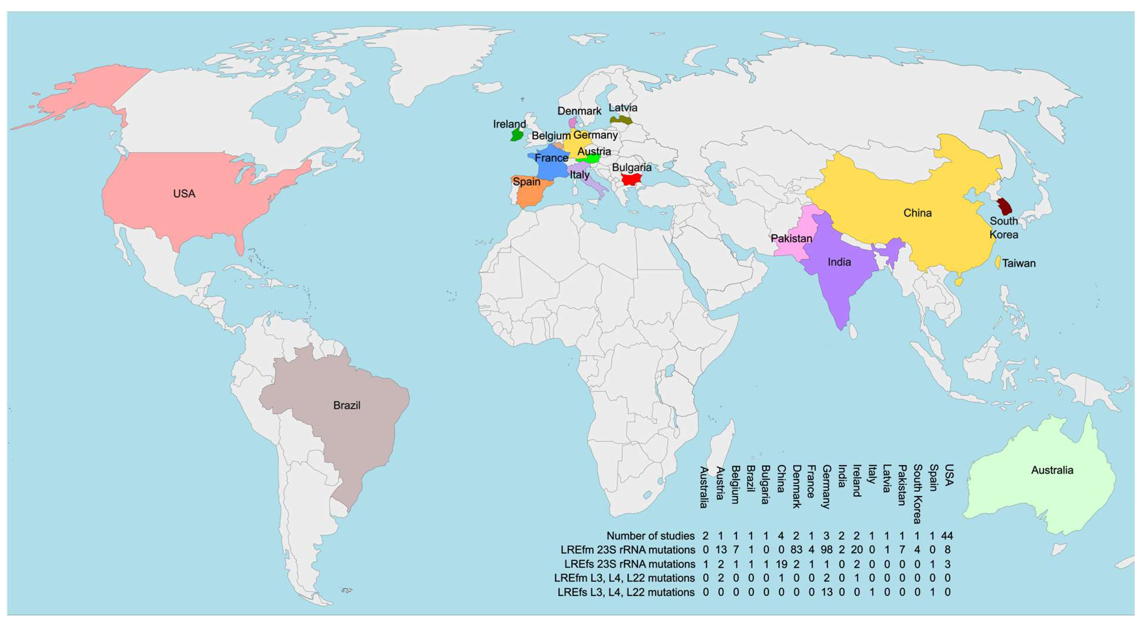
Another notable aspect of the studies identifying ribosomal protein mutations in LRE is their geographic distribution. Except for the SENTRY program, all studies originate from Europe, excluding the Chinese isolate with a L22 mutation. A combined map displaying all studies that have identified mutations in ribosomal structural components (rRNA and proteins) along with their geographical locations is presented in Figure 2.

3.3. Non-Mutational Mechanisms Conferring LNZ Resistance in Enterococci

To date, all identified LNZ resistance mechanisms in enterococci prevent the binding of the antibiotic to its binding pocket in the ribosomal peptidyl transferase center. The most direct ones, discussed in the previous subsection, involve alterations in ribosomal components—specifically, the 23S rRNA and all ribosomal proteins located near the binding pocket. They rely on spontaneous mutations that are not transferable via horizontal gene transfer.
In contrast, another class of resistance mechanisms indirectly prevents LNZ from binding to its target without altering the genetically encoded ribosomal structure. This group relies on the activity of proteins encoded by three main types of determinants—cfr, optrA, and poxtA [106]. A notable characteristic of these genes is their association with a wide range of mobile genetic elements including IS1216, Tn6218, and Tn1546 among others, which facilitates horizontal gene transfer and significantly increases the risk of widespread dissemination [107]. Since these determinants are not part of the core enterococcal genome and are significantly larger than point mutations, their detection via WGS is straightforward, even when using a de novo assembly approach. Many studies employ classical PCR-based screening of isolates before sequencing to select specific LRE subcategories for WGS-based resistome analysis. Primer combinations have been designed to simultaneously detect all acquired LNZ resistance gene types in clinical enterococcal isolates [108]. Such pre-selection should be considered when interpreting results, as it may introduce bias by favouring isolates with coexisting resistance mechanisms.
While detecting acquired LNZ resistance determinants in Enterococcus spp. isolates is simple and reliable, determining their precise location using only Illumina sequencing reads can be challenging. All three types of determinants are frequently plasmid-borne and often co-localize with transposons and other mobile genetic elements with multiple copies, leading to assembly gaps and localization near contig ends. To address this limitation, some studies reviewed in this section adopt a hybrid WGS approach, incorporating long-read sequencing to resolve the genetic context of these transferable elements. The long reads are typically generated using the MinION device from Oxford Nanopore Technologies, which offers excellent scalability and routinely produces reads exceeding 150 kilobases when high-molecular-weight DNA is used [109].
The following subsections will review all studies that have sequenced the genomes of clinical LRE isolates carrying at least one cfr, optrA, or poxtA gene.

3.3.1. LNZ Resistance in Enterococci via Target Modification Mechanisms

LNZ resistance in enterococci, as well as in other Gram-positive pathogens can be manifested via alterations in the modifications of specific nucleotides in the 23S rRNA placed at or near an antibiotic binding site which can affect drug binding to the ribosome. While certain housekeeping modifications at the peptidyl transferase center have been shown to influence LNZ susceptibility, there is only one known so far that confers transferable LNZ resistance [55]. It is mediated by the multiresistance gene cfr, which encodes an rRNA methyltransferase [110]. This enzyme catalyzes the methylation of the C-8 position of the 23S rRNA nucleotide A2503, a key component of the LNZ binding pocket (Figure 1) [111]. This modification grants resistance to five distinct antibiotic classes that bind to overlapping but nonidentical sites within the peptidyl transferase center. The resulting phenotype is known as PhLOPSA, referring to resistance against phenicols, lincosamides, oxazolidinones, pleuromutilins, and streptogramin A antibiotics [55]. The Cfr protein has evolved from the housekeeping rRNA methyltransferase RlmN, highlighting how proteins related to core ribosomal compounds can serve as a crucial reservoir for resistance evolution due to their inherent ability to interact with and modulate the antibiotic’s target [106].
Several variants of cfr have been reported to date, including cfr(B), cfr(C), cfr(D), and cfr(E) [112]. However, a recent study indicated that only cfr(B) and cfr(D) have been detected in enterococci through a search on NCBI PubMed [113]. Moreover, variants of the cfr gene have been infrequently identified among members of the genus Enterococcus in general [114]. Notably, nearly all reports associating LNZ resistance in enterococci with the cfr gene describe its presence in combination with other resistance determinants, such as optrA, poxtA, and/or mutations in the 23S rRNA gene. A widely cited case from Thailand describes a LREfs ST16 isolate recovered after prolonged treatment with the antibiotic. It is frequently presented as evidence implicating cfr in LNZ resistance in E. faecalis, as the isolate lacked mutations in the 23S rRNA gene and in the genes encoding ribosomal proteins L3 and L4 [115]. However, it is important to note that this report predates the identification of optrA and poxtA; therefore, their presence in the isolate cannot be definitively ruled out, particularly in the absence of WGS data necessary for retrospective resistome analysis. Furthermore, experiments involving the cloning and expression of Cfr(D) in E. faecium and E. faecalis did not confer any resistance, in contrast to similar experiments in E. coli, where the expected PhLOPSA phenotype was observed [112]. The expression of the constructs was validated in all three cases using RT-qPCR. This observation highlights that, unlike 23S rRNA mutations, the presence of a cfr gene variant does not necessarily guarantee an LNZ-resistant phenotype in enterococci. Moreover, analyses of clinical and environmental isolates have shown that, even when the MIC is elevated, it is typically not as high as in cases with multiple G2576U variants. LRE strains with high levels of resistance often employ additional mechanisms alongside cfr-mediated methylation.
A summary of the data from WGS studies that have identified cfr gene variants in LRE is showcased in Table 4.
As evident from the summarized data, a total of 17 studies have reported the identification of cfr variants in sequenced LRE genomes. The geographical distribution is highly biased toward the Northern Hemisphere, with studies covering countries in Asia, Europe, and North America. Notably, only Denmark and Germany are represented by more than one study.
Significantly more cfr-positive LREfm isolates have been reported compared to LREfs. Overall, the number of sequenced genomes remains low. This can, to some extent, be attributed to the study designs—among all acquired LNZ resistance determinants, cfr was the only one not used as a selection criterion for LRE WGS in any of the identified studies with larger number of sequenced genomes. A similar explanation applies to the high prevalence of cfr + optrA or cfr + poxtA combinations observed in the analyzed LRE genomes.
The recent study by Cinthi et al. is of particular interest, as it describes two E. faecium and one E. faecalis isolate that were resistant to both LNZ and vancomycin due to the presence of optrA, cfr(D), and vanA genes, all located on plasmids with a linear topology [52]. Notably, this study represents the first report of a linear plasmid in E. faecalis [52].
The highest number of described isolates (n = 7) originates from Pakistan, where the identified cfr(D) variants are accompanied by poxtA or, in one case, a combination of poxtA and optrA [46]. Given the presence of LRE isolates with G2576U mutations in Pakistan, as discussed in previous sections, the country emerges as a hotspot for LRE strains harboring diverse resistance mechanisms, including complex combinations of multiple determinants. This is likely attributable to Pakistan’s status as a high-burden country for multidrug-resistant tuberculosis, with one of the highest caseloads and incidence rates globally, as recognized by the WHO. The widespread use of LNZ in treatment regimens (e.g., bedaquiline, pretomanid, and LNZ, with or without moxifloxacin, in six-month all-oral therapies for Pakistan) has been instrumental in controlling tuberculosis. However, it has also contributed to a surge in LNZ resistance among enterococci.
Overall, the precise contribution of cfr gene variants to LNZ resistance in enterococci, if any, remains uncertain. Nevertheless, their presence in clinical Enterococcus isolates should not be neglected, as enterococci may serve as a reservoir for cfr and cfr-like genes, with the potential for horizontal transfer to staphylococcal species, where these determinants are clearly implicated in linezolid resistance [114].

3.3.2. LNZ Resistance in Enterococci via Target Protection Mechanisms

The final mechanism of LNZ resistance identified in clinical Enterococci relies on target protection by members of the ATP-binding cassette (ABC)-F protein subfamily. These proteins bind to the ribosome, facilitating the release of ribosome-targeted antibiotics, thus rescuing the translation apparatus from antibiotic-mediated inhibition [119]. What is particularly remarkable is that the exact mechanism by which these proteins confer resistance was not clarified until 2016, after a debate that lasted over a quarter of a century. Moreover, this group is far from small or isolated. On the contrary, members of the ABC-F protein subfamily mediate resistance to a broader range of antibacterial drug classes than any other single group of resistance proteins. They constitute a major source of clinical resistance to nearly all antibacterial drug classes targeting the 50S subunit of the ribosome, including lincosamides, macrolides, oxazolidinones, phenicols, pleuromutilins, and streptogramins groups A and B [106]. Despite this, the precise mechanism of action was not resolved until 2016, bringing an end to a debate that had lasted for over 25 years [120].
ABC-F proteins differ from most other members of the ATP-binding cassette (ABC) superfamily in that they lack the typical transmembrane regions. Instead, they consist of two ABC domains separated by a linker region, which has been identified as the P site tRNA interaction domain, playing a key role in antibiotic resistance [121]. Based on their antibiotic specificity, three major categories of ARE ABC-F proteins can be distinguished: the first group mediates resistance to group A streptogramins, lincosamides, and occasionally pleuromutilins; the second group confers resistance to group B streptogramins and macrolide antibiotics (and sometimes ketolides); and the third group, including Optr and Poxt, mediates resistance to oxazolidinones and phenicols [106]. These will be further discussed in relation to their role in LNZ resistance in enterococci. The molecular mechanism by which Optr and Poxt proteins are able to dislodge oxazolidinones and phenicols from the ribosome requires further investigation since these proteins have relatively short antibiotic resistance domains, which would not typically be expected to extend into the peptidyl transferase center [122].
Although the precise molecular mechanism of target protection by OptrA and PoxtA remains to be fully elucidated, their role in conferring LNZ resistance in clinical LRE isolates is undeniable. The optrA gene encodes an ABC-F protein that targets the ribosome of Gram-positive bacteria, mediating resistance to both phenicols and oxazolidinones through ribosomal protection [106]. First identified in clinical isolates in 2015 [123], optrA-carrying LRE strains have since been detected in a diverse range of hosts, including both hospitalized and healthy humans [124].
Additionally, an optrA reservoir has emerged in animal populations, largely due to the widespread use of phenicols in veterinary medicine to treat respiratory infections [125]. OptrA-producing isolates exhibit considerable genetic diversity and are associated with various chromosomal and plasmid genetic platforms. Notably, optrA has been more frequently reported in E. faecalis than in E. faecium [124]. Given that recent meta-analyses indicate a rapid increase in the frequency of LREfs isolates in recent years, it is evident that optrA poses the greatest threat to the continued effectiveness of LNZ as a last-resort treatment for severe enterococcal infections [26,34].
Noteworthy, in addition to the wild-type optrA gene identified in numerous LRE isolates from diverse sources, at least 69 optrA variants have been described to date. These variants differ by 1 to 20 amino acid substitutions, and no less than 35 of them have been identified in Enterococcus species to date [114]. A recent study, conducted at a tertiary care hospital in Hangzhou (China), identified eight distinct optrA variants among 15 optrA-positive clinical isolates, demonstrating that such genetic diversity is also present in clinical settings [126]. Furthermore, data from multiple studies examining linezolid MIC values in various Gram-positive bacteria suggest a correlation between certain optrA variants and elevated LNZ-resistance levels. Variants such as the wild-type, D, EDP, KD, KLDP, RD, RDK, and RDKP are more frequently found in isolates exhibiting MIC values ≥ 8 mg/L [114].
Since its initial identification in an MRSA clinical isolate from Italy in 2018, poxtA has been detected in enterococci across multiple countries [127]. To date, it has been primarily reported in E. faecium isolates of human origin in Europe (Greece, Spain, Portugal, Ireland), as well as in the USA, Pakistan, and Turkey, with a prevalence among LREfm ranging from 16% to 78% [128]. Otherwise, poxtA is more commonly detected in environmental samples and food-producing animals than in human isolates, again with E. faecium exhibiting a higher prevalence than E. faecalis [128]. A variant of poxtA, designated poxtA2, was identified on a 13,746 bp plasmid in the linezolid-resistant Enterococcus gallinarum isolate Eg-IV02, recovered from a fecal swab of a healthy child in the rural Bolivian Chaco region. Hybrid whole-genome sequencing using both short and long reads revealed that poxtA2 possesses a distinct genetic context compared to poxtA and likely represents its ancestor [129].
A summary of the data from WGS studies that have identified optrA gene and/or poxtA gene in LRE is showcased in Table 5.
A total of 42 studies from various countries worldwide have documented the global spread of optrA and poxtA among clinical LRE isolates. Notably, China leads with seven studies, followed by Spain with four. The most frequently detected isolate is LREfs carrying optrA, whereas poxtA is more commonly associated with LREfm, reinforcing previously observed biases in the distribution of these resistance determinants. Interestingly, a WGS-based study in the literature conducted its data processing before the identification of optrA and poxtA, failing to determine the LNZ resistance mechanism despite a clear resistant phenotype [146]. This highlights the potential value of reanalyzing previously sequenced genomes with unresolved resistance mechanisms, as it may lead to the discovery of previously unrecognized determinants or novel mutations.
The optrA and poxtA genes have been detected on both plasmids and chromosomal loci. Two studies even reported optrA detection on novel linear plasmids alongside vanA in both LREfm and LREfs isolates [51,52]. This finding is particularly concerning, as optrA dissemination is generally lower in E. faecium compared to E. faecalis, but the emergence of such novel vectors for horizontal gene transfer could alter this trend. Moreover, the co-presence of vanA further narrows the already limited treatment options.
An intriguing study from India employed a hybrid WGS approach using long Nanopore sequencing reads [84]. This analysis identified an LREfm isolate carrying two copies of optrA, one integrated into the chromosome and the other plasmid-borne, highlighting the potential of advanced NGS technologies in elucidating LNZ resistance mechanisms in Enterococcus spp. Similar findings for poxtA were reported by Lázaro-Perona et al., who identified a clinical LREfm isolate with an increased copy number of the plasmid carrying the poxtA gene [104]. Proteomic analysis confirmed the presence of the PoxtA and revealed elevated expression levels in the isolate.
Finally, the study by Hu et al. is particularly noteworthy, as it reports the first global identification and characterization of a clinical E. gallinarum strain harboring poxtA EF9F6 (LNZ MIC = 8 mg/L) [88]. Li et al. also describe a optrA KLPD-positive Enterococcus hirae isolate of human origin; however, their study was primarily designed to investigate optrA carriage among patients [126].
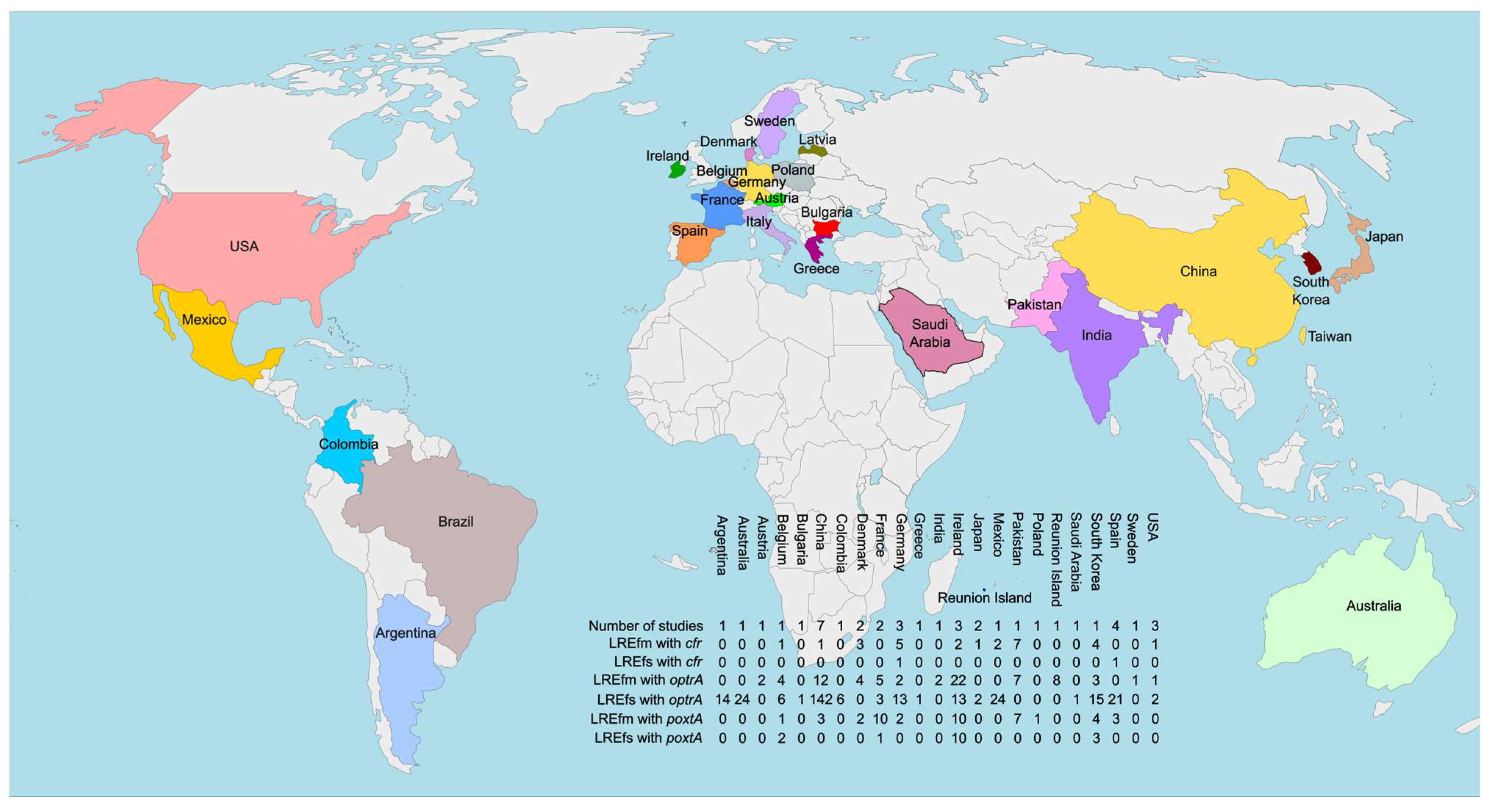
Figure 3 presents a consolidated map illustrating the geographical distribution of studies that have identified target protection resistance determinants in LRE.

3.4. Tedizolid—Resistance Mechanisms and Cross-Resistance with LNZ

Tedizolid (TDZ) is a second-generation oxazolidinone antibiotic developed after LNZ and approved in 2014 by the U.S. Food and Drug Administration for the treatment of acute bacterial skin and skin structure infections caused by susceptible Gram-positive bacteria [147]. Later, in 2015, TDZ was approved for medical use in the European Union by the European Medicines Agency [148]. It is administered as TDZ phosphate, a prodrug that is converted in vivo to the active compound, TDZ, through the action of phosphatases. This oxazolidinone represents a departure from the previously established structure–activity relationships described for LNZ. TDZ contains a hydroxymethyl side chain at the C-5 position, which was initially thought to reduce effectiveness [149]. However, this limitation was overcome by the incorporation of a fourth, para-oriented aromatic ring (the D-ring), which introduces additional hydrogen-bonding interactions and enhances stabilization of binding to the target site [150]. These structural modifications resulted in TDZ exhibiting strong in vitro activity against both MRSA and VRE. Moreover, it consistently demonstrated 4- to 8-fold greater potency compared to LNZ across a broad spectrum of Gram-positive microorganisms, including strains with reduced LNZ susceptibility [149,151].
According to The Surveillance of TDZ Activity and Resistance (STAR) program TDZ (MIC50/90, 0.25/0.25 mg/L; 99.9% susceptible) displayed activity similar to or greater than LNZ (MIC50/90, 1/2 mg/L; 99.5% susceptible), ampicillin (MIC50/90, 1/1 mg/L; 100.0% susceptible), daptomycin (MIC50/90, 0.5/1 mg/L; 99.6% susceptible), and vancomycin (MIC50/90, 1/2 mg/L; 98.1% susceptible) against a cohort of 4992 E. faecalis isolates [152]. Notably, in the same study TDZ demonstrated superior effectiveness against the vancomycin-resistant E. faecalis (VREfs) subset, showing 4- to 8-fold higher potency than LNZ (MIC50/90, 1/2 mg/L; 100.0% susceptible) and daptomycin (MIC50/90, 0.5/1 mg/L; 100.0% susceptible), respectively. In contrast, among the LNZ-non-susceptible E. faecalis isolates identified in the study, only 73.1% were inhibited by TDZ at concentrations ≤0.5 mg/L. Meanwhile, all of these isolates remained susceptible to ampicillin, daptomycin, and vancomycin at their respective clinical breakpoints, suggesting that TDZ and LNZ are likely to share certain resistance mechanisms. Furthermore, 6 of the 7 VREfs isolates exhibiting non-susceptibility to TDZ were found to be optrA-positive without accompanying 23S rRNA mutations, indicating a possible role of this determinant in the observed resistance phenotype [152]. Similar findings were reported in a recent study from Japan [153] in which three clinical LREfs isolates with TDZ MICs of 2–4 mg/L were also optrA-positive but lacked the G2576T mutation, whereas another three isolates harbored only this variant in two of their 23S rRNA loci and exhibited higher MICs of 8 mg/L. The analysis of 19 LREfm isolates from the same study revealed variable numbers of G2576T mutations across their genomes, with TDZ MICs ranging from 1 to 16 mg/L. As observed for LNZ, the number of affected 23S rRNA loci was not a reliable predictor of MIC values, since isolates with four mutated operons exhibited MICs of 2, 4, 8, and 16 mg/L, respectively, in the absence of other known LNZ-resistance determinants [153]. Conversely, the sole isolate harboring the T2504A variant in two of its 23S rRNA loci exhibited the highest observed MIC of 32 mg/L.
A large-scale study analyzing 916 E. faecium and 1342 E. faecalis isolates from invasive infections in hospitalized patients across U.S. and European medical centers (2015–2017) reported that TDZ inhibited all E. faecalis isolates except for one [151]. Among the E. faecium isolates, seven were LNZ-non-susceptible (MIC 4–8 mg/L), and five of these also exhibited TDZ nonsusceptibility (MIC ≥ 1 mg/L). Notably, one E. faecium isolate from Ankara, Turkey, demonstrated an elevated TDZ MIC of 0.5 mg/L despite lacking G2576T variants, while carrying both optrA and poxtA. This observation supports previous in vitro evidence indicating that poxtA can contribute to reduced susceptibility to TDZ [127].
One of the notable advantages of TDZ is that its MIC values appear largely unaffected by the presence of cfr genes, which have been implicated in several reported outbreaks of LNZ-resistant organisms [149]. However, this advantage is of limited relevance in LRE, where the role of this determinant in conferring LNZ resistance remains uncertain. The same applies to the Gly152Asp mutation of the 50S ribosomal protein L3, which has been found to solely confer cross-resistance to both LNZ and TDZ in MRSA isolates [154].
All findings on LNZ resistance mechanisms in enterococci and their impact on TDZ cross-resistance are summarized in Table 6.

3.5. Future Perspectives on WGS for Detecting and Investigating LNZ Resistance in Enterococci

With the decreasing costs of long-read NGS technologies such as Nanopore, new opportunities have emerged for studying LNZ resistance mechanisms associated with acquired genetic determinants. Notably, two of the most compelling discoveries, an LREfm isolate harboring two optrA copies in different genomic locations and another with an increased plasmid-borne optrA copy number, were made using sequencing data generated by a MinION device [84,104].
Moreover, a recent study by Coll et al. focused on evaluating and enhancing the accuracy of antibiotic resistance detection in E. faecium genomes for diagnostic and surveillance purposes [155]. The authors achieved near-complete genotype–phenotype concordance after re-evaluating false negatives, reporting a sensitivity of 100.0% [90.8–100.0] and a specificity of 98.3% [97.8–98.7]. Their findings align with the present literature review, where only one study entirely failed to identify the underlying LNZ resistance mechanism in the isolate described [146]. In a few other cases, individual isolates exhibited phenotypic LNZ resistance despite no identifiable resistance mechanism being detected through sequencing. However, such cases remain sporadic [105,156].
The study by Beh et al. aimed to elucidate the genomic epidemiology and population structure of LRE in Victoria, Australia, as well as at a global scale [157]. In a noteworthy shift in paradigm, the authors focused on isolates exhibiting phenotypic and/or genotypic resistance to linezolid, subjecting them to WGS to identify a range of resistance-associated determinants, including cfr, cfr(B), cfr(D), optrA, and poxtA. The resulting genomic data were compared with all publicly available sequencing reads and/or assemblies from comparable strains in the NCBI Pathogen Detection database and the Sequence Read Archive. These findings suggest that future research will likely emphasize genotypic characterization of LNZ resistance in enterococci, with epidemiological and population structure analyses increasingly conducted through the lens of modern bacterial genomics.
Another noteworthy application of NGS in diagnosing enterococcal infections is demonstrated in the work of Li et al. [158]. The authors present a metagenomic NGS (mNGS)-based approach for detecting E. faecalis in a urinary tract infection case. This innovative, culture-independent method proved to be rapid, sensitive, and highly accurate for pathogen identification while also providing valuable insights to guide clinicians in selecting an appropriate treatment. Additionally, mNGS enabled the assessment of treatment efficacy by comparing unique reads before and after LNZ administration. This suggests its potential for monitoring the patient’s microbiome for the occurrence of LRE, particularly in clinical indications requiring prolonged LNZ-based therapy.

4. Conclusions

The present review article emphasizes WGS as an indispensable tool for elucidating the molecular basis of LNZ resistance in enterococci, uncovering a complex interplay of resistance mechanisms. These include previously unreported 23S rRNA mutations, novel amino acid exchanges in ribosomal proteins of isolates lacking other known LNZ resistance determinants, and multiple copies of chromosomal- and plasmid-born ABC-F protein-encoding genes in single strains. Genomic analyses have shown that many LRE isolates harbor multiple resistance mechanisms, often in combination, particularly in geographical regions where LNZ is used in prolonged treatment courses for multi-drug-resistant tuberculosis. As LRE continue to evolve and acquire additional resistance traits, WGS-based research remains critical for tracking its dissemination, identifying novel resistance determinants and high-risk clones, and guiding antimicrobial stewardship efforts worldwide.
In the context of a growing pandemic of AMR, the emergence of LNZ resistance in clinical Enterococcus spp. isolates, especially in VRE, is a worrisome situation worldwide. There is no specific approved drug currently available for the treatment of infections due to those multidrug-resistant pathogens, although few studies have demonstrated the successful outcome using tigecycline and daptomycin separately [159]. Therefore, control of indiscriminate use of LNZ, robust global and regional surveillance studies on its resistance rates and mechanisms, as well as integration of antimicrobial stewardship programs into prevention strategies in hospital settings to limit the spread of LRE strains are necessary.

Author Contributions

S.P.: Design of the review content, critical overview of the literature, writing of the manuscript (original draft preparation), and coordinating the research project. B.K.: Design of the review content, writing (review and editing), funding. T.S.: Design of the review content, critical overview of the literature, writing (review and editing), and supervision. All authors have read and agreed to the published version of the manuscript.

Funding

The Bulgarian studies cited in the present review article were partially supported by the European Union-Next Generation EU, through the National Recovery and Resilience Plan of the Republic of Bulgaria, project № BG-RRP-2.004-0005 “Improving the research capacity and quality to achieve international recognition and resilience of the Technical University—Sofia” (IDEAS).

Institutional Review Board Statement

Not applicable.

Informed Consent Statement

Not applicable.

Data Availability Statement

No new data were created or analyzed in this study. Data sharing is not applicable to this article.

Conflicts of Interest

The authors declare no conflict of interest.

References

  1. Hashemian, S.M.; Farhadi, T.; Ganjparvar, M. Linezolid: A Review of Its Properties, Function, and Use in Critical Care. Drug Des. Dev. Ther. 2018, 12, 1759–1767. [Google Scholar] [CrossRef]
  2. Bibel, B.; Raskar, T.; Couvillion, M.; Lee, M.; Kleinman, J.I.; Takeuchi-Tomita, N.; Churchman, L.S.; Fraser, J.S.; Fujimori, D.G. Context-Specific Inhibition of Mitochondrial Ribosomes by Phenicol and Oxazolidinone Antibiotics. Nucleic Acids Res. 2025, 53, gkaf046. [Google Scholar] [CrossRef]
  3. Vishnu, V.Y.; Modi, M.; Goyal, M.K.; Lal, V. Linezolid Induced Reversible Peripheral Neuropathy. Am. J. Ther. 2016, 23, e1839–e1841. [Google Scholar] [CrossRef]
  4. Soriano, A.; Miró, O.; Mensa, J. Mitochondrial Toxicity Associated with Linezolid. N. Engl. J. Med. 2005, 353, 2305–2306. [Google Scholar] [CrossRef]
  5. De Vriese, A.S.; Van Coster, R.; Smet, J.; Seneca, S.; Lovering, A.; Van Haute, L.L.; Vanopdenbosch, L.J.; Martin, J.-J.; Ceuterick-de Groote, C.; Vandecasteele, S.; et al. Linezolid-Induced Inhibition of Mitochondrial Protein Synthesis. Clin. Infect. Dis. 2006, 42, 1111–1117. [Google Scholar] [CrossRef]
  6. Ramesh, V.; Gattu, S.; Maqsood, M.; Rao, V. Linezolid-Induced Lactic Acidosis. BMJ Case Rep. 2024, 17, e259335. [Google Scholar] [CrossRef] [PubMed]
  7. Kato, H.; Hagihara, M.; Asai, N.; Koizumi, Y.; Yamagishi, Y.; Mikamo, H. A Systematic Review and Meta-Analysis of Myelosuppression in Pediatric Patients Treated with Linezolid for Gram-Positive Bacterial Infections. J. Infect. Chemother. 2021, 27, 1143–1150. [Google Scholar] [CrossRef] [PubMed]
  8. Pfaller, M.A.; Mendes, R.E.; Streit, J.M.; Hogan, P.A.; Flamm, R.K. Five-Year Summary of In Vitro Activity and Resistance Mechanisms of Linezolid against Clinically Important Gram-Positive Cocci in the United States from the LEADER Surveillance Program (2011 to 2015). Antimicrob. Agents Chemother. 2017, 61, e00609-17. [Google Scholar] [CrossRef]
  9. Luna, V.A.; King, D.S.; Gulledge, J.; Cannons, A.C.; Amuso, P.T.; Cattani, J. Susceptibility of Bacillus anthracis, Bacillus cereus, Bacillus mycoides, Bacillus pseudomycoides and Bacillus thuringiensis to 24 Antimicrobials Using Sensititre(R) Automated Microbroth Dilution and Etest(R) Agar Gradient Diffusion Methods. J. Antimicrob. Chemother. 2007, 60, 555–567. [Google Scholar] [CrossRef] [PubMed]
  10. Barberis, C.M.; Sandoval, E.; Rodriguez, C.H.; Ramírez, M.S.; Famiglietti, A.; Almuzara, M.; Vay, C. Comparison Between Disk Diffusion and Agar Dilution Methods to Determine In Vitro Susceptibility of Corynebacterium spp. Clinical Isolates and Update of Their Susceptibility. J. Glob. Antimicrob. Resist. 2018, 14, 246–252. [Google Scholar] [CrossRef]
  11. Noll, M.; Kleta, S.; Al Dahouk, S. Antibiotic Susceptibility of 259 Listeria monocytogenes Strains Isolated from Food, Food-Processing Plants and Human Samples in Germany. J. Infect. Public. Health 2018, 11, 572–577. [Google Scholar] [CrossRef] [PubMed]
  12. Schumacher, A.; Trittler, R.; Bohnert, J.A.; Kümmerer, K.; Pagès, J.-M.; Kern, W.V. Intracellular Accumulation of Linezolid in Escherichia coli, Citrobacter freundii and Enterobacter aerogenes: Role of Enhanced Efflux Pump Activity and Inactivation. J. Antimicrob. Chemother. 2007, 59, 1261–1264. [Google Scholar] [CrossRef]
  13. Birmingham, M.C.; Rayner, C.R.; Meagher, A.K.; Flavin, S.M.; Batts, D.H.; Schentag, J.J. Linezolid for the Treatment of Multidrug-Resistant, Gram-Positive Infections: Experience from a Compassionate-Use Program. Clin. Infect. Dis. 2003, 36, 159–168. [Google Scholar] [CrossRef]
  14. Faella, F.; Pagliano, P.; Fusco, U.; Attanasio, V.; Conte, M. Combined Treatment with Ceftriaxone and Linezolid of Pneumococcal Meningitis: A Case Series Including Penicillin-Resistant Strains. Clin. Microbiol. Infect. 2006, 12, 391–394. [Google Scholar] [CrossRef]
  15. Thwaites, G.; Nguyen, N.V. Linezolid for Drug-Resistant Tuberculosis. N. Engl. J. Med. 2022, 387, 842–843. [Google Scholar] [CrossRef]
  16. Garazzino, S.; Krzysztofiak, A.; Esposito, S.; Castagnola, E.; Plebani, A.; Galli, L.; Cellini, M.; Lipreri, R.; Scolfaro, C.; Bertaina, C.; et al. Use of Linezolid in Infants and Children: A Retrospective Multicentre Study of the Italian Society for Paediatric Infectious Diseases. J. Antimicrob. Chemother. 2011, 66, 2393–2397. [Google Scholar] [CrossRef]
  17. Shi, Y.; Wu, H.-L.; Wu, Y.-H.; Li, S.; Zhang, L.-Y.; Xu, S.-S.; Huang, H.-Y.; Zhang, C.-H.; Yu, X.-B.; Cai, K.; et al. Safety and Clinical Efficacy of Linezolid in Children: A Systematic Review and Meta-Analysis. World J. Pediatr. 2023, 19, 129–138. [Google Scholar] [CrossRef]
  18. Dubin, K.; Pamer, E.G. Enterococci and Their Interactions with the Intestinal Microbiome. Microbiol. Spectr. 2017, 5, 1–16. [Google Scholar] [CrossRef]
  19. Komiyama, E.Y.; Lepesqueur, L.S.S.; Yassuda, C.G.; Samaranayake, L.P.; Parahitiyawa, N.B.; Balducci, I.; Koga-Ito, C.Y. Enterococcus Species in the Oral Cavity: Prevalence, Virulence Factors and Antimicrobial Susceptibility. PLoS ONE 2016, 11, e0163001. [Google Scholar] [CrossRef] [PubMed]
  20. Ghasemi, E.; Mansouri, S.; Shahabinejad, N. Vaginal Colonization and Susceptibility to Antibiotics of Enterococci During Late Pregnancy in Kerman City, Iran. Arch. Clin. Infect. Dis. 2016, 11, e35428. [Google Scholar] [CrossRef]
  21. Byappanahalli, M.N.; Nevers, M.B.; Korajkic, A.; Staley, Z.R.; Harwood, V.J. Enterococci in the Environment. Microbiol. Mol. Biol. Rev. 2012, 76, 685–706. [Google Scholar] [CrossRef]
  22. Ben Braïek, O.; Smaoui, S. Enterococci: Between Emerging Pathogens and Potential Probiotics. BioMed Res. Int. 2019, 2019, 5938210. [Google Scholar] [CrossRef] [PubMed]
  23. Fiore, E.; Van Tyne, D.; Gilmore, M.S. Pathogenicity of Enterococci. Microbiol. Spectr. 2019, 7, 1–23. [Google Scholar] [CrossRef]
  24. Lebreton, F.; Willems, R.J.L.; Gilmore, M.S. Enterococcus Diversity, Origins in Nature, and Gut Colonization. In Enterococci: From Commensals to Leading Causes of Drug Resistant Infection; Gilmore, M.S., Clewell, D.B., Ike, Y., Shankar, N., Eds.; Massachusetts Eye and Ear Infirmary: Boston, MA, USA, 2014. [Google Scholar]
  25. Ch’ng, J.-H.; Chong, K.K.L.; Lam, L.N.; Wong, J.J.; Kline, K.A. Biofilm-Associated Infection by Enterococci. Nat. Rev. Microbiol. 2019, 17, 82–94. [Google Scholar] [CrossRef]
  26. Dadashi, M.; Sharifian, P.; Bostanshirin, N.; Hajikhani, B.; Bostanghadiri, N.; Khosravi-Dehaghi, N.; van Belkum, A.; Darban-Sarokhalil, D. The Global Prevalence of Daptomycin, Tigecycline, and Linezolid-Resistant Enterococcus faecalis and Enterococcus faecium Strains from Human Clinical Samples: A Systematic Review and Meta-Analysis. Front. Med. 2021, 8, 720647. [Google Scholar] [CrossRef]
  27. Hegstad, K.; Mikalsen, T.; Coque, T.M.; Werner, G.; Sundsfjord, A. Mobile Genetic Elements and Their Contribution to the Emergence of Antimicrobial Resistant Enterococcus faecalis and Enterococcus faecium. Clin. Microbiol. Infect. 2010, 16, 541–554. [Google Scholar] [CrossRef]
  28. Arredondo-Alonso, S.; Top, J.; McNally, A.; Puranen, S.; Pesonen, M.; Pensar, J.; Marttinen, P.; Braat, J.C.; Rogers, M.R.C.; van Schaik, W.; et al. Plasmids Shaped the Recent Emergence of the Major Nosocomial Pathogen Enterococcus faecium. MBio 2020, 11, 1–17. [Google Scholar] [CrossRef] [PubMed]
  29. Hashimoto, Y.; Suzuki, M.; Kobayashi, S.; Hirahara, Y.; Kurushima, J.; Hirakawa, H.; Nomura, T.; Tanimoto, K.; Tomita, H. Enterococcal Linear Plasmids Adapt to Enterococcus faecium and Spread within Multidrug-Resistant Clades. Antimicrob. Agents Chemother. 2023, 67, e01619-22. [Google Scholar] [CrossRef]
  30. Miller, W.R.; Arias, C.A. ESKAPE Pathogens: Antimicrobial Resistance, Epidemiology, Clinical Impact and Therapeutics. Nat. Rev. Microbiol. 2024, 22, 598–616. [Google Scholar] [CrossRef]
  31. Iqbal, F.; Alocious, A.; Joy, S.C.; Stanly, E.A.R.; Rajesh, V.; Unnikrishnan, M.K.; Steinke, D.; Chandra, P. Vancomycin-Resistant Enterococci: A Rising Challenge to Global Health. Clin. Epidemiol. Glob. Health 2024, 28, 101663. [Google Scholar] [CrossRef]
  32. WHO. Publishes List of Bacteria for Which New Antibiotics Are Urgently Needed. Available online: https://www.who.int/news/item/27-02-2017-who-publishes-list-of-bacteria-for-which-new-antibiotics-are-urgently-needed (accessed on 23 March 2025).
  33. WHO. Bacterial Priority Pathogens List, 2024: Bacterial Pathogens of Public Health Importance to Guide Research, Development and Strategies to Prevent and Control Antimicrobial Resistance. Available online: https://www.who.int/publications/i/item/9789240093461 (accessed on 17 August 2025).
  34. Wada, Y.; Afolabi, H.A.; Al-Mhanna, S.B.; Bello, K.E.; Irekeola, A.A.; Wada, M.; Ahmed, N.; Harun, A.; Yean, C.Y.; Mohamad Nasir, N.S.; et al. Global Occurrence of Linezolid-Resistant Enterococcus (LRE): The First Systematic Review and Meta-Analysis. Microbe 2024, 2, 100041. [Google Scholar] [CrossRef]
  35. Bi, R.; Qin, T.; Fan, W.; Ma, P.; Gu, B. The Emerging Problem of Linezolid-Resistant Enterococci. J. Glob. Antimicrob. Resist. 2018, 13, 11–19. [Google Scholar] [CrossRef]
  36. Kainer, M.A.; Devasia, R.A.; Jones, T.F.; Simmons, B.P.; Melton, K.; Chow, S.; Broyles, J.; Moore, K.L.; Craig, A.S.; Schaffner, W. Response to Emerging Infection Leading to Outbreak of Linezolid-Resistant Enterococci. Emerg. Infect. Dis. 2007, 13, 1024–1030. [Google Scholar] [CrossRef]
  37. Jia, S.; Xu, X.; Qu, M.; Pei, Y.; Sun, S.; Liu, Y.; Dong, W.; Hu, Y.; Zhu, B.; Gao, G.F.; et al. Longitudinal Trends and Drivers of Antimicrobial Resistance in Campylobacter Worldwide (1954–2023). Zoonoses 2025, 5, 989. [Google Scholar] [CrossRef]
  38. Wang, Y.; Xu, X.; Jia, S.; Qu, M.; Pei, Y.; Qiu, S.; Zhang, J.; Liu, Y.; Ma, S.; Lyu, N.; et al. A Global Atlas and Drivers of Antimicrobial Resistance in Salmonella during 1900-2023. Nat. Commun. 2025, 16, 4611. [Google Scholar] [CrossRef]
  39. Waskito, L.A.; Rezkitha, Y.A.A.; Vilaichone, R.; Wibawa, I.D.N.; Mustika, S.; Sugihartono, T.; Miftahussurur, M. Antimicrobial Resistance Profile by Metagenomic and Metatranscriptomic Approach in Clinical Practice: Opportunity and Challenge. Antibiotics 2022, 11, 654. [Google Scholar] [CrossRef]
  40. Fines, M.; Leclercq, R. Activity of Linezolid against Gram-Positive Cocci Possessing Genes Conferring Resistance to Protein Synthesis Inhibitors. J. Antimicrob. Chemother. 2000, 45, 797–802. [Google Scholar] [CrossRef] [PubMed]
  41. Brumfitt, W.; Hamilton-Miller, J.M.T. In-Vitro Microbiological Activities of DuP 105 and DuP 721, Novel Synthetic Oxazolidinones. J. Antimicrob. Chemother. 1988, 21, 711–720. [Google Scholar] [CrossRef] [PubMed]
  42. Meka, V.G.; Gold, H.S. Antimicrobial Resistance to Linezolid. Clin. Infect. Dis. 2004, 39, 1010–1015. [Google Scholar] [CrossRef] [PubMed]
  43. Development of Linezolidresistant. Enterococcus faecium in Two Compassionate Use Program Patients Treated with Linezolid [Abstract 848]—New York University Health Sciences. Available online: https://search.hsl.med.nyu.edu (accessed on 24 March 2025).
  44. Klare, I.; Konstabel, C.; Mueller-Bertling, S.; Werner, G.; Strommenger, B.; Kettlitz, C.; Borgmann, S.; Schulte, B.; Jonas, D.; Serr, A.; et al. Spread of Ampicillin/Vancomycin-Resistant Enterococcus faecium of the Epidemic-Virulent Clonal Complex-17 Carrying the Genes esp and hyl in German Hospitals. Eur. J. Clin. Microbiol. Infect. Dis. 2005, 24, 815–825. [Google Scholar] [CrossRef]
  45. Theilacker, C.; Jonas, D.; Huebner, J.; Bertz, H.; Kern, W.V. Outcomes of Invasive Infection Due to Vancomycin-Resistant Enterococcus faecium during a Recent Outbreak. Infection 2009, 37, 540–543. [Google Scholar] [CrossRef]
  46. Nasir, S.A.R.; Zeeshan, M.; Ghanchi, N.; Saeed, N.; Ghayas, H.; Zaka, S.; Ashraf, J.; Jabeen, K.; Farooqi, J.; Hasan, Z.; et al. Linezolid-Resistant Enterococcus faecium Clinical Isolates from Pakistan: A Genomic Analysis. BMC Microbiol. 2024, 24, 347. [Google Scholar] [CrossRef]
  47. Abbo, L.; Shukla, B.S.; Giles, A.; Aragon, L.; Jimenez, A.; Camargo, J.F.; Simkins, J.; Sposato, K.; Tran, T.T.; Diaz, L.; et al. Linezolid- and Vancomycin-Resistant Enterococcus faecium in Solid Organ Transplant Recipients: Infection Control and Antimicrobial Stewardship Using Whole Genome Sequencing. Clin. Infect. Dis. 2019, 69, 259–265. [Google Scholar] [CrossRef] [PubMed]
  48. Almeida-Santos, A.C.; Novais, C.; Peixe, L.; Freitas, A.R. Vancomycin-Resistant Enterococcus faecium: A Current Perspective on Resilience, Adaptation, and the Urgent Need for Novel Strategies. J. Glob. Antimicrob. Resist. 2025, 41, 233–252. [Google Scholar] [CrossRef]
  49. Mortelé, O.; Van Kleef–van Koeveringe, S.; Vandamme, S.; Jansens, H.; Goossens, H.; Matheeussen, V. Epidemiology and Genetic Diversity of Linezolid-Resistant Enterococcus Clinical Isolates in Belgium from 2013 to 2021. J. Glob. Antimicrob. Resist. 2024, 38, 21–26. [Google Scholar] [CrossRef]
  50. Liu, P.; Zeng, B.; Wu, X.; Zheng, F.; Zhang, Y.; Liao, X. Risk Exploration and Prediction Model Construction for Linezolid-Resistant Enterococcus faecalis Based on Big Data in a Province in Southern China. Eur. J. Clin. Microbiol. Infect. Dis. 2024, 43, 259–268. [Google Scholar] [CrossRef] [PubMed]
  51. Kent, A.G.; Spicer, L.M.; Campbell, D.; Breaker, E.; McAllister, G.A.; Ewing, T.O.; Longo, C.; Balbuena, R.; Burroughs, M.; Burgin, A.; et al. Sentinel Surveillance Reveals Phylogenetic Diversity and Detection of Linear Plasmids Harboring vanA and optrA among Enterococci Collected in the United States. Antimicrob. Agents Chemother. 2024, 68, e00591-24. [Google Scholar] [CrossRef]
  52. Cinthi, M.; Coccitto, S.N.; Simoni, S.; Gherardi, G.; Palamara, A.T.; Di Lodovico, S.; Di Giulio, M.; Du, X.-D.; Vignaroli, C.; Brenciani, A.; et al. The optrA, cfr(D) and vanA Genes Are Co-Located on Linear Plasmids in Linezolid- and Vancomycin-Resistant Enterococcal Clinical Isolates in Italy. J. Antimicrob. Chemother. 2025, 80, 1362–1370. [Google Scholar] [CrossRef]
  53. Zahedi Bialvaei, A.; Rahbar, M.; Yousefi, M.; Asgharzadeh, M.; Samadi Kafil, H. Linezolid: A Promising Option in the Treatment of Gram-Positives. J. Antimicrob. Chemother. 2017, 72, 354–364. [Google Scholar] [CrossRef] [PubMed]
  54. Gemmell, C.G. Virulence Factor Expression by Gram-Positive Cocci Exposed to Subinhibitory Concentrations of Linezolid. J. Antimicrob. Chemother. 2002, 50, 665–672. [Google Scholar] [CrossRef]
  55. Long, K.S.; Vester, B. Resistance to Linezolid Caused by Modifications at Its Binding Site on the Ribosome. Antimicrob. Agents Chemother. 2012, 56, 603–612. [Google Scholar] [CrossRef]
  56. Turner, A.M.; Lee, J.Y.H.; Gorrie, C.L.; Howden, B.P.; Carter, G.P. Genomic Insights into Last-Line Antimicrobial Resistance in Multidrug-Resistant Staphylococcus and Vancomycin-Resistant Enterococcus. Front. Microbiol. 2021, 12, 637656. [Google Scholar] [CrossRef]
  57. Long, K.S.; Munck, C.; Andersen, T.M.B.; Schaub, M.A.; Hobbie, S.N.; Böttger, E.C.; Vester, B. Mutations in 23S rRNA at the Peptidyl Transferase Center and Their Relationship to Linezolid Binding and Cross-Resistance. Antimicrob. Agents Chemother. 2010, 54, 4705–4713. [Google Scholar] [CrossRef]
  58. Fleischmann, R.D.; Adams, M.D.; White, O.; Clayton, R.A.; Kirkness, E.F.; Kerlavage, A.R.; Bult, C.J.; Tomb, J.-F.; Dougherty, B.A.; Merrick, J.M.; et al. Whole-Genome Random Sequencing and Assembly of Haemophilus Influenzae Rd. Science 1995, 269, 496–512. [Google Scholar] [CrossRef]
  59. Beg, A.Z.; Khan, A.U. Exploring Bacterial Resistome and Resistance Dessemination: An Approach of Whole Genome Sequencing. Future Med. Chem. 2019, 11, 247–260. [Google Scholar] [CrossRef]
  60. Fuller, C.W.; Middendorf, L.R.; Benner, S.A.; Church, G.M.; Harris, T.; Huang, X.; Jovanovich, S.B.; Nelson, J.R.; Schloss, J.A.; Schwartz, D.C.; et al. The Challenges of Sequencing by Synthesis. Nat. Biotechnol. 2009, 27, 1013–1023. [Google Scholar] [CrossRef]
  61. Su, M.; Satola, S.W.; Read, T.D. Genome-Based Prediction of Bacterial Antibiotic Resistance. J. Clin. Microbiol. 2019, 57, e01405-18. [Google Scholar] [CrossRef] [PubMed]
  62. Florensa, A.F.; Kaas, R.S.; Clausen, P.T.L.C.; Aytan-Aktug, D.; Aarestrup, F.M. ResFinder—An Open Online Resource for Identification of Antimicrobial Resistance Genes in Next-Generation Sequencing Data and Prediction of Phenotypes from Genotypes. Microb. Genom. 2022, 8, 1–10. [Google Scholar] [CrossRef] [PubMed]
  63. Eyre, D.W.; De Silva, D.; Cole, K.; Peters, J.; Cole, M.J.; Grad, Y.H.; Demczuk, W.; Martin, I.; Mulvey, M.R.; Crook, D.W.; et al. WGS to Predict Antibiotic MICs for Neisseria gonorrhoeae. J. Antimicrob. Chemother. 2017, 72, 1937–1947. [Google Scholar] [CrossRef]
  64. Sinclair, A.; Arnold, C.; Woodford, N. Rapid Detection and Estimation by Pyrosequencing of 23S rRNA Genes with a Single Nucleotide Polymorphism Conferring Linezolid Resistance in Enterococci. Antimicrob. Agents Chemother. 2003, 47, 3620–3622. [Google Scholar] [CrossRef] [PubMed]
  65. Beukers, A.G.; Hasman, H.; Hegstad, K.; Van Hal, S.J. Recommendations to Address the Difficulties Encountered When Determining Linezolid Resistance from Whole-Genome Sequencing Data. Antimicrob. Agents Chemother. 2018, 62, e00613-18. [Google Scholar] [CrossRef]
  66. Hasman, H.; Clausen, P.T.L.C.; Kaya, H.; Hansen, F.; Knudsen, J.D.; Wang, M.; Holzknecht, B.J.; Samulioniené, J.; Røder, B.L.; Frimodt-Møller, N.; et al. LRE-Finder, a Web Tool for Detection of the 23S rRNA Mutations and the optrA, cfr, cfr(B) and poxtA Genes Encoding Linezolid Resistance in Enterococci from Whole-Genome Sequences. J. Antimicrob. Chemother. 2019, 74, 1473–1476. [Google Scholar] [CrossRef]
  67. Langmead, B.; Salzberg, S.L. Fast Gapped-Read Alignment with Bowtie 2. Nat. Methods 2012, 9, 357–359. [Google Scholar] [CrossRef] [PubMed]
  68. Li, H.; Durbin, R. Fast and Accurate Short Read Alignment with Burrows–Wheeler Transform. Bioinformatics 2009, 25, 1754–1760. [Google Scholar] [CrossRef]
  69. Mowlaboccus, S.; Daley, D.A.; Coombs, G.W. Genomic Characterisation of Linezolid-Resistant Enterococcus faecalis from Western Australia 2016–2021. Pathology 2023, 55, 397–399. [Google Scholar] [CrossRef] [PubMed]
  70. Kerschner, H.; Cabal, A.; Hartl, R.; Machherndl-Spandl, S.; Allerberger, F.; Ruppitsch, W.; Apfalter, P. Hospital Outbreak Caused by Linezolid Resistant Enterococcus faecium in Upper Austria. Antimicrob. Resist. Infect. Control 2019, 8, 150. [Google Scholar] [CrossRef]
  71. Kerschner, H.; Rosel, A.C.; Hartl, R.; Hyden, P.; Stoeger, A.; Ruppitsch, W.; Allerberger, F.; Apfalter, P. Oxazolidinone Resistance Mediated by optrA in Clinical Enterococcus faecalis Isolates in Upper Austria: First Report and Characterization by Whole Genome Sequencing. Microb. Drug Resist. 2021, 27, 685–690. [Google Scholar] [CrossRef]
  72. Do Prado, G.V.B.; Marchi, A.P.; Moreno, L.Z.; Rizek, C.; Amigo, U.; Moreno, A.M.; Rossi, F.; Guimaraes, T.; Levin, A.S.; Costa, S.F. Virulence and Resistance Pattern of a Novel Sequence Type of Linezolid-Resistant Enterococcus faecium Identified by Whole-Genome Sequencing. J. Glob. Antimicrob. Resist. 2016, 6, 27–31. [Google Scholar] [CrossRef] [PubMed]
  73. Strateva, T.V.; Hristova, P.; Stoeva, T.J.; Hitkova, H.; Peykov, S. First Detection and Genomic Characterization of Linezolid-Resistant Enterococcus faecalis Clinical Isolates in Bulgaria. Microorganisms 2025, 13, 195. [Google Scholar] [CrossRef]
  74. Yu, Z.; Chen, Z.; Cheng, H.; Zheng, J.; Li, D.; Deng, X.; Pan, W.; Yang, W.; Deng, Q. Complete Genome Sequencing and Comparative Analysis of the Linezolid-Resistant Enterococcus faecalis Strain DENG1. Arch. Microbiol. 2014, 196, 513–516. [Google Scholar] [CrossRef]
  75. Chen, M.; Pan, H.; Lou, Y.; Wu, Z.; Zhang, J.; Huang, Y.; Yu, W.; Qiu, Y. Epidemiological Characteristics and Genetic Structure of Linezolid-Resistant Enterococcus faecalis. Infect. Drug Resist. 2018, 11, 2397–2409. [Google Scholar] [CrossRef] [PubMed]
  76. Bao, P.; Zhang, Z.; Zhao, W.; Jiang, Y.; Wang, D. Isolation and Whole-Genome Sequencing of a New Type of Linezolid-Resistant Enterococcus faecalis from Two Cases of Infective Endocarditis Following Renal Transplantation. J. Glob. Antimicrob. Resist. 2020, 20, 346–347. [Google Scholar] [CrossRef]
  77. Brajerova, M.; Nyc, O.; Drevinek, P.; Krutova, M. Genomic Insights into the Spread of Vancomycin- and Tigecycline-Resistant Enterococcus faecium ST117. Ann. Clin. Microbiol. Antimicrob. 2025, 24, 36. [Google Scholar] [CrossRef] [PubMed]
  78. Misiakou, M.-A.; Hertz, F.B.; Schønning, K.; Häussler, S.; Nielsen, K.L. Emergence of Linezolid-Resistant Enterococcus faecium in a Tertiary Hospital in Copenhagen. Microb. Genom. 2023, 9, mgen001055. [Google Scholar] [CrossRef] [PubMed]
  79. Hammerum, A.M.; Karstensen, K.T.; Roer, L.; Kaya, H.; Lindegaard, M.; Porsbo, L.J.; Kjerulf, A.; Pinholt, M.; Holzknecht, B.J.; Worning, P.; et al. Surveillance of Vancomycin-Resistant Enterococci Reveals Shift in Dominating Clusters from vanA to vanB Enterococcus faecium Clusters, Denmark, 2015 to 2022. Eurosurveillance 2024, 29, 2300633. [Google Scholar] [CrossRef]
  80. Sassi, M.; Guérin, F.; Zouari, A.; Beyrouthy, R.; Auzou, M.; Fines-Guyon, M.; Potrel, S.; Dejoies, L.; Collet, A.; Boukthir, S.; et al. Emergence of optrA-Mediated Linezolid Resistance in Enterococci from France, 2006–2016. J. Antimicrob. Chemother. 2019, 74, 1469–1472. [Google Scholar] [CrossRef]
  81. Olearo, F.; Both, A.; Belmar Campos, C.; Hilgarth, H.; Klupp, E.-M.; Hansen, J.L.; Maurer, F.P.; Christner, M.; Aepfelbacher, M.; Rohde, H. Emergence of Linezolid-Resistance in Vancomycin-Resistant Enterococcus faecium ST117 Associated with Increased Linezolid-Consumption. Int. J. Med. Microbiol. 2021, 311, 151477. [Google Scholar] [CrossRef]
  82. Bender, J.K.; Fleige, C.; Lange, D.; Klare, I.; Werner, G. Rapid Emergence of Highly Variable and Transferable Oxazolidinone and Phenicol Resistance Gene optrA in German Enterococcus spp. Clinical Isolates. Int. J. Antimicrob. Agents 2018, 52, 819–827. [Google Scholar] [CrossRef]
  83. Bender, J.K.; Fleige, C.; Klare, I.; Fiedler, S.; Mischnik, A.; Mutters, N.T.; Dingle, K.E.; Werner, G. Detection of a cfr(B) Variant in German Enterococcus faecium Clinical Isolates and the Impact on Linezolid Resistance in Enterococcus spp. PLoS ONE 2016, 11, e0167042. [Google Scholar] [CrossRef]
  84. Bakthavatchalam, Y.D.; Vasudevan, K.; Babu, P.; Neeravi, A.R.; Narasiman, V.; Veeraraghavan, B. Genomic Insights of optrA-Carrying Linezolid-Resistant Enterococcus faecium Using Hybrid Assembly: First Report from India. J. Glob. Antimicrob. Resist. 2021, 25, 331–336. [Google Scholar] [CrossRef]
  85. Egan, S.A.; Shore, A.C.; O’Connell, B.; Brennan, G.I.; Coleman, D.C. Linezolid Resistance in Enterococcus faecium and Enterococcus faecalis from Hospitalized Patients in Ireland: High Prevalence of the MDR Genes optrA and poxtA in Isolates with Diverse Genetic Backgrounds. J. Antimicrob. Chemother. 2020, 75, 1704–1711. [Google Scholar] [CrossRef]
  86. Lazaris, A.; Coleman, D.C.; Kearns, A.M.; Pichon, B.; Kinnevey, P.M.; Earls, M.R.; Boyle, B.; O’Connell, B.; Brennan, G.I.; Shore, A.C. Novel Multiresistance cfr Plasmids in Linezolid-Resistant Methicillin-Resistant Staphylococcus epidermidis and Vancomycin-Resistant Enterococcus faecium (VRE) from a Hospital Outbreak: Co-Location of cfr and optrA in VRE. J. Antimicrob. Chemother. 2017, 72, 3252–3257. [Google Scholar] [CrossRef] [PubMed]
  87. Labecka, L.; Ķibilds, J.; Cīrulis, A.; Čeirāne, E.; Zeltiņa, I.; Reinis, A.; Vilima, B.; Rudzīte, D.; Erts, R.; Mauliņa, I.; et al. Evaluation of Antimicrobial Resistance in Clinical Isolates of Enterococcus spp. Obtained from Hospital Patients in Latvia. Medicina 2024, 60, 850. [Google Scholar] [CrossRef]
  88. Hu, Y.; Won, D.; Nguyen, L.P.; Osei, K.M.; Seo, Y.; Kim, J.; Lee, Y.; Lee, H.; Yong, D.; Choi, J.R.; et al. Prevalence and Genetic Analysis of Resistance Mechanisms of Linezolid-Nonsusceptible Enterococci in a Tertiary Care Hospital Examined via Whole-Genome Sequencing. Antibiotics 2022, 11, 1624. [Google Scholar] [CrossRef]
  89. Ruiz-Ripa, L.; Feßler, A.T.; Hanke, D.; Eichhorn, I.; Azcona-Gutiérrez, J.M.; Pérez-Moreno, M.O.; Seral, C.; Aspiroz, C.; Alonso, C.A.; Torres, L.; et al. Mechanisms of Linezolid Resistance Among Enterococci of Clinical Origin in Spain—Detection of optrA- and cfr(D)-Carrying Enterococcus faecalis. Microorganisms 2020, 8, 1155. [Google Scholar] [CrossRef] [PubMed]
  90. Pincus, N.B.; Joshi, T.; Gatesy, S.W.M.; Al-Heeti, O.; Moore, W.J.; Bachta, K.E.R. Breakthrough Daptomycin-, Linezolid-, Vancomycin-Resistant Enterococcus faecium Bacteremia during Protracted Daptomycin Therapy: A Case Report. IDCases 2022, 29, e01593. [Google Scholar] [CrossRef] [PubMed]
  91. Deshpande, L.M.; Ashcraft, D.S.; Kahn, H.P.; Pankey, G.; Jones, R.N.; Farrell, D.J.; Mendes, R.E. Detection of a New cfr-Like Gene, cfr (B), in Enterococcus faecium Isolates Recovered from Human Specimens in the United States as Part of the SENTRY Antimicrobial Surveillance Program. Antimicrob. Agents Chemother. 2015, 59, 6256–6261. [Google Scholar] [CrossRef]
  92. Chilambi, G.S.; Nordstrom, H.R.; Evans, D.R.; Ferrolino, J.A.; Hayden, R.T.; Marón, G.M.; Vo, A.N.; Gilmore, M.S.; Wolf, J.; Rosch, J.W.; et al. Evolution of Vancomycin-Resistant Enterococcus faecium during Colonization and Infection in Immunocompromised Pediatric Patients. Proc. Natl. Acad. Sci. USA 2020, 117, 11703–11714. [Google Scholar] [CrossRef]
  93. Chacko, K.I.; Sullivan, M.J.; Beckford, C.; Altman, D.R.; Ciferri, B.; Pak, T.R.; Sebra, R.; Kasarskis, A.; Hamula, C.L.; Van Bakel, H. Genetic Basis of Emerging Vancomycin, Linezolid, and Daptomycin Heteroresistance in a Case of Persistent Enterococcus faecium Bacteremia. Antimicrob. Agents Chemother. 2018, 62, e02007-17. [Google Scholar] [CrossRef]
  94. Gargis, A.S.; Spicer, L.M.; Kent, A.G.; Zhu, W.; Campbell, D.; McAllister, G.; Ewing, T.O.; Albrecht, V.; Stevens, V.A.; Sheth, M.; et al. Sentinel Surveillance Reveals Emerging Daptomycin-Resistant ST736 Enterococcus faecium and Multiple Mechanisms of Linezolid Resistance in Enterococci in the United States. Front. Microbiol. 2022, 12, 807398. [Google Scholar] [CrossRef]
  95. Wardenburg, K.E.; Potter, R.F.; D’Souza, A.W.; Hussain, T.; Wallace, M.A.; Andleeb, S.; Burnham, C.-A.D.; Dantas, G. Phenotypic and Genotypic Characterization of Linezolid-Resistant Enterococcus faecium from the USA and Pakistan. J. Antimicrob. Chemother. 2019, 74, 3445–3452. [Google Scholar] [CrossRef]
  96. Mendes, R.E.; Deshpande, L.; Streit, J.M.; Sader, H.S.; Castanheira, M.; Hogan, P.A.; Flamm, R.K. ZAAPS Programme Results for 2016: An Activity and Spectrum Analysis of Linezolid Using Clinical Isolates from Medical Centres in 42 Countries. J. Antimicrob. Chemother. 2018, 73, 1880–1887. [Google Scholar] [CrossRef]
  97. Marshall, S.H.; Donskey, C.J.; Hutton-Thomas, R.; Salata, R.A.; Rice, L.B. Gene Dosage and Linezolid Resistance in Enterococcus faecium and Enterococcus faecalis. Antimicrob. Agents Chemother. 2002, 46, 3334–3336. [Google Scholar] [CrossRef]
  98. Boumghar-Bourtchaï, L.; Dhalluin, A.; Malbruny, B.; Galopin, S.; Leclercq, R. Influence of Recombination on Development of Mutational Resistance to Linezolid in Enterococcus faecalis JH2-2. Antimicrob. Agents Chemother. 2009, 53, 4007–4009. [Google Scholar] [CrossRef]
  99. Miller, W.R.; Munita, J.M.; Arias, C.A. Mechanisms of Antibiotic Resistance in Enterococci. Expert. Rev. Anti-Infect. Ther. 2014, 12, 1221–1236. [Google Scholar] [CrossRef] [PubMed]
  100. Chen, H.; Wu, W.; Ni, M.; Liu, Y.; Zhang, J.; Xia, F.; He, W.; Wang, Q.; Wang, Z.; Cao, B.; et al. Linezolid-Resistant Clinical Isolates of Enterococci and Staphylococcus cohnii from a Multicentre Study in China: Molecular Epidemiology and Resistance Mechanisms. Int. J. Antimicrob. Agents 2013, 42, 317–321. [Google Scholar] [CrossRef]
  101. Yi, M.; Zou, J.; Zhao, J.; Tang, Y.; Yuan, Y.; Yang, B.; Huang, J.; Xia, P.; Xia, Y. Emergence of optrA-Mediated Linezolid Resistance in Enterococcus faecium: A Molecular Investigation in a Tertiary Hospital of Southwest China from 2014–2018. Infect. Drug Resist. 2022, 15, 13–20. [Google Scholar] [CrossRef] [PubMed]
  102. Baccani, I.; Antonelli, A.; Galano, A.; Bartalesi, F.; Bartoloni, A.; Rossolini, G.M. Linezolid-Resistant Enterococcus faecalis Infection Following Prolonged Low-Dosage Linezolid Treatment for Multidrug-Resistant Tuberculosis. Clin. Infect. Dis. 2017, 65, 2159–2160. [Google Scholar] [CrossRef] [PubMed]
  103. McHugh, M.P.; Parcell, B.J.; Pettigrew, K.A.; Toner, G.; Khatamzas, E.; El Sakka, N.; Karcher, A.M.; Walker, J.; Weir, R.; Meunier, D.; et al. Presence of optrA-Mediated Linezolid Resistance in Multiple Lineages and Plasmids of Enterococcus faecalis Revealed by Long Read Sequencing. Microbiology 2022, 168, 1–7. [Google Scholar] [CrossRef]
  104. Lázaro-Perona, F.; Navarro-Carrera, P.; Bloise, I.; Prieto-Casado, P.; García-Pérez, I.; Paradela, A.; Corrales, F.; Cacho-Calvo, J.; Mingorance, J. Multiple Mechanisms Drive Linezolid Resistance in Clinical Enterococcus faecium Isolates by Increasing poxtA Gene Expression. J. Glob. Antimicrob. Resist. 2025, 42, 113–119. [Google Scholar] [CrossRef]
  105. Deshpande, L.M.; Castanheira, M.; Flamm, R.K.; Mendes, R.E. Evolving Oxazolidinone Resistance Mechanisms in a Worldwide Collection of Enterococcal Clinical Isolates: Results from the SENTRY Antimicrobial Surveillance Program. J. Antimicrob. Chemother. 2018, 73, 2314–2322. [Google Scholar] [CrossRef]
  106. Wilson, D.N.; Hauryliuk, V.; Atkinson, G.C.; O’Neill, A.J. Target Protection as a Key Antibiotic Resistance Mechanism. Nat. Rev. Microbiol. 2020, 18, 637–648. [Google Scholar] [CrossRef]
  107. Brenciani, A.; Morroni, G.; Schwarz, S.; Giovanetti, E. Oxazolidinones: Mechanisms of Resistance and Mobile Genetic Elements Involved. J. Antimicrob. Chemother. 2022, 77, 2596–2621. [Google Scholar] [CrossRef]
  108. Bender, J.K.; Fleige, C.; Klare, I.; Werner, G. Development of a Multiplex-PCR to Simultaneously Detect Acquired Linezolid Resistance Genes cfr, optrA and poxtA in Enterococci of Clinical Origin. J. Microbiol. Methods 2019, 160, 101–103. [Google Scholar] [CrossRef]
  109. Wang, Y.; Zhao, Y.; Bollas, A.; Wang, Y.; Au, K.F. Nanopore Sequencing Technology, Bioinformatics and Applications. Nat. Biotechnol. 2021, 39, 1348–1365. [Google Scholar] [CrossRef] [PubMed]
  110. Kehrenberg, C.; Schwarz, S.; Jacobsen, L.; Hansen, L.H.; Vester, B. A New Mechanism for Chloramphenicol, Florfenicol and Clindamycin Resistance: Methylation of 23S Ribosomal RNA at A2503. Mol. Microbiol. 2005, 57, 1064–1073. [Google Scholar] [CrossRef]
  111. Giessing, A.M.B.; Jensen, S.S.; Rasmussen, A.; Hansen, L.H.; Gondela, A.; Long, K.; Vester, B.; Kirpekar, F. Identification of 8-Methyladenosine as the Modification Catalyzed by the Radical SAM Methyltransferase cfr That Confers Antibiotic Resistance in Bacteria. RNA 2009, 15, 327–336. [Google Scholar] [CrossRef] [PubMed]
  112. Guerin, F.; Sassi, M.; Dejoies, L.; Zouari, A.; Schutz, S.; Potrel, S.; Auzou, M.; Collet, A.; Lecointe, D.; Auger, G.; et al. Molecular and Functional Analysis of the Novel cfr(D) Linezolid Resistance Gene Identified in Enterococcus faecium. J. Antimicrob. Chemother. 2020, 75, 1699–1703. [Google Scholar] [CrossRef] [PubMed]
  113. Fu, Y.; Deng, Z.; Shen, Y.; Wei, W.; Xiang, Q.; Liu, Z.; Hanf, K.; Huang, S.; Lv, Z.; Cao, T.; et al. High Prevalence and Plasmidome Diversity of optrA-Positive Enterococci in a Shenzhen Community, China. Front. Microbiol. 2024, 15, 1505107. [Google Scholar] [CrossRef]
  114. Schwarz, S.; Zhang, W.; Du, X.-D.; Krüger, H.; Feßler, A.T.; Ma, S.; Zhu, Y.; Wu, C.; Shen, J.; Wang, Y. Mobile Oxazolidinone Resistance Genes in Gram-Positive and Gram-Negative Bacteria. Clin. Microbiol. Rev. 2021, 34, 1–63. [Google Scholar] [CrossRef]
  115. Diaz, L.; Kiratisin, P.; Mendes, R.E.; Panesso, D.; Singh, K.V.; Arias, C.A. Transferable Plasmid-Mediated Resistance to Linezolid Due to cfr in a Human Clinical Isolate of Enterococcus faecalis. Antimicrob. Agents Chemother. 2012, 56, 3917–3922. [Google Scholar] [CrossRef]
  116. Wang, Z.; Liu, D.; Zhang, J.; Liu, L.; Zhang, Z.; Liu, C.; Hu, S.; Wu, L.; He, Z.; Sun, H. Genomic Epidemiology Reveals Multiple Mechanisms of Linezolid Resistance in Clinical Enterococci in China. Ann. Clin. Microbiol. Antimicrob. 2024, 23, 41. [Google Scholar] [CrossRef]
  117. Kuroda, M.; Sekizuka, T.; Matsui, H.; Suzuki, K.; Seki, H.; Saito, M.; Hanaki, H. Complete Genome Sequence and Characterization of Linezolid-Resistant Enterococcus faecalis Clinical Isolate KUB3006 Carrying a cfr(B)-Transposon on Its Chromosome and optrA-Plasmid. Front. Microbiol. 2018, 9, 2576. [Google Scholar] [CrossRef]
  118. Martínez-Ayala, P.; Perales-Guerrero, L.; Gómez-Quiroz, A.; Avila-Cardenas, B.B.; Gómez-Portilla, K.; Rea-Márquez, E.A.; Vera-Cuevas, V.C.; Gómez-Quiroz, C.A.; Briseno-Ramírez, J.; De Arcos-Jiménez, J.C. Whole-Genome Sequencing of Linezolid-Resistant and Linezolid-Intermediate-Susceptibility Enterococcus faecalis Clinical Isolates in a Mexican Tertiary Care University Hospital. Microorganisms 2025, 13, 684. [Google Scholar] [CrossRef]
  119. Sharkey, L.K.R.; Edwards, T.A.; O’Neill, A.J. ABC-F Proteins Mediate Antibiotic Resistance through Ribosomal Protection. MBio 2016, 7, e01975-15. [Google Scholar] [CrossRef] [PubMed]
  120. Sharkey, L.K.R.; O’Neill, A.J. Antibiotic Resistance ABC-F Proteins: Bringing Target Protection into the Limelight. ACS Infect. Dis. 2018, 4, 239–246. [Google Scholar] [CrossRef]
  121. Crowe-McAuliffe, C.; Graf, M.; Huter, P.; Takada, H.; Abdelshahid, M.; Nováček, J.; Murina, V.; Atkinson, G.C.; Hauryliuk, V.; Wilson, D.N. Structural Basis for Antibiotic Resistance Mediated by the Bacillus subtilis ABCF ATPase VmlR. Proc. Natl. Acad. Sci. USA 2018, 115, 8978–8983. [Google Scholar] [CrossRef] [PubMed]
  122. Murina, V.; Kasari, M.; Takada, H.; Hinnu, M.; Saha, C.K.; Grimshaw, J.W.; Seki, T.; Reith, M.; Putrinš, M.; Tenson, T.; et al. ABCF ATPases Involved in Protein Synthesis, Ribosome Assembly and Antibiotic Resistance: Structural and Functional Diversification across the Tree of Life. J. Mol. Biol. 2019, 431, 3568–3590. [Google Scholar] [CrossRef] [PubMed]
  123. Wang, Y.; Lv, Y.; Cai, J.; Schwarz, S.; Cui, L.; Hu, Z.; Zhang, R.; Li, J.; Zhao, Q.; He, T.; et al. A Novel Gene, optrA, That Confers Transferable Resistance to Oxazolidinones and Phenicols and Its Presence in Enterococcus faecalis and Enterococcus faecium of Human and Animal Origin. J. Antimicrob. Chemother. 2015, 70, 2182–2190. [Google Scholar] [CrossRef]
  124. Freitas, A.R.; Tedim, A.P.; Novais, C.; Lanza, V.F.; Peixe, L. Comparative Genomics of Global optrA-Carrying Enterococcus faecalis Uncovers a Common Chromosomal Hotspot for optrA Acquisition within a Diversity of Core and Accessory Genomes. Microb. Genom. 2020, 6, e000350. [Google Scholar] [CrossRef]
  125. Elghaieb, H.; Freitas, A.R.; Abbassi, M.S.; Novais, C.; Zouari, M.; Hassen, A.; Peixe, L. Dispersal of Linezolid-Resistant Enterococci Carrying poxtA or optrA in Retail Meat and Food-Producing Animals from Tunisia. J. Antimicrob. Chemother. 2019, 74, 2865–2869. [Google Scholar] [CrossRef]
  126. Li, Y.; Jiang, T.; Mao, J.; Xu, F.; Zhang, R.; Yan, J.; Cai, J.; Xie, Y. Prevalence and Genetic Diversity of optrA-Positive Enterococci Isolated from Patients in an Anorectal Surgery Ward of a Chinese Hospital. Front. Microbiol. 2024, 15, 1481162. [Google Scholar] [CrossRef]
  127. Antonelli, A.; D’Andrea, M.M.; Brenciani, A.; Galeotti, C.L.; Morroni, G.; Pollini, S.; Varaldo, P.E.; Rossolini, G.M. Characterization of poxtA, a Novel Phenicol–Oxazolidinone–Tetracycline Resistance Gene from an MRSA of Clinical Origin. J. Antimicrob. Chemother. 2018, 73, 1763–1769. [Google Scholar] [CrossRef] [PubMed]
  128. Dejoies, L.; Sassi, M.; Schutz, S.; Moreaux, J.; Zouari, A.; Potrel, S.; Collet, A.; Lecourt, M.; Auger, G.; Cattoir, V. Genetic Features of the poxtA Linezolid Resistance Gene in Human Enterococci from France. J. Antimicrob. Chemother. 2021, 76, 1978–1985. [Google Scholar] [CrossRef] [PubMed]
  129. Baccani, I.; Antonelli, A.; Di Pilato, V.; Coppi, M.; Di Maggio, T.; Spinicci, M.; Villagran, A.L.; Revollo, C.; Bartoloni, A.; Rossolini, G.M. Detection of poxtA2, a Presumptive poxtA Ancestor, in a Plasmid from a Linezolid-Resistant Enterococcus gallinarum Isolate. Antimicrob. Agents Chemother. 2021, 65, 1–3. [Google Scholar] [CrossRef] [PubMed]
  130. Gagetti, P.; Faccone, D.; Ceriana, P.; Lucero, C.; Menocal, A.; Argentina, G.L.; Corso, A. Emergence of optrA-Mediated Linezolid Resistance in Clinical Isolates of Enterococcus faecalis from Argentina. J. Glob. Antimicrob. Resist. 2023, 35, 335–341. [Google Scholar] [CrossRef]
  131. Pan, P.; Sun, L.; Shi, X.; Huang, X.; Yin, Y.; Pan, B.; Hu, L.; Shen, Q. Analysis of Molecular Epidemiological Characteristics and Antimicrobial Susceptibility of Vancomycin-Resistant and Linezolid-Resistant Enterococcus in China. BMC Med. Genom. 2024, 17, 174. [Google Scholar] [CrossRef]
  132. Chen, H.; Wang, X.; Yin, Y.; Li, S.; Zhang, Y.; Wang, Q.; Wang, H. Molecular Characteristics of Oxazolidinone Resistance in Enterococci from a Multicenter Study in China. BMC Microbiol. 2019, 19, 162. [Google Scholar] [CrossRef]
  133. Wu, W.; Xiao, S.; Han, L.; Wu, Q. Antimicrobial Resistance, Virulence Gene Profiles, and Molecular Epidemiology of Enterococcal Isolates from Patients with Urinary Tract Infections in Shanghai, China. Microbiol. Spectr. 2025, 13, e01217-24. [Google Scholar] [CrossRef]
  134. Yang, P.; Li, J.; Lv, M.; He, P.; Song, G.; Shan, B.; Yang, X. Molecular Epidemiology and Horizontal Transfer Mechanism of optrA -Carrying Linezolid-Resistant Enterococcus faecalis. Pol. J. Microbiol. 2024, 73, 349–362. [Google Scholar] [CrossRef]
  135. Saavedra, S.Y.; Bernal, J.F.; Montilla-Escudero, E.; Torres, G.; Rodríguez, M.K.; Hidalgo, A.M.; Ovalle, M.V.; Rivera, S.; Perez-Gutierrez, E.; Duarte, C. Vigilancia nacional de aislamientos clínicos de Enterococcus faecalis resistentes al linezolid portadores del gen optrA en Colombia, 2014–2019. Rev. Panam. Salud Pública 2020, 44, 1. [Google Scholar] [CrossRef]
  136. Tsilipounidaki, K.; Gerontopoulos, A.; Papagiannitsis, C.; Petinaki, E. First Detection of an optrA-Positive, Linezolid-Resistant ST16 Enterococcus faecalis from Human in Greece. New Microbes New Infect. 2019, 29, 100515. [Google Scholar] [CrossRef]
  137. Egan, S.A.; Corcoran, S.; McDermott, H.; Fitzpatrick, M.; Hoyne, A.; McCormack, O.; Cullen, A.; Brennan, G.I.; O’Connell, B.; Coleman, D.C. Hospital Outbreak of Linezolid-Resistant and Vancomycin-Resistant ST80 Enterococcus faecium Harbouring an optrA-Encoding Conjugative Plasmid Investigated by Whole-Genome Sequencing. J. Hosp. Infect. 2020, 105, 726–735. [Google Scholar] [CrossRef]
  138. Segawa, T.; Hisatsune, J.; Ishida-Kuroki, K.; Sugawara, Y.; Masuda, K.; Tadera, K.; Kashiyama, S.; Yokozaki, M.; Le, M.N.-T.; Kawada-Matsuo, M.; et al. Complete Genome Sequence of optrA-Carrying Enterococcus faecalis Isolated from Open Pus in a Japanese Patient. J. Glob. Antimicrob. Resist. 2023, 33, 276–278. [Google Scholar] [CrossRef]
  139. Wardal, E.; Żabicka, D.; Skalski, T.; Kubiak-Pulkowska, J.; Hryniewicz, W.; Sadowy, E. Characterization of a Tigecycline-, Linezolid- and Vancomycin-Resistant Clinical Enteroccoccus faecium Isolate, Carrying vanA and vanB Genes. Infect. Dis. Ther. 2023, 12, 2545–2565. [Google Scholar] [CrossRef]
  140. Kamus, L.; Auger, G.; Gambarotto, K.; Houivet, J.; Ramiandrisoa, M.; Picot, S.; Lugagne-Delpon, N.; Jaffar-Bandjee, M.-C.; Zouari, A.; Birer, A.; et al. Investigation of a vanA Linezolid- and Vancomycin-Resistant Enterococcus faecium Outbreak in the Southwest Indian Ocean (Reunion Island). Int. J. Antimicrob. Agents 2022, 60, 106686. [Google Scholar] [CrossRef]
  141. Farman, M.; Yasir, M.; Al-Hindi, R.R.; Farraj, S.A.; Jiman-Fatani, A.A.; Alawi, M.; Azhar, E.I. Genomic Analysis of Multidrug-Resistant Clinical Enterococcus faecalis Isolates for Antimicrobial Resistance Genes and Virulence Factors from the Western Region of Saudi Arabia. Antimicrob. Resist. Infect. Control 2019, 8, 55. [Google Scholar] [CrossRef] [PubMed]
  142. On, Y.; Lee, S.Y.; Yoo, J.S.; Kim, J.W. Molecular Epidemiology and Genetic Context of optrA-Carrying Linezolid-Resistant Enterococci from Humans and Animals in South Korea. Antibiotics 2025, 14, 571. [Google Scholar] [CrossRef] [PubMed]
  143. Càmara, J.; Camoez, M.; Tubau, F.; Pujol, M.; Ayats, J.; Ardanuy, C.; Domínguez, M.Á. Detection of the Novel optrA Gene Among Linezolid-Resistant Enterococci in Barcelona, Spain. Microb. Drug Resist. 2019, 25, 87–93. [Google Scholar] [CrossRef]
  144. Rodríguez-Lucas, C.; Fernández, J.; Vázquez, X.; De Toro, M.; Ladero, V.; Fuster, C.; Rodicio, R.; Rodicio, M.R. Detection of the optrA Gene Among Polyclonal Linezolid-Susceptible Isolates of Enterococcus faecalis Recovered from Community Patients. Microb. Drug Resist. 2022, 28, 773–779. [Google Scholar] [CrossRef] [PubMed]
  145. Fang, H.; Fröding, I.; Ullberg, M.; Giske, C.G. Genomic Analysis Revealed Distinct Transmission Clusters of Vancomycin-Resistant Enterococcus faecium ST80 in Stockholm, Sweden. J. Hosp. Infect. 2021, 107, 12–15. [Google Scholar] [CrossRef] [PubMed]
  146. Lin, D.; Guo, Y.; Chen, C.; Fuzhu, Y.; Xiao, S.; Zhu, D.; Wang, M.; Xu, X. Draft Genome Sequence of Linezolid-Resistant Enterococcus faecalis Clinical Isolate HS0914. Genome Announc. 2014, 2, 1. [Google Scholar] [CrossRef]
  147. More Information for Sivextro (Tedizolid). FDA 2019. Available online: https://www.fda.gov/drugs/drug-approvals-and-databases/more-information-sivextro-tedizolid (accessed on 20 August 2025).
  148. Sivextro European Medicines Agency (EMA). Available online: https://www.ema.europa.eu/en/medicines/human/EPAR/sivextro (accessed on 20 August 2025).
  149. Rybak, J.M.; Roberts, K. Tedizolid Phosphate: A Next-Generation Oxazolidinone. Infect. Dis. Ther. 2015, 4, 1–14. [Google Scholar] [CrossRef] [PubMed]
  150. Im, W.B.; Choi, S.H.; Park, J.-Y.; Choi, S.H.; Finn, J.; Yoon, S.-H. Discovery of Torezolid as a Novel 5-Hydroxymethyl-Oxazolidinone Antibacterial Agent. Eur. J. Med. Chem. 2011, 46, 1027–1039. [Google Scholar] [CrossRef]
  151. Carvalhaes, C.G.; Sader, H.S.; Flamm, R.K.; Streit, J.M.; Mendes, R.E. Assessment of Tedizolid In Vitro Activity and Resistance Mechanisms against a Collection of Enterococcus spp. Causing Invasive Infections, Including Isolates Requiring an Optimized Dosing Strategy for Daptomycin from U.S. and European Medical Centers, 2016 to 2018. Antimicrob. Agents Chemother. 2020, 64, e00175-20. [Google Scholar] [CrossRef]
  152. Carvalhaes, C.G.; Sader, H.S.; Streit, J.M.; Mendes, R.E. Five-Year Analysis of the In Vitro Activity of Tedizolid against a Worldwide Collection of Indicated Species Causing Clinical Infections: Results from the Surveillance of Tedizolid Activity and Resistance (STAR) Programme. JAC-Antimicrob. Resist. 2022, 4, dlac088. [Google Scholar] [CrossRef]
  153. Shoji, R.; Maeda, M.; Yamaguchi, K.; Takuma, T.; On, R.; Ugajin, K.; Okatomi, D.; Fukuchi, K.; Tokimatsu, I.; Ishino, K. Resistance Mechanisms and Tedizolid Susceptibility in Clinical Isolates of Linezolid-Resistant Bacteria in Japan. JAC-Antimicrob. Resist. 2025, 7, dlaf097. [Google Scholar] [CrossRef]
  154. Goda, N.B.; El-Ganiny, A.M.; El-khamissy, T.R.; Najar, F.Z.; Kadry, A.A. Identification of Genetic Mutations Conferring Tedizolid Resistance in MRSA Mutants. Eur. J. Clin. Microbiol. Infect. Dis. 2025, 44, 1917–1924. [Google Scholar] [CrossRef]
  155. Coll, F.; Gouliouris, T.; Blane, B.; Yeats, C.A.; Raven, K.E.; Ludden, C.; Khokhar, F.A.; Wilson, H.J.; Roberts, L.W.; Harrison, E.M.; et al. Antibiotic Resistance Determination Using Enterococcus faecium Whole-Genome Sequences: A Diagnostic Accuracy Study Using Genotypic and Phenotypic Data. Lancet Microbe 2024, 5, e151–e163. [Google Scholar] [CrossRef]
  156. Wenzler, E.; Santarossa, M.; Meyer, K.A.; Harrington, A.T.; Reid, G.E.; Clark, N.M.; Albarillo, F.S.; Bulman, Z.P. In Vitro Pharmacodynamic Analyses Help Guide the Treatment of Multidrug-Resistant Enterococcus faecium and Carbapenem-Resistant Enterobacter cloacae Bacteremia in a Liver Transplant Patient. Open Forum Infect. Dis. 2020, 7, ofz545. [Google Scholar] [CrossRef]
  157. Beh, J.Q.; Daniel, D.S.; Judd, L.M.; Wick, R.R.; Kelley, P.; Cronin, K.M.; Sherry, N.L.; Howden, B.P.; Connor, C.H.; Webb, J.R. Genomics to Understand the Global Landscape of Linezolid Resistance in Enterococcus faecium and Enterococcus faecalis. Microb. Genom. 2025, 11, 1–12. [Google Scholar] [CrossRef] [PubMed]
  158. Li, M.; Yang, F.; Lu, Y.; Huang, W. Identification of Enterococcus faecalis in a Patient with Urinary-Tract Infection Based on Metagenomic Next-Generation Sequencing: A Case Report. BMC Infect. Dis. 2020, 20, 467. [Google Scholar] [CrossRef] [PubMed]
  159. Paudel, R.; Nepal, H.P. Linezolid Resistance in Vancomycin Resistant Enterococci: A Worrisome Situation. Int. J. Basic. Clin. Pharmacol. 2021, 10, 464. [Google Scholar] [CrossRef]
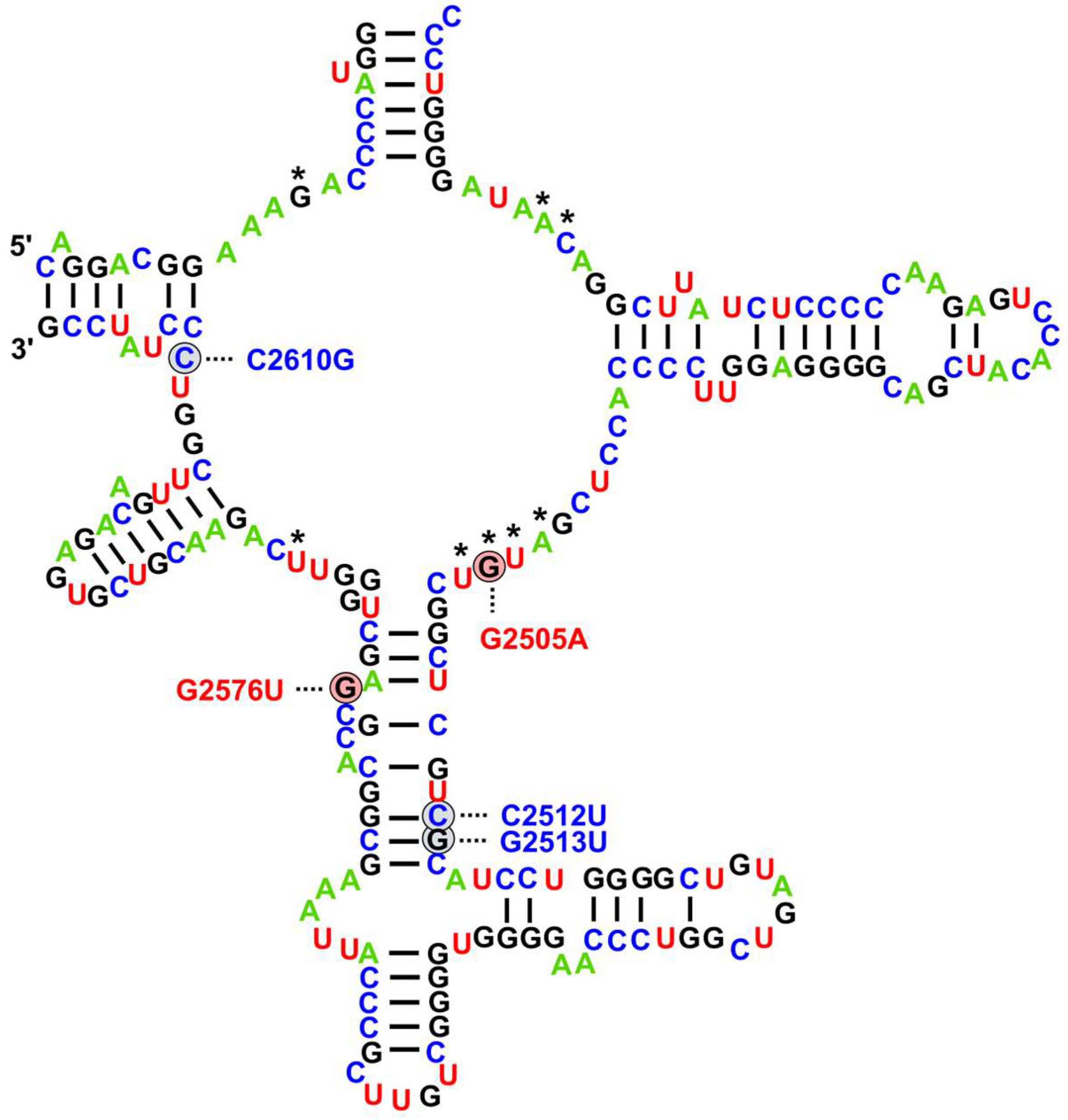
Figure 1. Linezolid resistance-associated mutations in enterococci affecting the peptidyl transferase loop of domain V in 23S rRNA. Base positions are referenced according to the Escherichia coli numbering. The nucleotides that contact directly with the linezolid are denoted by an asterisk. Mutations conferring resistance to linezolid are color-coded based on their significance: red for common variants with a well-established role, and blue for recently identified variants associated with advancements in sequencing technologies. Mutation positions are taken from Turner et al. [56].
Ijms 26 08207 g001
Figure 2. Geographical distribution of studies reporting sequenced genomes of linezolid-resistant E. faecium and/or E. faecalis strains harboring 23S rRNA mutations and/or mutations in genes encoding ribosomal proteins L3, L4, and L22.
Ijms 26 08207 g002
Figure 3. Geographical distribution of studies reporting sequenced genomes of linezolid-resistant E. faecium and/or E. faecalis strains harboring cfr, optrA or poxtA genes.
Ijms 26 08207 g003
Table 1. A compilation of studies utilizing whole-genome sequencing for the detection of 23S rRNA mutations associated with LNZ resistance in clinical Enterococcus spp. isolates.
CountryYearGenomes with 23S rRNA MutationsAdditional Resistance MechanismsSource
Australia2016–20211 LREfs with G2576U (3 loci affected)No Data[69]
Austria2014–201713 LREfm with G2576U and 3 of them also harbor A2598G variantNo Data[70]
Austria20171 LREfs with G388A/D130N (1 locus), T2802 C/I934M (1 locus) and T2838C/- (2 loci);
1 LREfs with G388A/D130N (3 loci), T2802 C/I934M (1 locus) and T2838C/- (2 loci)
optrA_4, T301C/F101L in L4;
optrA_3, T301C/F101L in L4
[71]
Belgium2013–20257 LREfm with G2576U1 LREFm with cfr[49]
Brazil20131 LREfm with G2576UNo Data[72]
Bulgaria20181 LREfs with G2576U (3 loci)
1 LREfs with C2163T (1 locus)
No Data
optrA
[73]
China-1 LREfs with G2576U (2 loci)No Data[74]
China2011–201516 LREfs with G2576U10 LREfs with optrA[75]
China20182 LREfs with novel mutationsNo Data[76]
Czech
Republic
20211 LREfm with G2576UNo Data[77]
Denmark2014–202352 LREfm with G2576U1 LREfm with cfr + poxtA[78]
Denmark2015–202231 LREfm with G2576U1 LREfm with optrA[79]
France2006–20164 LREfm with G2576U3 LREfm with optrA[80]
Germany2014–201896 LREfm with G2576U1 LREfm with poxtA[81]
Germany2007–20171 LREfs with G2576UoptrA[82]
Germany2013–20151 LREfm with G2576U (4 affected loci)
1 LREfm with G2576U (2 affected loci)
cfr(B)
cfr(B)
[83]
India20192 LREfm with G2592T2 LREfm with optrA[84]
Ireland2016–201919 LREfm with G2576U (1–5 affected loci)
2 LREfs with G2576U
1 LREfm with poxtA
No Data
[85]
Ireland2013–20141 LREfm with G2576U (4 loci affected)optrA, cfr, T150A in L3[86]
Latvia2021–20221 LREfm with G2576UNo Data[87]
Pakistan2021–20237 LREfm with G2576U6 LREfm with optrA[46]
South
Korea
2019–20204 LREfm with G2576UNo Data[88]
Spain2017–20181 LREfs with G2576UoptrA[89]
USA-3 LREfm with G2576U (2 × 3 loci affected and 1 × 2 loci affected)No Data[90]
USA2012–20131 LREfm with G2576U (1 locus affected)cfr(B)[91]
USA2009–20194 LREfm with G2576UNo Data[92]
USA20151 LREfm with G2576U (2–3 loci affected)No Data[93]
USA20174 LREfm with G2576UNo Data[47]
USA2013–20162 LREfm with G2576UNo Data[94]
USA2018–20193 LREfs with G2576UNo Data[51]
USA2015–201629 LREfm with G2576UNo Data[95]
ZAAPS
programme
-2 LREfm with G2576UNo Data[96]
LNZ: linezolid; LREfm: linezolid-resistant E. faecium; LREfs: linezolid-resistant E. faecalis.
Table 2. LNZ-resistant E. faecium isolates from Ireland with different copy numbers of the G2576U mutation—Egan et al. [85].
Isolate123456789101112131415161718
Sequence type80203787203203203171778778778978778911780808080
G2576 copy number233222451332423233
LNZ MIC (mg/L)
256

256

256

256
164848643232
256

256

256
32
256
641664
LNZ: linezolid; MIC: minimum inhibitory concentration.
Table 3. A compilation of studies utilizing whole-genome sequencing for the detection of missense mutations in the L3 and L4 ribosomal proteins associated with LNZ resistance in clinical Enterococcus spp. isolates.
CountryYearGenomes with L3 and/or L4 MutationsAdditional Resistance MechanismsSource
Austria20171 LREfm with T301C/F101L in L4
1 LREfm with T301C/F101L in L4 + C120T/- in L22
optrA + 23S mutations (see Table 1)
[71]
China2014–20181 LREfm with Ser77Thr in L22 No Data[101]
Germany2007–201713 LREfs with F101L in L4optrA[82]
Germany2013–20152 LREfm with 211GGT213
(glycine insertion) in L4
cfr(B)[83]
Ireland2013–20141 LREfm with T150A in L3G2576U (4 loci affected), optrA, cfr[86]
Italy20161 LREfs with 71Gly72 in L4No Data[102]
Scotland2014–20175 LREfs with T150A in L3 and F101L in L4
1 LREfs with T150A in L3 and F101L in L4
optrA
optrA + cfr(D)
[103]
Spain20231 LREfm with Lys68Glu in L4poxtA[104]
SENTRY Program2008–20164 LREfs with F101L in L4
1 LREfs with F101L in L4
optrA
optrA + cfr
[105]
LNZ: linezolid; LREfm: linezolid-resistant E. faecium; LREfs: linezolid-resistant E. faecalis.
Table 4. A compilation of studies utilizing whole-genome sequencing for the detection of cfr genes associated with LNZ resistance in clinical Enterococcus spp. isolates.
CountryYearGenomes Harboring cfrAdditional Resistance MechanismsSource
Belgium2013–20211 LREfm with cfr(B)G2576U[49]
China2011–20221 LREfm with cfr(D)optrA[116]
Denmark2014–20231 LREfm with cfr
1 LREfm with cfr
poxtA + G2576U
poxtA
[78]
Denmark2015–20221 LREfm with cfrBNo Data[79]
Germany2007–20171 LREfs with cfrNo Data[82]
Germany2013–20151 LREfm with cfr(B)
2 LREfm with cfr(B)
2 LREfm with cfr(B)
No Data
G2576U
211GGT213 insertion in the gene for L4
[83]
Ireland2016–20191 LREfm with cfr(D)optrA[85]
Ireland2013–20141 LREfm wit cfrG2576U (4 copies), T150A in L3,
optrA
[86]
Italy 2 LREfm with cfr(D)
1 LREfs with cfr(D)
optrA
optrA
[52]
Japan20171 LREfs with cfr(B)optrA[117]
Mexico2023–20242 LREfs with cfr(A)optrA[118]
Pakistan2021–20236 LREfm with cfr(D)
1 LREfm with cfr(D)
poxtA
optrA + poxtA
[46]
Scotland2014–20171 LREfs with cfr(D)optr(A) + T150A in L3 + F101L in L4[103]
South
Korea
2019–20203 LREfm with cfr(D)
1 LREfm with cfr(D)
poxtA
optrA + poxtA
[88]
Spain2017–20181 LREfs with cfr(D)optrA[89]
USA2012–20131 LREfm with cfr(B)No data[91]
SENTRY
Program
2008–20161 LREfs with cfr
1 LREfs with cfr
optrA
optrA + F101L in L4
[105]
LNZ: linezolid; LREfm: linezolid-resistant E. faecium; LREfs: linezolid-resistant E. faecalis.
Table 5. A compilation of studies utilizing whole-genome sequencing for the detection of optrA and poxtA genes associated with LNZ resistance in clinical Enterococcus spp. isolates.
CountryYearGenomes Harboring optrA/poxtAAdditional Resistance MechanismsSource
Argentina2016–202114 LREfs with optrANo Data[130]
Austarlia2016–202124 LREfs with optrANo Data[69]
Austria20172 LREfm with optrA23S rRNA mutations (Table 1) and L4 mutations (Table 3)[71]
Belgium2013–20213 LREfm with optrA
1 LREfm with optrA and poxtA
61 LREfs with optrA
2 LREfs with poxtA
No Data
No Data
No Data
No Data
[49]
Bulgaria20231 LEEfs with optrA23S rRNA mutation C2163T[73]
China2012–202111 LREfs with optrANo Data[131]
China2014–20176 LREfm with optrA
1 LREfm with optrA + poxtA
1 LREfm with poxtA
No Data
No Data
No Data
[101]
China 2011–20222 LREfm with optrA
1 LREfm with optrA
1 LREfm with poxtA
61 LREfs with optrA
No Data
cfr(D)
No Data
No Data
[116]
China2009–20131 LREfm with optrA
12 LREfs with optrA
No Data
No Data
[132]
China2022–20235 LREfs with optrANo Data[133]
China2011–201510 LREfs with optrA
13 LREfs with optrA
G2576U
No Data
[75]
China2018–202230 LREfs with optrANo Data[134]
Colombia2014–20196 LREFs with optrANo Data[135]
Denmark2014–20231 LREFm with poxtA
1 LREFm with poxtA
cfr
cfr + G3576U
[78]
Denmark2015–20223 LREfm with optrA
1 LREfm with optrA
No Data
G2576U
[79]
France2006–20163 LREfm with optrA
2 LREfm with optrA
3 LREfs with optrA
G2576U
No Data
No Data
[80]
France2016–202010 LREfm with poxtA
1 LREfs with poxtA
No Data
No Data
[128]
Germany2014–20181 LREfm with poxtA
1 LREfm with poxtA
G2567U
No Data
[81]
Germany2007–20171 LREfm with optrA
1 LREfm with optrA
13 LREfs with optrA
No Data
G2576U
No Data
[82]
Greece20181 LREfs with optrANo Data[136]
India20191 LREfm with optrA
1LREfm with optrA 2 copies
G2592T
G2592T
[84]
Ireland201919 LREfm with optrANo Data[137]
Ireland2016–20191 LREfm with optrA + poxtA
1 LREfm with optrA
1 LREfm with optrA
9LREfm with poxtA
13 LREfs with optrA
10 LREfs with poxtA
No Data
cfr(D)
No Data
No Data
No Data
No Data
[85]
Ireland2013–20141 LREfm with optrAG2576U (4 copies), T150A in L3, cfr[86]
Italy 2 LREfm with optrA
1 LREfs with optrA
cfr(D)
cfr(D)
[52]
Japan20171 LREfs with optrAcfr(B)[117]
Japan20211 LREfs with optrANo Data[138]
Mexico2023–202422 LREfs with optrA
2 LREfs with optrA
No Data
cfr(A)
[118]
Pakistan2021–20236 LREfm with optrA
1 LREfm with optrA + poxtA
6 LREfm with poxtA
G2576U
cfr(D)
cfr(D)
[46]
Poland20201 LREfm with poxtANo Data[139]
Reunion
Island
2015–20198LREfm with optrANo data[140]
Saudi
Arabia
2014–20151 LREfs with optrANo Data[141]
Scotland2014–20175 LREfs with optrA
1 LREfs with optrA
T150A in L3 + F101L in L4
cfr(D) + T150A in L3 + F101L in L4
[103]
South
Korea
2017–20192 LREfs with optrANo data[142]
South
Korea
2019–20202 LREfm with optrA
1 LREfm with optrA + poxtA
3 LREfm with poxtA
15 LREfs with optrA
3 LREfs with poxtA
No Data
cfr(D)
cfr(D)
No Data
No Data
[88]
Spain2016–20175 LREfs with optrANo Data[143]
Spain2017–201811 LREfs with optrA
1 LREfs with optrA
1 LREfs with optrA
No Data
cfr(D)
G2576U
[89]
Spain20232 LREfm with poxtA
1 LREfm with poxtA
No Data
Lys68Glu in L4
[104]
Spain20183 LREfs with optrANo Data[144]
Sweden2017–20201 LREfm with optrANo Data[145]
USA2013–20161 LREfs with optrANo Data[94]
USA2018–20191 LREfm with optrA
1 LREfs with optrA
No Data
No Data
[51]
SENTRY
Program
2008–20164 LREfs with optrA
1 LREfs with optrA
1 LREfs with optrA
F101L in L4
cfr + F101L
cfr
[105]
ZAAPS
Program
-8 LREfs with optrANo Data[96]
LNZ: linezolid; LREfm: linezolid-resistant E. faecium; LREfs: linezolid-resistant E. faecalis.
Table 6. A compilation of LNZ resistance mechanisms detected in clinical Enterococcus spp. isolates.
MechanismsInvolved Mutations/DeterminantsAssociated
Phenotypes
Geographic Distribution of WGS Studies and Clinical Significance
Modifications of LNZ binding pocket23S rRNA mutations; G2576U most frequentLNZ MICs: 8– ≥ 256, generally higher with more 23S rRNA loci affected, but many exceptions;
TDZ MICs: 4–8× lower, except T2504A (MIC 32)
Asia, Australia, Europe, North America, and South America; mainly LREfm; non-transferable
Modifications of LNZ binding pocketL3, L4 and L22 ribosomal proteinsLNZ MICs ≤8 in most cases;
TDZ—important in staphylococci
Asia, Europe; mainly LREfs; non-transferable
Target modificationcfr genesUnclear significance in LRE; low LNZ MICs;
TDZ unaffected, clinically important in staphylococci
Asia, Europe, North America; mainly LREfm; transferrable—reservoir for staphylococci
Ribosome protectionoptrA and poxtA variantsVarying MICs dependant on the variant;
TDZ MICs 4–8× lower
Global dissemination; mainly LREfs; easily transferrable (especially optrA); optrA is the most widespread LNZ resistance mechanism in clinical LREfs
LNZ: linezolid; TDZ: tedizolid; MIC: minimum inhibitory concentration; LREfm: linezolid-resistant E. faecium; LREfs: linezolid-resistant E. faecalis.
Disclaimer/Publisher’s Note: The statements, opinions and data contained in all publications are solely those of the individual author(s) and contributor(s) and not of MDPI and/or the editor(s). MDPI and/or the editor(s) disclaim responsibility for any injury to people or property resulting from any ideas, methods, instructions or products referred to in the content.