Abstract
Nucleic acid modifications play important roles in biological activities and disease occurrences, and have been considered as cancer biomarkers. Due to the relatively low amount of nucleic acid modifications in biological samples, it is necessary to develop sensitive and reliable qualitative and quantitative methods to reveal the content of any modifications. In this review, the key processes affecting the qualitative and quantitative analyses are discussed, such as sample digestion, nucleoside extraction, chemical labeling, chromatographic separation, mass spectrometry detection, and data processing. The improvement of the detection sensitivity and specificity of analytical methods based on mass spectrometry makes it possible to study low-abundance modifications and their biological functions. Some typical nucleic acid modifications and their potential as biomarkers are displayed, and efforts to improve diagnostic accuracy are discussed. Future perspectives are raised for this research field.
1. Introduction
Nucleic acid modifications play important roles in regulating gene expression, cell differentiation, and individual development [1,2]. These modifications do not change the gene sequence, but can expand genetic information [3]. Unlike gene sequences, these modifications dynamically change throughout an individual’s lifecycle thanks to various environmental factors. With the advance of research, more and more diseases have been proven to be related to changes caused by nucleic acid modification, and have been considered as potential targets for precise diagnosis and personalized treatment [4].
Commonly, a complete workflow for nucleic acid modification study can be divided into the following steps: (a) choose a biological model that may be influenced by nucleic acid modification; (b) determine the content of any nucleic acid modifications in the biological model using qualitative and quantitative strategies, and estimate the correlation between modified content and biological function; (c) screen target genes containing functional modification using sequencing technologies; (d) verify the functions of target genes in biological processes using molecular biology methods, and reveal the mechanisms of these functions; (e) verify the clinical performance and potential therapy among extensive clinical samples; (f) use the discovered nucleic acid modification as the biological model for diagnosis and therapy. It can be seen that the precise qualitative and quantitative analyses of nucleic acid modification provide almost the earliest direct evidence, and this result plays a strong guiding role for the subsequent research. Therefore, it is necessary to develop sensitive and reliable qualitative and quantitative methods.
Here, we will introduce DNA and RNA modifications, especially the well-known modifications. Then, we will introduce the detection strategies for nucleic modifications, focusing on mass spectrometry-based analytical methods. We will discuss the methods to enhance sensitivity based on appropriate enzymatic digestion and chemical labeling, as well as the latest research progress in disease diagnosis using nucleic modifications as biomarkers. We will also provide future perspectives for this research field.
2. Nucleic Acid Modifications
2.1. Modifications of DNA
As early as 1948, modified cytosine was first discovered on calf thymus DNA, and was inferred to be 5-methylcytosine (5mC) [5]. In 1975, researchers recognized that epigenetic information could be carried through chemical modification of cytosine [6,7]. As research continues, over 50 types of DNA modifications have been discovered in mammals, plants, and microorganisms [3,8]. The related chemical and biological properties of these DNA modifications are systematically organized in an open-source database, DNAmod (https://dnamod.hoffmanlab.org, accessed on 30 January 2024) [8]. After seven decades of development, the complete pathway of cytosine methylation and demethylation was discovered, including 5mC, 5-hydroxymethylcytosine (5hmC), 5-formylcytosine (5fC), 5-carboxylcytosine (5caC), and an abasic site (AP site) [9,10,11,12,13,14,15]. Other important DNA modifications include uracil (dU), 5-hydroxymethyluracil (5hmU), 5-formyluracil (5fU), β-D-glucosyl-5-hydroxymethyluracil (base J), N6-methyladenine (6mA), N6-hydroxymethyladenine (6hmA), N6-carbamoylmethyladenine (ncm6A), 2-aminoadenine (m2A), 8-oxo-7,8-dihydroguanine (8-oxo-G, OG), and 7-methylguanine (7mG) [3,8,16] (Figure 1A). Among them, the level of 5mC was reported as 2–7% of the genomic cytosine [17], while 5hmC was determined to be about 0.03–0.7% [15,18]. The contents were found to be 20 5fC and 3 5caC in every 106 C, respectively, which nearly touches the sensitivity limit of direct mass spectrometry detection [10]. 5hmU, 5fU, and OG were found to be at either one or several bases per 106 C level [19,20,21]. It can be seen that the discovery of modifications is related to the nucleic acid’s natural content, which indicates that improving the sensitivity of the method is of great importance for the discovery and research of new modifications [18].
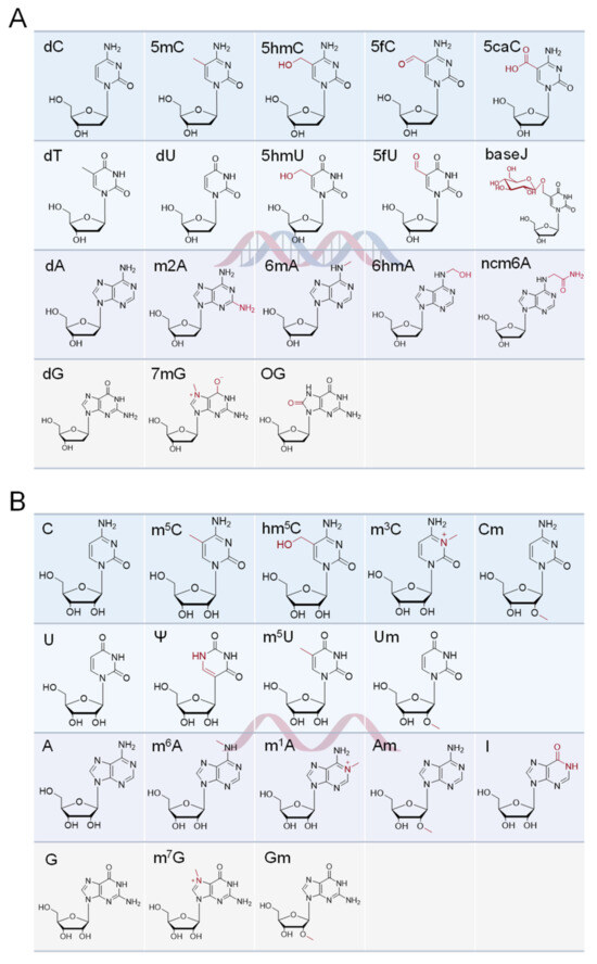
Figure 1.
The natural nucleosides and some of the most studied modified versions found in (A) DNA and (B) RNA.
2.2. Modifications of RNA
Compared to DNA modification, there are many more types of RNA modification. Over 170 RNA modifications have been found so far, which exist in almost all types of RNA, including messenger RNA (mRNA), transfer RNA (tRNA), ribosomal RNA (rRNA), and small nuclear RNA (snRNA) [22]. N6-methyladenine (m6A) is one of the most abundant RNA modifications in eukaryotes, with a content of 0.1–0.4% of total adenine, which is similar to pseudouridine (Ψ) content (0.2–0.6% of total uridine) [23,24]. Other important RNA modifications include N6-hydroxymethyladenine (hm6A), N1-methyladenosine (m1A), inosine (I), 2′-O-methyladenosine (Am), 5-methyluridine (m5U), 2′-O-methyluridine (Um), 5-methylcytosine (m5C), 5-hydroxymethylcytosine (hm5C), 3-methylcytosine (m3C), 2′-O-methylcytidine (Cm), 7-methylguanosine (m7G), and 2′-O-methylguanosine (Gm), et al. (Figure 1B). The related chemical and biological properties of these DNA modifications are systematically organized in an open-source database, MODOMICS (https://iimcb.genesilico.pl/modomics/, accessed on 30 January 2024), including LC–MS information, pathways, sequences, and related diseases [25,26].
3. Qualitative and Quantitative Analysis
The reported methods for nucleic acid modification quantification include thin-layer chromatography (TLC) [10], liquid chromatography (LC) or capillary electrophoresis (CE) based on optical detection [27,28,29], gas chromatography–mass spectrometry (GC–MS) [30], liquid chromatography–mass spectrometry (LC–MS), surface-enhanced Raman scattering (SERS) spectroscopy [31], immunoassay, and biosensing-based methods [32,33,34,35,36,37,38]. MS-based methods have been considered as the main analytical tool for nucleic acid modification quantification due to their wide applicability, excellent sensitivity, and wide linear range, providing comparative global compositional analysis of different biological samples. However, the low abundance of modifications limits the discovery of new modifications and the research of nucleic acid modifications as biomarkers or indicators. The main challenge is to establish simple, fast, and ultrasensitive global quantification methods to obtain more precise information for limited clinical samples. Effective sample preparation strategies, separation and detection processes, and data processing methods are important for accurate qualitative and high-sensitivity quantitative analysis, which help researchers to obtain reliable and comprehensive modification information (Figure 2).
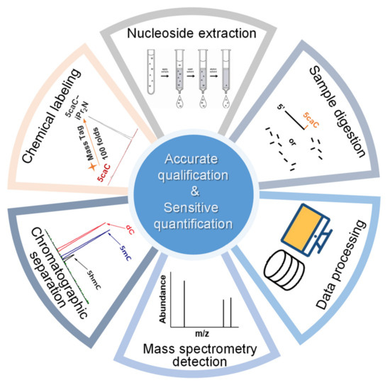
Figure 2.
Process of mass spectrometry-based method for accurate qualification and sensitive quantification of nucleic acid modification.
3.1. Preparation of Biological Samples
3.1.1. Hydrolysis
In mass spectrometry-based nucleic acid modification analysis, researchers first extract nucleic acids from biological samples, and use nucleases to hydrolyze the chain into individual nucleotides. Considering that the high hydrophilicity and negative charge of phosphate would decrease the ionization efficiency of mass spectrometry, phosphatase is used to remove phosphates and form deoxyribonucleosides or ribonucleosides. The classical digestion method developed by Crain and colleagues was divided into two steps: nuclease P1 or nuclease S1 first digests denatured DNA or RNA at 50 °C under pH 5 buffer, then the digestion solution is adjusted to pH 8, and phosphodiesterase and alkaline phosphatase are added to remove phosphates at 37 °C sequentially [39,40,41,42]. In this two-step method, nuclease P1 and nuclease S1 only recognize RNA or single-stranded DNA, and it is necessary to boil genomic DNA at 100 °C for denaturing first [43]. Quinlivan et al. developed a one-step digestion method, performed under an appropriate pH, by replacing nuclease P1 with endonuclease from Serratia marcescens, Benzonase, or DNase I, which has been widely commercialized owing to relatively fewer processing steps and a lower time consumption and dilution ratio [43,44]. In this one-step method, endonuclease from Serratia marcescens and Benzonase recognize single- and double-stranded DNA and RNA, and as the denaturing step is not essential, RNA samples could be hydrolyzed through the same workflow [43]. Directly hydrolyzing DNA or RNA into bases through heating under acidic conditions is also an alternative choice [45,46,47].
The aim of hydrolysis is to release nucleotides completely with an unbiased approach and reduce additional artificial modification and the loss of natural modification. The hydrolysis method should be carefully chosen for certain modifications, especially for low-abundance modifications, which might cause more bias in quantification results. Weinfeld et al. reported that several enzymes worked through stacking with aromatic nucleic acid bases [48]. Yuan et al. first reported the resistance of 5caC to phosphodiesterase I (PDE1), and found that commercial one-step digestion mix was suitable for 5caC hydrolysis [49]. Chu et al. reported that PDE1 released m7G fully, both in mRNA sequence and in the 5′ cap; contrarily, S1 nuclease released the internal m7G with high activity but released m7G from the 5′ cap with much lower activity [50]. Different digestion methods were recommended for studies of 2′-O-methylated ribonucleosides [51], RNA 5′ caps [52], et al. Besides the base structure, sequence specificity also needs to be considered; for instance, the formation of G-quadruplex (G4) inhibits the cleavage efficiency of nuclease [53,54]. Caution also needs to be used regarding digestion conditions during special modification quantification. Matuszewski et al. reported that hydantoin N6-threonylcarbamoyladenosine (ct6A) was converted to stereoisomer under mild alkaline conditions in several minutes [55]. The extreme pH, radical species, buffer types, and deaminase contamination may cause changes in modification type and intensity, including A to I, G to OG, m1A to m6A, and 5-methoxycarbonylmethyl-2-thiouridine (mcm5s2U) to 5-methoxycarbonylmethylisocytidine (mcm5isoC) artifacts, which can be avoided by adding metal-chelating reagent, antioxidant, or deaminase inhibitor and treating in mild conditions [40,44,56,57,58].
In addition, a simpler workflow and shorter preparation time are also desirable goals. Lai et al. reported that appropriate concentration of divalent Mg2+ and Ca2+ promoted DNA digestion catalyzed by the DNase set, but high concentration of monovalent Na+ and K+ inhibited DNA digestion [59]. Engineered nuclease mutant was produced for the hyperactive and unbiased release of DNA modifications, which better overcame the inhibition of buffer conditions [60,61,62]. To shorten the preparation time, Yin et al. designed a cascade bioreactor by immobilizing nucleases on a capillary silica monolith, and DNA was completely digested within 10 min [63].
3.1.2. Nucleoside Extraction
The purpose of nucleoside extraction is to improve separation and detection efficiency and obtain higher signals of the analytes by removing salts, proteins, and uninterested nucleosides, especially large amounts of normal nucleosides, from the matrix. The initial extraction methods enrich free nucleosides from biological samples such as serum and urine, normally based on hydrophobic or hydrophilic interactions, ion exchange, and affinity with 1,2-cis-diol compounds with commercial materials, such as HLB [64,65] and WCX [66], as well as new materials such as graphene [67], polymers [68], metal oxides [69,70,71], and boronate-decorated substrates [72,73,74], which are also used for the extraction of nucleoside products after enzymatic digestion to further increase signal response. The shapes of substrate and extraction devices influence extraction efficiency [75,76]. However, most of the above methods either lack selectivity for specific types of nucleoside modification or discriminate against DNA nucleosides and 2′-O-methylated ribonucleosides.
In order to extract specific structural nucleosides, various novel functional materials have been designed and synthesized. Ma et al. developed a pH-response covalent organic framework decorated with gold nanoparticles as a linker and glutathione as a functional group, which selectively captured m1A via electrostatic interaction and released it in an acidic condition to prevent m1A’s rearrangement to m6A [77]. Wang et al. developed cyclodextrin-based porous liquids, which showed a chiral recognition and separation ability of D-type and L-type pyrimidine nucleosides [78].
3.1.3. Chemical Labeling
The quantification of ultra-rare nucleic acid modifications always needs a large amount of sample to gain enough response from detectors. In contrast, the capacity of chromatographic columns and the linear range of mass spectrometry seem to be limited to the direct quantification of multiple nucleosides with large differences in content. Moreover, limited biological and clinical samples mean that direct detection is far from meeting the requirements of real research. Therefore, chemists have developed brilliant labeling strategies based on chemical and enzymatic catalyzed reactions to improve detection sensitivity. Table 1 summarizes the application of labeling reagents in modified nucleoside detection.
In one of the strategies, labeling reagents react indiscriminately with all nucleosides. Pertrimethylsilylation, permethylation and O-isopropylidenation are achieved by adding N-methyl-N-(trimethylsilyl)trifluoroacetamide (MSTFA), bis(trimethylsilyl)trifluoroacetamide (BSTFA), iodomethane, and acetone. These methods are usually simple, robust, and low-cost, and are mainly followed by GC–MS detection [30,71,79,80,81]. 8-(diazomethyl)quinoline (8-DMQ), dimethyl-p-phenylenediamine (DMPA), and 2-(diazomethyl)-N-methyl-N-phenyl-benzamide (2-DMBA) are synthesized and used to react with phosphate groups to enhance the mass spectrometry response of nucleoside triphosphates and nucleotides extracted from endogenous metabolites [82,83,84].
In other designs, labeling reagents only react with specific modified nucleosides. α-haloketones, including 2-bromo-1-(3,4-dimeth oxyphenyl)-ethanone (BDMOPE), 2-bromo-1-(4-methoxyphenyl)-ethanone (BMOPE), 2-bromo-1-(4-diethylaminophenyl)-ethanone (BDEPE), and 2-bromo-1-(4-dimethylamino-phenyl)-ethanone (BDAPE), react with the N3 and N4 positions of cytosine to form the cyclic derivatives [85,86]. Several more bases other than cytosine were found to be derived from α-haloketone reagent [87]. Hydrazine group-based reagents display high reactivity and selectivity to react with different modified nucleosides under different conditions. Yu et al. and Yuan et al. developed a series of reaction routes through labeling the aldehyde group with hydrazino-s-triazine-based reagents (Me2N, Et2N, and i-Pr2N) [88,89] directly, labeling the carboxyl group under catalysis, and converting the hydroxyl group to the reactive aldehyde group through oxidation before labeling (5hmC to 5fC, and 5hmU to 5fU), which increased the sensitivity of 5fC, 5caC, 5hmC, 5fU, and 5hmU by up to 850 folds (Figure 3) [90,91,92]. Other reported hydrazine-based labeling reagents include Girard’s reagents (GirP, GirT, GirD and 4-APC) [93,94] and rhodamine B hydrazine [95]. In addition, cationic xylyl-bromide (CAX-B) [96], N-dimethyl-amino naphthalene-1-sulfonyl chloride (Dns-Cl, Dens-Cl) [97], hydroxyl amine-based reagents [98], and N-cyclohexyl-N′-β-(4-methylmorpholinium) ethylcarbodiimide p-toluenesulfonate (CMCT) [99] play important roles in labeling and improving the sensitivity of nucleic acid modifications. Enzyme-based methods possess good specificity of recognition and enrichment, although they may change the original structure of the nucleosides. Tang et al. reported a method which covalently added a glucosyl group to 5-hmC using T4 β-glucosyltransferase and enriched the product by hydrophilic interaction [100]. Hu et al. reported an enrichment method for 5′ NAD+ modification, in which the NAD+ group was replaced with a click reaction group through the catalysis of adenosine diphosphate ribosylcyclase (ADPRC), and the product was enriched for quantification and sequencing [101].
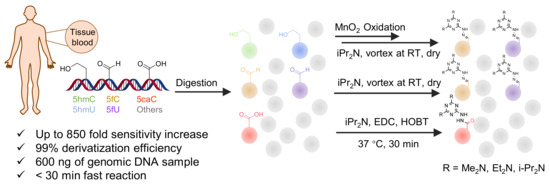
Figure 3.
Workflows of derivatization of five types of DNA modification (5hmC, 5fC, 5caC, 5hmU, and 5fU) by hydrazino-s-triazine-based reagents.
Some general characteristics of well-designed labeling reagents include an efficient and selective reaction group, a hydrophobic backbone group, and an easily charged tertiary or quaternary amine group. These function groups block the negatively charged and hydrophilic structure, enhance the organic solvent proportion during LC, and make the targets easy to distribute on the surfaces of droplets during ESI, resulting in the increase of spraying ability, ionization efficiency, and mass spectrometry response. In addition to ultrasensitive determination, labeling reaction is also important for relative quantification and structural identification. Several reagents with stable isotope labeling or similar structures were synthesized and applied in pairs [84,97].

Table 1.
Chemical labeling strategies in high-sensitivity detection of nucleic acid modifications.
Table 1.
Chemical labeling strategies in high-sensitivity detection of nucleic acid modifications.
| Labeling Reagent | Structure | Target Nucleoside | Reaction Condition | LOD | Sensitivity Increase Fold | Sample Consumption | Ref. |
|---|---|---|---|---|---|---|---|
| MSTFA | 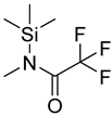 | all | 10 μL of methoxyamine hydrochloride as 20 mg/mL solution in pyridine, 90 μL of MSTFA | ND. | ND. | metabolites in 25 μL blood plasma, 5 × 10⁶ cells, or 5 mg tissues | [30] |
| acetone |  | ribonucleosides | 400 μL of acetone with p-toluene sulfonic acid (1 mg/mL), 50 °C, 2 h | 0.6–6.5 fmol | 7–30 folds | metabolites in 100 μL of urine | [71] |
| BSTFA |  | all | 120 μL of BSTFA, 70 °C, 2 h | ND. | ND. | 10 mg of freeze-dried leaves | [79] |
| acetone |  | ribonucleosides | 600 μL of acetone, 6 μL of HClO4, vortex for 30 s, −20 °C for 30 min | 0.026–0.16 ng/mL, 10 μL | ND. | metabolites in 100 μL urine | [80] |
| iodomethane-d3 |  | all | iodomethane-d3, on beads, room temperature, 10 min | 10 fmol/μL, 2 μL | ND. | 1 µg of purified DNA or RNA | [81] |
| 8-DMQ | 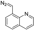 | nucleoside triphosphates | 50/1 molar ratio of 8-DMQ/analyte in 50 mM borate buffer (pH 6.9, 160 μL) with DMSO (40 μL), 25 °C,10 min | 0.4–1.3 fmol | 56–137 folds | metabolites in 1.0 × 10⁷ cells | [82] |
| DMPA |  | nucleotide | molar ratios of DMPA and EDC over nucleotides were set as 40,000 and 5000, with 100 μL of imidazole solution (1 mM, pH 6.0), 50 °C, 1.5 h | 0.12–0.47 fmol | 88–372 folds | metabolites in urine, tissue and cell line samples | [83] |
| 2-DMBA, d5-2DMBA | 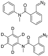 | nucleotides, nucleoside diphosphates, nucleoside triphosphates | 200 μL of 250 mg/L 2-DMBA in pH 7.0 borate buffer, 30 °C, 30 min | 0.07–0.39 fmol | 17–174 folds | metabolites in 20 mg of tissue and cell line samples | [84] |
| BDAPE |  | 5mC, 5hmC, 5fC, 5caC | 4 mM of BDAPE in 200 μL of ACN using 4 mM Et3N as the catalyst, 60 °C, 6 h | 0.06–0.23 fmol | 35–123 folds | 10 μg of genomic DNA | [85] |
| BDMOPE, BMOPE, BDEPE | 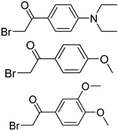 | m5Cm, hm5Cm, f5Cm, ca5Cm | 6 mM BDMOPE and 6 mM triethylamine, 60 °C, 6 h | 0.06–0.22 fmol by BDMOPE labeling | 46–462 folds | 10 μg of total RNA and small RNA | [86] |
| BrDPE |  | C, dC, A, dA, G, dG, T, dT, U | BrDPE/analyte ratio 200/1 and 4 mM triethylamine in 125 μL solvent, 40 °C, 3 h | 0.3–12.5 fmol | 31–107 folds | metabolites in 0.2 g of dry sample | [87] |
| Me2N, Et2N, and i-Pr2N | 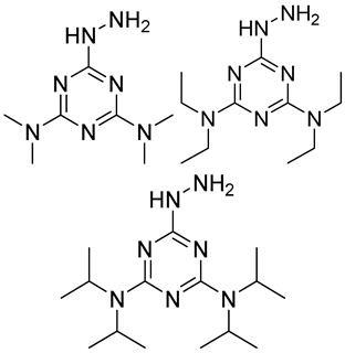 | 5fC, 5caC | 5 mM labeling reagents with 1% HAc in 20% MeOH votex for 10 s for 5fC; labeling reagents in 20 μL 50% ACN, 10 μL 4 mg/mL HOBT and 10 μL 50 mg/mL EDC, 37 °C, 30 min for 5caC | 10–25 amol | 100–125 folds | 600 ng of genomic DNA | [90] |
| i-Pr2N | 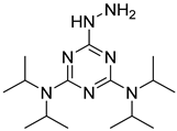 | 5hmC | 5 mg of MnO2 in 20 μL reaction volume, 50 °C, 1 h; 5 μL of oxidation product, 1 μL of HAc, 14 μL of 50 mM i-Pr2N solution, vortex and dry | 14 amol | 178 folds | 0.6–2.4 ng of cell-free DNA | [91] |
| i-Pr2N | 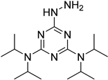 | 5fU, 5hmU, 5fC, 5hmC | 5 mg of MnO2, 2 μL FA, 50 °C, 1 h; 1 mg/mL i-Pr2N and 2 µL HAc, vortex | 26.0–44.4 amol | 275–850 folds | 2 μg of genomic DNA | [92] |
| GirP, GirT and 4-APC | 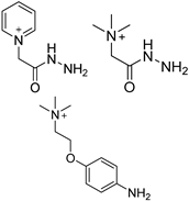 | 5fdC, 5frC, 5fdU, 5frU, 5frCm, 5frUm | GirP/analyte ratio 50/1, 30 °C, 5 min | 0.03–0.05 fmol | 115–880 folds | mixture of 10 μg genomic DNA and 10 μg total RNA | [93] |
| GirP, GirT and GirD | 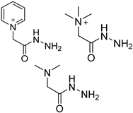 | 5fC, 5caC | GirD/analyte ratio 50/1–150/1, 40 °C, 5–40 min | 0.03–0.42 fmol | 52–260 folds | 20 μg of genomic DNA | [94] |
| rhodamine B hydrazine | 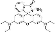 | 5fC | 10 μL of 5 mM labeling reagent in MeOH, 0.2 μL HAc, vortex and dry | 3 amol | 300 folds | total RNA in cell line sample | [95] |
| CAX-B | 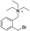 | bases that have active hydrogen | CAX-B in 50% ACN (20 mg/mL), with Et3N(20 μL/mL), was mixed 1:1 with the sample solution, 45 °C, 2 h | 160 amol thymidine | ND. | ND. | [96] |
| Dns-Cl, Dens-Cl | 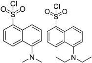 | C, dC, 5mdC, m5C, A, m1A, m6A | 100 μL of reaction buffer (pH 11) and 100 μL of Dns-Cl, 30 °C, 1 h | 0.001–0.01 μg/mL, 5 μL | 1.6–400 folds | metabolites in 106 cells | [97] |
| hydroxyl amine |  | AP sites, βE sites | 1.5 mM derivatization reagents in HEPES (20 mM, pH = 7.5) and Na2EDTA (0.1 mM), 37 °C, 40 min | 0.11 fmol | ND. | 5–20 μg of genomic DNA | [98] |
| CMCT |  | Ψ, U, m5U, m6U, mcm5U, hm5U, m1Ψ, mo5U | 50 mM CMCT in borate buffer (50 mM, pH 8.5), 40 °C, 14 h | 0.29–2.20 fmol | 6–1408 folds | 500 ng of mRNA | [99] |
ND, not determined. ACN, acetonitrile; βE sites, β-elimination sites; EDC, N-(3-(Dimethylamino)propyl)-N′-ethylcarbodiimide hydrochloride; Et3N, trimethylamine; FA, formic acid; HAc, acetic acid; HEPES, 2-[4-(2-hydroxyethyl)piperazin-1-yl]ethanesulfonic acid; HOBT, 1-hydroxybenzotriazole hydrate.
3.2. Chromatography-Coupled Mass Spectrometry Technique
3.2.1. LC–MS
Compared with nucleic acid determination based on immunoassay and optical detection, the liquid chromatography–mass spectrometry-based method provides better accuracy for qualification and higher sensitivity for quantification, better robustness for different sources of samples, and richer information regarding the discovery of unknown modifications, which is why it has become the most widely used method currently. The first discovery of 5hmC in mammalian cells was achieved using HPLC-MS in 2009 [9,15], and further studies have been performed continuously over the past decade, including the identification of new modifications [10,14,102]. In order to improve the detection efficiency, researchers focused on the improvement of the mass spectrometry mode, ionization mode, separation mode, and buffer addition, reducing the sample consumption to limited numbers of cells or even single cells.
With the development of mass spectrometry technology, the multiple reaction monitoring (MRM) mode of tandem mass spectrometry (MS/MS) was used for nucleic acid modification detection, and the limit of quantification (LOQ) of 5mC was 40 fmol in the report [41]. Targets were selected by precursor ions and fragment ions stepwise in the MRM mode, which improved the accuracy of structural identification and reduced the background, effectively improving the sensitivity. Multistage MS (MS/MS/MS) was introduced during modification identification to supplement MS/MS, especially for isomers which share precursor ions and fragment ions, such as m3U and m5U, or m1A and m6A [103,104]. The process of nucleoside electrospray ionization (ESI) was estimated, and the sensitivity was found to be limited by the formation of dimers, ion adducts, and in-source fragmentation, which could be prevented through the optimization of pipeline material, gas flow, heating temperature, collision energy, LC elution buffer, and appropriate derivatization reagent [95,105].
Since the development and commercialization of nanoliquid chromatography (nanoLC) and nanospray ESI, an extremely low flow rate of ~20 nL/min has been obtained, which helps to enhance desolvation, ionization efficiency, and the tolerance to salt, and greatly improves the sensitivity of nucleic acids [106,107,108]. Two-dimensional nanoLC involves a pre-column before the separation column. The nucleosides are trapped and concentrated on the pre-column, and then they enter the analysis column for separation, which is caused by gradually changing the elution solution [103,109,110,111]. The column stationary phases are also critical to separation [112], and are often replaced with novel materials that interact with nucleosides for online enrichment and improvement of separation [73,113].
Reasonable selection of the chromatographic mode can improve resolution and sensitivity, and reduce the signal overlap. The earliest research used reverse phase liquid chromatography (RP-LC) to separate nucleosides based on the C18 stationary phase [41]. Amide, perfluorinated phenyl (F5), and T3 bonding analytical chromatography columns were compared for chromatography separation [114]. Researchers compared different types of reverse phase stationary phases and introduced hydrophilic interaction liquid chromatography (HILIC), considering the relatively high hydrophilicity of most nucleosides [47,115]. The interaction between the target and the stationary phase and the detection sensitivity can be improved by adding formic acid [105], ammonium salts [64], nonafluoropentanoic acid [116], and malic acid [67,117] to the mobile phase.
3.2.2. CE–MS
Capillary electrophoresis is a separation technique driven by a high-voltage electric field in a capillary, which separates charged particles based on their mobility. It has higher separation efficiency, faster analysis speed, and lower sample consumption compared to liquid chromatography, and is especially suitable for the separation of polar nucleoside molecules. Early researchers used CE–UV to achieve the separation and detection of C and 5mC within 1.5 min [118,119]. Considering that the sensitivity of UV detectors was relatively low, fluorescence labeling methods were developed for highly sensitive detection using laser-induced fluorescence (LIF) detectors [120].
With the interface technology of capillary electrophoresis–mass spectrometry (CE–MS) becoming more mature [121,122,123,124,125,126], stability and sensitivity has become good enough to be applied to modified nucleoside detection [127,128]. Yuan et al. and Yu et al. developed ultrasensitive and simultaneous determination methods for DNA and RNA modified nucleosides. A sheathless interface prevented the dilution effect in these reports, a limit of detection (LOD) down to 2.5 amol was achieved, and sample consumption was reduced to a limited number of cells [129,130]. Lechner et al. analyzed RNA modifications at both the nucleoside level and the oligonucleotide level by using CE–MS, reached 68–97% sequencing coverage of the RNA, and separation of four methylated guanosine isomers (1-methylguanosine; N2-methylguanosine; Gm and m7G) was performed [131].
3.2.3. Other Mass Spectrometry-Based Techniques
Besides ESI and nanoESI introduced by LC and CE, electron ionization (EI) and chemical ionization (CI) combined with GC were also widely used [79,132,133], although reports suggested that high temperatures might cause changes to the target structure [134]. Direct injection [109] and ambient ionization, such as direct analysis in real time (DART) [135,136], create advantages for analysis speed. Ion mobility spectrometry (IMS) separates ions based on collision cross-sections in the millisecond time scale, and IMS has a sensitivity of 15 pmol of adenosine, displaying a potential ability to separate isomeric nucleotide and nucleoside variants in complex samples based on subtle differences in ion mobility behaviors, which is another dimension in addition to mass-to-charge ratios [137,138,139,140].
4. Data Analysis
Effective data processing helps to identify the structure of the analytes accurately, quantify the content of the analytes precisely, and discover unknown modifications, and it makes simultaneous and automated analyses possible.
In order to determine the analytes and avoid the interference from similar molecular weight substances and isomers, the following methods are usually used: (a) Use high-quality resolution mass spectrometry. Accurate mass-to-charge ratio excludes other types of molecules with similar molecular weights [109]. (b) Analyze the structure of secondary fragments. Isomers usually produce different fragment ions through tandem mass spectrometry, and multistage mass spectrometry is also used [136]. The structural analysis of fragments is also performed by comparing between naturally modified nucleosides and stable isotope-labeled nucleosides [141]. Chen et al. discovered that in-source fragmentation usually occurs in glycosidic bonds, and they found a correlation between glycosidic bond length and cleavage ratio through theoretical calculations, and proposed a qualitative method based on the mass spectrometry of parent ions and in-source cleavage fragments [95]. (c) Compare chromatographic retention times. Gonzalez et al. found that nucleoside retention times of chromatography follow a certain regular pattern according to the hydrophilicity, hydrophobicity, and other properties of nucleosides, which promotes the accuracy of qualification [104].
In order to process data in batches automatically for discovering and quantifying modified nucleosides, several types of software have been developed and applied. Commercial software, such as Compound Discoverer 3.0, is used to search for modified nucleosides using metabolite analysis workflows [142]. Specialized software, such as Nucleos’ID (https://github.com/MSARN/NucleosID, accessed on 30 January 2024) and NuMo Finder (https://github.com/ChenfengZhao/NuMoFinder, accessed on 30 January 2024), has been developed based on the retention time of liquid chromatography and capillary electrophoresis, as well as the mass of precursor ions and fragment ions provided in the database [81,143]. Nucleosides are classified into networks based on fragments through the establishment of a mass spectrometry database, which contributes to the discovery of unknown modifications [144]. These schemes are also used as part of mass spectrometry-based oligonucleotide sequencing [145,146]. The development of artificial intelligence (AI) brings more potential tools for nucleic acid modification data analysis [147]. The preparation and updating of databases are of great importance for automated processing engines, and MODOMICS and DNAmod are the most commonly used databases that contain nucleoside mass spectrometry information as mentioned above.
5. Disease Diagnoses Based on Nucleic Acid Modifications
Numerous studies show that nucleic acid modification is widely involved in important biological processes such as embryonic development, cell differentiation, and life rhythm by regulating gene transcription and expression. Abnormal nucleic acid modification is found to be associated with cancer [148], nervous system diseases [149], immune diseases [150], diabetes [151], and other diseases. Nucleic acid modification responds quickly to environmental stress and occurs in the early stages of most tumors, even before some oncogenic gene mutations occur, and is considered as an ideal biomarker for early diagnosis of cancer [152].
Thanks to the earliest studied modification of DNA, 5mC, researchers have reached a consensus on cancer biomarkers, which is that the methylation level of the entire genome of cancer cells is lower than that of normal cells, while the methylation level of specific genes is higher [4]. Various modifications, such as 5hmC of DNA, and m6A, Ψ, and m5C of RNA, have also been found to be associated with cancer, and their regulation mechanisms are still being studied [13,153,154]. In recent years, You et al. identified a new adenosine dual methylation modification, m1,6A, in mammalian cells, which has an abnormal upward trend in breast cancer tissue [155]. The discovery of these patterns of change and biological mechanisms has led nucleic acid modification to be increasingly regarded as a disease biomarker.
In addition to the application of a single biomarker, accumulating evidence suggests the advantage of combing of multiple biomarkers. Yu et al. developed an ultrahigh-sensitivity method for detecting multiple important modifications in cancer and adjacent tissues simultaneously, and found that the detection rates of 5hmC, 5hmU, and 5fU alone as a biomarker for breast cancer samples were 95%, 75%, and 85%, respectively; while by detecting these three cancer biomarkers simultaneously, two of the three were 100% consistent with the overall trend [92]. Therefore, simultaneous detection of multiple nucleic acid modifications as cancer biomarkers in clinical samples greatly improved the accuracy of cancer diagnosis. Tian et al. found the crosstalk between DNA 5mC and RNA m6A in hepatocellular carcinoma, and developed an epigenetic and epitranscriptomic module eigengene (EME) to optimize risk stratification better and predict the clinical outcomes and progression of patients [156].
The selection of biological tissues is important for modified nucleosides as tumor markers. The techniques for separating and preparing tissues into subdivided samples, such as cancer and adjacent tissues [92], circulating tumor cells [157], and cell-free DNA in blood [91], exosomes [158], and even single cells [159] have been widely studied, and the differences in modifications between different samples have been discovered. Yokoi et al. found that cancer cells secrete more exosomes that contain genomic DNA than normal cells [160], and Pan et al. found that the content of m6A in cancer-derived exosomal small RNAs is higher than that in the cells found by LC–MS/MS systematic profiling [158]. In addition, liquid biopsy could be achieved through separating cell-free DNA and exosomes, which has significant advantages, such as a simple and convenient sampling operation; low cost; more flexible and safe collection of samples throughout the entire disease process; dynamic monitoring of tumor progression and genetic changes; and detection of circulating biomarker targets generated in the early stages of cancer, achieving early diagnosis of cancer [161,162].
6. Conclusions and Perspectives
In conclusion, nucleic acid modification plays an important role in biological activity and disease occurrence. Exploring the relationship between modifications and diseases, searching for new biomarkers, and developing more accurate detection methods for diagnosis are urgent clinical needs. The overall quantification based on mass spectrometry can quickly obtain information about modification types and intensity, guiding research directions. Sensitive, fast, simple, and low-cost detection methods were reported for different specific applications by developing appropriate sample digestion, derivatization, and separation detection schemes.
In the future, more sensitive and selective quantification methods will still be one of the research focuses, promoting more precise zoning detection and single-cell detection to achieve accurate diagnoses. In addition, the unknown modifications are mainly confirmed through accidental discovery and chemical synthesis verification [57,155]. It is expected that the discovery of new modifications will be achieved through higher-sensitivity detection and better untargeted tandem mass spectrometry data processing methods. Meanwhile, quantification methods based on chemical labeling can be extended to the development of sequencing methods [49,163], obtaining information about the modifications to the genome and transcriptome, and investigating the mechanisms by which modifications affect biological functions. With more biological models validated, more analytical methods will be applied to life processes and diseases that are currently difficult to explore.
Author Contributions
Conceptualization, methodology, writing—original draft preparation, funding acquisition, Y.L.; investigation, J.-H.D., X.-Y.S., Y.-X.G., R.-H.Z., R.-Y.C. and Y.-H.L.; supervision, J.Z.; conceptualization, writing—review and editing, supervision, funding acquisition, Y.-L.Z.; conceptualization, supervision, funding acquisition, project administration, X.-X.Z. All authors have read and agreed to the published version of the manuscript.
Funding
This work was supported by the National Natural Science Foundation of China (no. 22076003, 22174002, and 22374003), the China Postdoctoral Science Foundation (2023M740068), and the BMS Junior Fellow of Beijing National Laboratory for Molecular Sciences (2023BMS10096).
Institutional Review Board Statement
Not applicable.
Informed Consent Statement
Not applicable.
Data Availability Statement
No new data were created or analyzed in this literature review. Data sharing is not applicable to this article.
Acknowledgments
We thank Research and Development Program of Scientific Instrument Innovation and Innovative Drug Ananlysis, Lianyungang Center of Institute for Molecular Engineering, PKU.
Conflicts of Interest
The authors declare no conflicts of interest.
References
- Wu, H.; Zhang, Y. Reversing DNA methylation: Mechanisms, genomics, and biological functions. Cell 2014, 156, 45–68. [Google Scholar] [CrossRef]
- Smith, Z.D.; Meissner, A. DNA methylation: Roles in mammalian development. Nat. Rev. Genet. 2013, 14, 204–220. [Google Scholar] [CrossRef] [PubMed]
- Raiber, E.A.; Hardisty, R.; van Delft, P.; Balasubramanian, S. Mapping and elucidating the function of modified bases in DNA. Nat. Rev. Chem. 2017, 1, 0069. [Google Scholar] [CrossRef]
- Robertson, K.D. DNA methylation and human disease. Nat. Rev. Genet. 2005, 6, 597–610. [Google Scholar] [CrossRef] [PubMed]
- Hotchkiss, R.D. The quantitative separation of purines, pyrimidines, and nucleosides by paper chromatography. J. Biol. Chem. 1948, 175, 315–332. [Google Scholar] [CrossRef]
- Holliday, R.; Pugh, J.E. DNA modification mechanisms and gene activity during development. Science 1975, 187, 226–232. [Google Scholar] [CrossRef] [PubMed]
- Riggs, A.D. X inactivation, differentiation, and DNA methylation. Cytogenet. Cell Genet. 1975, 14, 9–25. [Google Scholar] [CrossRef]
- Sood, A.J.; Viner, C.; Hoffman, M.M. DNAmod: The DNA modification database. J. Cheminformatics 2019, 11, 30. [Google Scholar] [CrossRef]
- Tahiliani, M.; Koh, K.P.; Shen, Y.H.; Pastor, W.A.; Bandukwala, H.; Brudno, Y.; Agarwal, S.; Iyer, L.M.; Liu, D.R.; Aravind, L.; et al. Conversion of 5-methylcytosine to 5-hydroxymethylcytosine in mammalian DNA by MLL partner TET1. Science 2009, 324, 930–935. [Google Scholar] [CrossRef]
- Ito, S.; Shen, L.; Dai, Q.; Wu, S.C.; Collins, L.B.; Swenberg, J.A.; He, C.; Zhang, Y. Tet proteins can convert 5-methylcytosine to 5-formylcytosine and 5-carboxylcytosine. Science 2011, 333, 1300–1303. [Google Scholar] [CrossRef]
- Kohli, R.M.; Zhang, Y. TET enzymes, TDG and the dynamics of DNA demethylation. Nature 2013, 502, 472–479. [Google Scholar] [CrossRef] [PubMed]
- Vilkaitis, G.; Merkiene, E.; Serva, S.; Weinhold, E.; Klimasauskas, S. The mechanism of DNA cytosine-5 methylation: Kinetic and mutational dissection of HhaI methyltransferase. J. Biol. Chem. 2001, 276, 20924–20934. [Google Scholar] [CrossRef] [PubMed]
- Chen, K.; Zhao, B.; He, C.A. Nucleic acid modifications in regulation of gene expression. Cell Chem. Biol. 2016, 23, 74–85. [Google Scholar] [CrossRef] [PubMed]
- He, Y.F.; Li, B.Z.; Li, Z.; Liu, P.; Wang, Y.; Tang, Q.Y.; Ding, J.P.; Jia, Y.Y.; Chen, Z.C.; Li, L.; et al. Tet-mediated formation of 5-carboxylcytosine and its excision by TDG in mammalian DNA. Science 2011, 333, 1303–1307. [Google Scholar] [CrossRef] [PubMed]
- Kriaucionis, S.; Heintz, N. The nuclear DNA base 5-hydroxymethylcytosine is present in Purkinje neurons and the brain. Science 2009, 324, 929–930. [Google Scholar] [CrossRef] [PubMed]
- Yuan, B.F. Assessment of DNA epigenetic modifications. Chem. Res. Toxicol. 2020, 33, 695–708. [Google Scholar] [CrossRef] [PubMed]
- Lister, R.; Pelizzola, M.; Dowen, R.H.; Hawkins, R.D.; Hon, G.; Tonti-Filippini, J.; Nery, J.R.; Lee, L.; Ye, Z.; Ngo, Q.M.; et al. Human DNA methylomes at base resolution show widespread epigenomic differences. Nature 2009, 462, 315–322. [Google Scholar] [CrossRef]
- Globisch, D.; Münzel, M.; Müller, M.; Michalakis, S.; Wagner, M.; Koch, S.; Brückl, T.; Biel, M.; Carell, T. Tissue distribution of 5-hydroxymethylcytosine and search for active demethylation intermediates. PLoS ONE 2010, 5, 15367. [Google Scholar] [CrossRef]
- Pfaffeneder, T.; Spada, F.; Wagner, M.; Brandmayr, C.; Laube, S.K.; Eisen, D.; Truss, M.; Steinbacher, J.; Hackner, B.; Kotljarova, O.; et al. Tet oxidizes thymine to 5-hydroxymethyluracil in mouse embryonic stem cell DNA. Nat. Chem. Biol. 2014, 10, 574–581. [Google Scholar] [CrossRef]
- Gedik, C.M.; Collins, A.; Dubois, J.; Duez, P.; Kouegnigan, L.; Rees, J.F.; Loft, S.; Moller, P.; Jensen, A.; Poulsen, H.; et al. Escodd, Establishing the background level of base oxidation in human lymphocyte DNA: Results of an interlaboratory validation study. FASEB J. 2005, 19, 82–84. [Google Scholar]
- Mangal, D.; Vudathala, D.; Park, J.H.; Lee, S.H.; Penning, T.M.; Blair, I.A. Analysis of 7,8-dihydro-8-oxo-2′-deoxyguanosine in cellular DNA during oxidative stress. Chem. Res. Toxicol. 2009, 22, 788–797. [Google Scholar] [CrossRef] [PubMed]
- Esteve-Puig, R.; Bueno-Costa, A.; Esteller, M. Writers, readers and erasers of RNA modifications in cancer. Cancer Lett. 2020, 474, 127–137. [Google Scholar] [CrossRef] [PubMed]
- Li, X.Y.; Zhu, P.; Ma, S.Q.; Song, J.H.; Bai, J.Y.; Sun, F.F.; Yi, C.Q. Chemical pulldown reveals dynamic pseudouridylation of the mammalian transcriptome. Nat. Chem. Biol. 2015, 11, 592–597. [Google Scholar] [CrossRef] [PubMed]
- Zhao, B.X.S.; Roundtree, I.A.; He, C. Post-transcriptional gene regulation by mRNA modifications. Nat. Rev. Mol. Cell Biol. 2017, 18, 31–42. [Google Scholar] [CrossRef] [PubMed]
- Machnicka, M.A.; Milanowska, K.; Oglou, O.O.; Purta, E.; Kurkowska, M.; Olchowik, A.; Januszewski, W.; Kalinowski, S.; Dunin-Horkawicz, S.; Rother, K.M.; et al. MODOMICS: A database of RNA modification pathways-2013 update. Nucleic Acids Res. 2013, 41, D262–D267. [Google Scholar] [CrossRef] [PubMed]
- Boccaletto, P.; Stefaniak, F.; Ray, A.; Cappannini, A.; Mukherjee, S.; Purta, E.; Kurkowska, M.; Shirvanizadeh, N.; Destefanis, E.; Groza, P.; et al. MODOMICS: A database of RNA modification pathways. 2021 update. Nucleic Acids Res. 2022, 50, D231–D235. [Google Scholar] [CrossRef] [PubMed]
- Basanta-Sanchez, M.; Temple, S.; Ansari, S.A.; D’Amico, A.; Agris, P.F. Attomole quantification and global profile of RNA modifications: Epitranscriptome of human neural stem cells. Nucleic Acids Res. 2016, 44, e26. [Google Scholar] [CrossRef] [PubMed]
- Ranogajec, A.; Beluhan, S.; Smit, Z. Analysis of nucleosides and monophosphate nucleotides from mushrooms with reversed-phase HPLC. J. Sep. Sci. 2010, 33, 1024–1033. [Google Scholar] [CrossRef]
- Sotgia, S.; Zinellu, A.; Pisanu, E.; Murgia, L.; Pinna, G.A.; Gaspa, L.; Deiana, L.; Carru, C. A hydrophilic interaction ultraperformance liquid chromatography (HILIC-UPLC) method for genomic DNA methylation assessment by UV detection. Anal. Bioanal. Chem. 2010, 396, 2937–2941. [Google Scholar] [CrossRef]
- Lai, Z.J.; Kind, T.; Fiehn, O. Using accurate mass gas chromatography-mass spectrometry with the MINE database for epimetabolite annotation. Anal. Chem. 2017, 89, 10171–10180. [Google Scholar] [CrossRef]
- Morla-Folch, J.; Xie, H.N.; Gisbert-Quilis, P.; Gómez-de Pedro, S.; Pazos-Perez, N.; Alvarez-Puebla, R.A.; Guerrini, L. Ultrasensitive direct quantification of nucleobase modifications in DNA by surface-enhanced Raman scattering: The case of cytosine. Angew. Chem.-Int. Ed. 2015, 54, 13650–13654. [Google Scholar] [CrossRef] [PubMed]
- Zhong, X.Y.; Zhou, Q.Y.; Dong, J.H.; Yu, Y.; Zhou, Y.L.; Zhang, X.X. Detection of average methylation level of specific genes by binary-probe hybridization. Talanta 2021, 234, 122630. [Google Scholar] [CrossRef] [PubMed]
- Zhao, M.; Zou, G.R.; Tang, J.; Guo, J.Y.; Wang, F.; Chen, Z.L. Probe-labeled electrochemical approach for highly selective detection of 5-carboxycytosine in DNA. Anal. Chim. Acta 2023, 1273, 341521. [Google Scholar] [CrossRef]
- Jiang, S.; Cai, Y.B.; Zhang, Q.Y.; Liu, Q.; Wang, Z.Y.; Zhang, C.Y. Bioorthogonal reaction-mediated enzymatic elongation-driven dendritic nanoassembly for genome-wide analysis of 5-hydroxymethyluracil in breast tissues. Nano Lett. 2023, 23, 10625–10632. [Google Scholar] [CrossRef] [PubMed]
- Shi, Y.; Wu, J.; Wu, W.X.; Luo, N.N.; Huang, H.; Chen, Y.H.; Sun, J.; Yu, Q.; Ao, H.; Xu, Q.Q.; et al. AuNPs@MoSe2 heterostructure as a highly efficient coreaction accelerator of electrocheluminescence for amplified immunosensing of DNA methylation. Biosens. Bioelectron. 2023, 222, 114976. [Google Scholar] [CrossRef]
- Guo, J.Y.; Zhao, M.; Chen, C.; Wang, F.; Chen, Z.L. A laser-induced graphene-based electrochemical immunosensor for nucleic acid methylation detection. Analyst 2023, 149, 137–147. [Google Scholar] [CrossRef] [PubMed]
- Kojima, N.; Suda, T.; Fujii, S.; Hirano, K.; Namihira, M.; Kurita, R. Quantitative analysis of global 5-methyl- and 5-hydroxymethylcytosine in TET1 expressed HEK293T cells. Biosens. Bioelectron. 2020, 167, 112472. [Google Scholar] [CrossRef]
- Chowdhury, B.; Cho, I.H.; Hahn, N.; Irudayaraj, J. Quantification of 5-methylcytosine, 5-hydroxymethylcytosine and 5-carboxylcytosine from the blood of cancer patients by an enzyme-based immunoassay. Anal. Chim. Acta 2014, 852, 212–217. [Google Scholar] [CrossRef]
- Crain, P.F. Preparation and enzymatic-hydrolysis of DNA and RNA for mass-spectrometry. Methods Enzymol. 1990, 193, 782–790. [Google Scholar]
- Dong, M.; Wang, C.; Deen, W.M.; Dedon, P.C. Absence of 2′-deoxyoxanosine and presence of abasic sites in DNA exposed to nitric oxide at controlled physiological concentrations. Chem. Res. Toxicol. 2003, 16, 1044–1055. [Google Scholar] [CrossRef]
- Liu, Z.F.; Liu, S.J.; Xie, Z.L.; Blum, W.; Perrotti, D.; Paschka, P.; Kilsovic, R.; Byrd, J.; Chan, K.K.; Marcucci, G. Characterization of in vitro and in vivo hypomethylating effects of decitabine in acute myeloid leukemia by a rapid, specific and sensitive LC-MS/MS method. Nucleic Acids Res. 2007, 35, e31. [Google Scholar] [CrossRef]
- Breyer, V.; Frischmann, M.; Bidmon, C.; Schemm, A.; Schiebel, K.; Pischetsrieder, M. Analysis and biological relevance of advanced glycation end-products of DNA in eukaryotic cells. FEBS J. 2008, 275, 914–925. [Google Scholar] [CrossRef] [PubMed]
- Quinlivan, E.P.; Gregory, J.F. DNA digestion to deoxyribonucleo side: A simplified one-step procedure. Anal. Biochem. 2008, 373, 383–385. [Google Scholar] [CrossRef] [PubMed]
- Su, D.; Chan, C.T.Y.; Gu, C.; Lim, K.S.; Chionh, Y.H.; McBee, M.E.; Russell, B.S.; Babu, I.R.; Begley, T.J.; Dedon, P.C. Quantitative analysis of ribonucleoside modifications in tRNA by HPLC-coupled mass spectrometry. Nat. Protoc. 2014, 9, 828–841. [Google Scholar] [CrossRef] [PubMed]
- Zhang, L.T.; Zhang, L.J.; Zhou, K.Y.; Ye, X.X.; Zhang, J.J.; Xie, A.M.; Chen, L.Y.; Kang, J.X.; Cai, C. Simultaneous determination of global DNA methylation and hydroxymethylation levels by hydrophilic interaction liquid chromatography-tandem mass spectrometry. J. Biomol. Screen. 2012, 17, 877–884. [Google Scholar] [CrossRef] [PubMed]
- Rossella, F.; Polledri, E.; Bollati, V.; Baccarelli, A.; Fustinoni, S. Development and validation of a gas chromatography/mass spectrometry method for the assessment of genomic DNA methylation. Rapid Commun. Mass Spectrom. 2009, 23, 2637–2646. [Google Scholar] [CrossRef] [PubMed]
- Rastegar, L.; Mighani, H.; Ghassempour, A. A comparison and column selection of hydrophilic interaction liquid chromatography and reversed-phase high-performance liquid chromatography for detection of DNA methylation. Anal. Biochem. 2018, 557, 123–130. [Google Scholar] [CrossRef]
- Weinfeld, M.; Soderlind, K.J.M.; Buchko, G.W. Influence of nucleic-acid base aromaticity on substrate reactivity with enzymes acting on single-stranded-DNA. Nucleic Acids Res. 1993, 21, 621–626. [Google Scholar] [CrossRef]
- Yuan, F.; Bi, Y.; Zhang, J.Y.; Zhou, Y.L.; Zhang, X.X.; Song, C.X. 5-Carboxylcytosine is resistant towards phosphodiesterase I digestion: Implications for epigenetic modification quantification by mass spectrometry. RSC Adv. 2019, 9, 29010–29014. [Google Scholar] [CrossRef]
- Chu, J.M.; Ye, T.T.; Ma, C.J.; Lan, M.D.; Liu, T.; Yuan, B.F.; Feng, Y.Q. Existence of Internal N7-methylguanosine modification in mRNA determined by differential enzyme treatment coupled with mass spectrometry analysis. ACS Chem. Biol. 2018, 13, 3243–3250. [Google Scholar] [CrossRef]
- Kaiser, S.; Byrne, S.R.; Ammann, G.; Atoi, P.A.; Borland, K.; Brecheisen, R.; DeMott, M.S.; Gehrke, T.; Hagelskamp, F.; Heiss, M.; et al. Strategies to avoid artifacts in mass spectrometry-based epitranscriptome analyses. Angew. Chem.-Int. Ed. 2021, 60, 23885–23893. [Google Scholar] [CrossRef] [PubMed]
- Wang, J.; Chew, B.L.A.; Lai, Y.; Dong, H.P.; Xu, L.; Liu, Y.; Fu, X.Y.; Lin, Z.G.; Shi, P.Y.; Lu, T.K.; et al. A systems-level mass spectrometry-based technique for accurate and sensitive quantification of the RNA cap epitranscriptome. Nat. Protoc. 2023, 18, 2671–2698. [Google Scholar] [CrossRef] [PubMed]
- Sun, D.; Hurley, L.H. The importance of negative superhelicity in inducing the formation of g-quadruplex and i-motif structures in the c-Myc promoter: Implications for drug targeting and control of gene expression. J. Med. Chem. 2009, 52, 2863–2874. [Google Scholar] [CrossRef] [PubMed]
- Zhou, Z.X.; Zhu, J.B.; Zhang, L.B.; Du, Y.; Dong, S.J.; Wang, E.K. G-quadruplex-based fluorescent assay of S1 nuclease activity and K+. Anal. Chem. 2013, 85, 2431–2435. [Google Scholar] [CrossRef] [PubMed]
- Matuszewski, M.; Wojciechowski, J.; Miyauchi, K.; Gdaniec, Z.; Wolf, W.M.; Suzuki, T.; Sochacka, E. A hydantoin isoform of cyclic N6-threonylcarbamoyladenosine (ct6A) is present in tRNAs. Nucleic Acids Res. 2017, 45, 2137–2149. [Google Scholar] [CrossRef] [PubMed]
- Taghizadeh, K.; McFaline, J.L.; Pang, B.; Sullivan, M.; Dong, M.; Plummer, E.; Dedon, P.C. Quantification of DNA damage products resulting from deamination, oxidation and reaction with products of lipid peroxidation by liquid chromatography isotope dilution tandem mass spectrometry. Nat. Protoc. 2008, 3, 1287–1298. [Google Scholar] [CrossRef] [PubMed]
- Jora, M.; Borland, K.; Abernathy, S.; Zhao, R.X.; Kelley, M.; Kellner, S.; Addepalli, B.; Limbach, P.A. Chemical amination/imination of carbonothiolated nucleosides during RNA hydrolysis. Angew. Chem.-Int. Ed. 2021, 60, 3961–3966. [Google Scholar] [CrossRef]
- Macon, J.B.; Wolfenden, R. 1-Methyladenosine. Dimroth rearrangement and reversible reduction. Biochemistry 1968, 7, 3453–3458. [Google Scholar] [CrossRef]
- Lai, W.Y.; Lyu, C.; Wang, H.L. Vertical ultrafiltration-facilitated DNA digestion for rapid and sensitive UHPLC-MS/MS detection of DNA modifications. Anal. Chem. 2018, 90, 6859–6866. [Google Scholar] [CrossRef]
- Song, X.R.; Song, X.Y.; Lai, W.Y.; Wang, H.L. Hyperactive DNA cutting for unbiased UHPLC-MS/MS quantification of epigenetic DNA marks by engineering DNase I mutants. Anal. Chem. 2022, 94, 17670–17676. [Google Scholar] [CrossRef]
- Delfino, D.; Mori, G.; Rivetti, C.; Grigoletto, A.; Bizzotto, G.; Cavozzi, C.; Malatesta, M.; Cavazzini, D.; Pasut, G.; Percudani, R. Actin-resistant DNase1L2 as a potential therapeutics for CF lung disease. Biomolecules 2021, 11, 410. [Google Scholar] [CrossRef] [PubMed]
- Pan, C.Q.; Lazarus, R.A. Engineering hyperactive variants of human deoxyribonuclease I by altering its functional mechanism. Biochemistry 1997, 36, 6624–6632. [Google Scholar] [CrossRef] [PubMed]
- Yin, J.F.; Chen, S.K.; Zhang, N.; Wang, H.L. Multienzyme cascade bioreactor for a 10 min digestion of genomic DNA into single nucleosides and quantitative detection of structural DNA modifications in cellular genomic DNA. ACS Appl. Mater. Interfaces 2018, 10, 21883–21890. [Google Scholar] [CrossRef] [PubMed]
- Yin, R.C.; Mo, J.Z.; Lu, M.L.; Wang, H.L. Detection of human urinary 5-hydroxymethylcytosine by stable isotope dilution HPLC-MS/MS analysis. Anal. Chem. 2015, 87, 1846–1852. [Google Scholar] [CrossRef] [PubMed]
- He, R.J.; Qiao, J.; Wang, X.X.; Chen, W.L.; Yin, T. A new quantitative method for pseudouridine and uridine in human serum and its clinical application in acute myeloid leukemia. J. Pharm. Biomed. Anal. 2022, 219, 114934. [Google Scholar] [CrossRef] [PubMed]
- Hsu, W.Y.; Lin, W.D.; Tsai, Y.H.; Lin, C.T.; Wang, H.C.; Jeng, L.B.; Lee, C.C.; Lin, Y.C.; Lai, C.C.; Tsai, F.J. Analysis of urinary nucleosides as potential tumor markers in human breast cancer by high performance liquid chromatography/electrospray ionization tandem mass spectrometry. Clin. Chim. Acta 2011, 412, 1861–1866. [Google Scholar] [CrossRef] [PubMed]
- Guo, C.; Hu, Y.Q.; Cao, X.J.; Wang, Y.S. HILIC-MS/MS for the determination of methylated adenine nucleosides in human urine. Anal. Chem. 2021, 93, 17060–17068. [Google Scholar] [CrossRef] [PubMed]
- Murakami, H.; Iida, K.; Oda, Y.; Umemura, T.; Nakajima, H.; Esaka, Y.; Inoue, Y.; Teshima, N. Hydrophilic interaction chromatography-type sorbent prepared by the modification of methacrylate-base resin with polyethyleneimine for solid-phase extraction of polar compounds. Anal. Sci. 2023, 39, 375–381. [Google Scholar] [CrossRef]
- Jiang, H.P.; Chu, J.M.; Lan, M.D.; Liu, P.; Yang, N.; Zheng, F.; Yuan, B.F.; Feng, Y.Q. Comprehensive profiling of ribonucleosides modification by affinity zirconium oxide-silica composite monolithic column online solid-phase microextraction—Mass spectrometry analysis. J. Chromatogr. A 2016, 1462, 90–99. [Google Scholar] [CrossRef]
- Wang, S.T.; Huang, W.; Lu, W.; Yuan, B.F.; Feng, Y.Q. TiO2-based solid phase extraction strategy for highly effective elimination of normal ribonucleosides before detection of 2′-deoxynucleosides/low-abundance 2′-O-modified ribonucleosides. Anal. Chem. 2013, 85, 10512–10518. [Google Scholar] [CrossRef]
- Chu, J.M.; Qi, C.B.; Huang, Y.Q.; Jiang, H.P.; Hao, Y.H.; Yuan, B.F.; Feng, Y.Q. Metal oxide-based selective enrichment combined with stable isotope labeling-mass spectrometry analysis for profiling of ribose conjugates. Anal. Chem. 2015, 87, 7364–7372. [Google Scholar] [CrossRef] [PubMed]
- Li, H.; Shan, Y.H.; Qiao, L.Z.; Dou, A.; Shi, X.Z.; Xu, G.W. Facile synthesis of boronate-decorated polyethyleneimine-grafted hybrid magnetic nanoparticles for the highly selective enrichment of modified nucleosides and ribosylated metabolites. Anal. Chem. 2013, 85, 11585–11592. [Google Scholar] [CrossRef] [PubMed]
- Gao, L.; Du, J.; Wang, C.Z.; Wei, Y.M. Fabrication of a dendrimer-modified boronate affinity material for online selective enrichment of cis-diol-containing compounds and its application in determination of nucleosides in urine. RSC Adv. 2015, 5, 106161–106170. [Google Scholar] [CrossRef]
- Wang, Z.Y.; Zou, T.; Feng, S.T.; Wu, F.S.; Zhang, J. Boronic acid-functionalized magnetic porphyrin-based covalent organic framework for selective enrichment of cis-diol-containing nucleosides. Anal. Chim. Acta 2023, 1278, 341691. [Google Scholar] [CrossRef] [PubMed]
- Wang, S.S.; He, Z.D.; Li, W.Z.; Zhao, J.Y.; Chen, T.; Shao, S.M.; Chen, H.M. Reshaping of pipette tip: A facile and practical strategy for sorbent packing-free solid phase extraction. Anal. Chim. Acta 2020, 1100, 47–56. [Google Scholar] [CrossRef] [PubMed]
- Gao, L.; Wang, C.Z.; Wei, Y.M. Enhanced binding capacity of boronate affinity fibrous material for effective enrichment of nucleosides in urine samples. RSC Adv. 2016, 6, 28470–28476. [Google Scholar] [CrossRef]
- Ma, Y.F.; Yuan, F.; Yu, Y.; Zhou, Y.L.; Zhang, X.X. Synthesis of a pH-responsive functional covalent organic framework via facile and rapid one-step postsynthetic modification and its application in highly efficient N1-methyladenosine extraction. Anal. Chem. 2020, 92, 1424–1430. [Google Scholar] [CrossRef]
- Wang, Y.; Sun, Y.W.; Bian, H.; Zhu, L.J.; Xia, D.H.; Wang, H.Z. Cyclodextrin porous liquid materials for efficient chiral recognition and separation of nucleosides. ACS Appl. Mater. Interfaces 2020, 12, 45916–45928. [Google Scholar] [CrossRef]
- Becker, M.; Zweckmair, T.; Forneck, A.; Rosenau, T.; Potthast, A.; Liebner, F. Evaluation of different derivatisation approaches for gas chromatographic-mass spectrometric analysis of carbohydrates in complex matrices of biological and synthetic origin. J. Chromatogr. A 2013, 1281, 115–126. [Google Scholar] [CrossRef]
- Li, S.F.; Jin, Y.B.; Tang, Z.; Lin, S.H.; Liu, H.X.; Jiang, Y.Y.; Cai, Z.W. A novel method of liquid chromatography-tandem mass spectrometry combined with chemical derivatization for the determination of ribonucleosides in urine. Anal. Chim. Acta 2015, 864, 30–38. [Google Scholar] [CrossRef]
- Xie, Y.X.; Vitorino, F.N.D.; Chen, Y.; Lempiäinen, J.K.; Zhao, C.F.; Steinbock, R.T.; Lin, Z.T.; Liu, X.Y.; Zahn, E.; Garcia, A.L.; et al. SWAMNA: A comprehensive platform for analysis of nucleic acid modifications. Chem. Commun. 2023, 59, 12499–12502. [Google Scholar] [CrossRef] [PubMed]
- Jiang, H.P.; Xiong, J.; Liu, F.L.; Ma, C.J.; Tang, X.L.; Yuan, B.F.; Feng, Y.Q. Modified nucleoside triphosphates exist in mammals. Chem. Sci. 2018, 9, 4160–4167. [Google Scholar] [CrossRef]
- Zeng, H.; Qi, C.B.; Liu, T.; Xiao, H.M.; Cheng, Q.Y.; Jiang, H.P.; Yuan, B.F.; Feng, Y.Q. Formation and determination of endogenous methylated nucleotides in mammals by chemical labeling coupled with mass spectrometry analysis. Anal. Chem. 2017, 89, 4153–4160. [Google Scholar] [CrossRef] [PubMed]
- Liu, F.L.; Qi, C.B.; Cheng, Q.Y.; Ding, J.H.; Yuan, B.F.; Feng, Y.Q. Diazo reagent labeling with mass spectrometry analysis for sensitive determination of ribonucleotides in living organisms. Anal. Chem. 2020, 92, 2301–2309. [Google Scholar] [CrossRef] [PubMed]
- Tang, Y.; Zheng, S.J.; Qi, C.B.; Feng, Y.Q.; Yuan, B.F. Sensitive and simultaneous determination of 5-methylcytosine and its oxidation products in genomic DNA by chemical derivatization coupled with liquid chromatography-tandem mass spectrometry analysis. Anal. Chem. 2015, 87, 3445–3452. [Google Scholar] [CrossRef] [PubMed]
- Feng, Y.; Ma, C.J.; Ding, J.H.; Qi, C.B.; Xu, X.J.; Yuan, B.F.; Feng, Y.Q. Chemical labeling—Assisted mass spectrometry analysis for sensitive detection of cytidine dual modifications in RNA of mammals. Anal. Chim. Acta 2020, 1098, 56–65. [Google Scholar] [CrossRef] [PubMed]
- Huang, X.R.; Zhang, L.; Wei, L.J.; Wang, M.L.; Li, B.W.; Guo, B.; Ma, M. One-pot derivatization for wide-scope detection of nucleobases and deoxyribosides in natural medicinal foods with liquid chromatography-tandem mass spectrometry. J. Agric. Food Chem. 2020, 68, 10200–10212. [Google Scholar] [CrossRef]
- Tie, C.; Zhang, X.X. A new labelling reagent for glycans analysis by capillary electrophoresis-mass spectrometry. Anal. Methods 2012, 4, 357–359. [Google Scholar] [CrossRef]
- Zhao, M.Z.; Zhang, Y.W.; Yuan, F.; Deng, Y.; Liu, J.X.; Zhou, Y.L.; Zhang, X.X. Hydrazino-s-triazine based labelling reagents for highly sensitive glycan analysis via liquid chromatography-electrospray mass spectrometry. Talanta 2015, 144, 992–997. [Google Scholar] [CrossRef]
- Yu, Y.; Yuan, F.; Zhang, X.H.; Zhao, M.Z.; Zhou, Y.L.; Zhang, X.X. Ultrasensitive determination of rare modified cytosines based on novel hydrazine labeling reagents. Anal. Chem. 2019, 91, 13047–13053. [Google Scholar] [CrossRef]
- Yuan, F.; Yu, Y.; Zhou, Y.L.; Zhang, X.X. 5hmC-MIQuant: Ultrasensitive quantitative detection of 5-hydroxymethylcytosine in low-input cell-free DNA samples. Anal. Chem. 2020, 92, 1605–1610. [Google Scholar] [CrossRef] [PubMed]
- Yu, Y.; Pan, H.Y.; Zheng, X.; Yuan, F.; Zhou, Y.L.; Zhang, X.X. Ultrasensitive simultaneous detection of multiple rare modified nucleosides as promising biomarkers in low-put breast cancer DNA samples for clinical multi-dimensional diagnosis. Molecules 2022, 27, 7041. [Google Scholar] [CrossRef] [PubMed]
- Jiang, H.P.; Liu, T.; Guo, N.; Yu, L.; Yuan, B.F.; Feng, Y.Q. Determination of formylated DNA and RNA by chemical labeling combined with mass spectrometry analysis. Anal. Chim. Acta 2017, 981, 1–10. [Google Scholar] [CrossRef]
- Tang, Y.; Xiong, J.; Jiang, H.P.; Zheng, S.J.; Feng, Y.Q.; Yuan, B.F. Determination of oxidation products of 5-methylcytosine in plants by chemical derivatization coupled with liquid chromatography/tandem mass spectrometry analysis. Anal. Chem. 2014, 86, 7764–7772. [Google Scholar] [CrossRef]
- Chen, Y.N.; Shen, X.Y.; Yu, Y.; Xue, C.Y.; Zhou, Y.L.; Zhang, X.X. In-source fragmentation of nucleosides in electrospray ionization towards more sensitive and accurate nucleoside analysis. Analyst 2023, 148, 1500–1506. [Google Scholar] [CrossRef] [PubMed]
- Wang, P.G.; Zhang, Q.; Yao, Y.Y.; Giese, R.W. Cationic xylene tag for increasing sensitivity in mass spectrometry. J. Am. Soc. Mass Spectrom. 2015, 26, 1713–1721. [Google Scholar] [CrossRef] [PubMed]
- Li, W.; Zhang, P.; Hou, X.Y.; Tang, T.; Li, S.Q.; Sun, R.Q.; Zhang, Z.J.; Xu, F.G. Twins labeling derivatization-based LC-MS/MS strategy for absolute quantification of paired prototypes and modified metabolites. Anal. Chim. Acta 2022, 1193, 339399. [Google Scholar] [CrossRef]
- Rahimoff, R.; Kosmatchev, O.; Kirchner, A.; Pfaffeneder, T.; Spada, F.; Brantl, V.; Müller, M.; Carell, T. 5-Formyl- and 5-carboxydeoxycytidines do not cause accumulation of harmful repair intermediates in stem cells. J. Am. Chem. Soc. 2017, 139, 10359–10364. [Google Scholar] [CrossRef]
- Cheng, Q.Y.; Xiong, J.; Ma, C.J.; Dai, Y.; Ding, J.H.; Liu, F.L.; Yuan, B.F.; Feng, Y.Q. Chemical tagging for sensitive determination of uridine modifications in RNA. Chem. Sci. 2020, 11, 1878–1891. [Google Scholar] [CrossRef]
- Tang, Y.; Chu, J.M.; Huang, W.; Xiong, J.; Xing, X.W.; Zhou, X.; Feng, Y.Q.; Yuan, B.F. Hydrophilic material for the selective enrichment of 5-hydroxymethylcytosine and its liquid chromatography-tandem mass spectrometry detection. Anal. Chem. 2013, 85, 6129–6135. [Google Scholar] [CrossRef]
- Hu, H.; Flynn, N.; Zhang, H.L.; You, C.J.; Hang, R.L.; Wang, X.F.; Zhong, H.; Chan, Z.L.; Xia, Y.J.; Chen, X.M. SPAAC-NAD-seq, a sensitive and accurate method to profile NAD+-capped transcripts. Proc. Natl. Acad. Sci. USA 2021, 118, e2025595118. [Google Scholar] [CrossRef] [PubMed]
- Fu, Y.; Jia, G.F.; Pang, X.Q.; Wang, R.N.; Wang, X.; Li, C.J.; Smemo, S.; Dai, Q.; Bailey, K.A.; Nobrega, M.A.; et al. FTO-mediated formation of N6-hydroxymethyladenosine and N6-formyladenosine in mammalian RNA. Nat. Commun. 2013, 4, 1798. [Google Scholar] [CrossRef]
- Fu, L.J.; Amato, N.J.; Wang, P.C.; McGowan, S.J.; Niedernhofer, L.J.; Wang, Y.S. Simultaneous quantification of methylated cytidine and adenosine in cellular and tissue RNA by nano-flow liquid chromatography-tandem mass spectrometry coupled with the stable isotope-dilution method. Anal. Chem. 2015, 87, 7653–7659. [Google Scholar] [CrossRef] [PubMed]
- Gonzalez, G.; Cui, Y.X.; Wang, P.C.; Wang, Y.S. Normalized retention time for scheduled liquid chromatography-multistage mass spectrometry analysis of epitranscriptomic modifications. J. Chromatogr. A 2020, 1623, 461181. [Google Scholar] [CrossRef]
- Song, L.G.; James, S.R.; Kazim, L.; Karpf, A.R. Specific method for the determination of genomic DNA methylation by liquid chromatography-electrospray ionization tandem mass spectrometry. Anal. Chem. 2005, 77, 504–510. [Google Scholar] [CrossRef] [PubMed]
- Wilm, M.; Mann, M. Analytical properties of the nanoelectrospray ion source. Anal. Chem. 1996, 68, 1–8. [Google Scholar] [CrossRef] [PubMed]
- Schmidt, A.; Karas, M.; Dülcks, T. Effect of different solution flow rates on analyte ion signals in nano-ESI MS, or: When does ESI turn into nano-ESI? J. Am. Soc. Mass Spectrom. 2003, 14, 492–500. [Google Scholar] [CrossRef]
- Sesták, J.; Moravcová, D.; Kahle, V. Instrument platforms for nano liquid chromatography. J. Chromatogr. A 2015, 1421, 2–17. [Google Scholar] [CrossRef]
- Sun, Y.; Stransky, S.; Aguilan, J.; Brenowitz, M.; Sidoli, S. DNA methylation and hydroxymethylation analysis using a high throughput and low bias direct injection mass spectrometry platform. Methodsx 2021, 8, 338880. [Google Scholar] [CrossRef]
- Embrechts, J.; Lemière, F.; Van Dongen, W.; Esmans, E.L.; Buytaert, P.; Van Marck, E.; Kockx, M.; Makar, A. Detection of estrogen DNA-adducts in human breast tumor tissue and healthy tissue by combined nano LC-nano ES tandem mass spectrometry. J. Am. Soc. Mass Spectrom. 2003, 14, 482–491. [Google Scholar] [CrossRef]
- Sarin, L.P.; Kienast, S.D.; Leufken, J.; Ross, R.L.; Dziergowska, A.; Debiec, K.; Sochacka, E.; Limbach, P.A.; Fufezan, C.; Drexler, H.C.A.; et al. Nano LC-MS using capillary columns enables accurate quantification of modified ribonucleosides at low femtomol levels. RNA 2018, 24, 1403–1417. [Google Scholar] [CrossRef] [PubMed]
- Mateos-Vivas, M.; Fanali, S.; Rodríguez-Gonzalo, E.; Carabias-Martínez, R.; Aturki, Z. Rapid determination of nucleotides in infant formula by means of nano-liquid chromatography. Electrophoresis 2016, 37, 1873–1880. [Google Scholar] [CrossRef] [PubMed]
- Qiu, D.Y.; Liu, G.Z.; Li, F.; Kang, J.W. Determination of 5-methyldeoxycytosine and oxidized derivatives by nano-liquid chromatography with zwitterionic monolithic capillary column. J. Chromatogr. A 2023, 1693, 463895. [Google Scholar] [CrossRef]
- Lin, X.Y.; Zhang, Q.H.; Qin, Y.C.; Zhong, Q.S.; Lv, D.Z.; Wu, X.P.; Fu, P.C.; Lin, H. Potential misidentification of natural isomers and mass-analogs of modified nucleosides by liquid chromatography-triple quadrupole mass spectrometry. Genes 2022, 13, 878. [Google Scholar] [CrossRef] [PubMed]
- Rackowska, E.; Bobrowska-Korczak, B.; Giebultowicz, J. Development and validation of a rapid LC-MS/MS method for determination of methylated nucleosides and nucleobases in urine. J. Chromatogr. B—Anal. Technol. Biomed. Life Sci. 2019, 1128, 121775. [Google Scholar] [CrossRef] [PubMed]
- Kok, R.M.; Smith, D.E.C.; Barto, R.; Spijkerman, A.M.W.; Teerlink, T.; Gellekink, H.J.; Jakobs, C.; Smulders, Y.M. Global DNA methylation measured by liquid chromatography-tandem mass spectrometry: Analytical technique, reference values and determinants in healthy subjects. Clin. Chem. Lab. Med. 2007, 45, 903–911. [Google Scholar] [CrossRef] [PubMed]
- Guo, C.; Xie, C.; Chen, Q.; Cao, X.J.; Guo, M.Z.; Zheng, S.; Wang, Y.S. A novel malic acid-enhanced method for the analysis of 5-methyl-2′-deoxycytidine, 5-hydroxymethyl-2′-deoxycytidine, 5-methylcytidine and 5-hydroxymethylcytidine in human urine using hydrophilic interaction liquid chromatography-tandem mass spectrometry. Anal. Chim. Acta 2018, 1034, 110–118. [Google Scholar] [CrossRef]
- Fraga, M.F.; Rodríguez, R.; Cañal, M.J. Rapid quantification of DNA methylation by high performance capillary electrophoresis. Electrophoresis 2000, 21, 2990–2994. [Google Scholar] [CrossRef]
- Sotgia, S.; Carru, C.; Franconi, F.; Fiori, P.B.; Manca, S.; Pettinato, S.; Magliona, S.; Ginanneschi, R.; Deiana, L.; Zinellu, A. Rapid quantification of total genomic DNA methylation degree by short-end injection capillary zone electrophoresis. J. Chromatogr. A 2008, 1185, 145–150. [Google Scholar] [CrossRef]
- Stephen, T.K.L.; Guillemette, K.L.; Green, T.K. Analysis of trinitrophenylated adenosine and inosine by capillary electrophoresis and γ-cyclodextrin-enhanced fluorescence detection. Anal. Chem. 2016, 88, 7777–7785. [Google Scholar] [CrossRef]
- Moini, M. Simplifying CE-MS operation. 2. Interfacing low-flow separation techniques to mass spectrometry using a porous tip. Anal. Chem. 2007, 79, 4241–4246. [Google Scholar] [CrossRef] [PubMed]
- Maxwell, E.J.; Chen, D.D.Y. Twenty years of interface development for capillary electrophoresis-electrospray ionization-mass spectrometry. Anal. Chim. Acta 2008, 627, 25–33. [Google Scholar] [CrossRef] [PubMed]
- Wojcik, R.; Dada, O.O.; Sadilek, M.; Dovichi, N.J. Simplified capillary electrophoresis nanospray sheath-flow interface for high efficiency and sensitive peptide analysis. Rapid Commun. Mass Spectrom. 2010, 24, 2554–2560. [Google Scholar] [CrossRef] [PubMed]
- Busnel, J.M.; Schoenmaker, B.; Ramautar, R.; Carrasco-Pancorbo, A.; Ratnayake, C.; Feitelson, J.S.; Chapman, J.D.; Deelder, A.M.; Mayboroda, O.A. High capacity capillary electrophoresis-electrospray ionization mass spectrometry: Coupling a porous sheathless interface with transient-isotachophoresis. Anal. Chem. 2010, 82, 9476–9483. [Google Scholar] [CrossRef] [PubMed]
- Faserl, K.; Sarg, B.; Kremser, L.; Lindner, H. Optimization and evaluation of a sheathless capillary electrophoresis-electrospray ionization mass spectrometry platform for peptide analysis: Comparison to liquid chromatography-electrospray ionization mass spectrometry. Anal. Chem. 2011, 83, 7297–7305. [Google Scholar] [CrossRef] [PubMed]
- Sun, L.L.; Zhu, G.J.; Zhang, Z.B.; Mou, S.; Dovichi, N.J. Third-generation electrokinetically pumped sheath-flow nanospray interface with improved stability and sensitivity for automated capillary zone electrophoresis-mass spectrometry analysis of complex proteome digests. J. Proteome Res. 2015, 14, 2312–2321. [Google Scholar] [CrossRef] [PubMed]
- Liu, J.X.; Aerts, J.T.; Rubakhin, S.S.; Zhang, X.X.; Sweedler, J.V. Analysis of endogenous nucleotides by single cell capillary electrophoresis-mass spectrometry. Analyst 2014, 139, 5835–5842. [Google Scholar] [CrossRef]
- Liu, C.C.; Huang, J.S.; Tyrrell, D.L.J.; Dovichi, N.J. Capillary electrophoresis-electrospray-mass spectrometry of nucleosides and nucleotides: Application to phosphorylation studies of anti-human immunodeficiency virus nucleosides in a human hepatoma cell line. Electrophoresis 2005, 26, 1424–1431. [Google Scholar] [CrossRef]
- Yuan, F.; Zhang, X.H.; Nie, J.; Chen, H.X.; Zhou, Y.L.; Zhang, X.X. Ultrasensitive determination of 5-methylcytosine and 5-hydroxymethylcytosine in genomic DNA by sheathless interfaced capillary electrophoresis-mass spectrometry. Chem. Commun. 2016, 52, 2698–2700. [Google Scholar] [CrossRef]
- Yu, Y.; Zhu, S.H.; Yuan, F.; Zhang, X.H.; Lu, Y.Y.; Zhou, Y.L.; Zhang, X.X. Ultrasensitive and simultaneous determination of RNA modified nucleotides by sheathless interfaced capillary electrophoresis-tandem mass spectrometry. Chem. Commun. 2019, 55, 7595–7598. [Google Scholar] [CrossRef]
- Lechner, A.; Wolff, P.; Leize-Wagner, E.; François, Y.N. Characterization of post-transcriptional RNA modifications by sheathless capillary electrophoresis-high resolution mass spectrometry. Anal. Chem. 2020, 92, 7363–7370. [Google Scholar] [CrossRef] [PubMed]
- Rouzer, C.A.; Chaudhary, A.K.; Nokubo, M.; Ferguson, D.M.; Reddy, G.R.; Blair, I.A.; Marnett, L.J. Analysis of the malondialdehyde-2′-deoxyguanosine adduct pyrimidopurinone in human leukocyte DNA by gas chromatography electron capture negative chemical ionization mass spectrometry. Chem. Res. Toxicol. 1997, 10, 181–188. [Google Scholar] [CrossRef] [PubMed]
- Dizdaroglu, M.; Jaruga, P.; Rodriguez, H. Measurement of 8-hydroxy-2′-deoxyguanosine in DNA by high-performance liquid chromatography-mass spectrometry: Comparison with measurement by gas chromatography-mass spectrometry. Nucleic Acids Res. 2001, 29, e12. [Google Scholar] [CrossRef] [PubMed]
- Fang, M.L.; Ivanisevic, J.; Benton, H.P.; Johnson, C.H.; Patti, G.J.; Hoang, L.T.; Uritboonthai, W.; Kurczy, M.E.; Siuzdak, G. Thermal degradation of small molecules: A global metabolomic investigation. Anal. Chem. 2015, 87, 10935–10941. [Google Scholar] [CrossRef] [PubMed]
- Curtis, M.; Minier, M.A.; Chitranshi, P.; Sparkman, O.D.; Jones, P.R.; Xue, L.A. Direct analysis in real time (DART) mass spectrometry of nucleotides and nucleosides: Elucidation of a novel fragment C5H5O+ and its in-source adducts. J. Am. Soc. Mass Spectrom. 2010, 21, 1371–1381. [Google Scholar] [CrossRef] [PubMed][Green Version]
- Yang, H.M.; Wan, D.B.; Song, F.R.; Liu, Z.Q.; Liu, S.Y. Argon direct analysis in real time mass spectrometry in conjunction with makeup solvents: A method for analysis of labile compounds. Anal. Chem. 2013, 85, 1305–1309. [Google Scholar] [CrossRef]
- Kanu, A.B.; Hampikian, G.; Brandt, S.D.; Hill, H.H. Ribonucleotide and ribonucleoside determination by ambient pressure ion mobility spectrometry. Anal. Chim. Acta 2010, 658, 91–97. [Google Scholar] [CrossRef][Green Version]
- Lagies, S.; Schlimpert, M.; Braun, L.M.; Kather, M.; Plagge, J.; Erbes, T.; Wittel, U.A.; Kammerer, B. Unraveling altered RNA metabolism in pancreatic cancer cells by liquid-chromatography coupling to ion mobility mass spectrometry. Anal. Bioanal. Chem. 2019, 411, 6319–6328. [Google Scholar] [CrossRef]
- Quinn, R.; Basanta-Sanchez, M.; Rose, R.E.; Fabris, D. Direct infusion analysis of nucleotide mixtures of very similar or identical elemental composition. J. Mass Spectrom. 2013, 48, 703–712. [Google Scholar] [CrossRef]
- Kenderdine, T.; Nemati, R.; Baker, A.; Palmer, M.; Ujma, J.; FitzGibbon, M.; Deng, L.; Royzen, M.; Langridge, J.; Fabris, D. High-resolution ion mobility spectrometry-mass spectrometry of isomeric/isobaric ribonucleotide variants. J. Mass Spectrom. 2020, 55, e4465. [Google Scholar] [CrossRef]
- Kellner, S.; Neumann, J.; Rosenkranz, D.; Lebedeva, S.; Ketting, R.F.; Zischler, H.; Schneider, D.; Helm, M. Profiling of RNA modifications by multiplexed stable isotope labelling. Chem. Commun. 2014, 50, 3516–3518. [Google Scholar] [CrossRef] [PubMed]
- Ross, R.L.; Yu, N.X.; Zhao, R.X.; Wood, A.; Limbach, P.A. Automated identification of modified nucleosides during hraM-LC-MS/MS using a metabolomics ID workflow with neutral loss detection. J. Am. Soc. Mass Spectrom. 2023, 34, 2785–2792. [Google Scholar] [CrossRef] [PubMed]
- Gosset-Erard, C.; Didierjean, M.; Pansanel, J.; Lechner, A.; Wolff, P.; Kuhn, L.; Aubriet, F.; Leize-Wagner, E.; Chaimbault, P.; François, Y.N. Nucleos’ID: A new search engine enabling the untargeted identification of RNA post-transcriptional modifications from tandem mass of nucleosides. Anal. Chem. 2023, 95, 1608–1617. [Google Scholar] [CrossRef] [PubMed]
- Jora, M.; Corcoran, D.; Parungao, G.G.; Lobue, P.A.; Oliveira, L.F.L.; Stan, G.; Addepalli, B.; Limbach, P.A. Higher-energy collisional dissociation mass spectral networks for the rapid, semi-automated characterization of known and unknown ribonucleoside modifications. Anal. Chem. 2022, 94, 13958–13967. [Google Scholar] [CrossRef] [PubMed]
- Lobue, P.A.; Yu, N.X.; Jora, M.; Abernathy, S.; Limbach, P.A. Improved application of RNAModMapper—An RNA modification mapping software tool—For analysis of liquid chromatography tandem mass spectrometry (LC-MS/MS) data. Methods 2019, 156, 128–138. [Google Scholar] [CrossRef] [PubMed]
- Nakayama, H.; Akiyama, M.; Taoka, M.; Yamauchi, Y.; Nobe, Y.; Ishikawa, H.; Takahashi, N.; Isobe, T. Ariadne: A database search engine for identification and chemical analysis of RNA using tandem mass spectrometry data. Nucleic Acids Res. 2009, 37, e47. [Google Scholar] [CrossRef] [PubMed]
- Caudai, C.; Galizia, A.; Geraci, F.; Le Pera, L.; Morea, V.; Salerno, E.; Via, A.; Colombo, T. AI applications in functional genomics. Comput. Struct. Biotechnol. J. 2021, 19, 5762–5790. [Google Scholar] [CrossRef]
- Baylin, S.B.; Jones, P.A. Epigenetic determinants of cancer. Cold Spring Harb. Perspect. Biol. 2016, 8, a019505. [Google Scholar] [CrossRef]
- Sanchez-Mut, J.V.; Heyn, H.; Vidal, E.; Moran, S.; Sayols, S.; Delgado-Morales, R.; Schultz, M.D.; Ansoleaga, B.; Garcia-Esparcia, P.; Pons-Espinal, M.; et al. Human DNA methylomes of neurodegenerative diseases show common epigenomic patterns. Transl. Psychiatry 2016, 6, e718. [Google Scholar] [CrossRef]
- Farh, K.K.H.; Marson, A.; Zhu, J.; Kleinewietfeld, M.; Housley, W.J.; Beik, S.; Shoresh, N.; Whitton, H.; Ryan, R.J.H.; Shishkin, A.A.; et al. Genetic and epigenetic fine mapping of causal autoimmune disease variants. Nature 2015, 518, 337–343. [Google Scholar] [CrossRef]
- Ling, C.; Rönn, T. Epigenetics in human obesity and type 2 diabetes. Cell Metab. 2019, 29, 1028–1044. [Google Scholar] [CrossRef] [PubMed]
- Verma, M.; Srivastava, S. Epigenetics in cancer: Implications for early detection and prevention. Lancet Oncol. 2002, 3, 755–763. [Google Scholar] [CrossRef] [PubMed]
- Sendinc, E.; Shi, Y. RNA m6A methylation across the transcriptome. Mol. Cell 2023, 83, 428–441. [Google Scholar] [CrossRef] [PubMed]
- Xue, C.; Chu, Q.F.; Zheng, Q.X.; Jiang, S.M.; Bao, Z.Y.; Su, Y.S.; Lu, J.; Li, L.J. Role of main RNA modifications in cancer: N6-methyladenosine, 5-methylcytosine, and pseudouridine. Signal Transduct. Target. Ther. 2022, 7, 142. [Google Scholar] [CrossRef] [PubMed]
- You, X.J.; Zhang, S.; Chen, J.J.; Tang, F.; He, J.G.; Wang, J.; Qi, C.B.; Feng, Y.Q.; Yuan, B.F. Formation and removal of 1,N6-dimethyladenosine in mammalian transfer RNA. Nucleic Acids Res. 2022, 50, 9858–9872. [Google Scholar] [CrossRef] [PubMed]
- Tian, Y.; Xiao, H.J.; Yang, Y.H.; Zhang, P.P.; Yuan, J.H.; Zhang, W.; Chen, L.J.; Fan, Y.B.; Zhang, J.Z.; Cheng, H.; et al. Crosstalk between 5-methylcytosine and N6-methyladenosine machinery defines disease progression, therapeutic response and pharmacogenomic landscape in hepatocellular carcinoma. Mol. Cancer 2023, 22, 5. [Google Scholar] [CrossRef]
- Huang, W.; Qi, C.B.; Lv, S.W.; Xie, M.; Feng, Y.Q.; Huang, W.H.; Yuan, B.F. Determination of DNA and RNA methylation in circulating tumor cells by mass spectrometry. Anal. Chem. 2016, 88, 1378–1384. [Google Scholar] [CrossRef]
- Pan, H.Y.; Yu, Y.; Cao, T.; Liu, Y.; Zhou, Y.L.; Zhang, X.X. Systematic profiling of exosomal small RNA epigenetic modifications by high-performance liquid chromatography-mass spectrometry. Anal. Chem. 2021, 93, 14907–14911. [Google Scholar] [CrossRef]
- Clark, K.D.; Rubakhin, S.S.; Sweedler, J.V. Single-neuron RNA modification analysis by mass spectrometry: Characterizing RNA modification patterns and dynamics with single-cell resolution. Anal. Chem. 2021, 93, 14537–14544. [Google Scholar] [CrossRef]
- Yokoi, A.; Villar-Prados, A.; Oliphint, P.A.; Zhang, J.H.; Song, X.Z.; De Hoff, P.; Morey, R.; Liu, J.S.; Roszik, J.; Clise-Dwyer, K.; et al. Mechanisms of nuclear content loading to exosomes. Sci. Adv. 2019, 5, eaax8849. [Google Scholar] [CrossRef]
- Li, W.Z.; Wang, H.Y.; Zhao, Z.J.; Gao, H.Q.; Liu, C.L.; Zhu, L.; Wang, C.; Yang, Y.L. Emerging nanotechnologies for liquid biopsy: The detection of circulating tumor cells and extracellular vesicles. Adv. Mater. 2019, 31, 1805344. [Google Scholar] [CrossRef]
- Yu, D.; Li, Y.X.; Wang, M.Y.; Gu, J.M.; Xu, W.R.; Cai, H.; Fang, X.J.; Zhang, X. Exosomes as a new frontier of cancer liquid biopsy. Mol. Cancer 2022, 21, 56. [Google Scholar] [CrossRef]
- Liu, H.; Wang, Y.F.; Zhou, X. Labeling and sequencing nucleic acid modifications using bio-orthogonal tools. RSC Chem. Biol. 2022, 3, 994–1007. [Google Scholar] [CrossRef]
Disclaimer/Publisher’s Note: The statements, opinions and data contained in all publications are solely those of the individual author(s) and contributor(s) and not of MDPI and/or the editor(s). MDPI and/or the editor(s) disclaim responsibility for any injury to people or property resulting from any ideas, methods, instructions or products referred to in the content. |
© 2024 by the authors. Licensee MDPI, Basel, Switzerland. This article is an open access article distributed under the terms and conditions of the Creative Commons Attribution (CC BY) license (https://creativecommons.org/licenses/by/4.0/).