
ICOSLG Is Associated with Anti-PD-1 and Concomitant Antihistamine Treatment Response in Advanced Melanoma

 ,
,  , , , , , , , ,
  ,
,
Abstract
1. Introduction
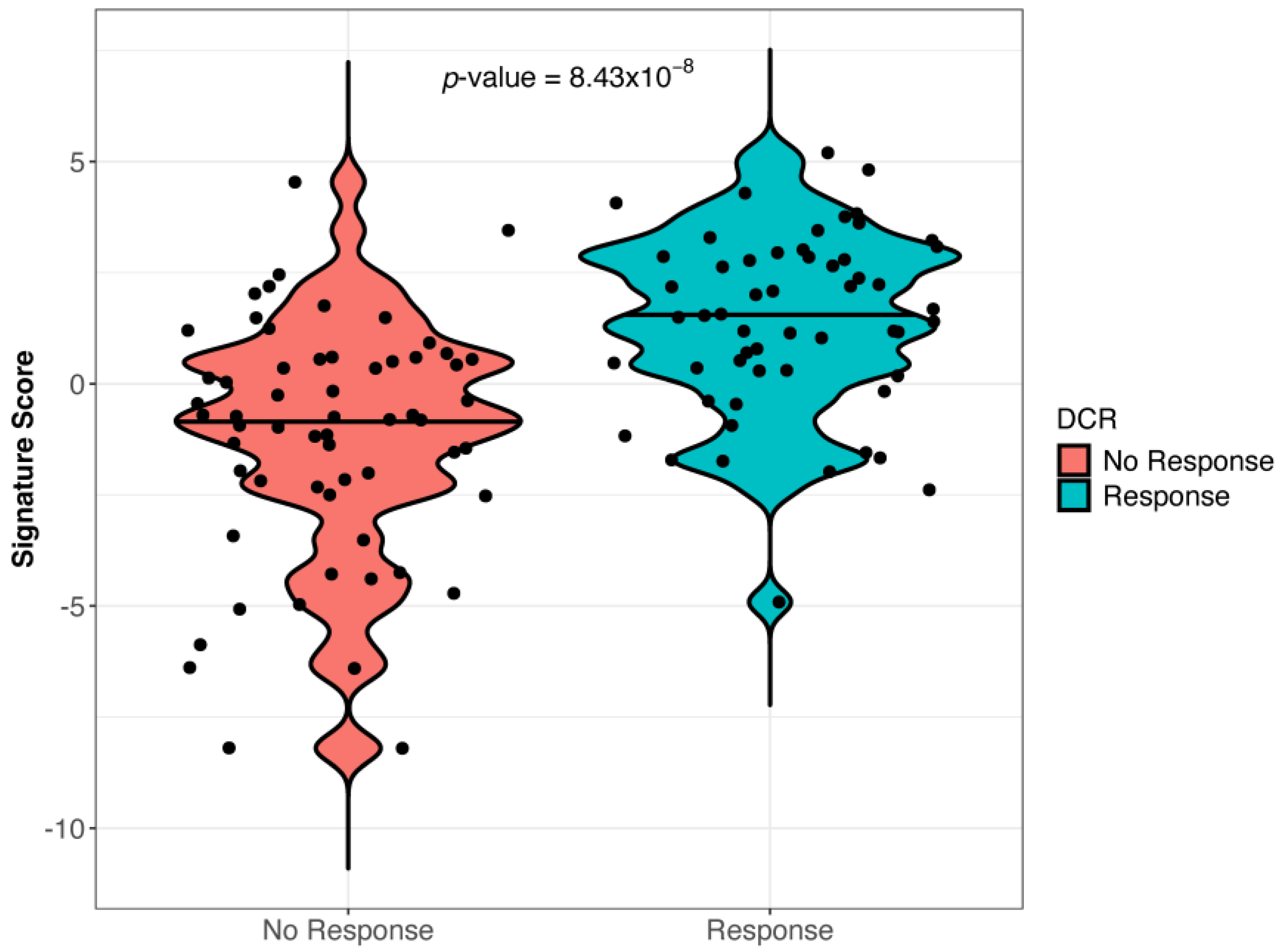
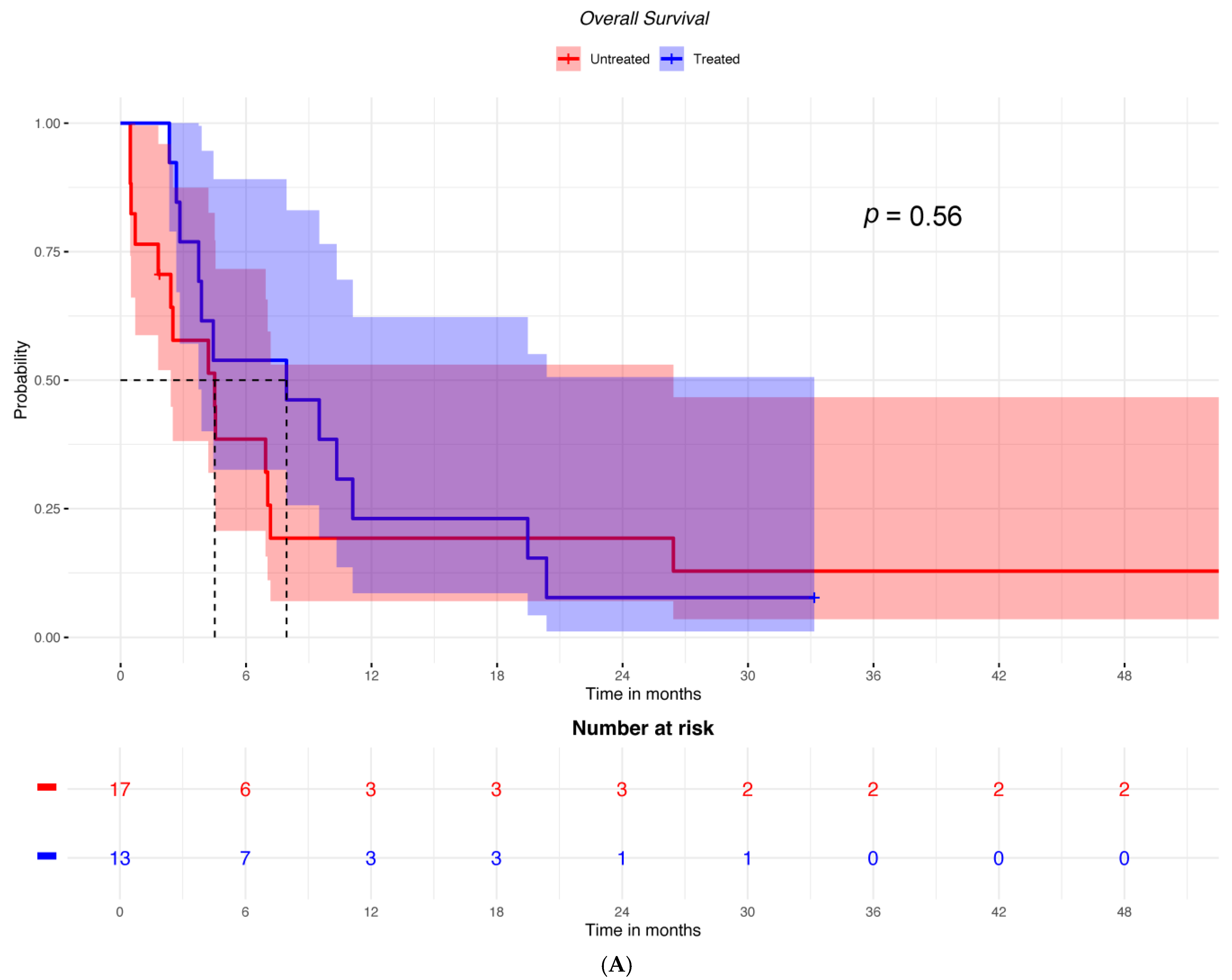
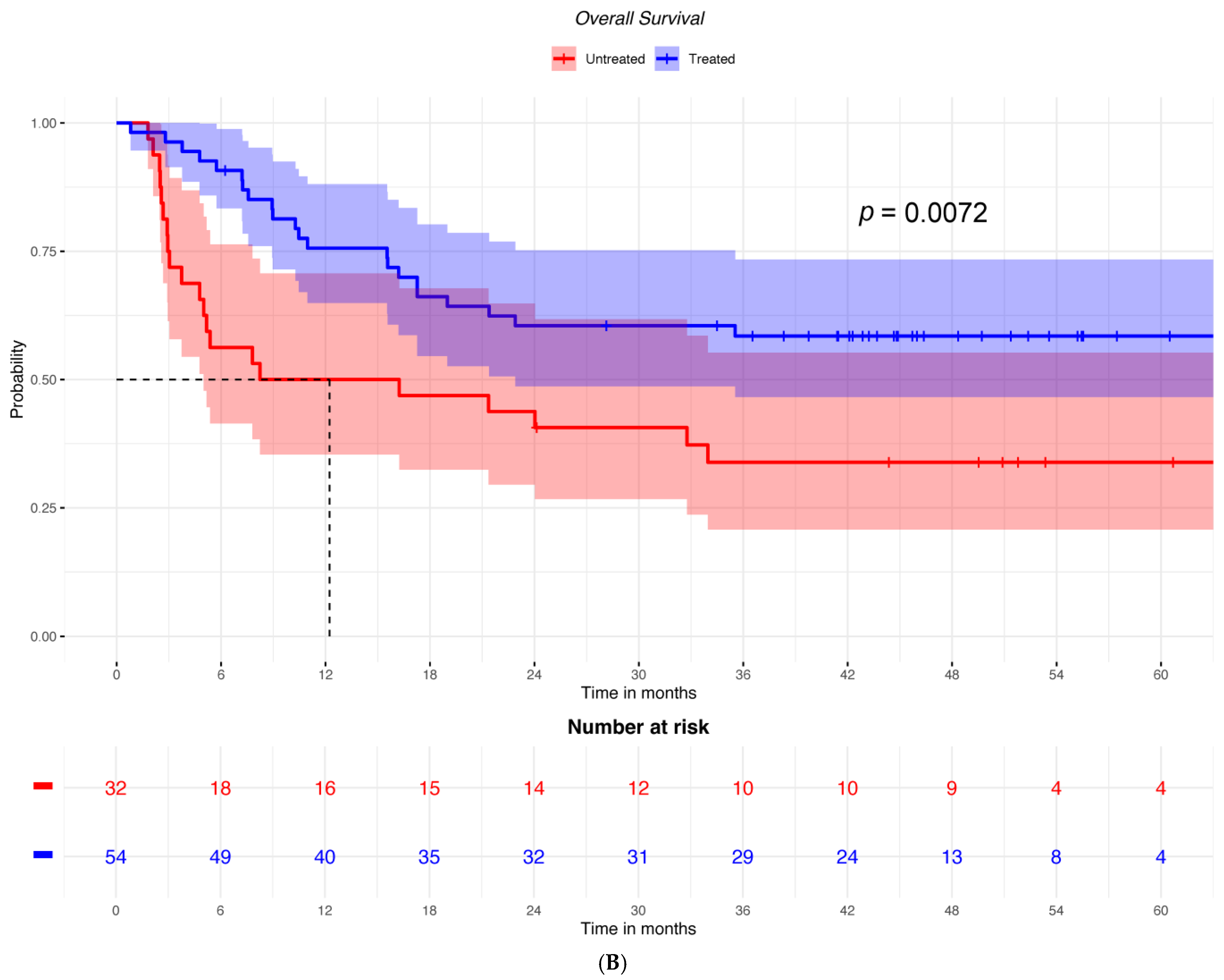
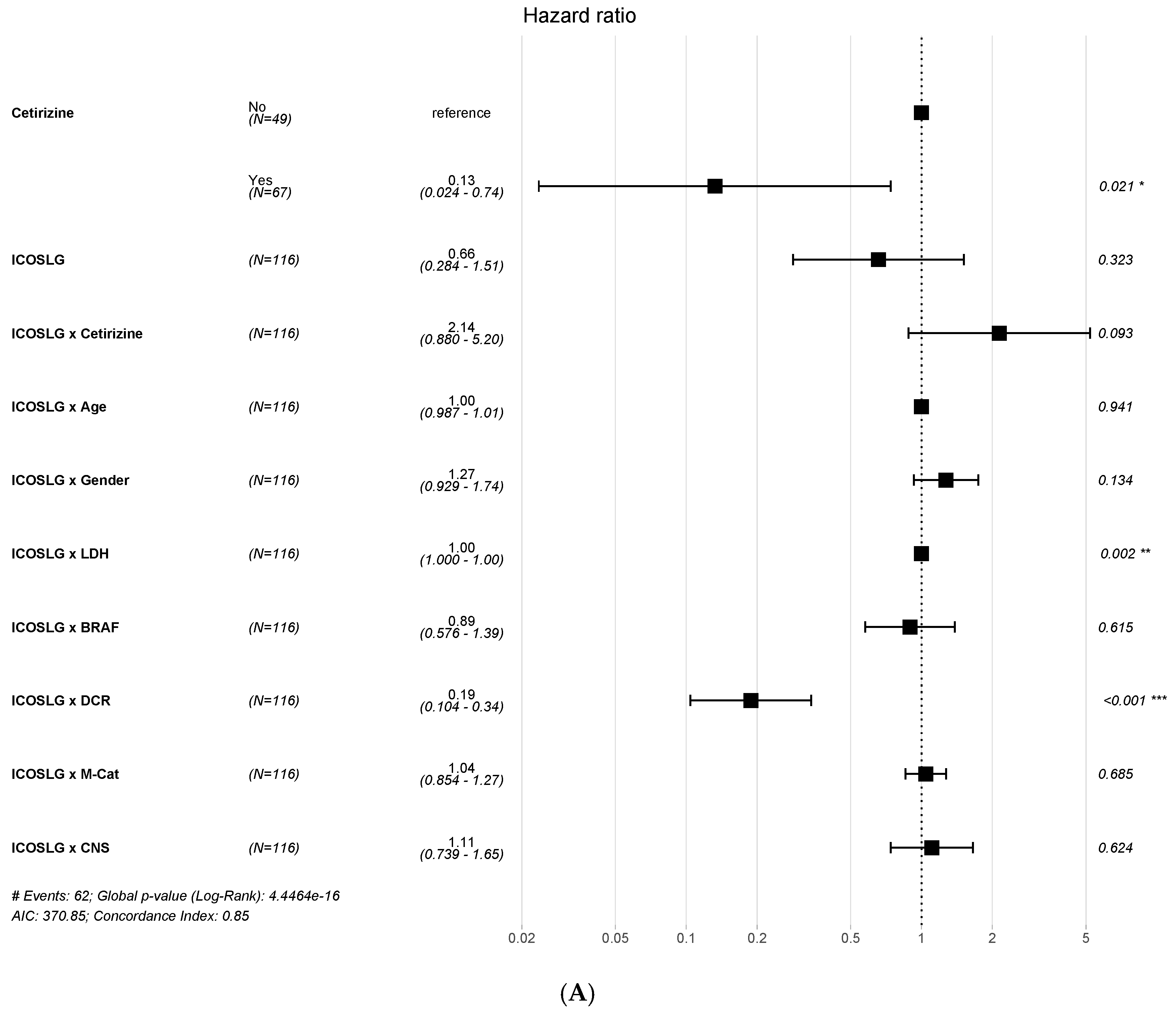
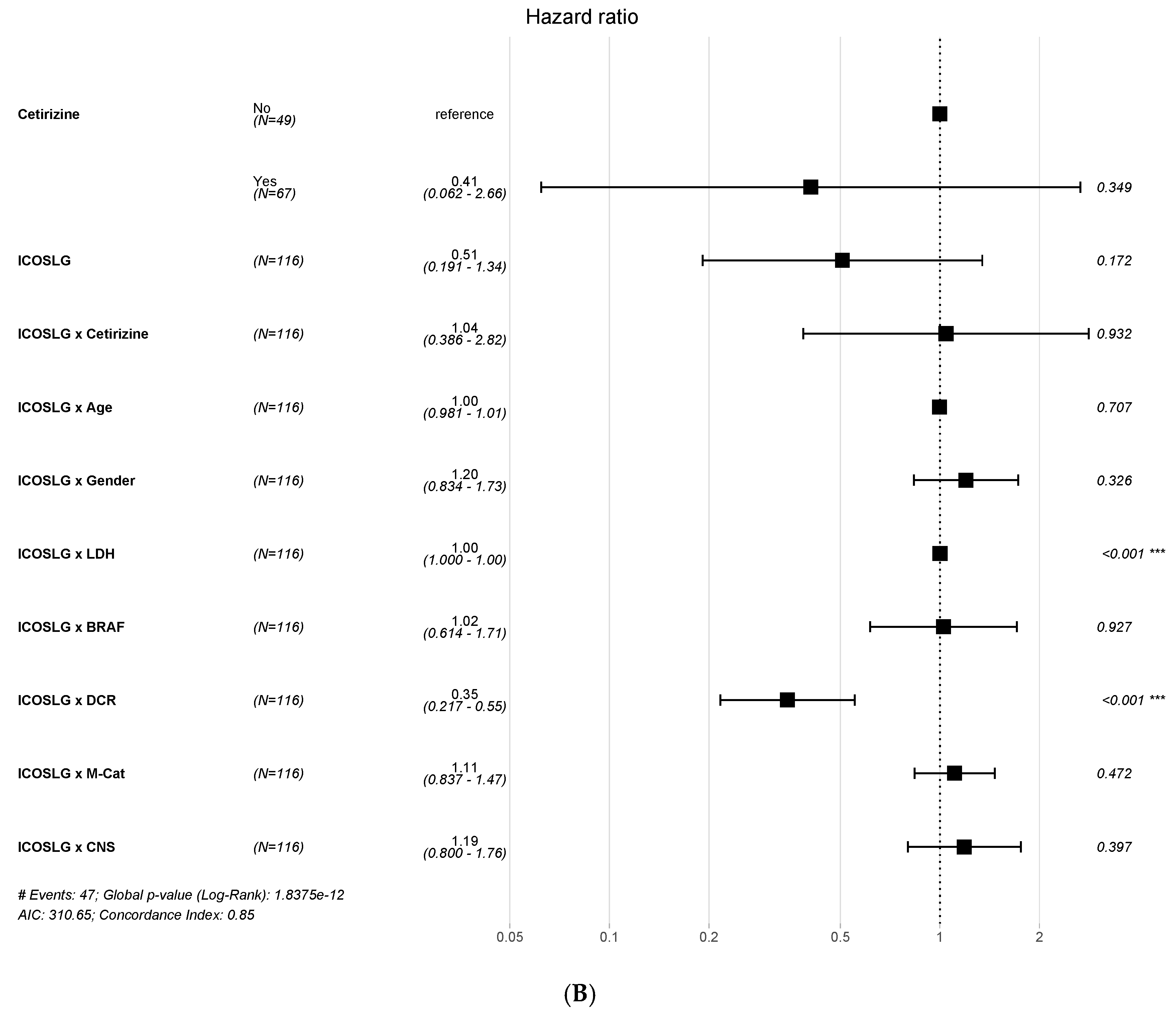
2. Results
2.1. Patient Characteristics and Outcomes
2.2. Gene Expression Signatures
3. Discussion
4. Materials and Methods
4.1. Study Design
4.2. Evaluation of Outcomes
4.3. Transcriptomic Analysis
4.4. Statistical Analysis
Supplementary Materials
Author Contributions
Funding
Institutional Review Board Statement
Informed Consent Statement
Data Availability Statement
Acknowledgments
Conflicts of Interest
References
- Topalian, S.L.; Hodi, F.S.; Brahmer, J.R.; Gettinger, S.N.; Smith, D.C.; McDermott, D.F.; Powderly, J.D.; Sosman, J.A.; Atkins, M.B.; Leming, P.D.; et al. Five-Year Survival and Correlates Among Patients with Advanced Melanoma, Renal Cell Carcinoma, or Non-Small Cell Lung Cancer Treated with Nivolumab. JAMA Oncol. 2019, 5, 1411–1420. [Google Scholar] [CrossRef]
- Robert, C.; Hwu, W.J.; Hamid, O.; Ribas, A.; Weber, J.S.; Daud, A.I.; Hodi, F.S.; Wolchok, J.D.; Mitchell, T.C.; Hersey, P.; et al. Long-term safety of pembrolizumab monotherapy and relationship with clinical outcome: A landmark analysis in patients with advanced melanoma. Eur. J. Cancer 2021, 144, 182–191. [Google Scholar] [CrossRef]
- Schachter, J.; Ribas, A.; Long, G.V.; Arance, A.; Grob, J.J.; Mortier, L.; Daud, A.; Carlino, M.S.; McNeil, C.; Lotem, M.; et al. Pembrolizumab versus ipilimumab for advanced melanoma: Final overall survival results of a multicentre, randomised, open-label phase 3 study (KEYNOTE-006). Lancet 2017, 390, 1853–1862. [Google Scholar] [CrossRef]
- Tang, Q.; Zhao, S.; Zhou, N.; He, J.; Zu, L.; Liu, T.; Song, Z.; Chen, J.; Peng, L.; Xu, S. PD-1/PD-L1 immune checkpoint inhibitors in neoadjuvant therapy for solid tumors (Review). Int. J. Oncol. 2023, 62, 49. [Google Scholar] [CrossRef]
- Versluis, J.M.; Menzies, A.M.; Sikorska, K.; Rozeman, E.A.; Saw, R.P.M.; van Houdt, W.J.; Eriksson, H.; Klop, W.M.C.; Ch’ng, S.; van Thienen, J.V.; et al. Survival update of neoadjuvant ipilimumab plus nivolumab in macroscopic stage III melanoma in the OpACIN and OpACIN-neo trials. Ann. Oncol. 2023, 34, 420–430. [Google Scholar] [CrossRef]
- Wolchok, J.D.; Chiarion-Sileni, V.; Gonzalez, R.; Grob, J.J.; Rutkowski, P.; Lao, C.D.; Cowey, C.L.; Schadendorf, D.; Wagstaff, J.; Dummer, R.; et al. Long-Term Outcomes with Nivolumab Plus Ipilimumab or Nivolumab Alone Versus Ipilimumab in Patients with Advanced Melanoma. J. Clin. Oncol. 2022, 40, 127–137. [Google Scholar] [CrossRef]
- Eljilany, I.; Castellano, E.; Tarhini, A.A. Adjuvant Therapy for High-Risk Melanoma: An In-Depth Examination of the State of the Field. Cancers 2023, 15, 4125. [Google Scholar] [CrossRef]
- Kinane, D.F.; Gabert, J.; Xynopoulos, G.; Guzeldemir-Akcakanat, E. Strategic approaches in oral squamous cell carcinoma diagnostics using liquid biopsy. Periodontol. 2000 2024, Online ahead of print. [Google Scholar] [CrossRef]
- Jamshidi, A.; Liu, M.C.; Klein, E.A.; Venn, O.; Hubbell, E.; Beausang, J.F.; Gross, S.; Melton, C.; Fields, A.P.; Liu, Q.; et al. Evaluation of cell-free DNA approaches for multi-cancer early detection. Cancer Cell. 2022, 40, 1537–1549.e12. [Google Scholar] [CrossRef]
- Oliveira, K.C.S.; Ramos, I.B.; Silva, J.M.C.; Barra, W.F.; Riggins, G.J.; Palande, V.; Pinho, C.T.; Frenkel-Morgenstern, M.; Santos, S.E.B.; Assumpcao, P.P.; et al. Current perspectives on circulating tumor DNA, precision medicine, and personalized clinical management of cancer. Mol. Cancer Res. 2020, 18, 517–528. [Google Scholar] [CrossRef]
- Lukacova, E.; Hanzlikova, Z.; Podlesnyi, P.; Sedlackova, T.; Szemes, T.; Grendar, M.; Samec, M.; Hurtova, T.; Malicherova, B.; Leskova, K.; et al. Novel liquid biopsy CNV biomarkers in malignant melanoma. Sci. Rep. 2024, 14, 15786. [Google Scholar] [CrossRef] [PubMed] [PubMed Central]
- Mallardo, D.; Simeone, E.; Vanella, V.; Vitale, M.G.; Palla, M.; Scarpato, L.; Paone, M.; De Cristofaro, T.; Borzillo, V.; Cortellini, A.; et al. Concomitant medication of cetirizine in advanced melanoma could enhance anti-PD-1 efficacy by promoting M1 macrophages polarization. J. Transl. Med. 2022, 20, 436. [Google Scholar] [CrossRef] [PubMed] [PubMed Central]
- Yunna, C.; Mengru, H.; Lei, W.; Weidong, C. Macrophage M1/M2 polarization. Eur. J. Pharmacol. 2020, 877, 173090. [Google Scholar] [CrossRef] [PubMed]
- Mallardo, D.; Giannarelli, D.; Vitale, M.G.; Galati, D.; Trillò, G.; Esposito, A.; Isgrò, M.A.; D’Angelo, G.; Festino, L.; Vanella, V.; et al. Nivolumab serum concentration in metastatic melanoma patients could be related to outcome and enhanced immune activity: A gene profiling retrospective analysis. J. Immunother. Cancer 2022, 10, e005132. [Google Scholar] [CrossRef]
- Mallardo, D.; Cortellini, A.; Capone, M.; Madonna, G.; Pinato, D.; Warren, S.; Simeone, E.; Ascierto, P. Concomitant type 2 diabetes mellitus (T2DM) in metastatic melanoma patients could be related to lower level of LAG-3: A transcriptomic analysis of a retrospective cohort. Ann. Oncol. 2022, 33, 445–447. [Google Scholar] [CrossRef]
- Lee, C.J.; An, H.J.; Cho, E.S.; Kang, H.C.; Lee, J.Y.; Lee, H.S.; Cho, Y.Y. Stat2 stability regulation, an intersection between immunity and carcinogenesis. Exp. Mol. Med. 2020, 52, 1526–1536. [Google Scholar] [CrossRef]
- Addeo, A.; Friedlaender, A.; Banna, G.L.; Weiss, G.J. TMB or not TMB as a biomarker: That is the question. Crit. Rev. Oncol. Hematol. 2021, 163, 103374. [Google Scholar] [CrossRef]
- Mo, S.F.; Cai, Z.Z.; Kuai, W.H.; Li, X.; Chen, Y.T. Universal cutoff for tumor mutational burden in predicting the efficacy of anti-PD-(L)1 therapy for advanced cancers. Front. Cell Dev. Biol. 2023, 11, 1209243. [Google Scholar] [CrossRef]
- Maio, M.; Ascierto, P.A.; Manzyuk, L.; Motola-Kuba, D.; Penel, N.; Cassier, P.A.; Bariani, G.M.; De Jesus Acosta, A.; Doi, T.; Longo, F.; et al. Pembrolizumab in microsatellite instability high or mismatch repair deficient cancers: Updated analysis from the phase II KEYNOTE-158 study. Ann. Oncol. 2022, 33, 929–938. [Google Scholar] [CrossRef]
- Zhang, Y.; Chen, A.; Li, D.; Yuan, Q.; Zhu, A.; Deng, J.; Wang, Y.; Liu, J.; Liang, C.; Li, W.; et al. Development of T follicular helper cell-independent nanoparticle vaccines for SARS-CoV-2 or HIV-1 by targeting ICOSL. NPJ Vaccines 2024, 9, 176. [Google Scholar] [CrossRef] [PubMed] [PubMed Central]
- Khayyamian, S.; Hutloff, A.; Büchner, K.; Gräfe, M.; Henn, V.; Kroczek, R.A.; Mages, H.W. ICOS-ligand, expressed on human endothelial cells, costimulates Th1 and Th2 cytokine secretion by memory CD4+ T cells. Proc. Natl. Acad. Sci. USA 2002, 99, 6198–6203. [Google Scholar] [CrossRef]
- Martin-Orozco, N.; Li, Y.; Wang, Y.; Liu, S.; Hwu, P.; Liu, Y.J.; Dong, C.; Radvanyi, L. Melanoma cells express ICOS ligand to promote the activation and expansion of T-regulatory cells. Cancer Res. 2010, 70, 9581–9590. [Google Scholar] [CrossRef]
- Strauss, L.; Bergmann, C.; Szczepanski, M.J.; Lang, S.; Kirkwood, J.M.; Whiteside, T.L. Expression of ICOS on human melanoma-infiltrating CD4+CD25highFoxp3+ T regulatory cells: Implications and impact on tumor-mediated immune suppression. J. Immunol. 2008, 180, 2967–2980. [Google Scholar] [CrossRef]
- Gigliotti, C.L.; Dianzani, C.; Stoppa, I.; Monge, C.; Sutti, S.; Sblattero, D.; Puricelli, C.; Rolla, R.; Dianzani, U.; Boggio, E. Differential Modulation of Human M1 and M2 Macrophage Activity by ICOS-Mediated ICOSL Triggering. Int. J. Mol. Sci. 2023, 24, 2953. [Google Scholar] [CrossRef] [PubMed] [PubMed Central]
- Kanungo, R.; Hippargi, S.B. CD64 Expression on Neutrophils (nCD64) as a Biomarker in Adult Patients with Sepsis: A Cross-Sectional Study. Cureus 2024, 16, e71912. [Google Scholar] [CrossRef] [PubMed] [PubMed Central]
- Takai, T. A novel recognition system for MHC class I molecules constituted by PIR. Adv. Immunol. 2005, 88, 161–192. [Google Scholar] [CrossRef] [PubMed]
- Hawthorne, B.C.; Engel, S.; McCarthy, M.B.R.; Cote, M.C.; Mazzocca, A.D.; Coyner, K.J. Biologic Adjuvants to Rotator Cuff Repairs Induce Anti-inflammatory Macrophage 2 Polarization and Reduce Inflammatory Macrophage 1 Polarization In Vitro. Arthroscopy 2024. Epub ahead of print. [Google Scholar] [CrossRef] [PubMed]
- Johnson, D.B.; Sullivan, R.J.; Ott, P.A.; Carlino, M.S.; Khushalani, N.I.; Ye, F.; Guminski, A.; Puzanov, I.; Lawrence, D.P.; Buchbinder, E.I.; et al. Ipilimumab Therapy in Patients with Advanced Melanoma and Preexisting Autoimmune Disorders. JAMA Oncol. 2016, 2, 234–240. [Google Scholar] [CrossRef] [PubMed]
- Maurer, D.M.; Adamik, J.; Santos, P.M.; Shi, J.; Shurin, M.R.; Kirkwood, J.M.; Storkus, W.J.; Butterfield, L.H. Dysregulated NF-κB-Dependent ICOSL Expression in Human Dendritic Cell Vaccines Impairs T-cell Responses in Patients with Melanoma. Cancer Immunol. Res. 2020, 8, 1554–1567. [Google Scholar] [CrossRef] [PubMed] [PubMed Central]
- Müller, E.; Christopoulos, P.F.; Halder, S.; Lunde, A.; Beraki, K.; Speth, M.; Øynebråten, I.; Corthay, A. Toll-Like receptor ligands and interferon-γ synergize for induction of antitumor M1 macrophages. Front. Immunol. 2017, 8, 1383. [Google Scholar] [CrossRef]
- Khunger, A.; Piazza, E.; Warren, S.; Smith, T.H.; Ren, X.; White, A.; Elliott, N.; Cesano, A.; Beechem, J.M.; Kirkwood, J.M.; et al. CTLA-4 blockade and interferon-α induce proinflammatory transcriptional changes in the tumor immune landscape that correlate with pathologic response in melanoma. PLoS ONE 2021, 16, e0245287. [Google Scholar] [CrossRef] [PubMed]
- Hazra, A.; Gogtay, N. Biostatistics Series Module 6: Correlation and Linear Regression. Indian J. Dermatol. 2016, 61, 593–601. [Google Scholar] [CrossRef] [PubMed] [PubMed Central]
- Lê Cao, K.A.; Boitard, S.; Besse, P. Sparse PLS discriminant analysis: Biologically relevant feature selection and graphical displays for multiclass problems. BMC Bioinform. 2011, 12, 253. [Google Scholar] [CrossRef] [PubMed]


| Cetirizine | |||
|---|---|---|---|
| Characteristics | No, n = 49 1 | Yes, n = 67 1 | Overall, n = 116 1 |
| Age | 66.00 (13.00) | 65.00 (26.50) | 66.00 (22.00) |
| Sex | |||
| Female | 22.0 (44.9%) | 29.0 (43.3%) | 51.0 (44.0%) |
| Male | 27.0 (55.1%) | 38.0 (56.7%) | 65.0 (56.0%) |
| BRAF | |||
| MUT | 13.0 (27.7%) | 9.0 (14.8%) | 22.0 (20.4%) |
| WT | 34.0 (72.3%) | 52.0 (85.2%) | 86.0 (79.6%) |
| Missing | 2 | 6 | 8 |
| LDH [U/L] | 333.00 (235.50) | 278.00 (204.75) | 304.00 (260.50) |
| Missing | 10 | 23 | 33 |
| M-Category | |||
| M0 | 2.0 (4.1%) | 3.0 (4.5%) | 5.0 (4.3%) |
| M1A | 5.0 (10.2%) | 9.0 (13.4%) | 14.0 (12.1%) |
| M1B | 6.0 (12.2%) | 9.0 (13.4%) | 15.0 (12.9%) |
| M1C | 36.0 (73.5%) | 46.0 (68.7%) | 82.0 (70.7%) |
| CNS | |||
| NO | 31.0 (67.4%) | 53.0 (81.5%) | 84.0 (75.7%) |
| YES | 15.0 (32.6%) | 12.0 (18.5%) | 27.0 (24.3%) |
| Missing | 3 | 2 | 5 |
| Anti-PD1 agent | |||
| Nivolumab | 32 (65.3%) | 42 (62.7%) | 74 (63.8%) |
| Pembrolizumab | 17 (34.7%) | 25 (37.3%) | 42 (36.2%) |
| First-line treatment | 39 (79.6%) | 49 (73.1%) | 88 (75.9%) |
| Pretreated with ipilimumab | 10 (20.4%) | 18 (26.9%) | 28 (24.1%) |
Disclaimer/Publisher’s Note: The statements, opinions and data contained in all publications are solely those of the individual author(s) and contributor(s) and not of MDPI and/or the editor(s). MDPI and/or the editor(s) disclaim responsibility for any injury to people or property resulting from any ideas, methods, instructions or products referred to in the content. |
© 2024 by the authors. Licensee MDPI, Basel, Switzerland. This article is an open access article distributed under the terms and conditions of the Creative Commons Attribution (CC BY) license (https://creativecommons.org/licenses/by/4.0/).
Share and Cite
Mallardo, D.; Fordellone, M.; Ottaviano, M.; Marano, G.; Vitale, M.G.; Mallardo, M.; Capasso, M.; De Cristofaro, T.; Capone, M.; Meinardi, T.; et al. ICOSLG Is Associated with Anti-PD-1 and Concomitant Antihistamine Treatment Response in Advanced Melanoma. Int. J. Mol. Sci. 2024, 25, 12439. https://doi.org/10.3390/ijms252212439
Mallardo D, Fordellone M, Ottaviano M, Marano G, Vitale MG, Mallardo M, Capasso M, De Cristofaro T, Capone M, Meinardi T, et al. ICOSLG Is Associated with Anti-PD-1 and Concomitant Antihistamine Treatment Response in Advanced Melanoma. International Journal of Molecular Sciences. 2024; 25(22):12439. https://doi.org/10.3390/ijms252212439
Chicago/Turabian StyleMallardo, Domenico, Mario Fordellone, Margaret Ottaviano, Giuseppina Marano, Maria Grazia Vitale, Mario Mallardo, Mariagrazia Capasso, Teresa De Cristofaro, Mariaelena Capone, Teresa Meinardi, and et al. 2024. "ICOSLG Is Associated with Anti-PD-1 and Concomitant Antihistamine Treatment Response in Advanced Melanoma" International Journal of Molecular Sciences 25, no. 22: 12439. https://doi.org/10.3390/ijms252212439
APA StyleMallardo, D., Fordellone, M., Ottaviano, M., Marano, G., Vitale, M. G., Mallardo, M., Capasso, M., De Cristofaro, T., Capone, M., Meinardi, T., Paone, M., Sabatelli, P., De Filippi, R., Cesano, A., Cavalcanti, E., Caracò, C., Warren, S., Budillon, A., Simeone, E., & Ascierto, P. A. (2024). ICOSLG Is Associated with Anti-PD-1 and Concomitant Antihistamine Treatment Response in Advanced Melanoma. International Journal of Molecular Sciences, 25(22), 12439. https://doi.org/10.3390/ijms252212439