Review

Three-Dimensional Cell Co-Culture Liver Models and Their Applications in Pharmaceutical Research

1
Department of Clinical Pharmacy, Key Laboratory of Basic Pharmacology of Guizhou Province and School of Pharmacy, Zunyi Medical University, Zunyi 563006, China
2
Key Laboratory of Basic Pharmacology of Ministry of Education and Joint International Research Laboratory of Ethnomedicine of Ministry of Education, Zunyi Medical University, Zunyi 563006, China
3
Key Laboratory of Clinical Pharmacy of Zunyi City, Zunyi Medical University, Zunyi 563006, China
4
Cancer Research UK Manchester Institute, The University of Manchester, Cheshire SK10 4TG, UK
5
School of Preclinical Medicine, Zunyi Medical University, Zunyi 563006, China
*
Author to whom correspondence should be addressed.
These authors contributed equally to this work.
Int. J. Mol. Sci. 2023, 24(7), 6248; https://doi.org/10.3390/ijms24076248
Submission received: 1 February 2023 / Revised: 14 March 2023 / Accepted: 22 March 2023 / Published: 26 March 2023
(This article belongs to the Special Issue Drug Metabolism and Drug-Induced Liver Injury)

Abstract

:
As the primary site for the biotransformation of drugs, the liver is the most focused on organ type in pharmaceutical research. However, despite being widely used in pharmaceutical research, animal models have inherent species differences, while two-dimensional (2D) liver cell monocultures or co-cultures and three-dimensional (3D) liver cell monoculture in vitro liver models do not sufficiently represent the complexity of the human liver’s structure and function, making the evaluation results from these tools less reliable. Therefore, there is a pressing need to develop more representative in vitro liver models for pharmaceutical research. Fortunately, an exciting new development in recent years has been the emergence of 3D liver cell co-culture models. These models hold great promise as in vitro pharmaceutical research tools, because they can reproduce liver structure and function more practically. This review begins by explaining the structure and main cell composition of the liver, before introducing the potential advantages of 3D cell co-culture liver models for pharmaceutical research. We also discuss the main sources of hepatocytes and the 3D cell co-culture methods used in constructing these models. In addition, we explore the applications of 3D cell co-culture liver models with different functional states and suggest prospects for their further development.

1. Introduction

The liver is the body’s largest digestive gland, responsible for more than 500 functions, including metabolism and detoxification [1,2]. It is mainly composed of hepatocytes and non-parenchymal cells, such as liver sinusoidal endothelial cells (LSECs), hepatic stellate cells (HSCs), and Kupffer cells (KCs), which work together to regulate liver function through direct or indirect cell crosstalk events [3,4].
As the primary organ of drug exposure, the liver plays a critical role in drug metabolism and toxicity, necessitating the development of liver models that can more accurately predict the efficacy and toxicity of drugs in humans for drug discovery [5,6,7,8,9]. In vitro liver models, including 2D and 3D liver cell monoculture and co-culture models, offer advantages over animal models, in terms of cost, ethical considerations, and efficiency. However, 2D liver cell models lack physiological relevance, while 3D liver cell monoculture models fail to represent the complex interactions between parenchymal and non-parenchymal cells [10,11,12]. Therefore, the development of 3D co-culture liver models that mimic the natural liver tissue structure and cell interactions offers the potential for more accurate in vitro prediction of drug response sensitivity in humans.
The previous frequently used prediction tools tended to fail to reliably predict drug efficacy and toxicity in humans, leading to high attrition rates in drug development [13]. For example, troglitazone, trovafloxacin, and nefazodone have been withdrawn from the market due to their severe hepatotoxicity [14]. Three-dimensional cell co-culture liver models can provide a greater predictability of drug response in humans and have been applied in many studies to predict the in vivo metabolism and clearance of candidate or marketed drugs [15,16]. For example, one study showed that Diclofenac exhibited phase I and phase II metabolic profiles and toxic effects similar to those in vivo in a microfluidic liver chip model [17]. A 3D cell co-culture liver model based on 3D bioprinting technology and microfluidic technology was applied to evaluate the efficacy of Metuzumab and showed similar results to those obtained in animal experiments and clinical trials of anti-cancer drugs with similar antitumor mechanisms [18]. Spheroid cultures of Primary human hepatocytes (PHHs), LSECs, KCs, and HSCs were used as models of nonalcoholic fatty liver disease (NASH), to evaluate the efficacy of the drug candidates Selonsertib and Firsocostat against NASH, with the results correlating with the effects in patients participating in clinical trials [19].
In this review, we first analyze the potential advantages of 3D cell co-culture liver models for pharmaceutical research based on the discussion of the unique structure and function of primary parenchymal and non-parenchymal cells in the liver; then, we introduce the main sources of hepatocytes in 3D cell co-culture liver models and the commonly used methods for the construction of 3D cell co-culture liver models; finally, we discuss 3D cell co-culture liver systems with different functions and their applications.

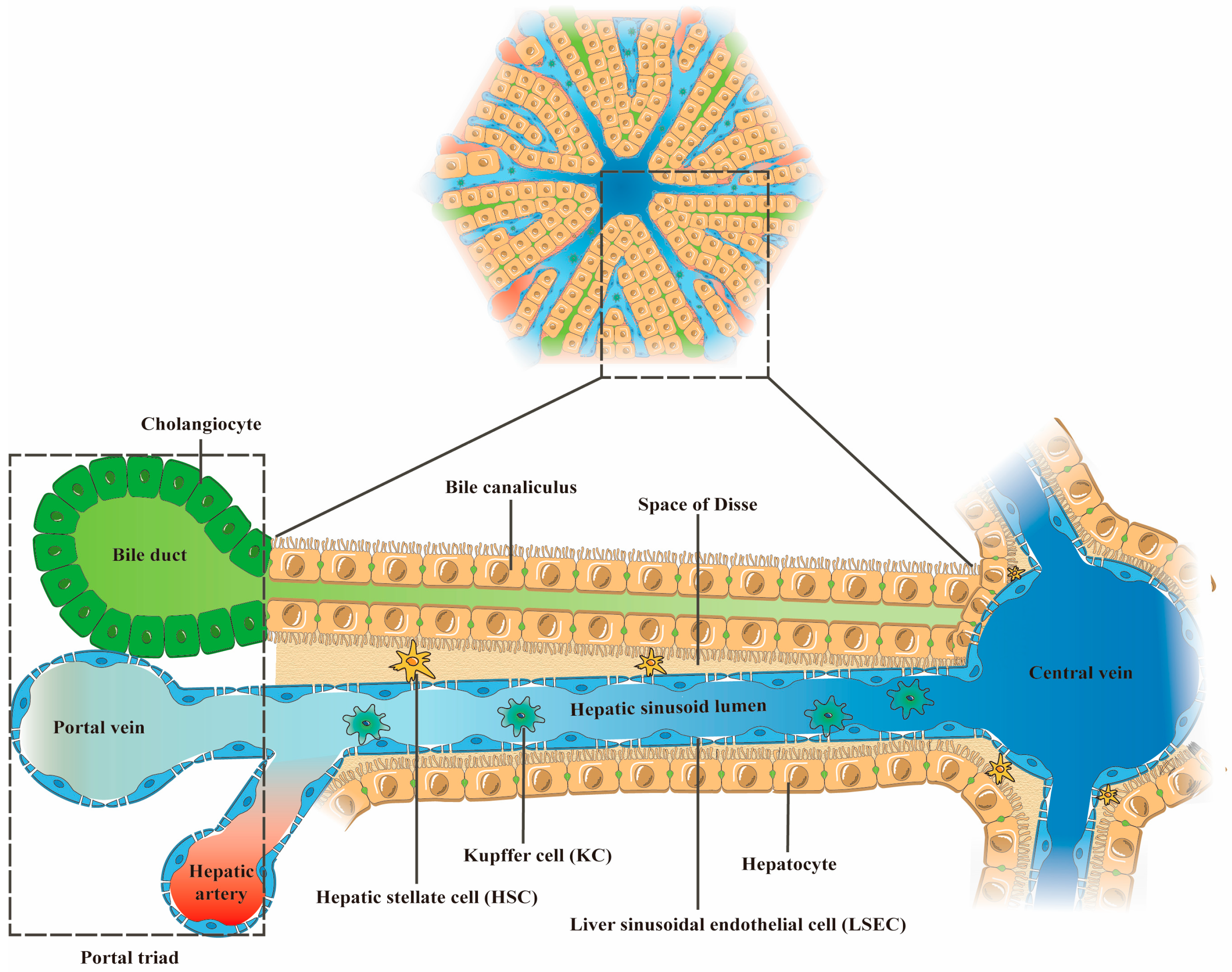
2. The Main Cellular Components of the Liver and Their Functions

Hepatic lobules with a hexagonal structure constitute the basic structural and functional unit of the liver (Figure 1). A central vein runs through the center of each lobule and is responsible for transporting intrahepatic metabolites. Surrounding the lobule is the portal triad, consisting of the hepatic portal vein, hepatic artery, and bile duct. The hepatic portal vein carries nutrient-rich blood to the liver, while the hepatic artery supplies oxygen to the liver [20].
Hepatocytes are the most abundant cell type in the liver, and they form hepatocyte plates that radiate out from the central vein. The adjacent liver plates are separated by hepatic sinusoids, which are capillary-like structures formed by the confluence of the terminal branches of the hepatic portal vein and the hepatic artery. In addition, adjacent hepatocytes are connected by the gaps that form bile canaliculi, which collect and transport bile. Liver sinusoidal endothelial cells (LSECs) form the liver sinusoidal wall and facilitate material exchange between the hepatocytes and the sinusoidal blood in the Disse space, which is the space between the hepatocytes and the LSECs [21]. Kupffer cells (KCs) are located in the hepatic sinusoid lumen and extend along the LSECs. Hepatic stellate cells (HSCs) are located in the space of Disse and, together with the space of Disse components, separate the hepatocytes from the LSECs [21]. It should be emphasized that the structure and function of hepatic lobules are regulated by the intricate interactions between the various liver cells (Table 1).

3. Potential Advantages of 3D Cell Co-Culture Liver Models for Drug Evaluation

It is necessary to further integrate the structure and function of the liver in vivo into liver models in vitro, as much as possible, to improve the reproducibility of in vitro experimental results in humans. In vitro liver models have evolved from 2D culture to the sandwich culture and then to the 3D culture of liver cells. Recently, the emergence of multicellular 3D cell co-culture models has revolutionized the construction of in vitro liver models. Although traditional 2D liver cell culture is inexpensive and easy to perform, the liver cells grow in a flat state under these conditions, resulting in cytoskeletal changes and loss of cell polarity within 2–3 days [33]. Most critically, drug metabolic enzymes, which play a key role in hepatic drug metabolism, have a low expression and activity under 2D conditions, which determines the ineffectiveness of 2D liver cell monoculture and co-culture models for research on the prediction of hepatic drug metabolism, drug interactions, and toxic reactions mediated by drug metabolic enzymes. Fortunately, the transformation from a 2D culture to a 3D culture mode for liver cells effectively overcomes the environmental homogeneity limitations of 2D systems [34,35]. Three-dimensional systems can be self-aggregated cell spheres, extracellular matrix (ECM)-like 3D network scaffolds made of natural or synthetic materials, and organ-like systems constructed using 3D technology, which have oxygen, nutrient, and various biological factor gradients and 3D tissue structures, where cell–cell and cell–ECM interactions can occur. This culture mode, which is closer to the complex microenvironment in vivo, largely contributes to the improved functional activity of liver cells in vitro [36].
However, it has recently been realized that 3D liver cell monoculture models ignore the direct or indirect signal exchange of heterotypic cells in liver tissue. Thus, 3D co-culture liver models that integrate hepatic heterotypic cell interactions and 3D microenvironmental signaling stand out, with many advantages. Several studies have demonstrated that 3D cell co-culture liver models exhibit better predictive ability than 2D and 3D monocultures and 2D co-cultures in drug-induced hepatotoxicity, hepatic drug metabolism, and drug interaction studies, since cell growing environment and signaling regulation between heterotypic cells in 3D cell co-culture allow them to exhibit more relevant liver environments and phenotypes in vivo, such as reproducible liver sinusoidal structures, long-term maintenance of liver cell activity and function, and increased expression of genes related to liver-specific functions [37,38,39]. The 3D co-culture model of hepatoma cells and fibroblasts constructed by Al Hrout et al. [40] exhibited an increased expression of genes and proteins associated with hepatocellular carcinoma development and a poor prognosis compared to 2D or 3D monocultures of hepatoma cells, and it more closely resembled the developmental characteristics of hepatocellular carcinoma in vivo and facilitated a more accurate assessment of the anticancer effects of anticancer drugs in vitro. In summary, 3D cell co-culture liver models can effectively improve the functional stability of hepatocytes in vitro and ensure the reliability of in vitro drug prediction results and thus are expected to provide a reliable new platform for pharmaceutical research.

4. Hepatocyte Sources of 3D Cell Co-Culture Liver Models

Hepatocytes are the predominant cell type in the liver and are responsible for most of its functions, making them the most important cell type in 3D cell co-culture liver models in vitro. Their value in pharmaceutical research lies in their ability to express specific liver functions, such as protein and urea synthesis, as well as drug metabolic enzyme expression levels, particularly CYP450 enzymes. Additionally, their sensitivity to stimuli such as toxins or drugs is critical for evaluating drug efficacy and toxicity.

4.1. Primary Hepatocytes

Primary hepatocytes (PHs) are widely considered the gold standard for constructing in vitro liver models, with both animal and human sources being utilized [41]. Primary human hepatocytes (PHHs), in particular, are preferred by researchers in drug interaction, drug metabolism, and drug toxicity studies, due to their secretion properties and metabolic enzyme activities being comparable to those of in vivo hepatocytes, and they can reliably reflect in vivo liver properties [42]. However, the stable expression of their functional properties is highly dependent on the signal regulation of the microenvironment, which means that PHHs will quickly experience phenotypic changes, decreased cell activity, and loss of cell membrane polarity under a single 2D environment [43].
The in vitro culture conditions of PHHs have been improved, to enhance their functional activity, with studies such as Hu et al. [44] and Mazzocchi et al. [45] utilizing a 3D culture environment composed of Matrigel and a 3D bio-printed model, respectively. These studies have shown that PHHs can maintain their morphology and function for several months, with improved sensitivity to hepatotoxic agents. However, applications have been greatly restricted by the limited number of donors, ethical constraints, and genetic variability among donors [46]. Animal PHs are more readily available than PHHs, but due to the species differences between animals and humans, their applications are also limited by poor reproducibility.

4.2. Hepatoma Cell Lines

HepaRG, HepG2, Huh7, and Hep3B are common hepatocellular carcinoma-derived cell lines used for constructing in vitro liver models because they are readily available, easy to culture, highly proliferative, and phenotypically stable, with less inter-donor variability compared to PHHs [47]. However, the high proliferative potential of these cell lines is usually accompanied by a hypodifferentiated phenotype, which leads to a lack of specific liver functions, especially in the level of enzyme expression (especially the phase I and II metabolic enzymes involved in drug metabolism), protein and urea synthesis, and a much lower sensitivity to toxic reactions than PHHs [48]. For example, HepG2, Huh7, and Hep3B only express human liver-specific CYP450 enzymes at low levels, about 5–15% of that of PHHs, which may limit their predictive ability for hepatotoxicity caused by parent drug metabolites. Moreover, although they have similar expression levels of most phase II metabolic enzymes to PHHs [49,50], their reliability as a model for drug-induced hepatotoxicity assessment should be further evaluated.
In contrast, HepaRG cells, which were identified from a patient with hepatocellular carcinoma associated with hepatitis C virus infection, can differentiate into hepatocyte-like and bile duct-like cells after high-density exposure to dimethyl sulfoxide (DMSO) [51,52]. They acquire a highly differentiated phenotype and typical expression of liver function, including CYP450 enzyme activity (except CYP2D6) and albumin and urea synthesis similar to PHHs. They also express nuclear receptors such as the pregnane X receptor and the homeodomain androstane receptor [46]. Furthermore, they can form bile duct structures and express hepatic sinusoidal and bile canalicular transporters that are not found in other hepatoma cell lines, which play an important role in endogenous and exogenous material transport [53,54]. These structures and signals make HepaRG cells the most promising alternative to PHHs for in vitro drug evaluation.
However, some studies have shown that HepaRG cell-derived hepatocyte-like cells (HepaRG-tdHep), when stimulated by inflammatory factors TNF-α, IL-6, and TGF-β, can retro-differentiate into progenitor cells with stem cell characteristics, accompanied by the decrease in expression of liver-specific functions. In particular, TNF-α can indirectly induce expression deficiency of liver-specific markers (ALDOB and CDH1) in HepaRG-tdHep through activation of the NFkB gene network, while IL-6 can directly reduce the expression levels of ALDOB and CDH1. Additionally, TGF-β can induce the epithelial-to-mesenchymal transition (EMT) of HepaRG-tdHep, to regulate their retro-differentiation process. Interestingly, the retro-differentiation of HepaRG-tdHep induced by TNF-α, IL-6, and TGF-β is not reversed by DMSO [55,56]. Furthermore, the expression level of the liver-specific functions in HepaRG cell lines can also be regulated by their seeding density, since HepaRG cells differentiated into hepatocyte-like cells can retro-differentiate into hepatic progenitor cells through low-density seeding [57]. Therefore, the phenomenon of retro-differentiation exhibited by HepaRG cells under the influence of multiple factors is a point to be noted during their application.

4.3. Stem Cells

Human induced pluripotent stem cells (HiPSCs), embryonic stem cells (ESCs), and adult stem cells (ASCs), such as mesenchymal stem cells, hepatic stem cells, and hematopoietic stem cells, have demonstrated strong self-replication and self-renewal capabilities in vitro. HiPSCs and ESCs, in particular, have the potential to differentiate into almost any cell type in the human body, under certain conditions. Moreover, HiPSCs can be obtained from patient biopsies for personalized research [58,59]. In the context of the global scarcity of liver donors, stem cells capable of differentiating into hepatocyte-like cells under specific conditions can serve as potential resources for liver disease treatment, regeneration, and in vitro modeling.
However, it can be difficult for stem cells to differentiate into functionally mature hepatocytes in vitro, and there can be challenges such as immune rejection and resource shortage [60]. Although HiPSCs can effectively avoid ethical, immunogenicity, and resource shortage problems, their ability to differentiate directionally into mature hepatocytes is limited, and their derived hepatocytes exhibit more of the phenotype and characteristics of fetal hepatocytes, such as expressing high levels of alpha-fetoprotein and lower mature liver-specific metabolic functions [59,61,62,63]. To improve the ability of stem cells to differentiate into mature hepatocytes in vitro, it is essential to optimize their in vitro differentiation conditions. For example, the CYP450 enzyme activity and protein expression in HiPSC-derived hepatocytes increased when cultured in a 3D environment consisting of cellulose nanofibril (CNF) gel [64]. However, despite the improvement in culture conditions, their degree of differentiation is still unsatisfactory, which is a crucial factor limiting their application. Therefore, it is important to further explore methods for improving the stable differentiation of stem cells into hepatocytes, making them a reliable source of hepatocytes for research and potential clinical applications.

5. Common Co-Culture Methods for 3D Cell Co-Culture Liver Models

The development of liver function is known to be closely related to the environment–cell complex and heterotypic or homotypic cell–cell communication, which includes both direct physical contact communication and indirect paracrine signaling exchange between cells. To more realistically reproduce the intercellular information transfer in the liver, 3D cell co-culture liver models can be mainly divided into direct 3D co-culture and indirect 3D co-culture, based on their cell culture conditions. These can be realized with various 3D techniques, such as self-aggregating multicellular spheroids, 3D liver organoids, and 3D scaffold co-culture of liver cells for achieving direct 3D co-culture, while indirect 3D co-culture can be achieved based on Transwell chambers. Furthermore, 3D bioprinting co-culture of liver cells and microfluidic multicellular liver chips can be either direct 3D cell co-culture liver models or indirect 3D cell co-culture liver models (Table 2).

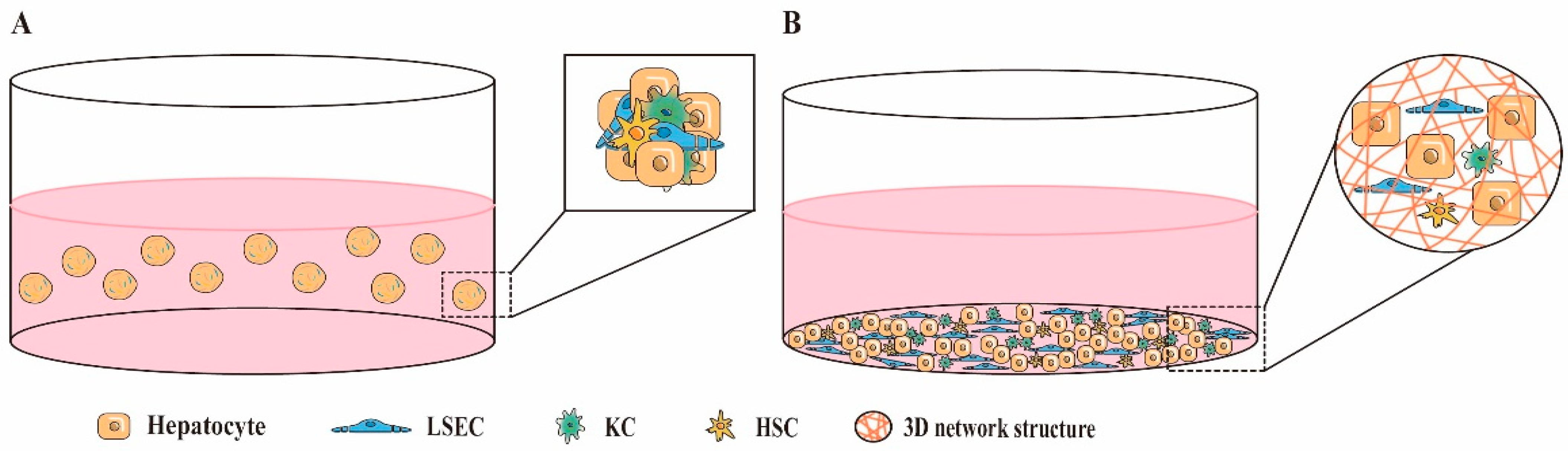
5.1. Direct 3D Co-Culture

Direct 3D co-culture involves mixing two or more different types of liver cells into self-assembling spheroids or culturing them together in a 3D environment that simulates the structure of native liver tissue (Figure 2). This 3D environment can typically be constructed with natural or synthetic materials that are low-toxicity or non-toxic, biocompatible and biodegradable, such as collagen, fibrin, alginate, and hydrogel. Pingitore et al. [69] generated mixed cell spheroids of HepG2 cells and immortalized hepatic stellate cells (LX-2) at a 1:1 or a physiological ratio of 24:1 in ultra-low attachment 96-well plates, to construct a non-alcoholic fatty liver-like organ system. In addition, Ahmed et al. [86] sequentially seeded primary human LSECs, HSCs, and hepatocytes on hollow fiber membranes (HF), to mimic the contact-layered structure between heterotypic cells in liver tissue.
Direct 3D co-culture allows for close contact between different liver cells, which enables signal communication between them through direct cell–cell adhesion, paracrine secretion of soluble cytokines, cell–ECM adhesion, and other mechanisms. In a multicellular organ such as the liver, direct contact between heterotypic cells plays an important role in full liver function. However, it is important to consider the actual distribution of co-cultured cells in the liver tissue in vivo when deciding on the rationality of direct co-culture. Direct contact co-culture in vitro of cell types that do not normally have direct contact in vivo may be unnecessary. Furthermore, tight junctions between heterotypic cells also affect the difficulty of cell separation, which can be disadvantageous for experiments requiring a single-cell type analysis. Therefore, the decision to use direct co-culture should be based on the type of co-culture cells and the purpose of the experiment.

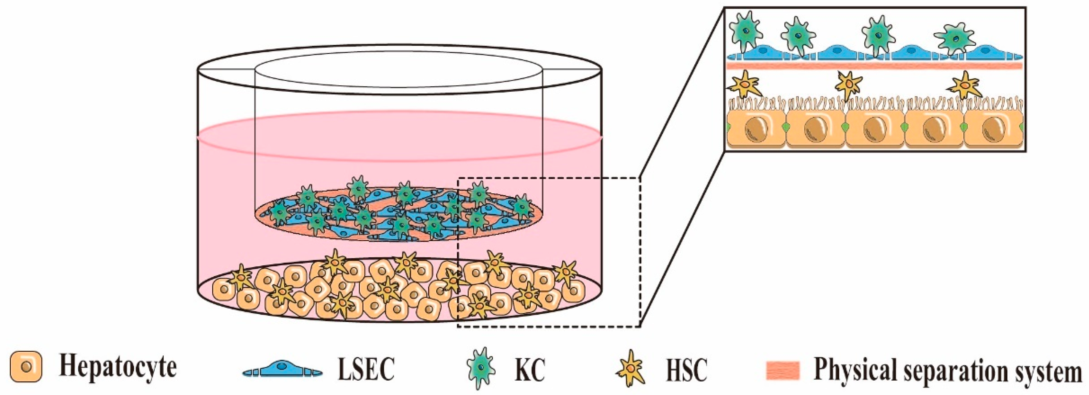
5.2. Indirect 3D Co-Culture

Indirect 3D co-culture involves the culture of two or more types of liver cells in a 3D environment with a physical separation system (Figure 3), which does not allow direct contact between the cells present on either side of the physical separation system, and signals between them are communicated through soluble cytokines. Common materials for physical separation systems are Transwell cell culture plates and various natural or synthetic materials. Otsuka et al. [87] constructed a 3D layered co-culture system from rat PHs and NIH/3T3 fibroblasts using Transwell cell culture plates as a physical separation system and explored the interaction between heterotypic cells. Kang et al. [23] seeded rat PHs and LSECs on both sides of a 6-well Transwell membrane precoated with rat tail collagen (LSECs on the top and hepatocytes on the bottom). They achieved a 3D layered co-culture of hepatocytes and LSECs, in which the 6-well Transwell membrane precoated with rat tail collagen mimicked the Disse spatial structure. The morphology of hepatocytes remained normal, and the expression of CYP450 enzymes remained similar during 39 days of culture, demonstrating the importance of soluble cytokines from LSECs for maintaining hepatocyte function. In addition, some researchers have also achieved indirect 3D co-culture of HepG2 and NIH/3T3 fibroblasts with the application of 3D bioprinting technology. They demonstrated that co-culture of HepG2 and NIH/3T3 fibroblasts exhibited a better hepatocyte function compared to monocultures of HepG2 [88]. In conclusion, indirect co-culture systems can effectively avoid unnecessary cell contact, facilitate the separation of co-cultured heterotypic cells, and may become promising tools for studying non-contact liver cell communication in vivo.

6. Types of 3D Cell Co-Culture Liver Model

The liver microenvironment is complex and dynamic, whereby the hepatocytes are surrounded by the ECM, which provides structural support and signaling molecules essential for their growth, proliferation, adhesion, and differentiation. Furthermore, hepatocytes communicate with non-parenchymal cells, either through direct cell–cell contact or indirect signaling mechanisms, leading to distinct functional states of the liver. Hence, different 3D co-culture combinations of hepatocyte and non-parenchymal cells can reproduce liver activity in diverse functional states in vivo, for different research purposes. This section outlines the 3D cell co-culture liver systems representing various functional states.

6.1. Three-Dimensional Cell Co-Culture Models for Enhanced Hepatocyte Function

In liver tissue, hepatocytes are surrounded by the hepatic sinusoidal network formed by LSECs, which is crucial for maintaining hepatocyte activity and functional homeostasis, as the basis of material exchange in hepatocytes [89]. Co-culture of LSECs and hepatocytes in vitro has been shown to be able to achieve a vascularized 3D liver tissue structure, where the presence of the vascular-like tubular network provides nutrients and oxygen to support the function of hepatocytes in vitro and maintain the elimination of metabolic waste products [90]. Additionally, LSECs can induce upregulation of vascular secretory factor Wnt2 and hepatocyte growth factor (HGF) through the Id1 pathway, both of which are essential for hepatocyte proliferation [89].
Studies have evaluated the effects of co-culturing LSECs with hepatocytes in 3D environments using various combinations of cells. Nahmias et al. [91] constructed monocultures of rat PHs and co-cultures with rat LSECs, human umbilical vein endothelial cells (HUVECs), or 3T3-J2 fibroblasts in a 3D environment of collagen gels, respectively. The ability of hepatocytes to clear low-density lipoprotein (LDL) in these cultures was assessed. The results showed that the 3D co-culture of PHs-LSECs not only reproduced the liver sinusoidal structure but also improved the ability of hepatocytes to clear LDL, which was not observed in other co-cultures. Ardalani H et al. [92] constructed mixed cell spheroids, including HiPSCs-derived hepatocytes (iHEP) and LSECs. The iHEP/LSECs spheroids showed a higher albumin and urea secretion capacity, as well as CYP3A4 and CYP2C9 activities, in the presence of DMSO, compared with other spheroids of iHEP, iHEP/HSCs, iHEP/cholangiocyte (CCs), iHEP/iPS-derived endothelial cells (iECs), or iHEP/LSECs/HSCs. Moreover, the presence of bile duct structures and capillary-like endothelial networks in iHEP/LSECs spheroids was confirmed through immunostaining, indicating that the 3D spheroid of LSECs and iHEP promoted the in vitro maturation of iHEP. German et al. [93] cultured HepaRG cells alone or in combination with LSECs or HUVECs in a 2D environment or a porous 3D environment composed of chitosan–gelatin. The results showed that the 3D co-culture of HepaRG/LSECs showed the highest HepaRG cell viability and APAP transformation capacity, whereas a higher CYP3A4 activity was exhibited in 3D co-cultures of HepaRG/HUVECs.
In summary, co-culturing LSECs with hepatocytes in 3D environments can recapitulate key structures of the liver sinusoids in vivo, which is important for research on enhancing in vitro hepatocyte function.

6.2. Three-Dimensional Cell Co-Culture Models of Liver Fibrosis

Hepatic fibrosis is a liver disease characterized by the accumulation of excess extracellular matrix proteins in the liver. The induction process is usually co-regulated by multiple types of liver cells. However, it is noteworthy that the activation of HSCs and their transformation to a myofibroblast-like cell phenotype are followed by excessive production of extracellular matrix proteins, which is the main mechanism by which hepatic fibrosis occurs. This dictates that the main target of hepatic fibrosis research is HSCs [94].
On this basis, a series of in vivo-relevant 3D cell co-culture liver fiber models have been developed in recent years. For instance, Prestigiacomo et al. [95] mixed rat PHs, HSCs, and KCs into 3D spheroid cultures using the hanging drop technique and explored the response of 3D spheroid cultures of PHs/HSC/KC and PHs with the stimulation of the pro-fibrotic factors TGF-β1 and LPS. When treated with TGF-β1 (1 ng/mL) or LPS, increased expression of α-SMA, an activating HSC-related gene, or an inflammatory response triggered by cytokines was detected in PHs/HSC/KC spheroid cultures. At the same time, a similar phenomenon was not observed in PHs spheroids. To construct a more representative in vitro model of liver fibrosis, Mannaerts et al. [96] prepared mixed cell spheroids of mouse PHs and HSCs in a 1:2 cell ratio. They exposed 2D cultures of HSCs and mixed spheroids of PHs and HSCs to APAP, to induce liver injury, to assess the activation of HSCs in a state of liver injury. It was found that mixed spheroids of PHs/HSCs better reproduced liver injury-dependent HSC activation, exhibiting similar gene upregulation of activated HSCs in vivo, such as Bicc1, Mfap4, Fmod, Oas1g, and Col6a3, whereas 2D cultures of HSCs showed low expression of these genes after the same treatment. Similarly, Leite et al. [97] developed a spheroid model of HepaRG/HSC. Their data showed that 3D spheroids of HepaRG/HSCs maintained functional activity for 21 days and allowed the drug to activate HSCs in a hepatocyte-dependent manner, as manifested by pro-fibrotic compound stimulation with massive collagen secretion and deposition. Existing studies support these experimental results; when liver injury occurs, hepatocytes can alter their gene expression and secretion profile, resulting in high levels of expression of the Notch protein, osteopontin, TGFβ, NADPH oxidase, and NLRP3 inflammasome proteins involved in HSC activation, which can further induce fibrosis [98,99,100]. Likewise, in a Transwell culture system, Yu et al. [101] co-cultured free fatty acid-induced senescent hepatocytes in a layer with primary HSCs. They demonstrated HSC activation and liver fibrosis associated with hepatocyte senescence. Studies have shown that the aging of hepatocytes can lead to a decline or loss of activity of the Nrf2-ARE antioxidant pathway, resulting in oxidative stress, which activates HSCs and induces fibrosis.
However, apart from hepatocytes, LSECs and KCs also play a vital role in HSC activation. Pathologically, LSECs undergo rapid loss of their fenestrated structure, transforming into a capillary phenotype, which is often accompanied by the production of factors that promote HSC activation, such as fibronectin (FN), sphingosine kinase 1 (SphK1), and adipocyte fatty acid-binding protein (A-FABP) [102,103,104]. Furthermore, since liver fibrosis is typically accompanied by a hepatic inflammatory response, KCs, as liver resident macrophages, are the primary players in this response and can induce HSC activation and the liver fibrosis process once activated. One study on a CCl4-induced mouse model of liver fibrosis highlighted significant upregulation of the KC-derived TIM-4, a class of pro-fibrotic factors [105]. In another study, it was confirmed that a damage-related factor, HMGB1, was derived from KCs during liver injury and could stimulate HSCs to secrete a large amount of type I collagen through the RAGE signaling pathway, participating in the liver fibrosis process [106]. Under oxidative stress, KCs can also generate reactive oxygen species (ROS), which activate HSCs and induce liver fibrosis [107].
In summary, liver fibrosis in vivo is regulated by various liver cells, including HSCs, hepatocytes, LSECs, and KCs. However, there are currently few co-culture models of liver fibrosis, and only a few studies have reported the co-culture of LSECs with HSCs, hepatocytes, and/or KCs, to develop in vitro models of liver fibrosis. Therefore, future studies must integrate different types of liver cells into a 3D environment for co-culture, to construct a more representative in vitro model of liver fibrosis.

6.3. Three-Dimensional Cell Co-Culture Model of Drug-Induced Liver Injury

During drug discovery, preclinical experimental models often fail to predict drug-induced hepatotoxicity, resulting in clinical trial failures and significant economic losses. Hepatocytes are typically used as the gold standard for drug screening models, due to their unique functional activities. However, the role of KCs as liver resident macrophages cannot be ignored, especially in immune-mediated drug-induced liver injury, as they perform potent scavenger and phagocytosis functions. Once the immune environment is dysregulated, they can directly contact hepatocytes or activate into the M1 phenotype and produce various inflammatory cytokines, growth factors, and ROS, leading to liver injury [108]. The homogeneity of hepatocyte monoculture models is one of the reasons for the low reliability of hepatotoxicity assay data. To address this issue, some investigators co-cultured iPSC-derived KCs (iKCs) with cryopreserved PHHs in a 3D system, which demonstrated improved sensitivity for predicting acetaminophen- and trovafloxacin-induced hepatotoxicity and chlorpromazine-induced cholestasis compared with 3D monocultures of PHH [109].
In short, KCs play a vital role in the hepatotoxicity response, and 3D co-culture systems containing hepatocytes and KCs have the potential to provide more accurate and efficient tools for preclinical drug-induced hepatotoxicity assessment in the future. However, more research is needed to confirm the functional properties of these co-culture systems for general applications in toxic drug screening.

7. Conclusions and Application Prospects of 3D Cell Co-Culture Models

In conclusion, 3D co-culture of heterotypic liver cells is a promising tool for preclinical drug screening, due to its ability to recapitulate the complex cellular communication and liver phenotype, functioning closer to in vivo compared with 2D monoculture or 2D co-culture of heterotypic liver cells. For instance, 3D liver co-culture systems containing HSCs and hepatocytes have demonstrated their potential in reproducing liver fibrosis onset and progression in vitro. Similarly, 3D co-culture systems comprising hepatocytes, and KCs may be a better choice to assess drug-induced liver injury, because most drug-induced hepatotoxicity is immune-mediated. In addition, the presence of more physiologically relevant hepatocyte functional activities in 3D co-culture models based on hepatocytes and LSECs could provide realistic in vitro experimental results to generalize hepatic drug interactions, drug metabolism, and enzyme induction in vivo. Finally, suitable liver cell co-culture models can be selectively constructed according to the purpose of the study, given the varying levels of function of hepatocytes induced by different hepatic non-parenchymal cells.
Furthermore, to improve the reproducibility of in vitro evaluation data for drugs in humans, future research will focus on developing in vitro liver models that mimic the complex cell crosstalk and cell growth microenvironment present in vivo. However, hepatic non-parenchymal cells, such as LSECs and HSCs, are currently less well-studied than hepatic parenchymal cells (PHHs or hepatocellular carcinoma cell lines). Unfortunately, in the absence of suitable in vitro culture conditions, they lose their characteristic phenotypes and functions after a short period of in vitro culture. Therefore, developing optimized in vitro culture conditions for hepatic non-parenchymal cells, to improve their in vitro expansion capacity and availability, is a major challenge and one that urgently needs to be overcome in future studies. Meanwhile, selecting optimized media components that simultaneously maintain the activity and functional stability of different co-cultured cell types is essential. Notably, developing in vitro cell culture systems with more liver ECM characteristics, to simulate the complex microenvironment and cellular signaling in vivo, may be necessary. Liver ECM components, such as collagen, fibronectin, laminin, hyaluronic acid, and some growth factors, play a pivotal role in cell adhesion, growth, migration, and differentiation processes, through various signaling modulations.
Moreover, the emergence of microfluidics in recent years has provided a new opportunity to improve cell culture conditions in vitro. Combining microfluidics with 3D cell culture technology could potentially overcome the limitations of traditional static 3D cell culture and allow a sustained blood perfusion physiological microenvironment similar to that in vivo for cell culture in vitro. This approach is promising for providing more sensitive nutrient and oxygen gradients, as well as metabolic waste excretion for cells. In particular, microfluidic liver chips, and even microfluidic multi-organ chips, with their more precise cellular and environmental regulation, are expected to improve the life activity and function of cells in vitro from several aspects, providing more systematic and realistic in vitro assessment data of the real response of drugs in humans, for drug development and post-marketing re-evaluation. However, the technical complexity and high cost of microfluidic chips are challenges that must be considered and overcome.
In conclusion, 3D cell co-culture liver models with more complex cellular communication and microenvironmental characteristics will become a powerful tool in future drug development and screening studies, improving the reliability of drug evaluation data in vitro. Addressing the challenges of in vitro culture conditions for liver cells and developing further liver ECM-like cell culture systems and microfluidic chips are essential steps in this direction.

Author Contributions

Y.M., Conceived and Writing—Original Draft; L.H., Drawing and modification; J.T., Review and Editing; W.G., Drawing and Editing; Y.F., Review and Editing; Y.L., Review and Editing; F.T., Conceived, Review, Editing and Supervision. All authors have read and agreed to the published version of the manuscript.

Funding

This work was supported by the National Natural Science Foundation of China (32160225, 31460246), the Project of Special Funds for Science and Technology Cooperation in Guizhou Province and Zunic City (Shengshikehe [2015] 53).

Data Availability Statement

Data sharing is not applicable.

Conflicts of Interest

The authors declare no conflict of interest.

References

  1. Ma, L.; Wu, Y.; Li, Y.; Aazmi, A.; Zhou, H.; Zhang, B.; Yang, H. Current Advances on 3D-Bioprinted Liver Tissue Models. Adv. Healthc. Mater. 2020, 9, e2001517. [Google Scholar] [CrossRef] [PubMed]
  2. Zhu, X.; Zhang, B.; He, Y.; Bao, J. Liver Organoids: Formation Strategies and Biomedical Applications. Tissue Eng. Regen. Med. 2021, 18, 573–585. [Google Scholar] [CrossRef] [PubMed]
  3. Janani, G.; Priya, S.; Dey, S.; Mandal, B.B. Mimicking Native Liver Lobule Microarchitecture In Vitro with Parenchymal and Non-parenchymal Cells Using 3D Bioprinting for Drug Toxicity and Drug Screening Applications. ACS Appl. Mater. Interfaces 2022, 14, 10167–10186. [Google Scholar] [CrossRef] [PubMed]
  4. Braet, F.; Taatjes, D.J.; Wisse, E. Probing the unseen structure and function of liver cells through atomic force microscopy. Semin. Cell Dev. Biol. 2018, 73, 13–30. [Google Scholar] [CrossRef]
  5. Calitz, C.; Hamman, J.H.; Fey, S.J.; Wrzesinski, K.; Gouws, C. Recent advances in three-dimensional cell culturing to assess liver function and dysfunction: From a drug biotransformation and toxicity perspective. Toxicol. Mech. Methods 2018, 28, 369–385. [Google Scholar] [CrossRef]
  6. Shao, T.; Chen, Z.; Rong, J.; Belov, V.; Chen, J.; Jeyarajan, A.; Deng, X.; Fu, H.; Yu, Q.; Rwema, S.H.; et al. [(18)F]MAGL-4-11 positron emission tomography molecular imaging of monoacylglycerol lipase changes in preclinical liver fibrosis models. Acta Pharm. Sin. B 2022, 12, 308–315. [Google Scholar] [CrossRef]
  7. Mandon, M.; Huet, S.; Dubreil, E.; Fessard, V.; Le Hégarat, L. Three-dimensional HepaRG spheroids as a liver model to study human genotoxicity in vitro with the single cell gel electrophoresis assay. Sci. Rep. 2019, 9, 10548. [Google Scholar] [CrossRef] [Green Version]
  8. Hiller, T.; Berg, J.; Elomaa, L.; Röhrs, V.; Ullah, I.; Schaar, K.; Dietrich, A.C.; Al-Zeer, M.A.; Kurtz, A.; Hocke, A.C.; et al. Generation of a 3D Liver Model Comprising Human Extracellular Matrix in an Alginate/Gelatin-Based Bioink by Extrusion Bioprinting for Infection and Transduction Studies. Int. J. Mol. Sci. 2018, 19, 3129. [Google Scholar] [CrossRef] [Green Version]
  9. Tian, Q.Q.; Zhu, Y.T.; Diao, X.X.; Zhang, X.L.; Xu, Y.C.; Jiang, X.R.; Shen, J.S.; Wang, Z.; Zhong, D.F. Species differences in the CYP3A-catalyzed metabolism of TPN729, a novel PDE5 inhibitor. Acta Pharmacol. Sin. 2021, 42, 482–490. [Google Scholar] [CrossRef]
  10. Wei, J.; Lu, J.; Chen, M.; Xie, S.; Wang, T.; Li, X. 3D spheroids generated on carbon nanotube-functionalized fibrous scaffolds for drug metabolism and toxicity screening. Biomater. Sci. 2019, 8, 426–437. [Google Scholar] [CrossRef]
  11. Wang, J.; Chen, F.; Liu, L.; Qi, C.; Wang, B.; Yan, X.; Huang, C.; Hou, W.; Zhang, M.Q.; Chen, Y.; et al. Engineering EMT using 3D micro-scaffold to promote hepatic functions for drug hepatotoxicity evaluation. Biomaterials 2016, 91, 11–22. [Google Scholar] [CrossRef]
  12. Zheng, Y.B.; Ma, L.D.; Wu, J.L.; Wang, Y.M.; Meng, X.S.; Hu, P.; Liang, Q.L.; Xie, Y.Y.; Luo, G.A. Design and fabrication of an integrated 3D dynamic multicellular liver-on-a-chip and its application in hepatotoxicity screening. Talanta 2022, 241, 123262. [Google Scholar] [CrossRef] [PubMed]
  13. Chen, M.; Borlak, J.; Tong, W. High lipophilicity and high daily dose of oral medications are associated with significant risk for drug-induced liver injury. Hepatology 2013, 58, 388–396. [Google Scholar] [CrossRef] [PubMed]
  14. Babai, S.; Auclert, L.; Le-Louët, H. Safety data and withdrawal of hepatotoxic drugs. Therapie 2021, 76, 715–723. [Google Scholar] [CrossRef]
  15. Yamada, K.M.; Cukierman, E. Modeling tissue morphogenesis and cancer in 3D. Cell 2007, 130, 601–610. [Google Scholar] [CrossRef] [PubMed] [Green Version]
  16. Breslin, S.; O’Driscoll, L. Three-dimensional cell culture: The missing link in drug discovery. Drug Discov. Today 2013, 18, 240–249. [Google Scholar] [CrossRef]
  17. Sarkar, U.; Ravindra, K.C.; Large, E.; Young, C.L.; Rivera-Burgos, D.; Yu, J.; Cirit, M.; Hughes, D.J.; Wishnok, J.S.; Lauffenburger, D.A.; et al. Integrated Assessment of Diclofenac Biotransformation, Pharmacokinetics, and Omics-Based Toxicity in a Three-Dimensional Human Liver-Immunocompetent Coculture System. Drug Metab. Dispos. 2017, 45, 855–866. [Google Scholar] [CrossRef] [Green Version]
  18. Li, Y.; Zhang, T.; Pang, Y.; Li, L.; Chen, Z.N.; Sun, W. 3D bioprinting of hepatoma cells and application with microfluidics for pharmacodynamic test of Metuzumab. Biofabrication 2019, 11, 034102. [Google Scholar] [CrossRef]
  19. Ströbel, S.; Kostadinova, R.; Fiaschetti-Egli, K.; Rupp, J.; Bieri, M.; Pawlowska, A.; Busler, D.; Hofstetter, T.; Sanchez, K.; Grepper, S.; et al. A 3D primary human cell-based in vitro model of non-alcoholic steatohepatitis for efficacy testing of clinical drug candidates. Sci. Rep. 2021, 11, 22765. [Google Scholar] [CrossRef]
  20. Berasain, C.; Avila, M.A. Regulation of hepatocyte identity and quiescence. Cell Mol. Life Sci. 2015, 72, 3831–3851. [Google Scholar] [CrossRef]
  21. Moradi, E.; Jalili-Firoozinezhad, S.; Solati-Hashjin, M. Microfluidic organ-on-a-chip models of human liver tissue. Acta Biomater. 2020, 116, 67–83. [Google Scholar] [CrossRef] [PubMed]
  22. Hu, L.; Li, H.; Chi, Z.; He, J. Loss of the RNA-binding protein Rbm15 disrupts liver maturation in zebrafish. J. Biol. Chem. 2020, 295, 11466–11472. [Google Scholar] [CrossRef] [PubMed]
  23. Kang, Y.B.; Rawat, S.; Cirillo, J.; Bouchard, M.; Noh, H.M. Layered long-term co-culture of hepatocytes and endothelial cells on a transwell membrane: Toward engineering the liver sinusoid. Biofabrication 2013, 5, 045008. [Google Scholar] [CrossRef] [PubMed] [Green Version]
  24. De Rudder, M.; Dili, A.; Stärkel, P.; Leclercq, I.A. Critical Role of LSEC in Post-Hepatectomy Liver Regeneration and Failure. Int. J. Mol. Sci. 2021, 22, 8053. [Google Scholar] [CrossRef]
  25. Gracia-Sancho, J.; Caparrós, E.; Fernández-Iglesias, A.; Francés, R. Role of liver sinusoidal endothelial cells in liver diseases. Nat. Rev. Gastroenterol. Hepatol. 2021, 18, 411–431. [Google Scholar] [CrossRef] [PubMed]
  26. Shetty, S.; Lalor, P.F.; Adams, D.H. Liver sinusoidal endothelial cells-gatekeepers of hepatic immunity. Nat. Rev. Gastroenterol. Hepatol. 2018, 15, 555–567. [Google Scholar] [CrossRef] [PubMed] [Green Version]
  27. DeLeve, L.D.; Maretti-Mira, A.C. Liver Sinusoidal Endothelial Cell: An Update. Semin. Liver Dis. 2017, 37, 377–387. [Google Scholar] [CrossRef]
  28. Lafoz, E.; Ruart, M.; Anton, A.; Oncins, A.; Hernández-Gea, V. The Endothelium as a Driver of Liver Fibrosis and Regeneration. Cells 2020, 9, 929. [Google Scholar] [CrossRef] [Green Version]
  29. Hammoutene, A.; Rautou, P.E. Role of liver sinusoidal endothelial cells in non-alcoholic fatty liver disease. J. Hepatol. 2019, 70, 1278–1291. [Google Scholar] [CrossRef] [Green Version]
  30. Ezhilarasan, D. Hepatic stellate cells in the injured liver: Perspectives beyond hepatic fibrosis. J. Cell Physiol. 2022, 237, 436–449. [Google Scholar] [CrossRef]
  31. Terkelsen, M.K.; Bendixen, S.M.; Hansen, D.; Scott, E.A.H.; Moeller, A.F.; Nielsen, R.; Mandrup, S.; Schlosser, A.; Andersen, T.L.; Sorensen, G.L.; et al. Transcriptional Dynamics of Hepatic Sinusoid-Associated Cells After Liver Injury. Hepatology 2020, 72, 2119–2133. [Google Scholar] [CrossRef] [PubMed] [Green Version]
  32. Krenkel, O.; Tacke, F. Liver macrophages in tissue homeostasis and disease. Nat. Rev. Immunol. 2017, 17, 306–321. [Google Scholar] [CrossRef] [PubMed]
  33. Brooks, A.; Liang, X.; Zhang, Y.; Zhao, C.X.; Roberts, M.S.; Wang, H.; Zhang, L.; Crawford, D.H.G. Liver organoid as a 3D in vitro model for drug validation and toxicity assessment. Pharmacol. Res. 2021, 169, 105608. [Google Scholar] [CrossRef] [PubMed]
  34. Tasnim, F.; Singh, N.H.; Tan, E.K.F.; Xing, J.; Li, H.; Hissette, S.; Manesh, S.; Fulwood, J.; Gupta, K.; Ng, C.W.; et al. Tethered primary hepatocyte spheroids on polystyrene multi-well plates for high-throughput drug safety testing. Sci. Rep. 2020, 10, 4768. [Google Scholar] [CrossRef] [PubMed] [Green Version]
  35. Corrado, B.; De Gregorio, V.; Imparato, G.; Attanasio, C.; Urciuolo, F.; Netti, P.A. A three-dimensional microfluidized liver system to assess hepatic drug metabolism and hepatotoxicity. Biotechnol. Bioeng. 2019, 116, 1152–1163. [Google Scholar] [CrossRef]
  36. Shiota, J.; Samuelson, L.C.; Razumilava, N. Hepatobiliary Organoids and Their Applications for Studies of Liver Health and Disease: Are We There Yet? Hepatology 2021, 74, 2251–2263. [Google Scholar] [CrossRef]
  37. Ya, S.; Ding, W.; Li, S.; Du, K.; Zhang, Y.; Li, C.; Liu, J.; Li, F.; Li, P.; Luo, T.; et al. On-Chip Construction of Liver Lobules with Self-Assembled Perfusable Hepatic Sinusoid Networks. ACS Appl. Mater. Interfaces 2021, 13, 32640–32652. [Google Scholar] [CrossRef]
  38. Lee, H.; Chae, S.; Kim, J.Y.; Han, W.; Kim, J.; Choi, Y.; Cho, D.W. Cell-printed 3D liver-on-a-chip possessing a liver microenvironment and biliary system. Biofabrication 2019, 11, 025001. [Google Scholar] [CrossRef]
  39. Lu, Y.; Ma, J.; Lin, G. Development of a two-layer transwell co-culture model for the in vitro investigation of pyrrolizidine alkaloid-induced hepatic sinusoidal damage. Food Chem. Toxicol. 2019, 129, 391–398. [Google Scholar] [CrossRef]
  40. Al Hrout, A.; Cervantes-Gracia, K.; Chahwan, R.; Amin, A. Modelling liver cancer microenvironment using a novel 3D culture system. Sci. Rep. 2022, 12, 8003. [Google Scholar] [CrossRef]
  41. Maepa, S.W.; Ndlovu, H. Advances in generating liver cells from pluripotent stem cells as a tool for modeling liver diseases. Stem Cells 2020, 38, 606–612. [Google Scholar] [CrossRef] [Green Version]
  42. Shoemaker, J.T.; Zhang, W.; Atlas, S.I.; Bryan, R.A.; Inman, S.W.; Vukasinovic, J. A 3D Cell Culture Organ-on-a-Chip Platform With a Breathable Hemoglobin Analogue Augments and Extends Primary Human Hepatocyte Functions in vitro. Front. Mol. Biosci. 2020, 7, 568777. [Google Scholar] [CrossRef]
  43. Underhill, G.H.; Khetani, S.R. Bioengineered Liver Models for Drug Testing and Cell Differentiation Studies. Cell Mol. Gastroenterol. Hepatol. 2018, 5, 426–439.e421. [Google Scholar] [CrossRef] [Green Version]
  44. Hu, H.; Gehart, H.; Artegiani, B.; LÖpez-Iglesias, C.; Dekkers, F.; Basak, O.; van Es, J.; Chuva de Sousa Lopes, S.M.; Begthel, H.; Korving, J.; et al. Long-Term Expansion of Functional Mouse and Human Hepatocytes as 3D Organoids. Cell 2018, 175, 1591–1606.e1519. [Google Scholar] [CrossRef] [Green Version]
  45. Mazzocchi, A.; Devarasetty, M.; Huntwork, R.; Soker, S.; Skardal, A. Optimization of collagen type I-hyaluronan hybrid bioink for 3D bioprinted liver microenvironments. Biofabrication 2018, 11, 015003. [Google Scholar] [CrossRef]
  46. Li, H.; Tang, Y.; Wei, W.; Yin, C.; Tang, F. Effects of saikosaponin-d on CYP3A4 in HepaRG cell and protein-ligand docking study. Basic Clin. Pharmacol. Toxicol. 2021, 128, 661–668. [Google Scholar] [CrossRef] [PubMed]
  47. Gerets, H.H.; Tilmant, K.; Gerin, B.; Chanteux, H.; Depelchin, B.O.; Dhalluin, S.; Atienzar, F.A. Characterization of primary human hepatocytes, HepG2 cells, and HepaRG cells at the mRNA level and CYP activity in response to inducers and their predictivity for the detection of human hepatotoxins. Cell Biol. Toxicol. 2012, 28, 69–87. [Google Scholar] [CrossRef] [PubMed] [Green Version]
  48. Sjogren, A.K.; Liljevald, M.; Glinghammar, B.; Sagemark, J.; Li, X.Q.; Jonebring, A.; Cotgreave, I.; Brolén, G.; Andersson, T.B. Critical differences in toxicity mechanisms in induced pluripotent stem cell-derived hepatocytes, hepatic cell lines and primary hepatocytes. Arch. Toxicol. 2014, 88, 1427–1437. [Google Scholar] [CrossRef] [PubMed]
  49. Lin, J.; Schyschka, L.; Mühl-Benninghaus, R.; Neumann, J.; Hao, L.; Nussler, N.; Dooley, S.; Liu, L.; Stöckle, U.; Nussler, A.K.; et al. Comparative analysis of phase I and II enzyme activities in 5 hepatic cell lines identifies Huh-7 and HCC-T cells with the highest potential to study drug metabolism. Arch. Toxicol. 2012, 86, 87–95. [Google Scholar] [CrossRef]
  50. Beckwitt, C.H.; Clark, A.M.; Wheeler, S.; Taylor, D.L.; Stolz, D.B.; Griffith, L.; Wells, A. Liver organ on a chip. Exp. Cell Res. 2018, 363, 15–25. [Google Scholar] [CrossRef] [Green Version]
  51. Nelson, L.J.; Morgan, K.; Treskes, P.; Samuel, K.; Henderson, C.J.; LeBled, C.; Homer, N.; Grant, M.H.; Hayes, P.C.; Plevris, J.N. Human Hepatic HepaRG Cells Maintain an Organotypic Phenotype with High Intrinsic CYP450 Activity/Metabolism and Significantly Outperform Standard HepG2/C3A Cells for Pharmaceutical and Therapeutic Applications. Basic Clin. Pharmacol. Toxicol. 2017, 120, 30–37. [Google Scholar] [CrossRef] [Green Version]
  52. Parent, R.; Marion, M.J.; Furio, L.; Trépo, C.; Petit, M.A. Origin and characterization of a human bipotent liver progenitor cell line. Gastroenterology 2004, 126, 1147–1156. [Google Scholar] [CrossRef] [PubMed]
  53. Mayati, A.; Moreau, A.; Le Vée, M.; Bruyère, A.; Jouan, E.; Denizot, C.; Parmentier, Y.; Fardel, O. Functional polarization of human hepatoma HepaRG cells in response to forskolin. Sci. Rep. 2018, 8, 16115. [Google Scholar] [CrossRef] [PubMed] [Green Version]
  54. Le Vee, M.; Jigorel, E.; Glaise, D.; Gripon, P.; Guguen-Guillouzo, C.; Fardel, O. Functional expression of sinusoidal and canalicular hepatic drug transporters in the differentiated human hepatoma HepaRG cell line. Eur. J. Pharm. Sci. 2006, 28, 109–117. [Google Scholar] [CrossRef] [PubMed]
  55. Dubois-Pot-Schneider, H.; Fekir, K.; Coulouarn, C.; Glaise, D.; Aninat, C.; Jarnouen, K.; Le Guével, R.; Kubo, T.; Ishida, S.; Morel, F.; et al. Inflammatory cytokines promote the retrodifferentiation of tumor-derived hepatocyte-like cells to progenitor cells. Hepatology 2014, 60, 2077–2090. [Google Scholar] [CrossRef] [PubMed]
  56. Fekir, K.; Dubois-Pot-Schneider, H.; Désert, R.; Daniel, Y.; Glaise, D.; Rauch, C.; Morel, F.; Fromenty, B.; Musso, O.; Cabillic, F.; et al. Retrodifferentiation of Human Tumor Hepatocytes to Stem Cells Leads to Metabolic Reprogramming and Chemoresistance. Cancer Res. 2019, 79, 1869–1883. [Google Scholar] [CrossRef] [PubMed] [Green Version]
  57. Cerec, V.; Glaise, D.; Garnier, D.; Morosan, S.; Turlin, B.; Drenou, B.; Gripon, P.; Kremsdorf, D.; Guguen-Guillouzo, C.; Corlu, A. Transdifferentiation of hepatocyte-like cells from the human hepatoma HepaRG cell line through bipotent progenitor. Hepatology 2007, 45, 957–967. [Google Scholar] [CrossRef] [PubMed]
  58. Wang, J.; Sun, M.; Liu, W.; Li, Y.; Li, M. Stem Cell-Based Therapies for Liver Diseases: An Overview and Update. Tissue Eng. Regen. Med. 2019, 16, 107–118. [Google Scholar] [CrossRef]
  59. Grandy, R.; Tomaz, R.A.; Vallier, L. Modeling Disease with Human Inducible Pluripotent Stem Cells. Annu. Rev. Pathol. 2019, 14, 449–468. [Google Scholar] [CrossRef]
  60. Hu, C.; Li, L. In vitro culture of isolated primary hepatocytes and stem cell-derived hepatocyte-like cells for liver regeneration. Protein Cell 2015, 6, 562–574. [Google Scholar] [CrossRef] [Green Version]
  61. Zhang, L.; Pu, K.; Liu, X.; Bae, S.D.W.; Nguyen, R.; Bai, S.; Li, Y.; Qiao, L. The Application of Induced Pluripotent Stem Cells Against Liver Diseases: An Update and a Review. Front. Med. 2021, 8, 644594. [Google Scholar] [CrossRef] [PubMed]
  62. Roy-Chowdhury, N.; Wang, X.; Guha, C.; Roy-Chowdhury, J. Hepatocyte-like cells derived from induced pluripotent stem cells. Hepatol. Int. 2017, 11, 54–69. [Google Scholar] [CrossRef] [PubMed]
  63. Nakamori, D.; Takayama, K.; Nagamoto, Y.; Mitani, S.; Sakurai, F.; Tachibana, M.; Mizuguchi, H. Hepatic maturation of human iPS cell-derived hepatocyte-like cells by ATF5, c/EBPα, and PROX1 transduction. Biochem. Biophys. Res. Commun. 2016, 469, 424–429. [Google Scholar] [CrossRef] [PubMed] [Green Version]
  64. Poorna, M.R.; Sudhindran, S.; Thampi, M.V.; Mony, U. Differentiation of induced pluripotent stem cells to hepatocyte-like cells on cellulose nanofibril substrate. Colloids Surf. B Biointerfaces 2021, 198, 111466. [Google Scholar] [CrossRef]
  65. Soret, P.A.; Magusto, J.; Housset, C.; Gautheron, J. In Vitro and In Vivo Models of Non-Alcoholic Fatty Liver Disease: A Critical Appraisal. J. Clin. Med. 2020, 10, 36. [Google Scholar] [CrossRef]
  66. Segovia-Zafra, A.; Di Zeo-Sánchez, D.E.; López-Gómez, C.; Pérez-Valdés, Z.; García-Fuentes, E.; Andrade, R.J.; Lucena, M.I.; Villanueva-Paz, M. Preclinical models of idiosyncratic drug-induced liver injury (iDILI): Moving towards prediction. Acta Pharm. Sin. B 2021, 11, 3685–3726. [Google Scholar] [CrossRef]
  67. Jin, M.; Yi, X.; Liao, W.; Chen, Q.; Yang, W.; Li, Y.; Li, S.; Gao, Y.; Peng, Q.; Zhou, S. Advancements in stem cell-derived hepatocyte-like cell models for hepatotoxicity testing. Stem Cell Res. Ther. 2021, 12, 84. [Google Scholar] [CrossRef]
  68. Abu-Absi, S.F.; Hansen, L.K.; Hu, W.S. Three-dimensional co-culture of hepatocytes and stellate cells. Cytotechnology 2004, 45, 125–140. [Google Scholar] [CrossRef] [Green Version]
  69. Pingitore, P.; Sasidharan, K.; Ekstrand, M.; Prill, S.; Lindén, D.; Romeo, S. Human Multilineage 3D Spheroids as a Model of Liver Steatosis and Fibrosis. Int. J. Mol. Sci. 2019, 20, 1629. [Google Scholar] [CrossRef] [Green Version]
  70. Dutta, D.; Heo, I.; Clevers, H. Disease Modeling in Stem Cell-Derived 3D Organoid Systems. Trends Mol. Med. 2017, 23, 393–410. [Google Scholar] [CrossRef]
  71. Li, Y.; Yang, X.; Plummer, R.; Hayashi, Y.; Deng, X.S.; Nie, Y.Z.; Taniguchi, H. Human Pluripotent Stem Cell-Derived Hepatocyte-Like Cells and Organoids for Liver Disease and Therapy. Int. J. Mol. Sci. 2021, 22, 471. [Google Scholar] [CrossRef] [PubMed]
  72. Azar, J.; Bahmad, H.F.; Daher, D.; Moubarak, M.M.; Hadadeh, O.; Monzer, A.; Al Bitar, S.; Jamal, M.; Al-Sayegh, M.; Abou-Kheir, W. The Use of Stem Cell-Derived Organoids in Disease Modeling: An Update. Int. J. Mol. Sci. 2021, 22, 7667. [Google Scholar] [CrossRef] [PubMed]
  73. Ho, B.X.; Pek, N.M.Q.; Soh, B.S. Disease Modeling Using 3D Organoids Derived from Human Induced Pluripotent Stem Cells. Int. J. Mol. Sci. 2018, 19, 936. [Google Scholar] [CrossRef] [Green Version]
  74. Zhang, X.; Jiang, T.; Chen, D.; Wang, Q.; Zhang, L.W. Three-dimensional liver models: State of the art and their application for hepatotoxicity evaluation. Crit. Rev. Toxicol. 2020, 50, 279–309. [Google Scholar] [CrossRef] [PubMed]
  75. Thanapirom, K.; Caon, E.; Papatheodoridi, M.; Frenguelli, L.; Al-Akkad, W.; Zhenzhen, Z.; Vilia, M.G.; Pinzani, M.; Mazza, G.; Rombouts, K. Optimization and Validation of a Novel Three-Dimensional Co-Culture System in Decellularized Human Liver Scaffold for the Study of Liver Fibrosis and Cancer. Cancers 2021, 13, 4936. [Google Scholar] [CrossRef]
  76. Wei, G.; Wang, J.; Lv, Q.; Liu, M.; Xu, H.; Zhang, H.; Jin, L.; Yu, J.; Wang, X. Three-dimensional coculture of primary hepatocytes and stellate cells in silk scaffold improves hepatic morphology and functionality in vitro. J. Biomed. Mater. Res. A 2018, 106, 2171–2180. [Google Scholar] [CrossRef]
  77. Bardsley, K.; Deegan, A.J.; El Haj, A.; Yang, Y. Current State-of-the-Art 3D Tissue Models and Their Compatibility with Live Cell Imaging. Adv. Exp. Med. Biol. 2017, 1035, 3–18. [Google Scholar] [CrossRef]
  78. Salloum, S.; Holmes, J.A.; Jindal, R.; Bale, S.S.; Brisac, C.; Alatrakchi, N.; Lidofsky, A.; Kruger, A.J.; Fusco, D.N.; Luther, J.; et al. Exposure to human immunodeficiency virus/hepatitis C virus in hepatic and stellate cell lines reveals cooperative profibrotic transcriptional activation between viruses and cell types. Hepatology 2016, 64, 1951–1968. [Google Scholar] [CrossRef] [Green Version]
  79. Liu, J.; Li, P.; Wang, L.; Li, M.; Ge, Z.; Noordam, L.; Lieshout, R.; Verstegen, M.M.A.; Ma, B.; Su, J.; et al. Cancer-Associated Fibroblasts Provide a Stromal Niche for Liver Cancer Organoids That Confers Trophic Effects and Therapy Resistance. Cell Mol. Gastroenterol. Hepatol. 2021, 11, 407–431. [Google Scholar] [CrossRef]
  80. Cuvellier, M.; Ezan, F.; Oliveira, H.; Rose, S.; Fricain, J.C.; Langouët, S.; Legagneux, V.; Baffet, G. 3D culture of HepaRG cells in GelMa and its application to bioprinting of a multicellular hepatic model. Biomaterials 2021, 269, 120611. [Google Scholar] [CrossRef]
  81. Taymour, R.; Kilian, D.; Ahlfeld, T.; Gelinsky, M.; Lode, A. 3D bioprinting of hepatocytes: Core-shell structured co-cultures with fibroblasts for enhanced functionality. Sci. Rep. 2021, 11, 5130. [Google Scholar] [CrossRef] [PubMed]
  82. Van Grunsven, L.A. 3D in vitro models of liver fibrosis. Adv. Drug Deliv. Rev. 2017, 121, 133–146. [Google Scholar] [CrossRef] [PubMed]
  83. Dellaquila, A.; Le Bao, C.; Letourneur, D.; Simon-Yarza, T. In Vitro Strategies to Vascularize 3D Physiologically Relevant Models. Adv. Sci. 2021, 8, e2100798. [Google Scholar] [CrossRef]
  84. Bircsak, K.M.; DeBiasio, R.; Miedel, M.; Alsebahi, A.; Reddinger, R.; Saleh, A.; Shun, T.; Vernetti, L.A.; Gough, A. A 3D microfluidic liver model for high throughput compound toxicity screening in the OrganoPlate®. Toxicology 2021, 450, 152667. [Google Scholar] [CrossRef] [PubMed]
  85. Du, Y.; Li, N.; Yang, H.; Luo, C.; Gong, Y.; Tong, C.; Gao, Y.; Lü, S.; Long, M. Mimicking liver sinusoidal structures and functions using a 3D-configured microfluidic chip. Lab A Chip 2017, 17, 782–794. [Google Scholar] [CrossRef] [Green Version]
  86. Ahmed, H.M.M.; Salerno, S.; Morelli, S.; Giorno, L.; De Bartolo, L. 3D liver membrane system by co-culturing human hepatocytes, sinusoidal endothelial and stellate cells. Biofabrication 2017, 9, 025022. [Google Scholar] [CrossRef]
  87. Otsuka, H.; Sasaki, K.; Okimura, S.; Nagamura, M.; Nakasone, Y. Micropatterned co-culture of hepatocyte spheroids layered on non-parenchymal cells to understand heterotypic cellular interactions. Sci. Technol. Adv. Mater. 2013, 14, 065003. [Google Scholar] [CrossRef]
  88. Wu, Y.; Wenger, A.; Golzar, H.; Tang, X.S. 3D bioprinting of bicellular liver lobule-mimetic structures via microextrusion of cellulose nanocrystal-incorporated shear-thinning bioink. Sci. Rep. 2020, 10, 20648. [Google Scholar] [CrossRef]
  89. Ding, B.S.; Nolan, D.J.; Butler, J.M.; James, D.; Babazadeh, A.O.; Rosenwaks, Z.; Mittal, V.; Kobayashi, H.; Shido, K.; Lyden, D.; et al. Inductive angiocrine signals from sinusoidal endothelium are required for liver regeneration. Nature 2010, 468, 310–315. [Google Scholar] [CrossRef] [Green Version]
  90. Alghuwainem, A.; Alshareeda, A.T.; Alsowayan, B. Scaffold-Free 3-D Cell Sheet Technique Bridges the Gap between 2-D Cell Culture and Animal Models. Int. J. Mol. Sci. 2019, 20, 4926. [Google Scholar] [CrossRef] [Green Version]
  91. Nahmias, Y.; Casali, M.; Barbe, L.; Berthiaume, F.; Yarmush, M.L. Liver endothelial cells promote LDL-R expression and the uptake of HCV-like particles in primary rat and human hepatocytes. Hepatology 2006, 43, 257–265. [Google Scholar] [CrossRef]
  92. Ardalani, H.; Sengupta, S.; Harms, V.; Vickerman, V.; Thomson, J.A.; Murphy, W.L. 3-D culture and endothelial cells improve maturity of human pluripotent stem cell-derived hepatocytes. Acta Biomater. 2019, 95, 371–381. [Google Scholar] [CrossRef] [PubMed]
  93. German, C.L.; Madihally, S.V. Type of endothelial cells affects HepaRG cell acetaminophen metabolism in both 2D and 3D porous scaffold cultures. J. Appl. Toxicol. 2019, 39, 461–472. [Google Scholar] [CrossRef] [PubMed]
  94. Zhang, J.; Li, Y.; Liu, Q.; Huang, Y.; Li, R.; Wu, T.; Zhang, Z.; Zhou, J.; Huang, H.; Tang, Q.; et al. Sirt6 Alleviated Liver Fibrosis by Deacetylating Conserved Lysine 54 on Smad2 in Hepatic Stellate Cells. Hepatology 2021, 73, 1140–1157. [Google Scholar] [CrossRef]
  95. Prestigiacomo, V.; Weston, A.; Suter-Dick, L. Rat multicellular 3D liver microtissues to explore TGF-β1 induced effects. J. Pharmacol. Toxicol. Methods 2020, 101, 106650. [Google Scholar] [CrossRef]
  96. Mannaerts, I.; Eysackers, N.; Anne van Os, E.; Verhulst, S.; Roosens, T.; Smout, A.; Hierlemann, A.; Frey, O.; Leite, S.B.; van Grunsven, L.A. The fibrotic response of primary liver spheroids recapitulates in vivo hepatic stellate cell activation. Biomaterials 2020, 261, 120335. [Google Scholar] [CrossRef]
  97. Leite, S.B.; Roosens, T.; El Taghdouini, A.; Mannaerts, I.; Smout, A.J.; Najimi, M.; Sokal, E.; Noor, F.; Chesne, C.; van Grunsven, L.A. Novel human hepatic organoid model enables testing of drug-induced liver fibrosis in vitro. Biomaterials 2016, 78, 1–10. [Google Scholar] [CrossRef]
  98. Gaul, S.; Leszczynska, A.; Alegre, F.; Kaufmann, B.; Johnson, C.D.; Adams, L.A.; Wree, A.; Damm, G.; Seehofer, D.; Calvente, C.J.; et al. Hepatocyte pyroptosis and release of inflammasome particles induce stellate cell activation and liver fibrosis. J. Hepatol. 2021, 74, 156–167. [Google Scholar] [CrossRef]
  99. Fabregat, I.; Moreno-Càceres, J.; Sánchez, A.; Dooley, S.; Dewidar, B.; Giannelli, G.; Ten Dijke, P. TGF-β signalling and liver disease. FEBS J. 2016, 283, 2219–2232. [Google Scholar] [CrossRef] [PubMed] [Green Version]
  100. Kisseleva, T.; Brenner, D. Molecular and cellular mechanisms of liver fibrosis and its regression. Nat. Rev. Gastroenterol. Hepatol. 2021, 18, 151–166. [Google Scholar] [CrossRef]
  101. Yu, H.; Jiang, X.; Dong, F.; Zhang, F.; Ji, X.; Xue, M.; Yang, F.; Chen, J.; Hu, X.; Bao, Z. Lipid accumulation-induced hepatocyte senescence regulates the activation of hepatic stellate cells through the Nrf2-antioxidant response element pathway. Exp. Cell Res. 2021, 405, 112689. [Google Scholar] [CrossRef]
  102. Marrone, G.; Shah, V.H.; Gracia-Sancho, J. Sinusoidal communication in liver fibrosis and regeneration. J. Hepatol. 2016, 65, 608–617. [Google Scholar] [CrossRef] [PubMed] [Green Version]
  103. Ye, Q.; Zhou, Y.; Zhao, C.; Xu, L.; Ping, J. Salidroside Inhibits CCl(4)-Induced Liver Fibrosis in Mice by Reducing Activation and Migration of HSC Induced by Liver Sinusoidal Endothelial Cell-Derived Exosomal SphK1. Front. Pharmacol. 2021, 12, 677810. [Google Scholar] [CrossRef] [PubMed]
  104. Wu, X.; Shu, L.; Zhang, Z.; Li, J.; Zong, J.; Cheong, L.Y.; Ye, D.; Lam, K.S.L.; Song, E.; Wang, C.; et al. Adipocyte Fatty Acid Binding Protein Promotes the Onset and Progression of Liver Fibrosis via Mediating the Crosstalk between Liver Sinusoidal Endothelial Cells and Hepatic Stellate Cells. Adv. Sci. 2021, 8, e2003721. [Google Scholar] [CrossRef] [PubMed]
  105. Wu, H.; Chen, G.; Wang, J.; Deng, M.; Yuan, F.; Gong, J. TIM-4 interference in Kupffer cells against CCL4-induced liver fibrosis by mediating Akt1/Mitophagy signalling pathway. Cell Prolif. 2020, 53, e12731. [Google Scholar] [CrossRef] [Green Version]
  106. Ge, X.; Arriazu, E.; Magdaleno, F.; Antoine, D.J.; Dela Cruz, R.; Theise, N.; Nieto, N. High Mobility Group Box-1 Drives Fibrosis Progression Signaling via the Receptor for Advanced Glycation End Products in Mice. Hepatology 2018, 68, 2380–2404. [Google Scholar] [CrossRef] [PubMed] [Green Version]
  107. Luangmonkong, T.; Suriguga, S.; Mutsaers, H.A.M.; Groothuis, G.M.M.; Olinga, P.; Boersema, M. Targeting Oxidative Stress for the Treatment of Liver Fibrosis. Rev. Physiol. Biochem. Pharmacol. 2018, 175, 71–102. [Google Scholar] [CrossRef]
  108. Chen, Q.; Xue, Y.; Sun, J. Kupffer cell-mediated hepatic injury induced by silica nanoparticles in vitro and in vivo. Int. J. Nanomed. 2013, 8, 1129–1140. [Google Scholar] [CrossRef] [Green Version]
  109. Tasnim, F.; Xing, J.; Huang, X.; Mo, S.; Wei, X.; Tan, M.H.; Yu, H. Generation of mature kupffer cells from human induced pluripotent stem cells. Biomaterials 2019, 192, 377–391. [Google Scholar] [CrossRef]
Figure 1. Schematic diagram of liver lobule structure.
Ijms 24 06248 g001
Figure 2. Examples of direct 3D cell co-culture liver models. (A) Mixed cell spheroid co-culture system. (B) Three-dimensional scaffold co-culture.
Ijms 24 06248 g002
Figure 3. An example of an indirect 3D cell co-culture liver model.
Ijms 24 06248 g003
Table 1. Structure and function of the major liver cell types.
Cell TypeNumber (% Total Liver Cells)Volume
(% Total)
Structural Features (Physiological State/Pathological State)Functions (Physiological State)Reference
Hepatocytes60%80%Physiological state: Rectangle; Sinusoidal surface with microvilli structureParticipate in the metabolism, synthesis, and secretion of exogenous and endogenous substances; Lipid storage; Transformation of toxic substances.[20,22,23]
Liver sinusoidal endothelial cells (LSECs)15–20%3%Physiological state: long spindle; Lacking basement membrane; Possessing sieve-like fenestrae structure through the cell
Pathological state: Basement membrane formation; Fenestration structure reduced or disappeared
Regulate vascular tone; Secrete NO; Present antigen; Filter toxins and antigenic substances in portal blood.[24,25,26,27,28,29]
Hepatic stellate cells (HSCs)3–8%1.5%Physiological state: Polygon; With raised cytoplasmic pseudopods
Pathological state: Myofibroblast-like phenotype
Store and release vitamin A in the body; Maintain the regeneration ability of liver tissue; Regulate the immune function of the liver; Maintain normal sinusoidal tone and liver stiffness by secreting pro-inflammatory and anti-inflammatory cytokines, as well as extracellular matrix.[21,25,30,31]
Kupffer cells (KCs)8–12%2%Physiological state: Elongate or nearly rounded; Morphological variability
Pathological state: the M1 phenotype
Modulate liver immune response; Maintain hepatic iron, cholesterol and bilirubin metabolism; Remove pathogens, toxins, senescent red blood cells and platelets from the blood.[4,32]
Table 2. Common 3D liver cell co-culture systems for direct 3D co-culture and/or indirect 3D co-culture, and their advantages and disadvantages.
3D Cell Co-Culture Method3D Cell Co-Culture SystemIntroductionAdvantagesDisadvantagesReference
Direct 3D co-culture (Physical contact and paracrine signaling interactions exist between heterotypic cells)Self-aggregating multicellular spheroidsLiver cells self-aggregate into cell spheroids in ultra-low adhesion plates or using the suspension drop techniqueEasy to operate; Low cost; High throughputHypoxia and necrosis of cells in the center of spheroid; Difficultly in controlling the size of the spheroid; Cells lack the support of exogenous matrix; The spheroid is loose; Uncontrollable spatial arrangement of cells; Cell separation is a tedious process[65,66,67,68,69]
3D liver organoidsStem cells differentiated by multiple lineages or hepatocyte-like cells derived from them are co-cultured with non-parenchymal cells to form self-organizing 3D structures resembling natural liver structuresComplex structure and communication similar to the natural liver; Multiple liver cells can be derived simultaneously; Long-term expansion; Genetic background can be preserved; Genes can be manipulated; High throughputCell maturity heterogeneity; Specific reagents are required to induce cell differentiation; Low liver phenotype and functional maturity; Uncontrollable spatial arrangement of cells; Poor repeatability; High cost; Time-consuming[70,71,72,73]
3D scaffold co-culture of liver cellsCells grow attached to porous 3D network scaffolds composed of natural materials (collagen, Matrigel, etc.) or synthetic materials (self-assembling peptides, polystyrene, etc.)Controllable size and shape; Presence of in vivo-like biochemical and biomechanical microenvironment; Adjustable mechanical and degradation properties of synthetic scaffolds; Existence of cell–ECM interactionsDifficulty in cell-scaffold separation; Difficulty in live cell imaging; Hypoxia and necrosis of cells in the center of the scaffold; Uncontrollable spatial arrangement of cells; Unknown composition and batch-to-batch variation of natural scaffolds; Biocompatibility and cytotoxicity issues with synthetic scaffolds[67,74,75,76,77]
Indirect 3D co-culture (Paracrine signaling interactions exist between cells, but no physical contact)3D liver cell co-culture system based on Transwell chambers3D layered co-culture of cells using Transwell chambers as a physical separation systemMimics the layered structure of natural liver sinusoids; Easy to study intercellular paracrine interactions independently; Avoids unnecessary intercellular contact; Controllable spatial arrangement of cells; Easy separation of co-cultured cells for individual analysis; High throughputHigh cost; Lack of physical contact between heterotypic cells[23,78,79]
Direct 3D co-culture/Indirect 3D co-culture3D bioprinting co-culture of liver cellsUsing printing technologies such as Laser based-, Inkjet based- and bio-extrusion, biological materials (bio-ink) that act as extracellular matrix and living cells can be precisely located layer by layer, to form 3D tissueHigh throughput; Excellent stability; Enables precise control of model structure and spatial arrangement of cellsHigh cost; Low resolution; Cell sedimentation during printing; Limited cell density; Scarcity of available bio-ink materials; The printing process can cause cell damage (phototoxicity and crush damage); The viscosity of bio-ink affects the printing performance; Deformation of scaffolds over time[80,81,82,83]
Microfluidic multicellular liver chipsCells are grown in a microarray with the continuous medium flow and microstructural features of liver lobulesSimilar physiological environment to liver lobules; Continuous culture-medium perfusion; Physiologically related oxygen and nutrient gradients; Continuous oxygen and metabolic waste delivery; Reproducing physiological shear stress; Controllable spatial arrangement of cellsLack of physical contact between heterotypic cells; High cost; Operating complexity; High technical requirements; Low cell recovery rate[12,66,84,85]
Disclaimer/Publisher’s Note: The statements, opinions and data contained in all publications are solely those of the individual author(s) and contributor(s) and not of MDPI and/or the editor(s). MDPI and/or the editor(s) disclaim responsibility for any injury to people or property resulting from any ideas, methods, instructions or products referred to in the content.

