Review

Unraveling the Role of Antimicrobial Peptides in Insects

by
Sylwia Stączek
,
Małgorzata Cytryńska
and
Agnieszka Zdybicka-Barabas
*
Department of Immunobiology, Institute of Biological Sciences, Faculty of Biology and Biotechnology, Maria Curie-Sklodowska University, Akademicka 19 St., 20-033 Lublin, Poland
*
Author to whom correspondence should be addressed.
Int. J. Mol. Sci. 2023, 24(6), 5753; https://doi.org/10.3390/ijms24065753
Submission received: 23 February 2023 / Revised: 14 March 2023 / Accepted: 15 March 2023 / Published: 17 March 2023
(This article belongs to the Special Issue Antimicrobial Peptides and Immunology)

Abstract

:
Antimicrobial peptides (AMPs) are short, mainly positively charged, amphipathic molecules. AMPs are important effectors of the immune response in insects with a broad spectrum of antibacterial, antifungal, and antiparasitic activity. In addition to these well-known roles, AMPs exhibit many other, often unobvious, functions in the host. They support insects in the elimination of viral infections. AMPs participate in the regulation of brain-controlled processes, e.g., sleep and non-associative learning. By influencing neuronal health, communication, and activity, they can affect the functioning of the insect nervous system. Expansion of the AMP repertoire and loss of their specificity is connected with the aging process and lifespan of insects. Moreover, AMPs take part in maintaining gut homeostasis, regulating the number of endosymbionts as well as reducing the number of foreign microbiota. In turn, the presence of AMPs in insect venom prevents the spread of infection in social insects, where the prey may be a source of pathogens.

1. Introduction

Antimicrobial peptides (AMPs) are indispensable components of insect innate immunity. They exhibit antibacterial, antifungal, antiviral, and antiparasitic activity. Since the discovery of the first insect AMP, cecropin in Hyalophora cecropia hemolymph [1], many AMPs with different biochemical and antimicrobial properties have been described in species representing different insect orders. Several families of AMPs have been identified in Drosophila melanogaster (Diptera), e.g., cecropins, defensin, drosocin, drosomycin, diptericins, metchnikowin, and attacins [2]. In lepidopteran species, e.g., the greater wax moth Galleria mellonella or the mulberry silkworm Bombyx mori, cecropins, defensins, gloverins, moricins, proline-rich peptides, attacins, and anionic antimicrobial peptides have been characterized [3,4,5,6]. The Antimicrobial Peptide Database [7] (https://aps.unmc.edu/; accessed on 11 February 2023) collecting AMPs of natural origin contains 3569 peptides, including 367 insect-derived molecules.
Most AMPs are amphipathic cationic molecules. Amphipathicity is important for the interaction of an AMP with microbial phospholipid membranes, which are regarded as the main targets of many AMPs. Binding with microbial membranes occurs through involvement of electrostatic and hydrophobic interactions. Many modes of AMP interactions with membranes have been described, e.g., (i) carpet model, (ii) barrel-stave model, (iii) thoroidal-pore model, (iv) aggregate model, (v) charged lipid clustering, (vi) membrane thinning/thickening, and (vii) formation of non-bilayer intermediates [6,8,9,10]. The peptide-membrane interaction often disturbs the membrane structure and leads to its depolarization and changes in permeability, eventually causing microbial cell death. Some AMPs can traverse the cell membrane and, once in the cell, they target intracellular components causing disturbance or inhibition of key metabolic processes: replication, transcription, protein biosynthesis and folding, and synthesis of cell wall components. In addition to cationic AMPs, which comprise the majority of natural AMPs characterized so far, anionic AMPs have been described. It is postulated that such peptides may have evolved in response to pathogens that developed resistance mechanisms against cationic AMPs [8,11,12]. Insect AMPs can be divided into three classes: (i) linear peptides without cysteines forming α-helices, (ii) peptides containing cysteines and stabilized by disulfide bridges, e.g., peptides with β-sheet structures or αβ motifs, and (iii) peptides with overrepresentation of one amino acid, usually glycine and/or proline. Despite the broad structural diversity, studies on the structure-activity relationships revealed that (i) net charge, (ii) hydrophobicity, and (iii) amphipathicity are the most important physicochemical and structural determinants of antimicrobial activity and effectiveness as well as selective toxicity of AMPs [6,8,13]. Moreover, using bioinformatic and pattern recognition methods, multidimensional structural signatures were identified in AMPs, which appeared to be the requisites for antimicrobial activity. One of such unifying signatures was found in eukaryotic α-helical AMPs. This unifying α-core signature contains a helical domain of 12 residues with a mean hydrophobic moment of 0.50 and favoring aliphatic over aromatic hydrophobic residues [14]. Similarly, a conserved signature, called the γ-core motif, was identified in AMPs stabilized with disulfide bridges. The γ-core motif is composed of two antiparallel β-sheets with a short turn region. The sequence signatures include (i) the length of 8–16 amino acid residues, and (ii) conserved GXC or CXG motifs within the sequence [15].
In insects, two main signaling pathways, Toll and Immune deficiency (Imd), are involved in the regulation of AMP gene expression in response to infection. The Toll and Imd pathways are activated by Lys-type peptidoglycan of Gram-positive bacteria and DAP-type peptidoglycan of Gram-negative bacteria (and Gram-positive bacteria Bacillus and Clostridium), respectively, after recognition by proper pattern recognition receptors (PRRs). The Toll pathway also responds to fungal infection after recognition of β-1,3-glucan. In Drosophila, Lys-type PG is recognized by extracellular peptidoglycan recognition proteins (PGRPs) PGRP-SA and -SD, whereas DAP-type PG is recognized by transmembrane PGRP-LC and intracellular PGRP-LE. Some PGRPs (PGRP-SB, -SC, -LB, -LD) possess amidase activity, which is essential in gut microbiota control [16]. Toll and Imd activation leads to nuclear translocation of Dorsal/Dif and Relish transcription factors, respectively, i.e., members of the NF-κB family. Induction of AMP gene expression occurs locally in epithelial cells, fat body cells, and hemocytes and results in a systemic response to infection. In epithelia, where AMPs can be expressed constitutively, the Imd pathway is mainly engaged. In response to infection, the Jak-STAT pathway can be activated as well (Figure 1) [17,18,19,20]. However, AMP gene expression can be induced by other signals, not necessarily connected with invading pathogens. In non-infected insects, metabolic changes, stressors, and aging can induce the expression of AMPs. Such a response is regulated by two other signaling pathways, i.e., the insulin-like signaling (ILS) pathway and the target of rapamycin (TOR) pathway, in which transcription factors from the Forkhead Box (Fox) family are involved [21].
The action of AMPs (also insect AMPs) serving as effective antimicrobials against various pathogens has recently been presented in several excellent reviews [6,11,22]. However, there is growing evidence that these molecules can play other physiological roles, being even dangerous for host tissues, e.g., regulation of microbiota, involvement in nervous system activity, and aging. Most of the evidence on these sometimes unexpected functions of AMPs has been obtained in research on D. melanogaster, since there are numerous different mutants and genetic tools available facilitating the development of Drosophila models of human diseases. However, there is some evidence from studies on other insect species, e.g., lepidopterans G. mellonella and B. mori or hymenopteran Apis mellifera. In this review, we will focus especially on other functions of AMPs in insects discovered to date (Figure 2).

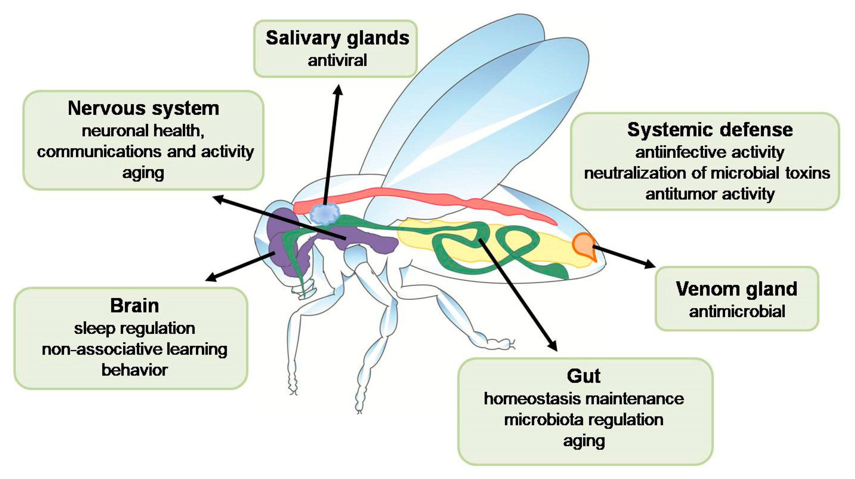
2. AMPs in the Nervous System

Research conducted so far shows that there is a connection between alterations in immune signaling and neuronal health, communication, and activity in insects. Changes in neuroimmune communication and glial cell signaling may contribute to behavioral changes in the insect. In addition, the insect nervous system can mount a local immune response to infection, and activation of signaling pathways may lead to neurodegeneration, malfunctioning neurons, and altered behavior [23]. Moreover, the AMP expression pattern is affected by aging independently of infection, and it has been postulated that an increased level of some AMPs produced in non-neuronal tissues during aging can mediate a signal initiating neuronal aging [24].
AMPs are important for regulation of normal functions of the insect brain, e.g., sleep and non-associative learning in Drosophila [25,26]. Results reported by Dissel et al. showed that the transcript levels of metchnikowin (Mtk), drosocin (Dro), and attacin (Att) were differentially increased in glia, neurons, and the head fat body, respectively, in sleep-deprived flies. They further demonstrated that the expression of Mtk in glia but not in neurons and the expression of Dro in neurons but not in glia had a negative effect on memory but modulated sleep in an opposite way. These AMPs were considered as candidates for conferring resilience/vulnerability to sleep deprivation [27]. A further link between sleep regulation and immune response in Drosophila was found by Toda et al. in their research on sleep mutants and the role of nemuri–a gene involved in sleep induction. Nemuri overexpression increased the sleep length and depth in Drosophila. Interestingly, nemuri encodes an AMP that can be secreted ectopically to drive prolonged sleep and to promote survival after infection. This peptide acts non-cell-autonomously to promote sleep. The nemuri peptide contains 64 amino acids, has sequence similarity to a Greenland cod (Gadus ogac) cathelicidin, and kills bacteria in vitro (Serratia marcescens, Escherichia coli) and in vivo. Drosophila adults overexpressing nemuri in neurons and infected with S. marcescens or Streptococcus pneumoniae survived longer, had a longer amount of sleep, and contained a lower bacterial load than control insects [28]. In another study, a Drosophila non-associative long-term memory (LTM) paradigm involving a natural predator of Drosophila, i.e., the endoparasitoid wasp Leptopilina heterotoma, was used for identification of novel memory genes during and after memory formation. The examination of transcriptional changes in the fly brain revealed that cecropin A1 (CecA1), CecA2, Dro, AttA, dipericin (Dpt), and DptB were differentially regulated at various time points. Specifically, CecA1, Dpt, and Dro were downregulated and upregulated in 7-h and 4-day exposed individuals, respectively [29]. Evidence that AMPs can directly affect brain function was also presented by Barajas-Azpeleta et al., who found 10-12-fold upregulation of diptericin B expression in the adult fly head following behavioral training. Moreover, using DptB null flies, they demonstrated that DptB was required for modulation of long-term memory in Drosophila. Interestingly, only silencing the DptB expression in the head fat body affected long-lasting memory, clearly indicating the head fat body as the only relevant source of DptB for behavioral modification [30].
Recently, the expression of two AMPs belonging to defensins, i.e., galiomicin and gallerimycin, has been detected in the brains of G. mellonella larvae after injection of Habrobracon hebetor venom or topical application of the entomopathogenic nematode Steinernema carpocapse. However, the role of these peptides in the G. mellonella nervous system has not been investigated yet [31]. In this study, the upregulation of the sericotropin gene and peptide was also detected in the brains of challenged larvae. Sericotropin functions in lepidopteran insects as a stimulator of silk production. It has been demonstrated that synthetic peptides derived from the N- and C-terminal parts of sericotropin exhibited antibacterial activity against Xenorhabdus spp. bacteria, i.e., symbionts of S. carpocapse, suggesting an antimicrobial role of sericotropin expressed in the G. mellonella brain [31]. In the brains of the honeybee A. meliffera infected with deformed wing virus (DWV), transcriptomic and metabolomic analyses revealed overexpression of genes encoding proline-rich AMPs, abaecin and apidaecin, as well as hymenoptaecin and an increased level of these peptides [32]. Results obtained in a study on the response of the central nervous system (CNS) of Locusta migratoria manilensis infected with the fungus Metarhizium acridum also indicated an important cross-talk between the CNS and the immune system. The expression patterns in the CNS responded rapidly to the infection and changed as the infection proceeded. Many differentially expressed genes, directly involved in immune function and regulation, were identified, including genes encoding defensin and diptericin, which were up-regulated in the CNS of M. acridum-challenged locusts [33].
The use of Drosophila models of Alzheimer’s disease (AD), traumatic brain injury (TBI), and ataxia-telangiectasia (AT) revealed a role of the Toll and Imd signaling pathways in neurodegeneration [34,35,36,37,38]. Although Toll signaling is engaged in proper development of the brain in Drosophila, it was demonstrated that suppression of Toll signaling protected against neurodegeneration. Moreover, Drosophila mutants in the negative regulator of the Imd pathway, dnr1 (defense repressor 1), had increased expression of AMP genes (Cec, Dpt, Att, drosomycin (Drs), Mtk), which was correlated with progressive age-dependent neuropathological changes and a shortened lifespan. It has also been demonstrated that neurodegeneration is dependent on the transcription factor Relish, and bacterial infection in the brain can trigger neurodegeneration through the neurotoxic effects of AMPs. On the other hand, Relish mutations inhibited upregulation of innate immune response genes and neurodegeneration in AT Drosophila mutants, whereas overexpression of active Relish in glial cells resulted in neurodegeneration [35,39]. Similar effects as those caused by mutations in dnr1 were observed in flies with mutations in other negative regulators of the Imd signaling pathway: zfh1 (transcription factor Zn finger homeodomain 1), Pirk (poor Imd response upon knock-in), trbd (deubiquitinase Trabid), and tg (Transglutaminase), strengthening the evidence for the negative role of AMPs in the development of diseases [40,41]. Moreover, the overexpression of Dro, AttC, and CecA1 in neurons or glia was accompanied not only by a shortened lifespan but also by early appearance of locomotor defects in comparison with controls, suggesting a causative relationship between high levels of AMPs and neurodegeneration in Drosophila [41]. In turn, Barati et al. studied the effect of amyloid-β 42 (Aβ42) and tau on the Imd pathway and neuroinflammation gene expression in a Drosophila model. They showed that the expression of genes involved in the Imd pathway, such as Relish and AMPs (AttA, DptB), increased with age in the W1118 control flies and in the embryonic nervous system of AD transgenic flies Aβ42, tauWT, or tauR406W, but the level of AMPs in glia remarkably decreased compared to W1118. The decline was higher in both tau flies compared to Aβ42 transgenic flies. The overexpression of AMPs in Drosophila leads to brain neurodegeneration and neuroinflammation [42]. Another example connecting overexpression of AMPs with neurodegeneration was provided by research on the role of the Yorkie transcription factor in polyglutamine (PolyQ)-mediated neurotoxicity (involved in Huntington’s disease in humans) in Drosophila. It was found that PolyQ expansion increased the expression of CecA, Att, Dpt, Dro, and Drs. It was postulated that upregulation of AMPs can participate in PolyQ-mediated neurotoxicity in Drosophila [43].
An important role of one of the AMPs, metchnikowin, in acute and chronic outcomes of TBI was demonstrated by Swanson et al. in a Drosophila TBI model. In an analysis of null mutations in 10 AMP genes (AttA, AttB, AttC, AttD, defensin (Def), Dro, Drs, Mtk, DptA, DptB), it was found that Mtk mutant flies exhibited reduced acute behavioral deficits and only the mutation in the Mtk gene protected flies from early mortality after TBI in different diet conditions. These results indicate that Mtk plays an infection-independent role in the nervous system and can promote neuropathological changes in the brain following TBI [44]. In another study conducted in a Drosophila Closed Head Injury (dCHI) model, an acute broad-spectrum immune response was detected in glia cells, where many AMP genes were up-regulated 24 h after TBI (Att, Cec, Def, Dpt, Dro, Drs, Mtk, listericin (Lys)). It was found that the deletions of most of these AMPs shortened insect survival after TBI. In contrast, loss of Def extended survival, indicating that the Drosophila immune response to TBI, including the induction of AMP expression, may result in different effects. Moreover, in this research, a link between the immune response and sleep in flies was demonstrated, as loss of Relish protected against impaired control of sleep and movement after TBI [26].
Progress of neurodegenerative processes is correlated in time with an age-dependent increase in AMP expression in the head tissue of Drosophila [41]. Using a transgenic Drosophila model of AD, Wang et al. provided evidence for distinct expression profiles of AMP genes in an aging model and in wild-type flies. Whole transcriptome profiles of wild-type Drosophila heads revealed upregulation of 12 AMP genes: AttA, AttB, AttC, AttD, CecA2, CecC, DptA, DptB, Drs, Def, Dro, and Mtk. A gradual increase in expression of these AMPs was observed in the wild-type flies during the normal aging process, whereas an initial decrease and a subsequent increase in the expression levels was found during aging of the AD flies. Interestingly, significant correlations between the abnormal amyloid β (Aβ) peptide concentration and abnormal expression of AttB, AttC, CecA, Drs, Mtk, and LysS were detected, suggesting contribution of AMP dysregulation to AD progression by inducing deposition of Aβ peptide aggregates [45]. However, in other studies, lysozyme was demonstrated to be beneficial in different Drosophila models of AD. For example, co-expression of human lysozyme rescued the rough eye phenotype indicative of toxicity in flies that expressed Aβ1-42 in the eyes. Moreover, lysozyme binding with Aβ1-42 in the Drosophila eye was detected, suggesting that such an interaction prevented from the toxic effects of Aβ1-42 [46]. Interestingly, antimicrobial activity of the Aβ peptide was reported, suggesting that it is an AMP in the nervous system and that neurodegenerative Alzheimer’s disease may be partially an infectious disease [47]. Increased activation of innate immunity mechanisms reflected by overexpression of AMP genes was also postulated as a cause of long-term deleterious effects leading to neurological deficits, persistent cell death in brains, and premature aging in a Drosophila model of radiation-induced neurotoxicity. In this study, upregulation of Drs, DroA, Dpt, AttC, Cec, and Mtk was detected in brains of 5-day- and 15-day-old adult flies after radiation exposure during larval development (late third larval instar), indicating contribution of persistent activation of immune signaling pathways to neurological deficits in adult flies [48].
The connection between immunity and nervous system is also evident at the level of neuropeptides. Among human neuropeptides, antimicrobial activity has been reported for enkelytin, substance P, neuropeptide Y, bombesin, adrenomedullin, vasoactive intestinal peptide, and pituitary adenylate cyclase activating polypeptide (PACAP) [49,50]. Besides regulation of neurodevelopment, emotion, and certain stress responses, the PACAP neuropeptide acts as an antimicrobial agent. Although its antimicrobial function was demonstrated in mammals, the phylogenetic analysis predicted antimicrobial properties of eleven PACAP homologs, including PACAP from the cockroach Periplaneta americana [51].

3. AMPs in Aging

Aging is a complex process involving accumulation of harmful changes resulting in an overall decline in several vital functions. It leads to progressive deterioration in the physiological condition of the organism such as changes in metabolism and behavior, impaired neuronal function, and reduced stress resistance, reproductive and immune capacity, or barrier function in the gut, which eventually cause disease and death. Many results important for understanding the molecular basis of aging were provided by research conducted in a D. melanogaster model [52].
A well-documented hallmark of aging in Drosophila is the decline in immune functions accompanied by an increase in AMP gene expression. Aging is associated with expansion of the AMP repertoire and loss of their specificity, possibly caused by immune dysregulation [2,53,54]. It was reported that the expression level of AMP genes, including AttA, AttB, AttC, AttD, CecC, Dpt, Def, Dro and Met as well as transcription factor Relish, increased considerably with age in Drosophila flies [55,56]. Initially, elevated AMP levels in old flies were associated with an impaired ability to clear infections effectively, but later studies showed little correlation between the AMP expression level and the microbial load. Badinloo et al. showed that decreased expression of Relish in the fat body of old Drosophila flies extended longevity, which was correlated with lower activation of AMP genes, whereas overexpression of AMP genes controlled by Relish was accompanied with a significantly shortened lifespan. Particularly, the overexpression of four AMPs: attacin A, defensin, metchnikowin, and cecropinA1, significantly decreased the lifespan of the flies, while the overexpression of drosomycin and drosocin had no significant impact on longevity. Increased expression of AMPs was observed in different body parts, including the gut, muscles, and fat body. High AMP levels correlated with depolarization of mitochondrial membranes and apoptosis of cells in different tissues. Hence, AMPs may have an impact on aging by causing changes in the mitochondrial membranes that lead to apoptosis [57]. The death of olfactory receptor neurons, particularly Or42b, was demonstrated to be connected with increased expression of AMPs during non-pathological aging in Drosophila. It was found that these neurons died as a result of age-related caspase 3-like protease activation associated with systemic activation of innate immunity signaling pathways, leading to enhanced expression of AMPs. Interestingly, Dro was sufficient for activation of caspase 3-like protease and cell death in Or42b neurons [58]. Overexpression of AMP genes was also connected with a shortened lifespan when the circadian clock-controlled Achilles gene was characterized in Drosophila. Achilles encodes a putative RNA-binding protein; its activity is rhythmic at the mRNA level, and it suppresses the expression of AMPs. Knocking-down of Achilles in Drosophila neurons led to dramatically increased systemic expression of immune related genes, including Mtk and Drs, and significantly reduced the overall lifespan of adult flies in the absence of immune challenge [59], again indicating a role for AMPs in lifespan shortening.
On the other hand, activation of a single AMP, Dro, in the gut or ubiquitously in adult Drosophila caused a significant extension of the fly lifespan accompanied by lower activity of immune signaling pathways over lifetime, decreased stress response, and slower loss of gut barrier integrity. No such effect was observed when Dro was expressed in the gut of flies with a diminished endogenous bacterial load in the gut, indicating a cross-talk between innate immunity, intestinal homeostasis, and aging. Similarly, increased ubiquitous expression of CecA1 positively influenced the Drosophila lifespan [60]. A role of AMPs, mainly Dpt, in increasing tolerance to oxidative stress correlated with a prolonged lifespan of Drosophila adults was reported in earlier studies. Overexpression of Dpt, Att, Cec, Drs, and Mtk was detected in flies that tolerated enormous oxidative stress. Then, it was demonstrated that overexpression of even one AMP (Dpt, Att) can be sufficient for better survival of wild-type flies in hyperoxia [61,62].
Recently, using Drosophila lines with different combinations of knocked out AMP genes, the pathogen-specific roles of AMPs have been reported, where microbicidal activity against a particular pathogen depended mainly on a certain AMP, e.g., the Dpt gene family alone provided effective defense against Providencia rettgeri, drosocin contributed to defense against Enterobacter cloacae, whereas buletin (also encoded by the Dro gene) was effective against Providencia burhodogranariea [63,64]. In addition to these findings, it turned out that when considering the role of AMPs in Drosophila response to infection, age and sexual dimorphism should be taken into account given the documented strong differences in immune response at the basal level upon infection and during aging between sexes [65,66]. Hanson et al. showed that Dpt expression was sufficient to protect young male flies against P. rettgeri; however, Shit et al. demonstrated that reintroduction of functional DptA and DptB (∆AMPs+Dpt) fully restored survival and decreased the P. rettgeri load only in young males but not in females or older males [63,66]. Aging had negative effects on the fitness and pathogen clearance ability of Drosophila females but not males. Infection of flies with P. rettgeri resulted in higher mortality and increased bacterial burden in older females than in younger females, while both young and old males had similar mortality rates and bacterial loads. Most mutant flies lacking a certain AMP, or a combination of AMPs, became highly susceptible to infection with age, with the exception of female mutants lacking Def. This effect may be related to the sexual dimorphism, where defensin is important for defense in males but not in females [66].
As reported by Badinloo et al., AMP induction in old flies appeared to be independent of recognition of PAMPs upstream Imd, but the activity of Ird5, i.e., a component of the IKK complex, was essential for AMP expression. The up-regulation of AMPs associated with aging was reduced in the ird5 mutant but was unaffected in the imd mutant flies, indicating that aging activates other pathways involved in induction of AMP expression [57]. Much earlier, Becker et al. showed that the AMP expression in non-infected Drosophila flies may be independent of the Toll and Imd signaling pathways but may depend on the transcription factor Forkhead Box O (FoxO), which is known as a key downstream regulator of the ILS pathway involved in metabolism, stress resistance, and aging. The AMP upregulation was lost in starved foxo mutant larvae, while foxo overexpression led to increased AMP expression. Direct binding of FoxO to the Drs regulatory region in Drosophila cells suggested direct regulation of AMP genes by FoxO, depending on the energy status of the cell, and indicated a cross-regulation of metabolism and innate immunity [67]. Conserved FoxO-binding sites have been identified in silico in Drosophila promoter regions of Cec and Dro [68]. These authors also demonstrated that AMPs were produced in Drosophila enterocytes in a FoxO- but not Relish-dependent manner. Furthermore, they showed the FoxO regulatory role in gut AMP expression after Drosophila oral infection with the Gram-negative bacterium S. marcescens. After infection, upregulation of Dpt, AttA, and AttB was detected in gut epithelia of wild type flies, while no significant increase in the transcript levels of these genes was noticed in the foxo mutants. The impaired FoxO signaling diminished the resistance of Drosophila to intestinal infection due to an insufficiently high level of AMP expression, which eventually caused a decline in survival [68]. Recently, involvement of FoxO in the activation of AMP gene expression has been demonstrated in the fat body of starved B. mori larvae [69]. Similarly to FoxO, a role of Forkhead (FKH), i.e., another transcription factor from the same family, in the regulation of AMP expression in the Drosophila gut was demonstrated. The results indicated that the TOR signaling pathway, responding mainly to protein availability, is also engaged in the cross-talk between metabolism and innate immunity by regulation of the expression of Dpt and Mtk [70].

4. AMPs as Antitumor Agents

Some insect AMPs exhibit selective in vitro cytotoxicity against different cancer cell lines, e.g., melanoma, lymphoma, leukemia, breast cancer, lung cancer, and bladder cancer [71,72,73,74]. Besides in vitro studies on the anticancer activity of insect AMPs, there are reports indicating in vivo involvement of AMPs in antitumor action, particularly based on research carried out using Drosophila models.
In vivo studies conducted by Parvy et al. showed the anticancer effect of defensin in a D. melanogaster model. The presence of tumor cells in the D. melanogaster body activates a cellular response, which leads to release of Eiger, i.e., a Tumor Necrosis Factor (TNF), from hemocytes. TNF is required for the exposure of phosphatidylserine on the surface of tumor cells. The presence of this phospholipid on the surface of cells gives them a negative charge, making them the target of cationic defensin produced, among others, in fat body cells. The effect of this interaction is cancer cell death and tumor regression. The Imd pathway in the trachea is responsible for expression of the Def gene in D. melanogaster and the Toll and Imd pathways in the fat body. Deletion of the defensin-encoding gene resulted in death of fewer cancer cells and enlargement of the tumor [75]. It was also found that AMP genes Drs, Def, Dpt, Mtk, AttA, and CecA2 were upregulated in Drosophila mxcmbn1 mutants with malignant hyperplasia in lymph glands. The downregulation of the Toll or Imd pathways increased, while the ectopic expression of each of the five different AMPs (Drs, Def, Dpt, Mtk, AttA) in the fat body suppressed the tumor phenotype in the mxc mutants. Moreover, drosomycin and defensin secreted by the fat body were taken up by circulating hemocytes and accumulated in their cytoplasm. These hemocytes were recruited into the tumor. Another AMP, diptericin, was directly localized at the tumor region. The AMPs increased apoptosis only in the tumor cells [76,77]. Literature data also indicate a link between anti-infection and anti-cancer mechanisms. Jacqueline et al. reported that oral infection with the Gram-negative bacterium Pectobacterium carotovorum accompanying tumor in Drosophila resulted in a reduction in the tumor size, compared to insects without infection. This effect was related to an increase in the expression of genes encoding diptericin and drosomycin AMPs, leading to increased death of tumor cells [78].

5. AMPs and Symbiotic Microbiota

Many insects possess mutualistic symbiotic microorganisms in the gut lumen and the body cavity or inside cells (Table 1). Most symbionts are vertically transmitted from mother to offspring, but gut symbiotic bacteria can be horizontally acquired in every generation from the environment, such as soil, water, and food. Some symbionts are obligate because they are essential for host survival, while others are facultative. All insects harbor facultative bacterial endosymbionts. Usually, these symbionts are beneficial to the host, providing essential nutrients, defending against pathogens, and facilitating adaptation to environmental conditions. They supplement the insect host’s diet with vitamins and cofactors and are a source of important amino acids [79]. For long-term survival in the host body, symbionts use different strategies that allow them to withstand the adverse effects of the host immune defense mechanisms and simultaneously be safe for the host [80,81,82,83,84]. Nevertheless, symbionts can become harmful to the host if they reproduce and grow uncontrollably, as they consume more nutrients than in a normal symbiotic relationship. Therefore, their number in the insect body should be controlled by the host immune system and a disturbance in their load can lead to dysbiosis. In some insect species, endosymbionts are housed within specialized host cells named bacteriocytes, which make up an organ called the bacteriome located around the host midgut and germ cells, from which they are transmitted to their progeny. Compartmentalization of symbionts in bacteriomes is considered a strategy used by the host to control beneficial symbionts in a limited space, whereas host immune mechanisms acting in other host tissues prevent pathogenic infection [2,85,86,87,88]. In insect-bacterial symbiosis, specific factors are produced by the host immune system to modulate the population of symbionts. AMPs, participating not only in the systemic but also local epithelial immune response are such factors [2,89,90]. In the insect gut, the Imd pathway together with the Jak-STAT pathway and the Duox-ROS system play a key role in the fight against pathogens and simultaneous control of symbiotic bacteria. Therefore, dysregulation of these pathways exerts a negative or positive influence on the gut microbial growth [89,91,92,93,94,95].
It has been reported that symbiotic bacteria do not induce the synthesis of intestinal AMPs, although they activate Relish. The low basal level of AMPs is associated with negative regulation of Relish by Caudal (Cad) (Figure 1). Caudal has been identified as a homeobox transcription factor essential for the formation of the anteroposterior body axis in Drosophila embryos, while in adult flies it is predominantly expressed in the posterior midgut, where it is involved in the maintenance of a low level of midgut-specific AMP gene transcription. The antagonistic relationship between Cad and Relish providing balance between immune defense and microbiota homeostasis in the insect gastrointestinal tract has been reported in D. melanogaster, G. mellonella, and Anopheles gambiae [85,96,97,98]. In Drosophila, Caudal is responsible for the constitutive low local expression of cecropin and diptericin in a tissue-specific manner. In flies lacking Caudal, the gut microbiota continuously activates the Imd pathway inducing AMP overexpression, which results in increased apoptosis of gut epithelial cells and a shorter lifespan [96]. RNAi-mediated silencing of Cad in A. gambiae resulted in a decreased prevalence and altered the species composition of midgut microbiota due to the increased expression of Relish2-controlled AMPs: cecropin-1, cecropin-3, gambicin, and defensin-1. In addition, after Gram-positive Staphylococcus aureus bacterial infection, an increased level of defensin-1 and gambicin was observed, which contributed to better survival of mosquitoes [97].
Enterococcus mundtii (syn. Streptococcus faecalis Andrewes and Horder) is considered a dominant bacterium in the midgut of G. mellonella larvae; it is transmitted vertically−from mother to offspring [99,100,101]. Krams et al. showed that the quality and quantity of the diet affected the number of Enterococci and had a significant effect on the expression level of AMP genes in the midgut of G. mellonella larvae. An elevated level of CecD, galiomicin, gallerimycin, gloverin, and 6-tox expression was correlated with an increased number of Enterococcus symbionts and diet diversity. It was postulated that an increased basal expression level of genes encoding galiomicin and particularly gloverin, i.e., an AMP with activity against Gram-positive bacteria such as Enterococcus, may be a mechanism that controls the symbionts and acts prophylactically against opportunistic pathogens in G. mellonella [101]. It has been shown that the Imd pathway also plays an important role in G. mellonella gut immunity, because the expression level of Imd pathway genes (Imd, Relish), in contrast to those related to the Toll pathway (Spätzle, Dif/Dorsal), was relatively higher in the gut (Figure 1) [98]. Moreover, it was observed that silencing of Relish resulted in downregulation of the gut expression of CecD, gloverin, gallerimycin, transferin, and galiomicin and bacterial overpopulation in the G. mellonella gut. Peroral treatment of the larvae with antibiotics eliminated the gut microbiota and significantly lowered the expression of Imd pathway genes, including all AMP genes. In such conditions, the Caudal expression in the gut was decreased as well, probably due to the low activity of the Imd pathway and the low level of AMPs [98,101].
The gut microbiota of the honeybee A. mellifera is a highly specialized bacterial community composed of e.g., Snodgrassella alvi, Gilliamella apicola, Frischella perrara, Bifidobacterium spp., and Lactobacillus spp. (Table 1). Frischella perrara is the first to colonize the gut immediately after adult emergence, is harbored by old forager bees, and strongly stimulates the host immune system in the presence of other gut bacterial species. A transcriptome analysis showed that F. perrara induced the host gene expression more potently than S. alvi, in particular genes known to be activated in response to bacteria, including AMP genes encoding apidaecin type 14 (Apid1), apidaecin type 73 (Apid73), abaecin (abaecin), and defensin 1 (Def1) [102]. Another study showed that the microbiota present in the gut of honeybees induced the expression of Apid and hymenoptaecin genes. A significant increase in the expression of these two AMPs was observed in the gut tissue of bees fed the gut symbiont S. alvi or normal gut microbiota, compared to bees lacking the gut microflora. No such differences were detected for the expression of abaecin and Def. However, bees lacking their normal gut microbiota have lower basal expression of AMPs, which may negatively affect their ability to fight infection. Interestingly, resident gut bacteria are relatively resistant to bee AMPs, compared to foreign species [103]. Additionally, different levels of resistance of A. mellifera microbiota species to different AMPs were found. In general, A. mellifera microbiota was more resistant to apidaecin Ia than Ib, whereas S. alvi was more resistant to apidaecin Ib than G. apicola. Gram-positive bacteria (Lactabacillus, Bifidobacterium) were highly resistant to both apidaecin and hymenoptaecin, while Gram-negative species, particularly S. alvi, were more sensitive to hymenoptaecin [103].
Referring to the intestinal microbiota of the honeybee, it is also worth paying attention to peptides with antimicrobial activity present in royal jelly. This is important because royal jelly, secreted by the pharyngeal glands of worker bees, is the food for larvae on the first three days of their development and the queen throughout its life; hence, its antimicrobial action is crucial. Several peptides with antimicrobial activity have been identified in royal jelly, e.g., royalisin and jelleines [104,105]. A. mellifera royalisin has broad antifungal and antibacterial properties, for instance against Paenibacillus larvae, i.e., an inducer of microbial epidemic in honeybee larvae. Royalisin is a typical cysteine-rich defensin-like peptide composed of 51 amino acids and stabilized by three disulfide bridges. This peptide shows high homology to Sarcophaga peregrina sapecin and Protophormia terraenovae phormicins, which are peptides with antibacterial activity [106]. Most jelleines (I-IV) are derived from the C-terminus of Major Royal Jelly Protein-1 (MRJP-1). They are short peptides composed of 8–9 amino acids. Jelleines, particularly jelleines I and II, have antibacterial and antifungal activity [104,107,108,109].
Modulation of the immune response to maintain endosymbionts was reported in carpenter ants of the genus Camponotus (Hymenoptera). These ants host their primary endosymbiont, a Gram-negative bacterium Blochmannia, which resides free in the cytoplasm of bacteriocytes in the midgut tissue and in matured oocytes. This endosymbiont is crucial for ant colonies, ensuring proper growth, development, and high fecundity of its host, which was demonstrated in studies of ant colonies with experimentally-reduced Blochmannia [110,111]. Camptonotus floridanus ants reduce the immune response within midgut tissue and ovaries in order to allow survival of Blochmannia floridanus, while the immune gene expression in other tissues is maintained at a normal level (Table 1) [112]. Low expression of genes encoding lysozymes and such AMPs as hymenopteacin and defensin-1 in bacteriocytes was demonstrated. C. floridanus hymenopteacin has activity against Gram-negative bacteria; therefore, its excessive synthesis can harm the Gram-negative endosymbionts. In addition, since the expression level of two PRR genes, PGRP-LB and PGRP-SC2, in the midguts of untreated insects was high, it was suggested that the products of these genes down-modulate the immune response of this tissue, which ensures tolerance of endosymbionts. These PGRPs are known to possess amidase activity responsible for peptidoglycan degradation [16], which prevents the activation of the immune response towards endosymbionts, including AMP synthesis in C. floridanus. Both these genes were much more strongly expressed in the midgut tissue than in other tissues. However, when Blochmannia is recognized in the hemocoel, an immune response is activated to prevent the spread of symbionts [112]. More recently, compartmentalized PGRP expression has been detected in the gut of the oriental fruit fly Bactrocera dorsalis (Diptera). High expression of PGRP-LC was found in the B. dorsalis foregut, while PGRP-LB and PGRP-SB, i.e., encoding receptors with amidase activity, were mainly expressed in the anterior and middle midgut. This expression pattern correlated with the presence of symbiotic Enterobacteriaceae in the B. dorsalis midgut. The knockdown of PGRP-LB and PGRP-SB led to increased expression of midgut AMPs and, consequently, to a decreased number of symbiotic bacteria and an elevated number of opportunistic microbes. Such regional expression of different PGRPs provides protection for symbiotic bacteria [113].
The Gram-negative bacterium Sodalis pierantonius is an endosymbiont of the cereal weevils Sitophilus spp. (Coleoptera) maintained in the bacteriome. When S. pierantonius bacteria are injected into the hemocoel, they induce systemic expression of AMP genes. Nevertheless, in physiological conditions, only the coleoptericin A (ColA) AMP gene is expressed in the bacteriome, while the other AMP genes are expressed only slightly or not at all, allowing the endosymbiont to survive. In symbiotic insects, ColA was expressed in all tissues harboring the endosymbionts, while in aposymbiotic insects ColA is constitutively present in gut epithelia, the fat body, and under the cuticle [114]. ColA and ColB have been identified only in coleopteran species, both having bacteriostatic activity against Gram-positive and Gram-negative bacteria. ColA impairs Escherichia coli cell division and leads to bacterial gigantism through specific binding with OmpA, which allows the peptide to enter the bacterial cell, where it interacts with a 60 kDa chaperonin GroEL. A similar phenotype was described for S. pierantonius cells in the weevil bacteriome [114]. Inhibition of ColA expression by RNAi in Sitophilus weevils was shown to result in partial loss of endosymbiont control, allowing them to leave the bacteriocytes and infect the surrounding tissues. Moreover, it was found that the ColA transcript level in weevils was correlated with the S. pierantonius load and that the expression of ColA (and ColB) in the bacteriome could be modulated by systemic infection of insects by exogenous bacteria [87,115,116].
The bean bug Riptortus pedestris (Hemiptera) with its monospecific gut symbionts, i.e., the genus Burkholderia, is a useful gut symbiosis model. The midgut of R. pedestris is divided into five sections M1, M2, M3, M4B, and M4. In the M4 region, the symbiotic organ contains a specific symbiont Burkholderia insecticola, which is orally acquired from the environment by second-instar nymphs [117]. Park et al. showed that the intestinal immunity of R. pedestris is related to an AMP called rip-thanatin and that the expression of the rip-thanatin gene upon systemic bacterial infection significantly increased in the M4 crypt [118]. When rip-thanatin was silenced by RNAi, the number of symbiotic bacteria also increased in the midgut of the bean bug, suggesting that thanatin controls the population of gut-colonizing Burkholderia symbionts. Indeed, it was shown that rip-thanatin has high activity against E. coli and Staphylococcus aureus bacteria but lower activity against symbiotic Burkholderia. The Burkholderia population has to be controlled in the midgut; when overgrown and present in the hemolymph of host insects, Burkholderia exhibit insecticidal activity [117,118,119].
Insect gut microbiota and resistance to infection can also be affected by endosymbionts that reside outside the gut. Drosophila flies naturally harbor two such endosymbiotic bacteria: Spiroplasma and Wolbachia [120,121]. The cell wall-less Spiroplasma poulsonii and its relatives are vertically transmitted facultative symbionts residing extracellularly in hemolymph of several Drosophila species. The absence of the host immune response to Spiroplasma is not caused by suppression of the immune system but is related to the fact that Spiroplasma cells are not detected by these flies [122,123,124]. It has been demonstrated that Spiroplasma generally did not affect the production of AMPs in Drosophila and triggered only mild and chronic synthesis of immune factors. However, proteomic analyses revealed enrichment of attacin and bomanin Bc3 in hemolymph of Spiroplasma-harboring insects. Nevertheless, the overexpression of selected AMP genes did not influence the endosymbiont titer, and flies with deleted 10 AMP genes contained Spiroplasma titers comparable with the control flies [116]. A comparison of the survival rate of Spiroplasma-free and Spiroplasma-harboring flies infected with different bacteria and the fungus Beauveria bassiana revealed that Spiroplasma decreased the resistance of the flies to infection by certain bacterial pathogens, mainly Gram-negative bacteria (Erwinia carotovora, Enterobacter cloacae) [124].
Table 1. Insects and their gut endosymbionts.
OrderHost InsectSymbiontsReference
LepidopteraBombyx moriProteus vulgaris, Erwinia sp.
Klebsiella pneumoniae
Citobacter freundii
Pseudomonas fluorescens
[125]
Galleria mellonellaStreptococcus faecalis
(Enterecoccus mundii)
[99,126]
Plutella xylostellaEnterococcus sp.
Enterobacter sp., Serratia sp.
[127]
Spodoptera lituraSerratia sp.[128]
DipteraAedes aegyptiWolbachia[129]
Anopheles gambiaeEnterobacter asburiae, Serratia sp.
Microbacterium sp.
Sphingomonas sp.
Chryseobacterium meningosepticum
[130]
Drosophila melanogasterSpiroplasma poulsonii
Acetobacter thailandicus
Lactobacillus plantarum
[96,131,132,133]
Drosophila nebulosaSpiroplasma poulsonii[124,134]
Glossinia spp.Sodalis glossimidia
Wigglesworthia sp.
Wigglesworthia glossinia
[79,135,136,137,138]
Melophagus ovinusArsenophonus melophagi
Sodalis melophagi
[139,140]
HymenopteraApis melliferaGilliamella apicola, Snodgrassiella sp.
Frischella perrara, Snodgrassella alvi
Bartonella apis
[102,103,141,142,143,144,145,146]
Bombus spp.Giliamella bombicola
Snodgrassiella sp.
[146,147]
Camponotus floridanusBlochmannia floridanus[112]
ColeopteraAcyrthosiphon pisumBuchnera aphidicola[148,149,150]
Cyrtotrachelus buquetiLactococcus sp., Serratia sp.
Dysgonomonas sp., Enterrococus sp.
[151]
Holotrichia parallelaPseudomonas sp., Ochrobacterium sp.
Cellulosimicrobrium sp.
[152]
Hylobius abietisErwinia sp., Rabnella sp., Serratia sp.[153,154]
Nicrophorus vespilloidesProvidencia sp., Morganella sp.
Vagococcus sp., Proteus sp.
Koukoulia sp., Serratia sp.
[155,156,157]
Pachyrhynchus infernalisNardonella sp.[123]
Paradieuches dissimilisCaballeronia, Symbiopectobacterium Wolbachia, Rickettsiella[158]
Riptortus pedestrisBurkholderia[159,160]
Sirex noctilioStreptomyces sp.[161]
Sitophilus oryzeSodalis pierantonius[162]
Sitophilus weevilsSodalis pierantonius[163]
BlattodeaBlattella germanicaBlattabacterium cuenoti[164]
The effect of Wolbachia, residing mainly in the fly germline, on D. melanogaster gut commensal microbiota was reported as well. Wolbachia is an intracellular inherited bacterium naturally infecting more than 60% of all insect species worldwide and affecting host populations, e.g., through its positive influence on host fitness [165]. In Wolbachia-infected flies, significantly reduced titers of Acetobacter species were detected, whereas the titers of other commensal bacteria from the genus Lactobacillus were not affected [166]. Interestingly, Wolbachia protects D. melanogaster against enteric viral infections and those caused by the opportunistic pathogen Pseudomonas aeruginosa [167,168]. Therefore, it is regarded as “a supplementary immune system” protecting the host against infection. In turn, other data indicate that Wolbachia does not affect the survival and immune response of D. melanogaster or D. simulans during systemic infection by P. aeruginosa, E. carotovora, or S. marcescens [169] and by intracellular bacteria such as Listeria monocytogenes and Salmonella typhimurium or extracellular bacterial pathogens P. rettgeri [170]. Later, it was found that a key role in the protection of Drosophila by Wolbachia is played by the route of infection; the protection is ensured in the case of enteric but not systemic infection by P. aeruginosa. Gupta et al. observed that the survival of flies after oral infection with P. aeruginosa increased when they were Wolbachia-positive, in comparison to Wolbachia-negative ones. Additionally, it was found that this protective effect was sexually dimorphic, as male flies harboring Wolbachia were able to clear bacteria within a week, whereas females stopped infection clearing after 96 h and maintained a stable bacterial load. Expression studies of Imd-mediated genes involved in antibacterial immune response have shown that they are upregulated during enteric bacterial infection in flies. In females, significantly increased expression of PGRP-LC in the anterior fly midgut and PGRP-LE in the posterior midgut was observed. In turn, increased expression of AttA, which is mediated by the Imd pathway, was shown in males [168].

6. Non-Classical AMPs and Infection

In addition to peptides exhibiting the characteristics of AMPs, recent research on Drosophila has indicated a role of so-called non-classical AMPs in infection. Some of them can confer infection resistance. Interestingly, the expression of these peptides is under the control of the Toll pathway [171]. Taxonomically-restricted Drosophila-specific bomanins, a peptide family encoded by 12 Bom genes activated by the Toll pathway are an example of non-classical AMPs involved in resistance. It was reported that BomΔ55C mutant flies lacking 10 Bom genes succumbed to Enterobacter faecalis, Candida glabrata, and Fusarium oxysporum more quickly than wild-type flies due to the defect in resistance [172]. A further study revealed that the individual short-form bomanins can provide resistance against C. glabrata when present in Drosophila hemolymph in the absence of the remaining 9 Bom peptides. This effect was dependent on the level of transcription rather than on the peptide sequence [173]. Moreover, Drosophila mutants in the Bom55C locus exhibited susceptibility to Aspergillus fumigatus mycotoxins, verruculogen, and restrictocin. The susceptibility of these mutant flies was rescued by overexpression of bomanins. For example, flies expressing BomS6 in the nervous system survived better and recovered faster from tremors caused by verruculogen injection, indicating that specific bomanins are able to neutralize mycotoxins instead of acting as classical AMPs [174]. Recently, it has been demonstrated that, from among the D. melanogaster Baramicin genes, namely BaraA, BaraB, and BaraC, only BaraA is involved in immunity and is upregulated by the Toll signaling pathway upon infection. The peptide products of this gene, i.e., IM10-like peptides, exhibit antifungal activity in vitro [175]. However, results reported by Huang et al. connected the antimicrobial activity of BaraA-encoded peptides with neutralization or counteraction of microbial toxins (e.g., E. faecalis EntV bacteriocin, Metarhizium robertsii destruxin A) rather than with direct antimicrobial action. Interestingly, the two other genes, BaraB and BaraC, encode truncated forms and are nervous system-specific genes in Drosophila [175,176]. Daisho1 and Daisho2, i.e., other taxonomically-restricted genes encoding Drosophila-specific non-classical AMPs, have also interesting contribution to antifungal defense. These two genes encode peptides that belong to effectors activated by Toll signaling, likewise Bom and Baramicin genes. They appeared to be particularly involved in defense against some subset of filamentous fungi, e.g., some species from the genera Fusarium and Aspergillus. Although in vitro binding of Daisho2 to F. oxysporum hyphae was demonstrated, whether Daisho peptides possess direct antifungal activity remains to be elucidated [177].

7. AMPs in Antiviral Response

Like all living organisms, insects are exposed to viruses during their lifetime. Studies of interactions between insects and viruses focused initially on species used economically by humans, such as the silkworm B. mori or the honeybee A. mellifera. A special role in the study of insect immune mechanisms, also in the context of antiviral defense, is played by the fruit fly D. melanogaster [178,179].
The primary mechanism involved in the antiviral response in insects is the activation of the RNAi pathway. This pathway is triggered by the presence of viral double-stranded RNA (dsRNA) formed during replication of RNA viruses or, in the case of DNA viruses, formed between complementary transcripts from convergent transcription [180]. In the RNAi pathway, small RNA is produced by cleavage of viral dsRNA. There are three main types of small RNAs: small interfering RNAs (siRNAs), microRNAs (miRNAs), and PIWI interacting RNAs (piRNAs). Damaged viral RNA prevents its replication [179,181]. Activation of RNA-based immunity has been described in a recently published review [179]. In addition, insects engage melanization, encapsulation, endocytosis, and autophagy in response to viral infection [182]. As a result of viral infection, the Toll, Imd, and Jak-STAT signaling pathways are also activated, which leads to induction of the synthesis of active molecules, including AMPs [183].
Representatives of the order Diptera, including many species of mosquitoes, are vectors for human pathogens, e.g., Zika, yellow fever, dengue, and Chikungunya viruses. Viruses in these insects occur in a persistent form, which is associated with the risk of transmission to humans during a bite [184]. Therefore, there are numerous studies on the mechanisms of the antiviral response in insects and the ability to eliminate viruses by the insect’s organism and thus reduce the risk of transmission [185]. Most mosquito viruses are RNA viruses belonging to the family Flaviviridae (yellow fever virus, dengue-DENV, Japanese encephalitis virus, West Nile virus, Zika virus), Togaviridae (Chikungunya virus, Sindbis virus), and Bunyaviridae. Viruses enter the mosquito’s body while feeding, multiply, and then reach its salivary glands. From there, they are transmitted to the new host. The transfer from the midgut to the salivary glands is not fully understood. It is likely that the virus replicates in the midgut epithelium, from where it enters the hemocoel. Then, along with the hemolymph, it can migrate to other tissues, i.e., salivary glands, fat body, trachea, muscles, or neural tissue. The mosquito’s immune system must protect the host by limiting the virus load, which is also a key factor limiting the transmission of the virus to humans [181].
Particular attention is focused on immunological reactions in the mosquito salivary glands, as this is the last stage before the transmission of the virus to humans. Genomic analyses of salivary glands of uninfected Aedes aegypti and those infected with DENV showed that the infection causes an increase in the expression of genes activated by the Toll and Imd pathways, including the gene encoding cecropin-like peptide. Both the pre-mature and mature forms of the peptide showed antiviral activity against the DENV and Chikungunya virus [186]. AMPs were also found in the Ae. aegypti midgut, fat body, and hemocytes. Activation of the Toll and Jak-STAT pathways in Ae. aegypti has been shown to reduce the level of DENV replication in the midgut [187]. Pan et al. confirmed the antiviral effect of AMPs in Ae. aegypti against DENV. Their research showed that defensins and cecropins made the Wolbachia-infected mosquito resistant to this virus. Mutations in the genes encoding defensin C, defensin D, cecropin N, and lysozyme C resulted in a higher viral load in the mosquito’s body. There was no antiviral activity of defensin A, cecropin G, cecropin D, cecropin E, and gambicin [185,188,189,190].
Activation of the Toll pathway in D. melanogaster is triggered after infection with Drosophila X virus (DXV), Drosophila C virus (DCV), Cricket paralysis virus (CrPV), Flock House virus (FHV), and Nora virus. Research conducted by Ferreira et al. indicated that the Toll pathway determined the immunity of Drosophila after natural oral infection with DCV, CrPV, FHV, and Nora virus. Toll pathway mutants showed a higher viral load [191]. It has also been shown that Drosophila Dif mutants were more sensitive to DXV [192]. This was not found for Dif or Dif/Dorsal mutants against DCV or Sindbis virus (SINV) [193,194]. In bees, knocking out the Dorsal-encoding gene increased the load of DWV [195].
In their research on D. melanogaster infected with DNA Kallithea virus, Palmer et al. indicated an evolutionarily conserved role of the Toll pathway in the antiviral response to DNA viruses. They showed that RNAi mutants and Imd mutants were susceptible to infection with this virus. Interestingly, no such observations were reported for Toll mutants. The researchers identified the Kallithea virus gp83 protein, which inhibits the Toll pathway by regulating the activity of NF-κB transcription factors. A similar effect was found for the Drosophila innubila nudivirus (DiNV) gp83 protein [196].
Huang et al. showed that infection of Drosophila with SINV activated the Imd and Jak-STAT pathways (Figure 1). The activation of the Imd pathway resulted in an increased number of transcripts of genes encoding metchnikowin (Mtk), diptericin B (DptB), and attacin C (AttC). This study also indicated that Drosophila with the knockdown of AttC and DptB genes showed a higher level of SINV virus [184]. The exact mechanism of the antiviral action of these AMPs is not fully known. It is hypothesized that attC may inhibit viral replication by interacting with viral or cellular components of the replication complex. In contrast, dptB does not affect the viral replication process; however, the synthesis of this peptide is associated with a lower viral load in Drosophila infected with SINV. In addition, an important antiviral role of dptB was found during the insect development. A mutation in the dptB-encoding gene resulted in abnormal development of flies when accompanied by SINV infection [184]. The Jak-STAT pathway was activated in response to infections with DENV, DCV, West Nile virus, and SINV. In both mosquitoes and Drosophila, inactivation of Jak kinase resulted in the development of viral infection [184]. It is hypothesized that this pathway is activated in Drosophila in response to cell damage due to viral infection rather than in response to viral signaling molecules [179].
The interesting results published by Zhang et al. indicated a non-canonical pathway leading to the synthesis of AMPs in response to viral infection. As mentioned earlier, RNAi is the main antiviral defense mechanism in insects. In order to counteract this mechanism, viruses produce viral suppressors of RNAi (VSRs), which can target all steps of the RNAi pathway. The authors described Drosophila VSR-interacting long non-coding RNA (VINR), which is involved in the induction of AMP gene expression upon detection of dsRNA-binding VSR of Drosophila C virus via a non-canonical Cactin-Deaf1 pathway [197].
AMPs are also involved in the antiviral response of B. mori [198]. This insect species is exposed to attack of e.g., B. mori nucleopolyhedrovirus (BmNPV), B. mori densovirus type 1 (BmDNV-1), and B. mori bidesovirus (BmBDV) also referred to as B. mori densovirus type 2 (BmDNV-2). BmNPV infection of both susceptible and resistant insects was found to result in the induction of AMPs, i.e., gloverin-1, -2, -3, lebocin, cecropin, attacin, and lysozyme. Since the increased expression of the gene encoding gloverin-4 was detected in resistant insects only, this peptide was recognized as an AMP conferring B. mori resistance to BmNPV [198,199]. In Helicoverpa armigera infected with the baculovirus Autographa californica multiple nucleocapsid nucleopolyhedrovirus (AcMNPV), the expression of five genes, including those encoding gloverin, cecropin-1, galiomycin, and lysozyme, was increased [200]. Moreno-Habel et al. used two gloverins from Trichoplusia ni hemocytes infected with AcMNPV. They pointed to the probable mechanism of the antiviral activity of gloverins, which may cause destruction of the BV envelope by accumulating on the surface of BVs [201]. Although G. mellonella (an apiary pest) is a commonly used model organism to study bacterial and fungal pathogens, there are insufficient literature data on the immune response of G. mellonella to viral infections. Traiyasut et al. showed that the total RNA of both Apis cerana japonica worker bees and G. mellonella contained nucleotide sequences of Israeli Acute Paralysis Virus (IAPV) and Black Queen Cell Virus (BQCV) [202]. Software used to predict the antiviral activity of AMPs indicated that some G. mellonella AMPs may have such activity [203]. The results obtained by us using Meta-iAVP (available at http://codes.bio/meta-iavp/; accessed on 9 December 2022) are presented in Table 2.
Like other insects, bees and bumblebees are not defenseless against viral pathogens. This is of particular economic importance from the human point of view due to the invaluable role of these representatives of the Hymenoptera order as pollinators. Most of the viruses that attack A. mellifera are RNA viruses, such as Dicistroviruses (BQCV, IAPV) and Iflaviruses (DWV, sacbrood virus, slow bee paralysis virus). The antiviral response involves similar mechanisms to those in Diptera and Lepidoptera. The activation of the Toll and Imd signaling pathways leads to production of many AMPs, including Hymenoptera-specific hymenoptaecin, abaecin, and apidaecin as well as other AMPs like defensin or lysozyme [182]. In the bumblebee Bombus terrestris, the level of hymenoptaecin gene expression increased after injection of IAPV [204]. Similarly, A. mellifera infection with DWV as well as concomitant exposure to DWV type A and a pesticide thiamethoxam resulted in increased expression of the hymenoptaecin gene [205,206]. Mookhploy et al. showed that the level of hymenoptaecin and defensin expression increased after injection of DWV into A. mellifera pupae, while abaecin and hymenoptaecin genes were upregulated in newly emerged adult bees [207].

8. AMPs in Insect Venoms

Among insects, venom is produced by representatives of the order Hymenoptera. Insect venom secreted by the venom glands is a rich source of bioactive molecules, including proteins, peptides, nucleotides, free amino acids, amines, and inorganic salts. In addition to large proteins with enzymatic activity, e.g., phospholipases, there are also peptides with a mass below 10 kDa [208]. The venom composition depends on its function. In social hymenopteran species, the venom is used both to defend the insect against predators and to incapacitate the prey. In solitary and parasitic wasps, the venom additionally contains neurotoxic substances. It also has antimicrobial functions preventing the spread of infection, the source of which may be the prey brought to the nest. The antimicrobial functions are connected e.g., with the presence of AMPs in the venom. As other peptides with antimicrobial activity, such peptides are usually amphipathic molecules.
The most common venom-producing animals are ants belonging to the family Formicidae [209]. Because ants live in high-density colonies, they are at high risk of infection and spread of pathogens. A potential source of infection in predatory species can be food; therefore, in addition to cytolytic peptides, potent antimicrobial molecules must be present in the venom. Of major importance are linear cationic peptides with antibacterial activity. Particularly rich in AMPs are the venoms of ants from the subfamilies Ponerinae, Paraponerinae, Myrmicinae, Myrmeciinae, Pseudomyrmecinae, Ectatomminae. The tropical ponerine ants Neoponera goeldii (previously Pachycondyla goeldii) produce amphipathic α-helical ponericins [210]. They have bactericidal effects against Gram-positive and Gram-negative bacteria as well as cytolytic and insecticidal activity. They are classified into three main families, depending on the sequence homology to previously characterized AMPs, e.g., ponericin G shows similarity to insect cecropin-like peptides. Ponericin W shows homology to bee melittin and frog gaegurin, while ponericin L is similar to dermaseptin of Phasmahyla and Phyllomedusa frogs [211,212,213]. The Australian ant Myrmecia pilosula is responsible for 90% of life-threatening allergic reactions to ant stings. Two main protein allergens from the venom of these ants have been identified: pilosulin 1, which exhibits cytolytic and hemolytic activity, and pilosulin 2 (pilosulin 3a), which occurs in the venom only in the form of a heterodimer (with pilosulin 3b) called pilosulin 3. It constitutes 80% of the peptides contained in venom and exhibits antibacterial and antifungal activity [214,215]. The venom of ants living in South America, Dinoponera quadriceps and Dinoponera australis, contains ponericin- and pilosulin-like peptides collectively called dinoponeratoxins, which are classified into six groups. Group III dinoponeratoxins show homology to AMPs from the temporin family, originally isolated from skin secretions of frog Rana temporaria. The high homology in the distribution of proline and leucine residues in both peptides suggests that these dinoponeratoxins exhibit antimicrobial activity similar to that of frog temporins. Temporins increase microbial membrane permeability and influence intracellular metabolic processes, probably without affecting cell integrity. Group V contains antibacterial and antifungal peptides isolated from D. quadriceps, which are similar to ponericin W, dinoponeratoxins of D. australis, and poneratoxins. Multifunctional dinoponeratoxins with antimicrobial, hemolytic, and histamine-releasing properties have been characterized. They were demonstrated to be active against bacteria, fungi, and parasites [209,216,217,218]. The dominant peptide in A. mellifera venom is melittin, constituting 40–60% of its dry weight [219]. Melittin is composed of 26 amino acids, which are spatially arranged in an α-helical structure. With an increasing peptide concentration and pH value in the environment, melittin monomers form tetrameric structures. Due to its amphipathic character, melittin interacts with cell membranes, causing their disintegration. It can act synergistically with phospholipases, facilitating hydrolysis of fatty acids. In addition to its broad antibacterial and antifungal activity, melittin exhibits strong hemolytic action [220]. Apamin and mast cell degranulating peptide (MCDP) have also been found in honeybee venom. Apamin and MCDP consist of 18 and 22 amino acids, respectively. The structure of both peptides is stabilized by two disulfide bridges. Apamin and MCDP have a neurotoxic effect by blocking Ca2+-dependent potassium channels or binding to specific receptors [221,222,223]. Secapins are other peptides found in the venom of the honeybees: secapin-1 from European and Chinese bees and secapin-2 from African bees. These 25 amino acid peptides contain one disulfide bridge and function as serine protease inhibitor-like peptides. Interestingly, A. cerrana secapin-1 was demonstrated to have antimicrobial activity. The peptide can bind to bacterial and fungal cell surfaces [224,225]. Melectin is an AMP isolated from the venom of the cleptoparasitic bee Melecta albifrons. This peptide consists of 18 amino acids and forms an α-helical structure. It can bind to lipopolysaccharide or lipoteichoic acid, leading to rapid death of a broad spectrum of Gram-negative and Gram-positive bacteria through membrane permeabilization. In contrast to melittin, melectin exhibits low hemolytic activity [226,227].
Bombolitins are functional equivalents of melittin in the venom of bumblebees of the genus Bombus. They share structural and biological properties with melittin. Depending on the species, they contain 17–19 amino acid residues [228]. In general, their interaction with synthetic membranes induces formation of α-helical structures integrated into lipid bilayers; however, differences in antimicrobial activity between bombolitins from different species have been demonstrated [229]. In addition to antimicrobial activity, they have the ability to lyse erythrocytes and liposomes, increase the activity of phospholipases, and release histamine from mast cells [210].
In the venom of various species of wasps and hornets from the Vespidae family, mastoparans capable of degranulating mast cells are functional analogs of melittin and bombolitin. Mastoparans typically contain 14 amino acids with high content of hydrophobic and basic residues; they form amphipathic α-helical structures. Similar peptides were found in the venoms of social wasps of the genus Polistes: mastoparan in Polistes jadwiga and dominulin A and B in P. dominulus. These peptides exhibit activity against Gram-positive and Gram-negative bacteria based on the formation of pores in the cell membrane [230]. Another example of venom peptides with antimicrobial activity is the 13 amino acid long crabrolin, originally described in the venom of the European hornet Vespa crabro and classified as a chemotactic factor. Its activity against selected bacteria is determined by the presence of hydrophobic groups and the positive charge, while the α-helical conformation is necessary to maintain hemolytic activity [231,232].

9. Conclusions

The involvement of AMPs in insect immune response against invading pathogens has long been recognized as a primary role of these molecules in the insect body. AMPs, synthesized particularly in the fat body and secreted into hemolymph, are essential components of systemic immune response to microbial infections. However, due to the specific properties and proper regulation of their gene expression, AMPs not only are able to fight pathogenic infections but also can control symbiotic microflora in the insect gut. Their strong antimicrobial properties make AMPs essential components of both royal jelly and insect venoms, protecting insect colonies against spread of infections. The ability of AMPs to interact with phospholipid membranes facilitates their binding to altered membranes of some cancer cells, which results in anticancer activity in vivo. Although the modes of the antiviral action of insect AMPs are still not well understood, the increased expression of particular AMP genes in response to viral infection clearly indicates their role in fighting viral pathogens. In addition to their purely antimicrobial action, AMPs are multifunctional molecules that highly contribute to proper functioning of the insect brain and the whole nervous system and to maintenance of intestinal homeostasis by preventing microbiota dysbiosis. Dysregulation of AMP expression may result in shortening of the insect lifespan and contributes to aging. AMPs are important players in the cross-talk between innate immunity, nervous system activity, microbiota homeostasis, and metabolism in insects.

Author Contributions

Conceptualization, writing—review and editing, S.S., M.C. and A.Z.-B. All authors have read and agreed to the published version of the manuscript.

Funding

This research received no external funding.

Data Availability Statement

Not applicable.

Conflicts of Interest

The authors declare no conflict of interest.

References

  1. Steiner, H.; Hultmark, D.; Engström, A.; Bennich, H.; Boman, H.G. Sequence and specificity of two antibacterial proteins involved in insect immunity. Nature 1981, 292, 246–248. [Google Scholar] [CrossRef] [PubMed]
  2. Hanson, M.A.; Lemaitre, B. New insights on Drosophila antimicrobial peptide function in host defense and beyond. Curr. Opin. Immunol. 2020, 62, 22–30. [Google Scholar] [CrossRef]
  3. Cytryńska, M.; Mak, P.; Zdybicka-Barabas, A.; Suder, P.; Jakubowicz, T. Purification and characterization of eight peptides from Galleria mellonella immune hemolymph. Peptides 2007, 28, 533–546. [Google Scholar] [CrossRef] [PubMed]
  4. Wojda, I. Immunity of the greater wax moth Galleria mellonella. Insect Sci. 2017, 24, 342–357. [Google Scholar] [CrossRef]
  5. Nesa, J.; Sadat, A.; Buccini, D.F.; Kati, A.; Mandal, A.K.; Franco, O.L. Antimicrobial peptides from Bombyx mori: A splendid immune defense response in silkworms. RSC Adv. 2020, 10, 512–523. [Google Scholar] [CrossRef] [PubMed] [Green Version]
  6. Wojda, I.; Cytryńska, M.; Zdybicka-Barabas, A.; Kordaczuk, J. Insect defense proteins and peptides. Subcell. Biochem. 2020, 94, 81–121. [Google Scholar]
  7. Wang, G.; Li, X.; Wang, Z. APD3: The antimicrobial peptide database as a tool for research and education. Nucleic Acids Res. 2016, 44, D1087–D1093. [Google Scholar] [CrossRef] [Green Version]
  8. Cytryńska, M.; Zdybicka-Barabas, A. Defense peptides: Recent developments. Biomol. Concepts 2015, 6, 237–251. [Google Scholar] [CrossRef]
  9. Malanovic, N.; Marx, L.; Blondelle, S.E.; Pabst, G.; Semeraro, E.F. Experimental concepts for linking the biological activities of antimicrobial peptides to their molecular modes of action. Biochim. Biophys. Acta Biomembr. 2020, 1862, 183275. [Google Scholar] [CrossRef]
  10. Ramazi, S.; Mohammadi, N.; Allahverdi, A.; Khalili, E.; Abdolmaleki, P. A review on antimicrobial peptides databases and the computational tools. Database 2022, 2022, baac011. [Google Scholar] [CrossRef]
  11. Eleftherianos, I.; Zhang, W.; Heryanto, C.; Mohamed, A.; Contreras, G.; Tettamanti, G.; Wink, M.; Bassal, T. Diversity of insect antimicrobial peptides and proteins—A functional perspective: A review. Int. J. Biol. Macromol. 2021, 191, 277–287. [Google Scholar] [CrossRef] [PubMed]
  12. Luo, Y.; Song, Y. Mechanism of antimicrobial peptides: Antimicrobial, anti-inflammatory and antibiofilm activities. Int. J. Mol. Sci. 2021, 22, 11401. [Google Scholar] [CrossRef]
  13. Hafeez, A.B.; Jiang, X.; Bergen, P.J.; Zhu, Y. Antimicrobial peptides: An update on classifications and databases. Int. J. Mol. Sci. 2021, 22, 11691. [Google Scholar] [CrossRef]
  14. Yount, N.Y.; Weaver, D.C.; Lee, E.Y.; Lee, M.W.; Wang, H.; Chan, L.C.; Wong, G.C.L.; Yeaman, M.R. Unifying structural signature of eukaryotic α-helical host defense peptides. Proc. Natl. Acad. Sci. USA 2019, 116, 6944–6953. [Google Scholar] [CrossRef] [Green Version]
  15. Yount, N.Y.; Yeaman, M.R. Multidimensional signatures in antimicrobial peptides. Proc. Natl. Acad. Sci. USA 2004, 101, 7363–7368. [Google Scholar] [CrossRef] [PubMed] [Green Version]
  16. Kordaczuk, J.; Sułek, M.; Wojda, I. General overview on the role of Peptidoglycan Recognition Proteins in insect immunity. Acta Biochim. Pol. 2020, 67, 319–326. [Google Scholar] [CrossRef]
  17. Charroux, B.; Royet, J. Drosophila immune response: From systemic antimicrobial peptide production in fat body cells to local defense in the intestinal tract. Fly 2010, 4, 40–47. [Google Scholar] [CrossRef] [PubMed] [Green Version]
  18. Zhang, W.; Tettamanti, G.; Bassal, T.; Heryanto, C.; Eleftherianos, I.; Mohamed, A. Regulators and signalling in insect antimicrobial innate immunity: Functional molecules and cellular pathways. Cell. Signal. 2021, 83, 110003. [Google Scholar] [CrossRef]
  19. Bai, S.; Yao, Z.; Raza, M.F.; Cai, Z.; Zhang, H. Regulatory mechanisms of microbial homeostasis in insect gut. Insect Sci. 2021, 28, 286–301. [Google Scholar] [CrossRef]
  20. Zhai, Z.; Huang, X.; Yin, Y. Beyond immunity: The Imd pathway as a coordinator of host defense, organismal physiology and behavior. Dev. Comp. Immunol. 2018, 83, 51–59. [Google Scholar] [CrossRef]
  21. Bland, M.L. Regulating metabolism to shape immune function: Lessons from Drosophila. Semin. Cell Dev. Biol. 2023, 138, 128–141. [Google Scholar] [CrossRef]
  22. Manniello, M.D.; Moretta, A.; Salvia, R.; Scieuzo, C.; Lucchetti, D.; Vogel, H.; Sgambato, A.; Falabella, P. Insect antimicrobial peptides: Potential weapons to counteract the antibiotic resistance. Cell Mol. Life Sci. 2021, 78, 4259–4282. [Google Scholar] [CrossRef]
  23. Mangold, C.A.; Hughes, D.P. Insect behavioral change and the potential contributions of neuroinflammation—A call for future research. Genes 2021, 12, 465. [Google Scholar] [CrossRef]
  24. Stuart, B.A.R.; Franitza, A.L.; Lezi, E. Regulatory roles of antimicrobial peptides in the nervous system: Implications for neuronal aging. Front. Cell Neurosci. 2022, 16, 843790. [Google Scholar] [CrossRef]
  25. Montanari, M.; Royet, J. Impact of microorganisms and parasites on neuronally controlled Drosophila behaviours. Cells 2021, 10, 2350. [Google Scholar] [CrossRef] [PubMed]
  26. van Alphen, B.; Stewart, S.; Iwanaszko, M.; Xu, F.; Li, K.; Rozenfeld, S.; Ramakrishnan, A.; Itoh, T.Q.; Sisobhan, S.; Qin, Z.; et al. Glial immune-related pathways mediate effects of closed head traumatic brain injury on behavior and lethality in Drosophila. PLoS Biol. 2022, 20, e3001456. [Google Scholar] [CrossRef] [PubMed]
  27. Dissel, S.; Seugnet, L.; Thimgan, M.S.; Silverman, N.; Angadi, V.; Thacher, P.V.; Burnham, M.M.; Shaw, P.J. Differential activation of immune factors in neurons and glia contribute to individual differences in resilience/vulnerability to sleep disruption. Brain Behav. Immun. 2015, 47, 75–85. [Google Scholar] [CrossRef] [Green Version]
  28. Toda, H.; Williams, J.A.; Gulledge, M.; Sehgal, A. A sleep-inducing gene, nemuri, links sleep and immune function in Drosophila. Science 2019, 363, 509–515. [Google Scholar] [CrossRef] [PubMed]
  29. Bozler, J.; Kacsoh, B.Z.; Chen, H.; Theurkauf, W.E.; Weng, Z.; Bosco, G. A systems level approach to temporal expression dynamics in Drosophila reveals clusters of long term memory genes. PLoS Genet. 2017, 13, e1007054. [Google Scholar] [CrossRef] [PubMed] [Green Version]
  30. Barajas-Azpeleta, R.; Wu, J.; Gill, J.; Welte, R.; Seidel, C.; McKinney, S.; Dissel, S.; Si, K. Antimicrobial peptides modulate long-term memory. PLoS Genet. 2018, 14, e1007440. [Google Scholar] [CrossRef] [Green Version]
  31. Shaik, H.A.; Mishra, A.; Sehadová, H.; Kodrík, D. Responses of sericotropin to toxic and pathogenic challenges: Possible role in defense of the wax moth Galleria mellonella. Comp. Biochem. Physiol. C Toxicol. Pharmacol. 2020, 227, 108633. [Google Scholar] [CrossRef]
  32. Pizzorno, M.C.; Field, K.; Kobokovich, A.L.; Martin, P.L.; Gupta, R.A.; Mammone, R.; Rovnyak, D.; Capaldi, E.A. Transcriptomic responses of the honey bee brain to infection with deformed wing virus. Viruses 2021, 13, 287. [Google Scholar] [CrossRef]
  33. Zhang, W.; Chen, J.; Keyhani, N.O.; Jin, K.; Wei, Q.; Xia, Y. Central nervous system responses of the oriental migratory, Locusta migratoria manilensis, to fungal infection. Sci. Rep. 2017, 7, 10340. [Google Scholar] [CrossRef] [Green Version]
  34. Katzenberger, R.J.; Loewen, C.A.; Wassarman, D.R.; Petersen, A.J.; Ganetzky, B.; Wassarman, D.A. A Drosophila model of closed head traumatic brain injury. Proc. Natl. Acad. Sci. USA 2013, 110, E4152–E4159. [Google Scholar] [CrossRef] [PubMed] [Green Version]
  35. Petersen, A.J.; Katzenberger, R.J.; Wassarman, D.A. The innate immune response transcription factor relish is necessary for neurodegeneration in a Drosophila model of ataxia-telangiectasia. Genetics 2013, 194, 133–142. [Google Scholar] [CrossRef] [PubMed] [Green Version]
  36. Bolus, H.; Crocker, K.; Boekhoff-Falk, G.; Chtarbanova, S. Modeling neurodegenerative disorders in Drosophila melanogaster. Int. J. Mol. Sci. 2020, 21, 3055. [Google Scholar] [CrossRef]
  37. Nayak, N.; Mishra, M. Drosophila melanogaster as a model to understand the mechanisms of infection mediated neuroinflammation in neurodegenerative diseases. J. Integr. Neurosci. 2022, 21, 66. [Google Scholar] [CrossRef] [PubMed]
  38. Yu, S.; Luo, F.; Xu, Y.; Zhang, Y.; Jin, L.H. Drosophila innate immunity involves multiple signaling pathways and coordinated communication between different tissues. Front. Immunol. 2022, 13, 905370. [Google Scholar] [CrossRef]
  39. Cao, Y.; Chtarbanova, S.; Petersen, A.J.; Ganetzky, B. Dnr1 mutations cause neurodegeneration in Drosophila by activating the innate immune response in the brain. Proc. Natl. Acad. Sci. USA 2013, 110, E1752–E1760. [Google Scholar] [CrossRef] [PubMed] [Green Version]
  40. Myllymäki, H.; Rämet, M. Transcription factor zfh1 downregulates Drosophila Imd pathway. Dev. Comp. Immunol. 2013, 39, 188–197. [Google Scholar] [CrossRef] [PubMed]
  41. Kounatidis, I.; Chtarbanova, S.; Cao, Y.; Hayne, M.; Jayanth, D.; Ganetzky, B.; Ligoxygakis, P. NF-κB immunity in the brain determines fly lifespan in healthy aging and age-related neurodegeneration. Cell Rep. 2017, 19, 836–848. [Google Scholar] [CrossRef]
  42. Barati, A.; Masoudi, R.; Yousefi, R.; Mosefi, M.; Mirshafiey, A. Tau and amyloid beta differentially affect the innate immune genes expression in Drosophila models of Alzheimer’s disease and b-D monnuroic acid (M2000) modulates the dysregulation. Gene 2022, 808, 145972–145984. [Google Scholar] [CrossRef]
  43. Dubey, S.K.; Tapadia, M.G. Yorkie regulates neurodegeneration through canonical pathway and innate immune response. Mol. Neurobiol. 2018, 55, 1193–1207. [Google Scholar] [CrossRef] [PubMed]
  44. Swanson, L.C.; Rimkus, S.A.; Ganetzky, B.; Wassarman, D.A. Loss of the antimicrobial peptide metchnikowin protects against traumatic brain injury outcomes in Drosophila melanogaster. G3 Genes Genomes Genet. 2020, 10, 3109–3119. [Google Scholar] [CrossRef]
  45. Wang, M.; Peng, I.F.; Li, S.; Hu, X. Dysregulation of antimicrobial peptide expression distinguishes Alzheimer’s disease from normal aging. Aging 2020, 12, 690–706. [Google Scholar] [CrossRef] [PubMed]
  46. Sandin, L.; Bergkvist, L.; Nath, S.; Kielkopf, C.; Janefjord, C.; Helmfors, L.; Zetterberg, H.; Blennow, K.; Li, H.; Nilsberth, C.; et al. Beneficial effects of increased lysozyme levels in Alzheimer’s disease modelled in Drosophila melanogaster. FEBS J. 2016, 283, 3508–3522. [Google Scholar] [CrossRef] [PubMed] [Green Version]
  47. Vojtechova, I.; Machacek, T.; Kristofikova, Z.; Stuchlik, A.; Petrasek, T. Infectious origin of Alzheimer’s disease: Amyloid beta as a component of brain antimicrobial immunity. PLoS Pathog. 2022, 18, e1010929. [Google Scholar] [CrossRef]
  48. Sudmeier, L.J.; Samudrala, S.S.; Howard, S.P.; Ganetzky, B. Persistent activation of the innate immune response in adult Drosophila following radiation exposure during larval development. G3 Genes Genomes Genet. 2015, 5, 2299–2306. [Google Scholar]
  49. Schluesener, H.J.; Su, Y.; Ebrahimi, A.; Pouladsaz, D. Antimicrobial peptides in the brain: Neuropeptides and amyloid. Front. Biosci. 2012, 4, 1375–1380. [Google Scholar] [CrossRef]
  50. Augustyniak, D.; Kramarska, E.; Mackiewicz, P.; Orczyk-Pawiłowicz, M.; Lundy, F.T. Mammalian Neuropeptides as Modulators of Microbial Infections: Their dual role in defense versus virulence and pathogenesis. Int. J. Mol. Sci. 2021, 22, 3658. [Google Scholar] [CrossRef]
  51. Lee, E.Y.; Chan, L.C.; Wang, H.; Lieng, J.; Hung, M.; Srinivasan, Y.; Wang, J.; Waschek, J.A.; Ferguson, A.L.; Lee, K.F.; et al. PACAP is a pathogen-inducible resident antimicrobial neuropeptide affording rapid and contextual molecular host defense of the brain. Proc. Natl. Acad. Sci. USA 2021, 118, e1917623117. [Google Scholar] [CrossRef]
  52. Piper, M.D.W.; Partridge, L. Drosophila as a model for ageing. Biochim. Biophys. Acta Mol. Basis Dis. 2018, 1864, 2707–2717. [Google Scholar] [CrossRef]
  53. Garschall, K.; Flatt, T. The interplay between immunity and aging in Drosophila. F1000Res 2018, 7, 160. [Google Scholar] [CrossRef]
  54. Maruzs, T.; Simon-Vecsei, Z.; Kiss, V.; Csizmadia, T.; Juhász, G. On the fly: Recent progress on autophagy and aging in Drosophila. Front. Cell Dev. Biol. 2019, 7, 140. [Google Scholar] [CrossRef] [Green Version]
  55. Lai, C.-Q.; Parnell, L.D.; Lyman, R.F.; Ordovas, J.M.; Mackay, T.F.C. Candidate genes affecting Drosophila life span identified by integrating microarray gene expression analysis and QTL mapping. Mech. Ageing Dev. 2007, 28, 237–249. [Google Scholar] [CrossRef] [PubMed]
  56. Ganesan, S.; Aggarwal, K.; Paquette, N.; Silverman, N. NF-κB/Rel proteins and the humoral immune responses of Drosophila melanogaster. NF-Kb Health Dis. 2011, 349, 25–60. [Google Scholar]
  57. Badinloo, M.; Nguyen, E.; Suh, W.; Alzahrani, F.; Castellanos, J.; Klichko, V.I.; Orr, W.C.; Radyuk, S.N. Overexpression of antimicrobial peptides contributes to aging through cytotoxic effects in Drosophila tissues. Arch. Insect Biochem. Physiol. 2018, 98, e21464. [Google Scholar] [CrossRef] [PubMed]
  58. Takeuchi, K.I.; Honda, D.; Okumura, M.; Miura, M.; Chihara, T. Systemic innate immune response induces death of olfactory receptor neurons in Drosophila. Genes Cells 2022, 27, 113–123. [Google Scholar] [CrossRef]
  59. Li, J.; Terry, E.E.; Fejer, E.; Gamba, D.; Hartmann, N.; Logsdon, J.; Michalski, D.; Rois, L.E.; Scuderi, M.J.; Kunst, M.; et al. Achilles is a circadian clock-controlled gene that regulates immune function in Drosophila. Brain Behav. Immun. 2017, 61, 127–136. [Google Scholar] [CrossRef] [Green Version]
  60. Loch, G.; Zinke, I.; Mori, T.; Carrera, P.; Schroer, J.; Takeyama, H.; Hoch, M. Antimicrobial peptides extend lifespan in Drosophila. PLoS ONE 2017, 12, e0176689. [Google Scholar] [CrossRef] [Green Version]
  61. Zhao, H.W.; Zhou, D.; Nizet, V.; Haddad, G.G. Experimental selection for Drosophila survival in extremely high O2 environments. PLoS ONE 2010, 5, e11701. [Google Scholar] [CrossRef]
  62. Zhao, H.W.; Zhou, D.; Haddad, G.G. Antimicrobial peptides increase tolerance to oxidant stress in Drosophila melanogaster. J. Biol. Chem. 2011, 286, 6211–6218. [Google Scholar] [CrossRef] [PubMed] [Green Version]
  63. Hanson, M.A.; Lemaitre, B.; Unckless, R.L. Dynamic evolution of antimicrobial peptides underscores trade-offs between immunity and ecological fitness. Front. Immunol. 2019, 10, 2620. [Google Scholar] [CrossRef] [Green Version]
  64. Carboni, A.L.; Hanson, M.A.; Lindsay, S.A.; Wasserman, S.A.; Lemaitre, B. Cecropins contribute to Drosophila host defense against a subset of fungal and Gram-negative bacterial infection. Genetics 2022, 220, iyab188. [Google Scholar] [CrossRef]
  65. Belmonte, R.L.; Corbally, M.K.; Duneau, D.F.; Regan, J.C. Sexual dimorphisms in innate immunity and responses to infection in Drosophila melanogaster. Front. Immunol. 2020, 10, 3075. [Google Scholar] [CrossRef] [Green Version]
  66. Shit, B.; Prakash, A.; Sarkar, S.; Vale, P.F.; Khan, I. Ageing leads to reduced specificity of antimicrobial peptide responses in Drosophila melanogaster. Proc. Biol. Sci. 2022, 289, 20221642. [Google Scholar]
  67. Becker, T.; Loch, G.; Beyer, M.; Zinke, I.; Aschenbrenner, A.C.; Carrera, P.; Inhester, T.; Schultze, J.L.; Hoch, M. FOXO-dependent regulation of innate immune homeostasis. Nature 2010, 436, 369–373. [Google Scholar] [CrossRef] [PubMed]
  68. Fink, C.; Hoffmann, J.; Knop, M.; Li, Y.; Isermann, K.; Roeder, T. Intestinal FoxO signaling is required to survive oral infection in Drosophila. Mucosal Immunol. 2016, 9, 927–936. [Google Scholar] [CrossRef] [Green Version]
  69. Zhang, J.; Yang, W.; Xu, J.; Yang, W.; Li, Q.; Zhong, Y.; Cao, Y.; Yu, X.Q.; Deng, X. Regulation of antimicrobial peptide genes via insulin-like signaling pathway in the silkworm Bombyx mori. Insect Biochem. Mol. Biol. 2018, 103, 12–21. [Google Scholar] [CrossRef] [PubMed]
  70. Varma, D.; Bülow, M.H.; Pesch, Y.Y.; Loch, G.; Hoch, M. Forkhead, a new cross regulator of metabolism and innate immunity downstream of TOR in Drosophila. J. Insect Physiol. 2014, 69, 80–88. [Google Scholar] [CrossRef]
  71. Suttmann, H.; Retz, M.; Paulsen, F.; Harder, J.; Zwergel, U.; Kamradt, J.; Wullich, B.; Unteregger, G.; Stöckle, M.; Lehmann, J. Antimicrobial peptides of the Cecropin-family show potent antitumor activity against bladder cancer cells. BMC Urol. 2008, 8, 5. [Google Scholar] [CrossRef] [Green Version]
  72. Kao, F.S.; Pan, Y.R.; Hsu, R.Q.; Chen, H.M. Efficacy verification and microscopic observations of an anticancer peptide, CB1a, on single lung cancer cell. Biochim. Biophys. Acta 2012, 1818, 2927–2935. [Google Scholar] [CrossRef] [Green Version]
  73. Tonk, M.; Vilcinskas, A.; Rahnamaeian, M. Insect antimicrobial peptides: Potential tools for the prevention of skin cancer. Appl. Microbiol. Biotechnol. 2016, 100, 7397–7405. [Google Scholar] [CrossRef] [Green Version]
  74. Ramos-Martín, F.; Herrera-León, C.; D’Amelio, N. Bombyx mori Cecropin D could trigger cancer cell apoptosis by interacting with mitochondrial cardiolipin. Biochim. Biophys. Acta. Biomembr. 2022, 1864, 184003. [Google Scholar] [CrossRef] [PubMed]
  75. Parvy, J.P.; Yu, Y.; Dostalova, A.; Kondo, S.; Kurjan, A.; Bulet, P.; Lemaitre, B.; Vidal, M.; Cordero, J.B. The antimicrobial peptide defensin cooperates with tumour necrosis factor to drive tumour cell death in Drosophila. eLife 2019, 8, e45061. [Google Scholar] [CrossRef] [PubMed]
  76. Araki, M.; Kurihara, M.; Kinoshita, S.; Awane, R.; Sato, T.; Ohkawa, Y.; Inoue, Y.H. Anti-tumour effects of antimicrobial peptides, components of the innate immune system, against haematopoietic tumours in Drosophila mxc mutants. Dis. Model. Mech. 2019, 12, dmm037721. [Google Scholar] [CrossRef] [PubMed] [Green Version]
  77. Kinoshita, S.; Takarada, K.; Kinoshita, Y.; Inoue, Y.H. Drosophila hemocytes recognize lymph gland tumors of mxc mutants and activate the innate immune pathway in a reactive oxygen species-dependent manner. Biol. Open. 2022, 11, bio059523. [Google Scholar] [CrossRef]
  78. Jacqueline, C.; Parvy, J.P.; Rollin, M.L.; Faugère, D.; Renaud, F.; Missé, D.; Thomas, F.; Roche, B. The role of innate immunity in the protection conferred by a bacterial infection against cancer: Study of an invertebrate model. Sci. Rep. 2020, 10, 10106. [Google Scholar] [CrossRef]
  79. Zaidman-Rémy, A.; Vigneron, A.; Weiss, B.L.; Heddi, A. What can a weevil teach a fly, and reciprocally? Interaction of host immune systems with endosymbionts in Glossina and Sitophilus. BMC Microbiol. 2018, 18, 279–292. [Google Scholar] [CrossRef] [Green Version]
  80. Mateos, M.; Castrezana, S.J.; Nankivell, B.J.; Estes, A.M.; Markow, T.A.; Moran, N.A. Heritable endosymbionts of Drosophila. Genetics 2006, 174, 363–376. [Google Scholar] [CrossRef] [Green Version]
  81. Gross, R.; Vavre, F.; Heddi, A.; Hurst, G.D.; Zchori-Fein, E.; Bourtzis, K. Immunity and symbiosis. Mol. Microbiol. 2009, 73, 751–759. [Google Scholar] [CrossRef]
  82. Douglas, A.E. Lessons from studying insect symbiosis. Cell Host Microbe 2011, 10, 359–367. [Google Scholar] [CrossRef] [Green Version]
  83. Kikuchi, Y.; Hosokawa, T.; Fukatsu, T. An ancient but promiscuous host-symbiont association between Burkholderia gut symbionts and their heteropteran hosts. ISME J. 2011, 5, 446–460. [Google Scholar] [CrossRef] [Green Version]
  84. Weiss, B.; Aksoy, S. Microbiome influences on insect host vector competence. Trends Parasitol. 2011, 27, 514–522. [Google Scholar] [CrossRef] [Green Version]
  85. Kuraishi, T.; Hori, A.; Kurata, S. Host-microbe interactions in the gut of Drosophila melanogaster. Front. Physiol. 2013, 4, 375. [Google Scholar] [CrossRef] [PubMed] [Green Version]
  86. Lee, K.-A.; Lee, W.-J. Drosophila as a model for intestinal dysbiosis and chronic inflammatory diseases. Dev. Comp. Immunol. 2014, 42, 102–110. [Google Scholar] [CrossRef]
  87. Masson, F.; Zaidman-Rémy, A.; Heddi, A. Antimicrobial peptides and cell processes tracking endosymbiont dynamics. Philos. Trans. R. Soc. B 2016, 371, 20150298. [Google Scholar] [CrossRef] [PubMed] [Green Version]
  88. Schmidt, K.; Engel, P. Mechanisms underlying gut microbiota-host interactions in insects. J. Exp. Biol. 2021, 224, jeb207696. [Google Scholar] [CrossRef] [PubMed]
  89. Buchon, N.; Broderick, N.A.; Lemaitre, B. Gut homeostasis in a microbial word: Insights from Drosophila melanogaster. Nat. Rev. Microbiol. 2013, 11, 615–626. [Google Scholar] [CrossRef] [Green Version]
  90. Margaert, P. Role of antimicrobial peptides in controlling symbiotic bacterial populations. Nat. Prod. Rep. 2018, 35, 336–356. [Google Scholar] [CrossRef]
  91. Lemaitre, B.; Hoffman, J. The host defense of Drosophila melanogaster. Annu. Rev. Immunol. 2007, 25, 697–743. [Google Scholar] [CrossRef] [Green Version]
  92. Bosco-Drayon, V.; Poidevin, M.; Boneca, I.G.; Narbonne-Reveau, K.; Royet, J.; Charroux, B. Peptidoglican sensing by the receptor PGRP-LE in the Drosophila melanogaster gut induces immune response to infectious bacteria and tolerance to microbiota. Cell. Host Microbe 2012, 12, 153–165. [Google Scholar] [CrossRef] [PubMed] [Green Version]
  93. Broderic, N.A.; Buchon, N.; Lemaitre, B. Microbiota-induced changes in Drosophila melanogaster host gene expression and gut morphology. mBio 2014, 5, e01117-14. [Google Scholar] [CrossRef] [PubMed] [Green Version]
  94. Marra, A.; Hanson, M.A.; Kondo, S.; Erkosar, B.; Lemaitre, B. Drosophila antimicrobial peptides and lysozyme regulate gut microbiota composition and abudance. mBio 2021, 12, e00824-21. [Google Scholar] [CrossRef] [PubMed]
  95. Zeng, T.; Jaffar, S.; Xu, Y.; Qi, Y. The intestinal immune defense system in insects. Int. J. Mol. Sci. 2022, 23, 15132. [Google Scholar] [CrossRef] [PubMed]
  96. Ryu, J.H.; Kim, S.H.; Lee, H.Y.; Bai, J.Y.; Nam, Y.D.; Bae, J.W.; Lee, D.G.; Shin, S.C.; Ha, E.M.; Lee, W.J. Innate immune homeostasis by the homeobox gene caudal and commensal-gut mutualism in Drosophila. Science 2008, 319, 777–782. [Google Scholar] [CrossRef] [Green Version]
  97. Clayton, A.M.; Cirimotich, C.M.; Dong, Y.; Dimopoulos, G. Caudal is a negative regulator of the Anopheles IMD pathway that controls resistance to P. falciparum infection. Dev. Comp. Immunol. 2013, 39, 323–332. [Google Scholar] [CrossRef] [Green Version]
  98. Sarvari, M.; Mikani, A.; Mehrabadi, M. The innate immune gene Relish and Caudal jointly contribute to the gut immune homeostasis by regulating antimicrobial peptides in Galleria mellonella. Dev. Comp. Immunol. 2020, 110, 103732. [Google Scholar] [CrossRef]
  99. Jarosz, J. Gut flora of Galleria mellonella suppressing ingested bacteria. J. Invertebr. Pathol. 1979, 34, 192–198. [Google Scholar] [CrossRef]
  100. Gohl, P.; LeMoine, C.M.; Cassone, B.J. Diet and ontogeny drastically alter the larval microbiome of the invertebrate model Galleria mellonella. Can. J. Microbiol. 2022, 68, 594–604. [Google Scholar] [CrossRef]
  101. Krams, I.A.; Kecko, S.; Jõers, P.; Trakimas, G.; Elferts, D.; Krams, R.; Luoto, S.; Rantala, M.J.; Inashkina, I.; Gudrā, D.; et al. Microbiome symbionts and diet diversity incur costs on the immune system of insect larvae. J. Exp. Biol. 2017, 220, 4204–4212. [Google Scholar] [CrossRef] [Green Version]
  102. Emery, O.; Schmit, K.; Engel, P. Immune system stimulation by the gut symbiont Frischella perrara in the honey bee (Apis mellifera). Mol. Ecol. 2017, 26, 2576–2590. [Google Scholar] [CrossRef]
  103. Kwong, W.K.; Mancenido, A.L.; Moran, N.A. Immune system stimulation by the native gut microbiota of honey bees. R. Soc. Open Sci. 2017, 4, 170003. [Google Scholar] [CrossRef] [PubMed] [Green Version]
  104. Ramanathan, A.N.K.G.; Nair, A.J.; Sugunan, V.S. A review on royal jelly proteins and peptides. J. Funct. Foods 2018, 44, 255–264. [Google Scholar] [CrossRef]
  105. Collazo, N.; Carpena, M.; Nuñez-estevez, B.; Otero, P.; Simal-Gandara, J.; Prieto, M.A. Health promoting properties of bee royal jelly: Food of the queens. Nutrients 2021, 13, 543. [Google Scholar] [CrossRef]
  106. Bílikova, K.; Huang, S.-C.; Linb, I.-P.; Simuth, J.; Peng, C.-C. Peptides structure and antimicrobial activity relationship of royalisin, an antimicrobial peptide from royal jelly of Apis mellifera. Peptides 2015, 68, 190–196. [Google Scholar] [CrossRef]
  107. Jia, F.; Wang, J.; Peng, J.; Zhao, P.; Kong, Z.; Wang, K.; Yan, W.; Wang, R. The in vitro, in vivo antifungal activity and the action mode of Jelleine-I against Candida species. Amino Acids 2018, 50, 229–239. [Google Scholar] [CrossRef] [PubMed]
  108. Tian, W.; Li, M.; Guo, H.; Peng, W.; Xue, X.; Hu, Y.; Liu, Y.; Zhao, Y.; Fang, X.; Wang, K.; et al. Architecture of the native major royal jelly protein 1 oligomer. Nat. Commun. 2018, 9, 3373. [Google Scholar] [CrossRef] [Green Version]
  109. Jia, F.; Wang, J.; Zhang, L.; Zhou, J.; He, Y.; Lu, Y.; Liu, K.; Yan, W.; Wang, K. Multiple action mechanism and in vivo antimicrobial efficacy of antimicrobial peptide Jelleine-I. J. Pept. Sci. 2021, 27, e3294. [Google Scholar] [CrossRef]
  110. de Souza, D.J.; Bézier, A.; Depoix, D.; Drezen, J.M.; Lenoir, A. Blochmannia endosymbionts improve colony growth and immune defence in the ant Camponotus fellah. BMC Microbiol. 2009, 9, 29. [Google Scholar] [CrossRef] [Green Version]
  111. Sinotte, V.M.; Freedman, S.N.; Ugelvig, L.V.; Seid, M.A. Camponotus floridanus ants incur a trade-off between phenotypic development and pathogen susceptibility from their mutualistic endosymbiont Blochmannia. Insects 2018, 9, 58. [Google Scholar] [CrossRef] [PubMed] [Green Version]
  112. Ratzka, C.; Gross, R.; Feldhaar, H. Gene expression analysis of the endosymbiont-bearing midgut tissue during ontogeny of the carpenter ant Camponotus floridanus. J. Insect Physiol. 2013, 59, 611–623. [Google Scholar] [CrossRef] [PubMed]
  113. Yao, Z.; Cai, Z.; Ma, Q.; Bai, S.; Wang, Y.; Zhang, P.; Guo, Q.; Gu, J.; Lemaitre, B.; Zhang, H. Compartmentalized PGRP expression along the dipteran Bactrocera dorsalis gut forms a zone of protection for symbiotic bacteria. Cell Rep. 2022, 41, 111523. [Google Scholar] [CrossRef]
  114. Login, F.H.; Balmand, S.; Vallier, A.; Vincent-Monégat, C.; Vigneron, A.; Weiss-Gayet, M.; Rochat, D.; Heddi, A. Antimicrobial peptides keep insect endosymbionts under control. Science 2011, 334, 362–365. [Google Scholar] [CrossRef] [PubMed]
  115. Maire, J.; Vincent-Monégat, C.; Masson, F.; Zaidman-Rémy, A.; Heddi, A. An IMD-like pathway mediates both endosymbiont control and host immunity in the cereal weevil Sitophilus spp. Microbiome 2018, 6, 6. [Google Scholar] [CrossRef] [Green Version]
  116. Masson, F.; Rommelaere, S.; Marra, A.; Schüpfer, F.; Lemaitre, B. Dual proteomics of Drosophila melanogaster hemolymph infected with the heritable endosymbiont Spiroplasma poulsonii. PLoS ONE 2021, 16, e0250524. [Google Scholar] [CrossRef] [PubMed]
  117. Lee, J.; Cha, W.H.; Lee, D.-W. Multiple precursor proteins of thanatin isoforms, an antimicrobial peptide associated with the gut symbiont of Riptorius pedestris. Front. Microbiol. 2022, 12, 796548. [Google Scholar] [CrossRef]
  118. Park, K.E.; Jang, S.H.; Lee, J.S.; Lee, A.; Kikuchi, Y.; Seo, Y.S.; Lee, B.L. The roles of antimicrobial peptide, rip-thanatin, in the midgut of Riptortus pedestris. Dev. Comp. Immunol. 2018, 78, 83–90. [Google Scholar] [CrossRef]
  119. Dash, R.; Bhattacharjya, S. Thanatin: An emerging host defense antimicrobial peptide with multiple modes of action. Int. J. Mol. Sci. 2021, 22, 1522. [Google Scholar] [CrossRef]
  120. Bourtiz, K.; Nirgianaki, A.; Onyango, P.; Savakis, C. A prokaryotic dnaA sequence in Drosophila melanogaster Wolbachia infection and cytoplasmatic incompatibility among laboratory strains. Insect Mol. Biol. 1994, 3, 131–142. [Google Scholar]
  121. Hamilton, P.T.; Perlman, S.J. Host defense via symbiosis in Drosophila. PLoS Pathog. 2013, 9, e1003808. [Google Scholar] [CrossRef] [Green Version]
  122. Hurst, G.D.D.; Anbutsu, H.; Kutsukake, M.; Fukatsu, T. Hidden from the host: Spiroplasma bacteria infecting Drosophila do not cause an immune response, but are suppressed by ectopic immune activation. Insect Mol. Biol. 2003, 12, 93–97. [Google Scholar] [CrossRef] [PubMed]
  123. Anbutsu, H.; Moriyama, M.; Nikoh, N.; Hosokawa, T.; Futahashi, R.; Tanahashi, M.; Meng, X.Y.; Kuriwada, T.; Mori, N.; Oshima, K.; et al. Small genome symbiont underlies cuticule hardness in beetles. Proc. Natl. Acad. Sci. USA 2017, 114, E8382–E8391. [Google Scholar] [CrossRef] [PubMed] [Green Version]
  124. Herren, J.K.; Lemaitre, B. Spiroplasma and host immunity: Activation of humoral immune responses increases endosymbiont load and susceptibility to certain Gram-negative bacterial pathogens in Drosophila melanogaster. Cell. Microbiol. 2011, 13, 1385–1396. [Google Scholar] [CrossRef] [PubMed] [Green Version]
  125. Anand, A.A.; Vennison, S.J.; Sankar, S.G.; Prabhu, D.I.; Vasan, P.T.; Raghurman, T.; Geoffrey, C.J.; Vendan, S.E. Isolation and characterization of bacteria from the gut of Bombyx mori that degrade cellulose, xylan, pectin and starch and their impact on digestion. J. Insect Sci. 2010, 10, 107. [Google Scholar] [CrossRef] [Green Version]
  126. Johnston, P.R.; Rolff, J. Host and symbiont jointly control gut microbiota during complete metamorphosis. PloS Pathog. 2015, 11, e1005246. [Google Scholar] [CrossRef] [Green Version]
  127. Xia, X.; Sun, B.; Gurr, G.M.; Vasseur, L.; Xue, M.; You, M. Gut microbiota mediate insecticide resistance in the diamondback moth, Plutella xylostella. Front. Microbiol. 2018, 9, 25. [Google Scholar] [CrossRef] [Green Version]
  128. Subhashini, D.V. Role of gut bacteria associated with the chlorpyrifos resistant tobacco leaf eating caterpillar Spodoptera litura on the efficacy of entomopathogenic fungi Beauvaria bassiana and Poecilomyces spp. Biol. Control. 2015, 29, 98–112. [Google Scholar] [CrossRef]
  129. Caragata, E.P.; Rancès, E.; O’Neill, S.L.; McGraw, E.A. Competition for amino acids between Wolbachia and the mosquito host, Aedes aegypti. Microb. Ecol. 2014, 67, 205–218. [Google Scholar] [CrossRef]
  130. Dong, Y.; Manfredini, F.; Dimopoulos, G. Implication of the mosquito midgut microbiota in the defense against malaria parasites. PLoS Pathog. 2009, 5, e1000423. [Google Scholar] [CrossRef] [Green Version]
  131. Paredes, J.C.; Herren, J.K.; Schüpfer, F.; Lamaitre, B. The role of lipid competition for endosymbiont-mediated protection against parasitoid wasp in Drosophila. mBio 2016, 7, e01006-16. [Google Scholar] [CrossRef] [PubMed] [Green Version]
  132. Pais, I.S.; Valente, R.S.; Sporniak, M.; Teixeira, L. Drosophila melanogaster establishes a species-specific mutualistic interaction with stable gut-colonizing bacteria. PLoS Biol. 2018, 16, e2005710. [Google Scholar] [CrossRef] [PubMed] [Green Version]
  133. Storelli, G.; Strigini, M.; Grenier, T.; Bozonnet, L.; Schwarzer, M.; Daniel, C.; Matos, R.; Leulier, F. Drosophila perpetuates nutritional mutualism by promoting the fitness of its intestinal symbiont Lactabacillus plantarum. Cell. Metab. 2018, 27, 362–377.e8. [Google Scholar] [CrossRef] [Green Version]
  134. Counce, S.J.; Poulson, D.F. The developmental effects of hereditary infections in Drosophila. Am. Zool. 1961, 1, 443. [Google Scholar]
  135. Weiss, B.L.; Maltz, M.; Aksoy, S. Obligate symbionts activate immune system development in the tsetse fly. J. Immunol. 2012, 188, 3395–3403. [Google Scholar] [CrossRef] [PubMed] [Green Version]
  136. Wang, J.; Weiss, B.L.; Aksoy, S. Tsetse fly microbiome: Form and function. Front. Cell. Infect. Microbiol. 2013, 3, 69. [Google Scholar] [CrossRef] [PubMed] [Green Version]
  137. Aksoy, F.; Telleria, E.L.; Echodu, R.; Wu, Y.; Okedi, L.M.; Weiss, B.L.; Aksoy, S.; Caccone, A. Analysis of multiple tsetse fly populations in Uganda reveals limited diversity and species-specific gut microbiota. Appl. Environ. Microbiol. 2014, 10, 20140407. [Google Scholar] [CrossRef] [Green Version]
  138. Griffith, B.C.; Weiss, B.L.; Aksoy, E.; Mireji, P.O.; Auma, J.E.; Wamwiri, F.N.; Echodu, R.; Murilla, G.; Aksoy, S. Analysis of the gut-specific microbiome from field-captured tsetse flies, and its potential relevance to host trypanosome vector competence. BMC Microbiol. 2018, 18, 146. [Google Scholar] [CrossRef] [Green Version]
  139. Nováková, E.; Husnik, F.; Šochová, E.; Hypša, V. Arsenophonus and Sodalis symbionts in louse flies: An analogy to the Wigglesworthia and Sodalis system in tsetse flies. Appl. Environ. Microbiol. 2015, 81, 6189–6199. [Google Scholar] [CrossRef] [Green Version]
  140. Husnik, F.; Hypsa, V.; Darby, A. Insect-symbiont gene expression in the midgut bacteriocytes of blood-sucking parasite. Genome Biol. Evol. 2020, 12, 429–442. [Google Scholar] [CrossRef] [PubMed] [Green Version]
  141. Engel, P.; Martinson, V.G.; Moran, N.A. Functional diversity within the simple gut microbiota of the honey bee. Proc. Natl. Acad. Sci. USA 2012, 109, 11002–11007. [Google Scholar] [CrossRef] [Green Version]
  142. Martinson, V.G.; Moy, J.; Moran, A. Establishment of characteristic gut bacteria during development of the honeybee worker. Appl. Environ. Microbiol. 2012, 78, 2830–2840. [Google Scholar] [CrossRef] [Green Version]
  143. Kwong, W.K.; Moran, N.A. Cultivation and characterization of the gut symbionts of honey bees and bumble bees: Description of Snodgrassella alvi gen. nov., sp. nov., a member of the family Neisseriaceae of the Betaprobacteria, and Gilliamella apicola gen. nov., sp. nov., a member of Orbaceae fam. nov., Orbales ord. nov., a sister taxon to the order ‘Enterbacteriales’ of the Gammaproteobacteria. Int. J. Syst. Evol. Microbiol. 2013, 63, 2008–2018. [Google Scholar] [PubMed]
  144. Kešnerová, L.; Mortiz, R.; Engel, P. Bartonella apis sp. nov., a honey bee gut symbiont of the class Alphaproteobacteria. Int. J. Syst. Evol. Microbiol. 2016, 66, 414–421. [Google Scholar] [CrossRef] [PubMed]
  145. Kwong, W.K.; Moran, N.A. Gut microbial communities of social bees. Nat. Rev. Microbiol. 2016, 14, 374–384. [Google Scholar] [CrossRef] [PubMed] [Green Version]
  146. Zheng, H.; Nishida, A.; Kwong, W.K.; Koch, H.; Engel, P.; Steele, M.I.; Moran, N.A. Metabolism of toxic sugars by strains of the bee gut symbiont Gilliamella apicola. mBio 2016, 7, e01326-16. [Google Scholar] [CrossRef] [Green Version]
  147. Koch, H.; Schmid-Hempel, P. Socially transmitted gut microbiota protect bumble bees against an intestinal parasite. Proc. Natl. Acad. Sci. USA 2011, 108, 19288–19292. [Google Scholar] [CrossRef] [Green Version]
  148. Oliver, K.M.; Degnan, P.H.; Burke, G.R.; Moran, N.A. Facultative symbionts in aphids and the horizontal transfer of ecologically important traits. Annu. Rev. Entomol. 2010, 55, 247–266. [Google Scholar] [CrossRef] [Green Version]
  149. Skaljac, M. Bacterial symbionts of aphids (Hemiptera: Aphididae). In Biology and Ecology of Aphids; CRC Press: Boca Raton, FL, USA, 2016; pp. 100–125. [Google Scholar]
  150. Luna-Ramirez, K.; Skaljac, M.; Grotmann, J.; Kirfel, P.; Vilcinskas, A. Orally delivered scorpion antimicrobial peptides exhibit activity against pea aphid (Acyrthosiphon pisum) and its bacterial symbionts. Toxins 2017, 9, 261. [Google Scholar] [CrossRef] [Green Version]
  151. Luo, C.; Li, Y.; Chen, Y.; Fu, C.; Long, W.; Xiao, X.; Liao, H.; Yang, Y. Bamboo lignocellulose degradation by gut symbiotic microbiota of the bamboo snout beetle Cyrtotrachelus buqueti. Biotechnol. Biofuels 2019, 12, 70. [Google Scholar] [CrossRef]
  152. Huang, S.; Sheng, P.; Zhang, H. Isolation and identification of cellulolytic bacteria from the gut of Holotrichia parallela larvae (Coleoptera: Scarabaeidae). Int. J. Mol. Sci. 2012, 13, 2563–2577. [Google Scholar] [CrossRef]
  153. Berasategui, A.; Axelsson, K.; Nordlander, G.; Schmidt, A.; Borg-Karlson, A.K.; Gershenzon, J.; Terenius, O.; Kaltenpoth, M. The gut microbiota of the pine weevil is similar across Europe and resembles that of other conifer-feeding beetles. Mol. Ecol. 2016, 25, 4014–4031. [Google Scholar] [CrossRef] [PubMed]
  154. Berasategui, A.; Salem, H.; Paetz, C.; Santoro, M.; Geshenzon, J.; Kaltenpoth, M.; Schmidt, A. Gut microbiota of the pine weevil degrades conifer diterpenes and increases insect fitness. Mol. Ecol. 2017, 26, 4099–4110. [Google Scholar] [CrossRef]
  155. Wang, Y.; Rosen, D. Gut microbiota colonization and transmission in the burying beetle Nicrophorus vespilloides throughout development. Appl. Environ. Microbiol. 2017, 83, e03250-16. [Google Scholar] [CrossRef] [PubMed] [Green Version]
  156. Shukla, S.P.; Plata, C.; Reichelt, M.; Steiger, S.; Heckel, D.G.; Kaltenpoth, M.; Vilcinskas, A.; Vogel, H. Mirobiome-assisted carrion preservation aids larval development in a burying beetle. Proc. Natl. Sci. USA 2018, 115, 11274–11279. [Google Scholar] [CrossRef] [Green Version]
  157. Heise, P.; Liu, Y.; Degenkolb, T.; Vogel, H.; Schäberle, T.F.; Vilcinskas, A. Antibiotic-producing beneficial bacteria in gut of the burying bettle Nicrophorus vespilloides. Front. Microbiol. 2019, 10, 1178. [Google Scholar] [CrossRef] [Green Version]
  158. Ishigami, K.; Jang, S.; Itoh, H.; Kikuchi, Y. Obligate gut symbiotic association with Caballeronia in the mulberry seed bug Paradieuches dissimilis (Lygaeoidea: Rhyparochromidae). Microb. Ecol. 2022, 1–12. [Google Scholar] [CrossRef]
  159. Kim, J.K.; Han, S.H.; Kim, C.-H.; Jo, Y.H.; Funtahashi, R.; Kikuchi, Y.; Fukatsu, T.; Lee, B.L. Molting-associated suppression of symbiont population and up-regulation of the Riptortus-Burkholderia symbionts. Dev. Comp. Immunol. 2014, 43, 10–14. [Google Scholar] [CrossRef]
  160. Kim, J.K.; Lee, J.B.; Jang, H.A.; Han, Y.S.; Fukatsu, T. Understanding regulation of the host-mediated gut symbiont population and the symbiont-mediated host immunity in the Riptortus-Burkholderia symbiosis system. Dev. Comp. Immunol. 2016, 64, 75–81. [Google Scholar] [CrossRef] [PubMed]
  161. Adams, A.S.; Jordan, M.S.; Adams, S.M.; Suen, G.; Goodwin, L.A.; Davenport, K.W.; Currie, C.R.; Raffa, K.F. Cellulose-degrading bacteria associated with the invasive woodwasp Sirex noctilio. ISME J. 2011, 5, 1323–1331. [Google Scholar] [CrossRef]
  162. Heddi, A.; Charles, H.; Khatchadourin, C.; Bonnot, G.; Nardon, P. Molecular characterization of the principal symbiotic bacteria of the weevil Sitophilus oryze: A peculiar G + C content of an endocytobiotic DNA. J. Mol. Evol. 1998, 47, 52–61. [Google Scholar] [CrossRef]
  163. Oakeson, K.F.; Gil, R.; Clayton, A.L.; Dunn, D.M.; von Niederhausern, A.C.; Hamil, C.; Aoyagi, A.; Duval, B.; Baca, A.; Silva, F.J.; et al. Genome degeneration and adaptation in a nascent stage of symbiosis. Genome Biol. Evol. 2014, 6, 76–93. [Google Scholar] [CrossRef] [PubMed]
  164. Zuber, L.; Dominguez-Santos, R.; Garcia-Ferris, C.; Silva, F.J. Identification of the gene repetitorie of the IMD pathway and expression of antimicrobial peptides genes in several tissues and hemolymph of the cocroach Blattella germanica. Int. J. Mol. Sci. 2022, 23, 8444. [Google Scholar] [CrossRef]
  165. Weinert, L.A.; Araujo-Jnr, E.V.; Ahmed, M.Z.; Welch, J.J. The incidence of bacterial endosymbionts in terrestrial arthropods. Proc. Biol. Sci. 2015, 282, 20150249. [Google Scholar] [CrossRef]
  166. Simhadri, R.K.; Fast, F.M.; Guo, R.; Schultz, M.J.; Vaisman, N.; Ortiz, I.; Bybee, J.; Slatko, B.E.; Frydman, H.M. The gut commensal microbiome of Drosophila melanogaster is modified by the endosymbiont Wolbachia. mSphere 2017, 2, e00287-17. [Google Scholar] [CrossRef] [Green Version]
  167. Teixeira, L.; Ferreira, A.; Ashburner, M. The bacterial symbiont Wolbachia induces resistance to RNA viral infections in Drosophila melanogaster. PLoS Biol. 2008, 6, 2753–2763. [Google Scholar] [CrossRef] [PubMed] [Green Version]
  168. Gupta, V.; Vasanthakrishnan, R.B.; Siva-Jothy, J.; Monteih, K.M.; Brown, S.P.; Vale, P.F. The route of infection determines Wolbachia antibacterial protection in Drosophila. Proc. Biol. Sci. 2017, 284, 20170809. [Google Scholar] [CrossRef] [Green Version]
  169. Wong, Z.S.; Hedges, L.M.; Brownlie, J.C.; Johnson, K.N. Wolbachia-mediated antibacterial protection and immune gene regulation in Drosophila. PLoS ONE 2011, 6, e25430. [Google Scholar] [CrossRef] [PubMed] [Green Version]
  170. Rottschaefer, S.M.; Lazzaro, B.P. No effect of Walbacia on resistance to intracellular infection by pathogenic bacteria in Drosophila melanogaster. PLoS ONE 2012, 7, e40500. [Google Scholar] [CrossRef] [PubMed] [Green Version]
  171. Lin, S.J.H.; Cohen, L.B.; Wasserman, S.A. Effector specificity and function in Drosophila innate immunity: Getting AMPed and dropping Boms. PLoS Pathog. 2020, 16, e1008480. [Google Scholar] [CrossRef]
  172. Clemmons, A.W.; Lindsay, S.A.; Wasserman, S.A. An effector Peptide family required for Drosophila toll-mediated immunity. PLoS Pathog. 2015, 11, e1004876. [Google Scholar] [CrossRef] [Green Version]
  173. Lindsay, S.A.; Lin, S.J.H.; Wasserman, S.A. Short-form bomanins mediate humoral immunity in Drosophila. J. Innate Immun. 2018, 10, 306–314. [Google Scholar] [CrossRef] [PubMed]
  174. Xu, R.; Lou, Y.; Tidu, A.; Bulet, P.; Heinekamp, T.; Martin, F.; Brakhage, A.; Li, Z.; Liégeois, S.; Ferrandon, D. The Toll pathway mediates Drosophila resilience to Aspergillus mycotoxins through specific bomanins. EMBO Rep. 2022, 24, e56036. [Google Scholar] [CrossRef]
  175. Hanson, M.A.; Cohen, L.B.; Marra, A.; Iatsenko, I.; Wasserman, S.A.; Lemaitre, B. The Drosophila baramicin polypeptide gene protects against fungal infection. PLoS Pathog. 2021, 17, e1009846. [Google Scholar] [CrossRef] [PubMed]
  176. Huang, J.; Lou, Y.; Liu, J.; Bulet, P.; Jiao, R.; Hoffmann, J.A.; Liégeois, S.; Li, Z.; Ferrandon, D. A Toll pathway effector protects Drosophila specifically from distinct toxins secreted by a fungus or a bacterium. bioRxiv 2022. [Google Scholar] [CrossRef]
  177. Cohen, L.B.; Lindsay, S.A.; Xu, Y.; Lin, S.J.H.; Wasserman, S.A. The daisho peptides mediate Drosophila defense against a subset of filamentous fungi. Front. Immunol. 2020, 11, 9. [Google Scholar] [CrossRef] [PubMed] [Green Version]
  178. West, C.; Silverman, N. p38b and JAK-STAT signaling protect against Invertebrate iridescent virus 6 infection in Drosophila. PLoS Pathog. 2018, 14, e1007020. [Google Scholar] [CrossRef] [PubMed]
  179. Schneider, J.; Imler, J.L. Sensing and signalling viral infection in Drosophila. Dev. Comp. Immunol. 2021, 117, 103985. [Google Scholar] [CrossRef]
  180. Guo, Z.; Li, Y.; Ding, S.W. Small RNA-based antimicrobial immunity. Nat. Rev. Immunol. 2019, 19, 31–44. [Google Scholar] [CrossRef]
  181. Lee, W.S.; Webster, J.A.; Madzokere, E.T.; Stephenson, E.B.; Herrero, L.J. Mosquito antiviral defense mechanisms: A delicate balance between innate immunity and persistent viral infection. Parasites Vectors 2019, 12, 165. [Google Scholar] [CrossRef] [Green Version]
  182. McMenamin, A.J.; Daughenbaugh, K.F.; Parekh, F.; Pizzorno, M.C.; Flenniken, M.L. Honey bee and bumble bee antiviral defense. Viruses 2018, 10, 395. [Google Scholar] [CrossRef] [Green Version]
  183. Mondotte, J.A.; Saleh, M.C. Antiviral immune response and the route of infection in Drosophila melanogaster. Adv. Virus Res. 2018, 100, 247–278. [Google Scholar] [PubMed]
  184. Huang, Z.; Kingsolver, M.B.; Avadhanula, V.; Hardy, R.W. An antiviral role for antimicrobial peptides during the arthropod response to alphavirus replication. J. Virol. 2013, 87, 4272–4280. [Google Scholar] [CrossRef] [PubMed] [Green Version]
  185. Machado, S.R.; van der Most, T.; Miesen, P. Genetic determinants of antiviral immunity in dipteran insects—Compiling the experimental evidence. Dev. Comp. Immunol. 2021, 119, 104010. [Google Scholar] [CrossRef]
  186. Luplertlop, N.; Surasombatpattana, P.; Patramool, S.; Dumas, E.; Wasinpiyamongkol, L.; Saune, L.; Hamel, R.; Bernard, E.; Sereno, D.; Thomas, F. Induction of a peptide with activity against a broad spectrum of pathogens in the Aedes aegypti salivary gland, following infection with dengue virus. PLoS Pathog. 2011, 7, e1001252. [Google Scholar] [CrossRef]
  187. Xi, Z.; Ramirez, J.L.; Dimopoulos, G. The Aedes aegypti toll pathway controls dengue virus infection. PLoS Pathog. 2008, 4, e1000098. [Google Scholar] [CrossRef] [PubMed]
  188. Ramirez, J.L.; Dimopoulos, G. The Toll immune signaling pathway control conserved anti-dengue defenses across diverse Ae. aegypti strains and against multiple dengue virus serotypes. Dev. Comp. Immunol. 2010, 34, 625–629. [Google Scholar] [CrossRef] [Green Version]
  189. Pan, X.; Zhou, G.; Wu, J.; Bian, G.; Lu, P.; Raikhel, A.S.; Xi, Z. Wolbachia induces reactive oxygen species (ROS)-dependent activation of the Toll pathway to control dengue virus in the mosquito Aedes aegypti. Proc. Natl. Acad. Sci. USA 2012, 109, E23–E31. [Google Scholar] [CrossRef] [Green Version]
  190. Xiao, X.; Liu, Y.; Zhang, X.; Wang, J.; Li, Z.; Pang, X.; Wang, P.; Cheng, G. Complement-related proteins control the flavivirus infection of Aedes aegypti by inducing antimicrobial peptides. PLoS Pathog. 2014, 10, e1004027. [Google Scholar] [CrossRef] [PubMed] [Green Version]
  191. Ferreira, A.G.; Naylor, H.; Esteves, S.S.; Pais, I.S.; Martins, N.E.; Teixeira, L. The Toll-dorsal pathway is required for resistance to viral oral infection in Drosophila. PLoS Pathog. 2014, 10, e1004507. [Google Scholar] [CrossRef] [PubMed] [Green Version]
  192. Zambon, R.A.; Nandakumar, M.; Vakharia, V.N.; Wu, L.P. The Toll pathway is important for an antiviral response in Drosophila. Proc. Natl. Acad. Sci. USA 2005, 102, 7257–7262. [Google Scholar] [CrossRef] [PubMed] [Green Version]
  193. Sabatier, L.; Jouanguy, E.; Dostert, C.; Zachary, D.; Dimarcq, J.L.; Bulet, P.; Imler, J.L. Pherokine-2 and-3: Two Drosophila molecules related to pheromone/odor-binding proteins induced by viral and bacterial infections. Eur. J. Biochem. 2003, 270, 3398–3407. [Google Scholar] [CrossRef]
  194. Avadhanula, V.; Weasner, B.P.; Hardy, G.G.; Kumar, J.P.; Hardy, R.W. A novel system for the launch of alphavirus RNA synthesis reveals a role for the Imd pathway in arthropod antiviral response. PLoS Pathog. 2009, 5, e1000582. [Google Scholar] [CrossRef] [PubMed] [Green Version]
  195. Nazzi, F.; Brown, S.P.; Annoscia, D.; Del Piccolo, F.; Di Prisco, G.; Varricchio, P.; Vedova, G.D.; Cattonaro, F.; Caprio, E.; Pennacchio, F. Synergistic parasite-pathogen interactions mediated by host immunity can drive the collapse of honeybee colonies. PLoS Pathog. 2012, 8, e1002735. [Google Scholar] [CrossRef] [Green Version]
  196. Palmer, W.H.; Joosten, J.; Overheul, G.J.; Jansen, P.W.; Vermeulen, M.; Obbard, D.J.; Van Rij, R.P. Induction and suppression of NF-κB signalling by a DNA virus of Drosophila. J. Virol. 2019, 93, e01443-18. [Google Scholar] [CrossRef] [Green Version]
  197. Zhang, L.; Xu, W.; Gao, X.; Li, W.; Qi, S.; Guo, D.; Ajayi, O.E.; Ding, S.W.; Wu, Q. IncRNA sensing of a viral suppressor of RNAi activates non-canonical innate immune signaling in Drosophila. Cell Host Microbe 2020, 27, 115–128. [Google Scholar] [CrossRef]
  198. Lü, P.; Pan, Y.; Yang, Y.; Zhu, F.; Li, C.; Guo, Z.; Yao, Q.; Chen, K. Discovery of anti-viral molecules and their vital functions in Bombyx mori. J. Invertebr. Pathol. 2018, 154, 12–18. [Google Scholar] [CrossRef] [PubMed]
  199. Bao, Y.Y.; Tang, X.D.; Lv, Z.Y.; Wang, X.Y.; Tian, C.H.; Xu, Y.P.; Zhang, C.X. Gene expression profiling of resistant and susceptible Bombyx mori strains reveals nucleopolyhedrovirus-associated variations in host gene transcript levels. Genomics 2009, 94, 138–145. [Google Scholar] [CrossRef] [Green Version]
  200. Wang, Q.; Liu, Y.; He, H.J.; Zhao, X.F.; Wang, J.X. Immune responses of Helicoverpa armigera to different kinds of pathogens. BMC Immunol. 2010, 11, 1–12. [Google Scholar] [CrossRef] [Green Version]
  201. Moreno-Habel, D.A.; Biglang-awa, I.M.; Dulce, A.; Luu, D.D.; Garcia, P.; Weers, P.M.; Haas-Stapleton, E.J. Inactivation of the budded virus of Autographa californica M nucleopolyhedrovirus by gloverin. J. Invertebr. Pathol. 2012, 110, 92–101. [Google Scholar] [CrossRef] [Green Version]
  202. Traiyasut, P.; Mookhploy, W.; Kimura, K.; Yoshiyama, M.; Khongphinitbunjong, K.; Chantawannakul, P.; Chukeatirote, E. First detection of honey bee viruses in wax moth. Chiang Mai J. Sci. 2016, 43, 695–698. [Google Scholar]
  203. Feng, M.; Fei, S.; Xia, J.; Labropoulou, V.; Swevers, L.; Sun, J. Antimicrobial peptides as potential antiviral factors in insect antiviral immune response. Front. Immunol. 2020, 11, 2030. [Google Scholar] [CrossRef]
  204. Wang, H.; Smagghe, G.; Meeus, I. The Single von Willebrand factor C-domain protein (SVC) coding gene is not involved in the hymenoptaecin upregulation after Israeli acute paralysis virus (IAPV) injection in the bumblebee Bombus terrestris. Dev. Comp. Immunol. 2018, 81, 152–155. [Google Scholar] [CrossRef]
  205. Quintana, S.; Brasesco, C.; Negri, P.; Marin, M.; Pagnuco, I.; Szawarski, N.; Reynaldi, F.; Larsen, A.; Eguaras, M.; Maggi, M. Up-regulated pathways in response to deformed wing virus infection in Apis mellifera (Hymenoptera: Apidae). Rev. De La Soc. Entomológica Argent. 2019, 78, 1–11. [Google Scholar] [CrossRef] [Green Version]
  206. Phokasem, P.; Mookhploy, W.; Krongdang, S.; Sinpoo, C.; Chantawannakul, P. Interaction between thiamethoxam and deformed wing virus type A on wing characteristics and expression of immune and apoptosis genes in Apis mellifera. Insects 2022, 13, 515. [Google Scholar] [CrossRef] [PubMed]
  207. Mookhploy, W.; Krongdang, S.; Chantawannakul, P. Effects of deformed wing virus infection on expressions of immune-and apoptosis-related genes in western honeybees (Apis mellifera). Insects 2021, 12, 82. [Google Scholar] [CrossRef] [PubMed]
  208. Moran, D.; Dutta, U.; Kunnumakkara, A.B.; Daimari, E.; Deka, B. Insect venoms and their bioactive components: A novel therapeutic approach in chronic diseases and cancer. J. Cancer Sci. Clin. Ther. 2022, 6, 360–382. [Google Scholar] [CrossRef]
  209. Aili, S.R.; Touchard, A.; Escoubas, P.; Padula, M.P.; Orivel, J.; Dejean, A.; Nicholson, G.M. Diversity of peptide toxins from stinging ant venoms. Toxicon 2014, 92, 166–178. [Google Scholar] [CrossRef]
  210. dos Santos-Pinto, J.R.A.; Perez-Riverol, A.; Lasa, A.M.; Palma, M.S. Diversity of peptidic and proteinaceous toxins from social Hymenoptera venoms. Toxicon 2018, 148, 172–196. [Google Scholar] [CrossRef] [Green Version]
  211. Orivel, J.; Redeker, V.; Le Caer, J.P.; Krier, F.; Revol-Junelles, A.M.; Longeon, A.; Chaffotte, A.; Dejean, A.; Rossier, J. Ponericins, new antibacterial and insecticidal peptides from the venom of the ant Pachycondyla goeldii. J. Biol. Chem. 2001, 276, 17823–17829. [Google Scholar] [CrossRef] [Green Version]
  212. He, S.; Stone, T.A.; Deber, C.M. Uncoupling amphipathicity and hydrophobicity: Role of charge clustering in membrane interactions of cationic antimicrobial peptides. Biochemistry 2021, 60, 2586–2592. [Google Scholar] [CrossRef]
  213. Schifano, N.P.; Caputo, G.A. Investigation of the role of hydrophobic amino acids on the structure-activity relationship in the antimicrobial venom peptide ponericin L1. J. Membr. Biol. 2022, 255, 537–551. [Google Scholar] [CrossRef]
  214. Wanandy, T.; Gueven, N.; Davies, N.W.; Brown, S.G.; Wiese, M.D. Pilosulins: A review of the structure and mode of action of venom peptides from an Australian ant Myrmecia pilosula. Toxicon 2015, 98, 54–61. [Google Scholar] [CrossRef]
  215. Yacoub, T.; Rima, M.; Karam, M.; Sabatier, J.M.; Fajloun, Z. Antimicrobials from venomous animals: An overview. Molecules 2020, 25, 2402. [Google Scholar] [CrossRef]
  216. Rádis-Baptista, G.; Dodou, H.V.; Prieto-da-Silva, Á.R.B.; Zaharenko, A.J.; Kazuma, K.; Nihei, K.I.; Inagaki, H.; Mori-Yasumoto, K.; Konno, K. Comprehensive analysis of peptides and low molecular weight components of the giant ant Dinoponera quadriceps venom. Biol. Chem. 2020, 401, 945–954. [Google Scholar] [CrossRef] [PubMed]
  217. Torres, A.F.; Huang, C.; Chong, C.M.; Leung, S.W.; Prieto-da-Silva, A.R.; Havt, A.; Quinet, Y.P.; Martins, A.M.; Lee, S.M.; Radis-Baptista, G. Transcriptome analysis in venom gland of the predatory giant ant Dinoponera quadriceps: Insights into the polypeptide toxin arsenal of hymenopterans. PLoS ONE 2014, 9, e87556. [Google Scholar] [CrossRef] [PubMed] [Green Version]
  218. Dodou Lima, H.V.; de Paula Cavalcante, C.S.; Rádis-Baptista, G. Antifungal in vitro activity of pilosulin- and ponericin-like peptides from the giant ant Dinoponera quadriceps and synergistic effects with antimycotic drugs. Antibiotics 2020, 9, 354. [Google Scholar] [CrossRef]
  219. Huang, S.; Jianhua, W.A.N.G.; Xiaozhong, W.A.N.G.; Chenghong, L.I. Melittin: A key composition of honey bee venom with diverse pharmaceutical function. In Proceedings of the International Conference on Biological Engineering and Pharmacy 2016 (BEP 2016), Shanghai, China, 9–11 December 2016; Atlantis Press: Dordrecht, The Netherlands, 2016; pp. 193–197. [Google Scholar]
  220. Wang, A.; Zheng, Y.; Zhu, W.; Yang, L.; Yang, Y.; Peng, J. Melittin-based nano-delivery systems for cancer therapy. Biomolecules 2022, 12, 118. [Google Scholar] [CrossRef]
  221. Wehbe, R.; Frangieh, J.; Rima, M.; El Obeid, D.; Sabatier, J.M.; Fajloun, Z. Bee Venom: Overview of main compounds and bioactivities for therapeutic interests. Molecules 2019, 24, 2997. [Google Scholar] [CrossRef] [PubMed] [Green Version]
  222. Carpena, M.; Nuñez-Estevez, B.; Soria-Lopez, A.; Simal-Gandara, J. Bee Venom: An updating review of its bioactive molecules and its health applications. Nutrients 2020, 12, 3360. [Google Scholar] [CrossRef] [PubMed]
  223. Gu, H.; Han, S.M.; Park, K.-K. Therapeutic effects of apamin as a bee venom component for non-neoplastic disease. Toxins 2020, 12, 195. [Google Scholar] [CrossRef] [Green Version]
  224. Lee, K.S.; Kim, B.Y.; Yoon, H.J.; Choi, Y.S.; Jin, B.R. Secapin, a bee venom peptide, exhibits anti-fibrinolytic, anti-elastolytic, and anti-microbial activities. Dev. Comp. Immunol. 2016, 63, 27–35. [Google Scholar] [CrossRef] [PubMed]
  225. Doublet, V.; Poeschl, Y.; Gogol-Döring, A.; Alaux, C.; Annoscia, D.; Aurori, C.; Barribeau, S.M.; Bedoya-Reina, O.C.; Brown, M.J.; Bull, J.C.; et al. Unity in defence: Honeybee workers exhibit conserved molecular responses to diverse pathogens. BMC Genom. 2017, 18, 1–17. [Google Scholar] [CrossRef] [PubMed] [Green Version]
  226. Ko, S.J.; Park, E.; Asandei, A.; Choi, J.Y.; Lee, S.C.; Seo, C.H.; Luchian, T.; Park, Y. Bee venom-derived antimicrobial peptide melectin has broad-spectrum potency, cell selectivity, and salt-resistant properties. Sci. Rep. 2020, 10, 10145. [Google Scholar] [CrossRef] [PubMed]
  227. Ratajczak, M.; Kaminska, D.; Matuszewska, E.; Hołderna-Kedzia, E.; Rogacki, J.; Matysiak, J. Promising antimicrobial properties of bioactive compounds from different honeybee products. Molecules 2021, 26, 4007. [Google Scholar] [CrossRef]
  228. Shi, N.; Szanto, T.G.; He, J.; Schroeder, C.I.; Walker, A.A.; Deuis, J.R.; Vetter, I.; Panyi, G.; King, G.F.; Robinson, S.D. Venom composition and pain-causing toxins of the Australian great carpenter bee Xylocopa aruana. Sci. Rep. 2022, 12, 22168. [Google Scholar] [CrossRef]
  229. Roberson, M.G.; Smith, D.K.; White, S.M.; Wallace, I.S.; Tucker, M.J. Interspecies bombolitins exhibit structural diversity upon membrane binding, leading to cell specificity. Biophys. J. 2019, 116, 1064–1074. [Google Scholar] [CrossRef] [Green Version]
  230. Rajesh, R.P.; Arun, R.; Selvam, M.M.; Alphonse, C.R.W.; Rajasekar, M.; Franklin, J.B. Identification and characterisation of novel wasp mastoparans and chemotactic peptides from the venom of social wasp Polistes stigma (Hymenoptera: Vespidae: Polistinae). J. Venom Res. 2021, 11, 16–20. [Google Scholar]
  231. Aschi, M.; Perini, N.; Bouchemal, N.; Luzi, C.; Savarin, P.; Migliore, L.; Bozzi, A.; Sette, M. Structural characterization and biological activity of Crabrolin peptide isoforms with different positive charge. Biochim. Biophys. Acta Biomembr. 2020, 1862, 183055. [Google Scholar] [CrossRef]
  232. Cantini, F.; Luzi, C.; Bouchemal, N.; Savarin, P.; Bozzi, A.; Sette, M. Effect of positive charges in the structural interaction of crabrolin isoforms with lipopolysaccharide. J. Pept. Sci. 2020, 26, e3271. [Google Scholar] [CrossRef]
Figure 1. Main signaling pathways involved in AMP gene regulation in response to infection (simplified scheme). The Jak-STAT (Janus kinase/signal transducers and activators of transcription) pathway is induced by septic injury/damage signals or bacterial or viral infection through binding of Upd (Unpaired) cytokine to the receptor Dome (Domeless). Next, Hop (Jak kinase Hopscotch) phosphorylates Dome and STAT (Stat92E) bound to the receptor. STAT disconnects, dimerizes, and translocates into the nucleus, where it induces gene expression, e.g., TEP. The Toll pathway is activated by binding the ligand Spz (Spaetzle), which is formed from pro-Spaetzle by SPE (Spaetzle-processing enzyme) cleavage when extracellular recognition receptors PGRP-SA, -SD, and GNBP-1 recognize Gram-positive bacteria (Lys-type PGN) or GNBP-3 recognizes fungi (β-1,3-glucan), as well as after recognizing viral infection. The activated Toll receptor (dimer) binds the adaptor protein MyD88, which recruits Tube and the kinase Pelle to form a MyD88-Tube-Pelle complex. Pelle phosphorylates Cactus (IκB) leading to its degradation. Dorsal/Dif transcription factors released from the complex with Cactus translocate into the nucleus, where they activate the expression of AMPs. The Imd pathway is activated by transmembrane PGRP-LC receptors after recognition of Gram-negative bacteria (DAP-type PG). An adaptor protein Imd interacts with dFADD (Drosophila Fas-associated death domain) and Dredd (Death related ced-3/Nedd2-like caspase). Activated Tak1 kinase (TGF-beta activated kinase 1) activates IKK. The NFκB factor Relish is phosphorylated by active IKK and cleaved by Dredd to release the Rel-68 domain, which translocates into the nucleus to activate transcription of target genes. Negative regulators of the Imd pathway are indicated by Ijms 24 05753 i001
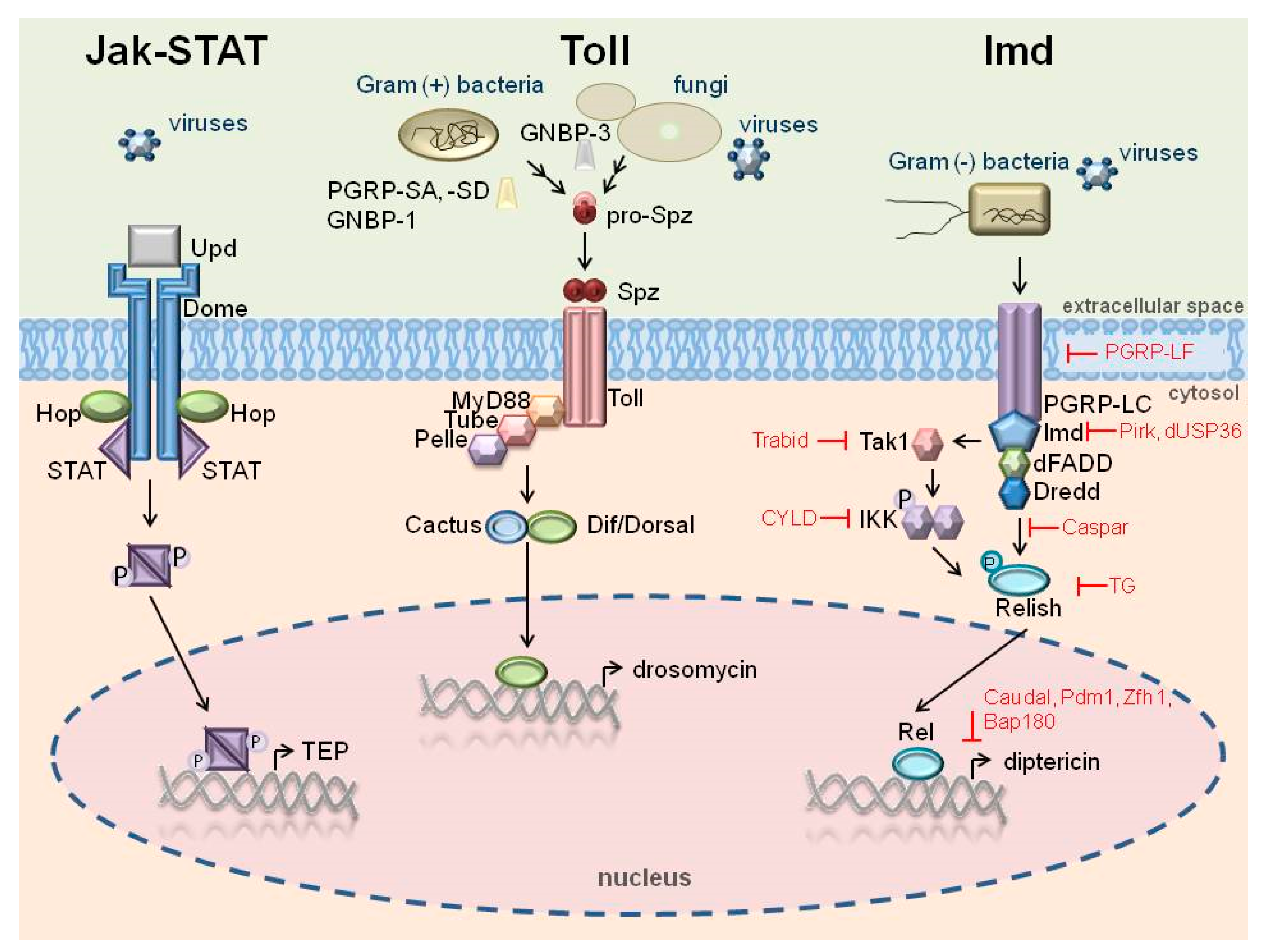
. The Imd pathway can also be activated by viruses.
Ijms 24 05753 g001
Figure 2. Different functions of AMPs in insects.
Ijms 24 05753 g002
Table 2. Predicted antiviral activity of selected G. mellonella AMPs.
G. mellonella AMPAccession Number/
Peptide ID
Predicted Antiviral ActivityAVP
Value
Non-AVP
Value
gallerimycinXP_026765221.1+10
lebocin-like anionic peptide 1P85211.1+0.980.02
cecropin D-like peptideP85210.1+0.930.07
G. mellonella cecropinXP_026754247.1+0.7460.254
apolipophoricinP80703+0.6980.302
proline rich peptide 2P85212.10.3240.676
defensin-like peptideP852150.2720.728
defensinP852130.2680.732
proline rich peptide 1P85214.10.0040.996
anionic peptide 2P8501601
The amino acid sequences and accession numbers of G. mellonella AMPs were obtained using the NCBI Reference Sequences database. The antiviral potential (AVP) was predicted using Meta-iAVP available at http://codes.bio/meta-iavp/; accessed on 9 December 2022).
Disclaimer/Publisher’s Note: The statements, opinions and data contained in all publications are solely those of the individual author(s) and contributor(s) and not of MDPI and/or the editor(s). MDPI and/or the editor(s) disclaim responsibility for any injury to people or property resulting from any ideas, methods, instructions or products referred to in the content.

