
Vitamin D Deficiency: An Underestimated Factor in Sepsis?


Abstract
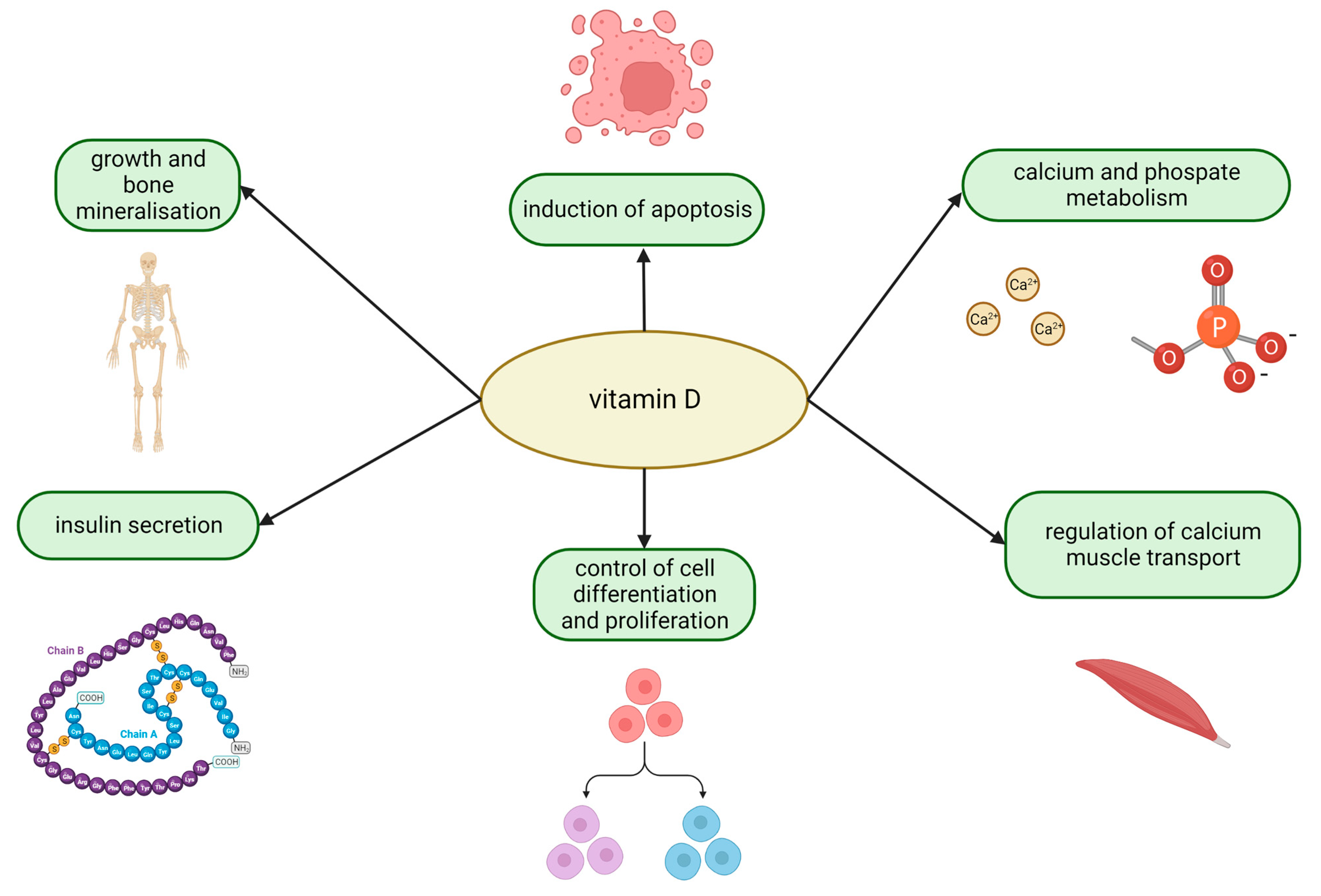
1. Introduction
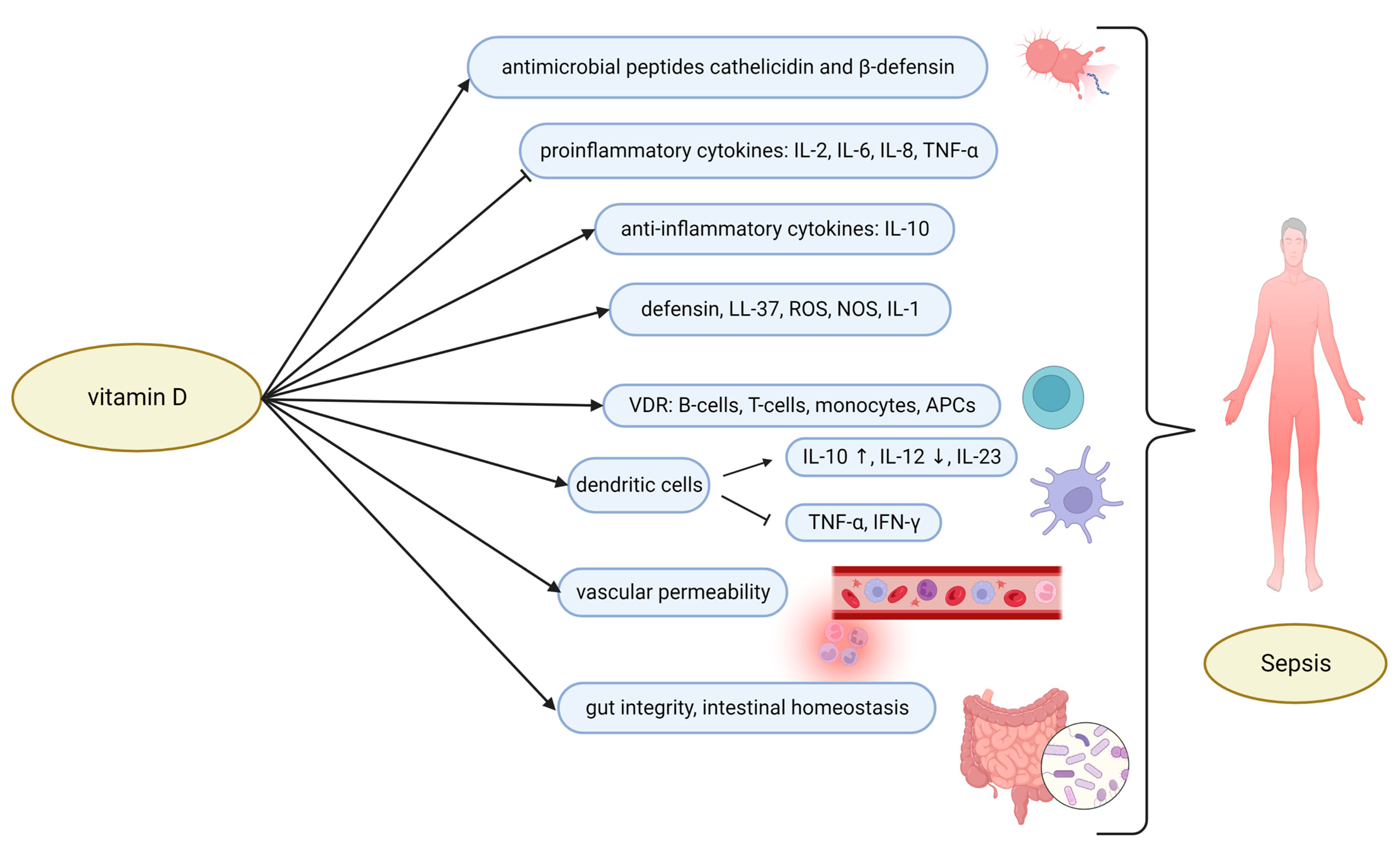
2. The Role of Vitamin D and Sepsis
3. Neonates and Children
3.1. Vitamin D Deficiency
3.2. Vitamin D Supplementation in Neonatal Sepsis
3.3. Vitamin D Supplementation in Children with Sepsis
4. Adults
4.1. Vitamin D Deficiency
4.2. Vitamin D Supplementation in Adults with Sepsis
4.3. Vitamin D Supplementation in Patients with COVID-19
5. Conclusions
Author Contributions
Funding
Institutional Review Board Statement
Informed Consent Statement
Data Availability Statement
Conflicts of Interest
References
- Chae, B.; Kim, Y.-J.; Kim, S.M.; Hong, S.-I.; Shin, Y.S.; Kim, J.-S.; Ryoo, S.M.; Kim, W.Y. Vitamin D Deficiency on Admission to the Emergency Department Is a Mortality Predictor for Patients with Septic Shock Treated with Early Protocol-Driven Resuscitation Bundle Therapy. Am. J. Med. Sci. 2022, S0002962922004347. [Google Scholar] [CrossRef] [PubMed]
- Adams, J.S.; Hewison, M. Unexpected Actions of Vitamin D: New Perspectives on the Regulation of Innate and Adaptive Immunity. Nat. Clin. Pract. Endocrinol. Metab. 2008, 4, 80–90. [Google Scholar] [CrossRef] [PubMed]
- Jones, K.S.; Assar, S.; Harnpanich, D.; Bouillon, R.; Lambrechts, D.; Prentice, A.; Schoenmakers, I. 25(OH)D2 Half-Life Is Shorter than 25(OH)D3 Half-Life and Is Influenced by DBP Concentration and Genotype. J. Clin. Endocrinol. Metab. 2014, 99, 3373–3381. [Google Scholar] [CrossRef]
- Braun, A.B.; Gibbons, F.K.; Litonjua, A.A.; Giovannucci, E.; Christopher, K.B. Low Serum 25-Hydroxyvitamin D at Critical Care Initiation Is Associated with Increased Mortality. Crit. Care Med. 2012, 40, 63–72. [Google Scholar] [CrossRef]
- Braun, A.; Chang, D.; Mahadevappa, K.; Gibbons, F.K.; Liu, Y.; Giovannucci, E.; Christopher, K.B. Association of Low Serum 25-Hydroxyvitamin D Levels and Mortality in the Critically Ill. Crit. Care Med. 2011, 39, 671–677. [Google Scholar] [CrossRef]
- Lee, P.; Eisman, J.A.; Center, J.R. Vitamin D Deficiency in Critically Ill Patients. N. Engl. J. Med. 2009, 360, 1912–1914. [Google Scholar] [CrossRef]
- Venkatram, S.; Chilimuri, S.; Adrish, M.; Salako, A.; Patel, M.; Diaz-Fuentes, G. Vitamin D Deficiency Is Associated with Mortality in the Medical Intensive Care Unit. Crit. Care 2011, 15, R292. [Google Scholar] [CrossRef] [PubMed]
- Moromizato, T.; Litonjua, A.A.; Braun, A.B.; Gibbons, F.K.; Giovannucci, E.; Christopher, K.B. Association of Low Serum 25-Hydroxyvitamin D Levels and Sepsis in the Critically Ill. Crit. Care Med. 2014, 42, 97–107. [Google Scholar] [CrossRef] [PubMed]
- Bone, R.C.; Balk, R.A.; Cerra, F.B.; Dellinger, R.P.; Fein, A.M.; Knaus, W.A.; Schein, R.M.; Sibbald, W.J. Definitions for Sepsis and Organ Failure and Guidelines for the Use of Innovative Therapies in Sepsis. Chest 1992, 101, 1644–1655. [Google Scholar] [CrossRef] [PubMed]
- Angus, D.C.; Linde-Zwirble, W.T.; Lidicker, J.; Clermont, G.; Carcillo, J.; Pinsky, M.R. Epidemiology of Severe Sepsis in the United States: Analysis of Incidence, Outcome, and Associated Costs of Care. Crit. Care Med. 2001, 29, 1303–1310. [Google Scholar] [CrossRef]
- Dugar, S.; Choudhary, C.; Duggal, A. Sepsis and Septic Shock: Guideline-Based Management. Cleve. Clin. J. Med. 2020, 87, 53–64. [Google Scholar] [CrossRef] [PubMed]
- Levy, M.M.; Evans, L.E.; Rhodes, A. The Surviving Sepsis Campaign Bundle: 2018 Update. Crit. Care Med. 2018, 46, 997–1000. [Google Scholar] [CrossRef]
- Flynn, L.; Zimmerman, L.H.; McNorton, K.; Dolman, M.; Tyburski, J.; Baylor, A.; Wilson, R.; Dolman, H. Effects of Vitamin D Deficiency in Critically Ill Surgical Patients. Am. J. Surg. 2012, 203, 379–382. [Google Scholar] [CrossRef] [PubMed]
- Yu, W.; Ying, Q.; Zhu, W.; Huang, L.; Hou, Q. Vitamin D Status Was Associated with Sepsis in Critically Ill Children: A PRISMA Compliant Systematic Review and Meta-Analysis. Medicine 2021, 100, e23827. [Google Scholar] [CrossRef] [PubMed]
- He, M.; Cao, T.; Wang, J.; Wang, C.; Wang, Z.; Abdelrahim, M.E.A. Vitamin D Deficiency Relation to Sepsis, Paediatric Risk of Mortality III Score, Need for Ventilation Support, Length of Hospital Stay, and Duration of Mechanical Ventilation in Critically Ill Children: A Meta-Analysis. Int. J. Clin. Pract. 2021, 75, e13908. [Google Scholar] [CrossRef] [PubMed]
- Holick, M.F. Vitamin D Deficiency. N. Engl. J. Med. 2007, 357, 266–281. [Google Scholar] [CrossRef]
- Liu, P.T.; Stenger, S.; Li, H.; Wenzel, L.; Tan, B.H.; Krutzik, S.R.; Ochoa, M.T.; Schauber, J.; Wu, K.; Meinken, C.; et al. Toll-like Receptor Triggering of a Vitamin D-Mediated Human Antimicrobial Response. Science 2006, 311, 1770–1773. [Google Scholar] [CrossRef] [PubMed]
- Liu, P.T.; Stenger, S.; Tang, D.H.; Modlin, R.L. Cutting Edge: Vitamin D-Mediated Human Antimicrobial Activity against Mycobacterium Tuberculosis Is Dependent on the Induction of Cathelicidin. J. Immunol. 2007, 179, 2060–2063. [Google Scholar] [CrossRef]
- Bhalla, A.K.; Amento, E.P.; Krane, S.M. Differential Effects of 1,25-Dihydroxyvitamin D3 on Human Lymphocytes and Monocyte/Macrophages: Inhibition of Interleukin-2 and Augmentation of Interleukin-1 Production. Cell Immunol. 1986, 98, 311–322. [Google Scholar] [CrossRef]
- Dürr, U.H.N.; Sudheendra, U.S.; Ramamoorthy, A. LL-37, the Only Human Member of the Cathelicidin Family of Antimicrobial Peptides. Biochim. Biophys. Acta 2006, 1758, 1408–1425. [Google Scholar] [CrossRef]
- Pinheiro da Silva, F.; Machado, M.C.C. Antimicrobial Peptides: Clinical Relevance and Therapeutic Implications. Peptides 2012, 36, 308–314. [Google Scholar] [CrossRef] [PubMed]
- Dellinger, R.P.; Levy, M.M.; Rhodes, A.; Annane, D.; Gerlach, H.; Opal, S.M.; Sevransky, J.E.; Sprung, C.L.; Douglas, I.S.; Jaeschke, R.; et al. Surviving Sepsis Campaign: International Guidelines for Management of Severe Sepsis and Septic Shock, 2012. Intensive Care Med. 2013, 39, 165–228. [Google Scholar] [CrossRef]
- Agrawal, T.; Gupta, G.K.; Agrawal, D.K. Vitamin D Supplementation Reduces Airway Hyperresponsiveness and Allergic Airway Inflammation in a Murine Model. Clin. Exp. Allergy 2013, 43, 672–683. [Google Scholar] [CrossRef] [PubMed]
- Youssef, D.A.; Miller, C.W.; El-Abbassi, A.M.; Cutchins, D.C.; Cutchins, C.; Grant, W.B.; Peiris, A.N. Antimicrobial Implications of Vitamin D. Derm. Endocrinol. 2011, 3, 220–229. [Google Scholar] [CrossRef]
- Zhang, Y.; Leung, D.Y.M.; Richers, B.N.; Liu, Y.; Remigio, L.K.; Riches, D.W.; Goleva, E. Vitamin D Inhibits Monocyte/Macrophage Proinflammatory Cytokine Production by Targeting MAPK Phosphatase-1. J. Immunol. 2012, 188, 2127–2135. [Google Scholar] [CrossRef] [PubMed]
- Sadeghi, K.; Wessner, B.; Laggner, U.; Ploder, M.; Tamandl, D.; Friedl, J.; Zügel, U.; Steinmeyer, A.; Pollak, A.; Roth, E.; et al. Vitamin D3 Down-Regulates Monocyte TLR Expression and Triggers Hyporesponsiveness to Pathogen-Associated Molecular Patterns. Eur. J. Immunol. 2006, 36, 361–370. [Google Scholar] [CrossRef]
- Cantorna, M.T.; Snyder, L.; Lin, Y.-D.; Yang, L. Vitamin D and 1,25(OH)2D Regulation of T Cells. Nutrients 2015, 7, 3011–3021. [Google Scholar] [CrossRef]
- Mocanu, V.; Oboroceanu, T.; Zugun-Eloae, F. Current Status in Vitamin D and Regulatory T Cells—Immunological Implications. Rev. Med. Chir. Soc. Med. Nat. Iasi. 2013, 117, 965–973. [Google Scholar]
- Mao, X.; Hu, B.; Zhou, Z.; Xing, X.; Wu, Y.; Gao, J.; He, Y.; Hu, Y.; Cheng, Q.; Gong, Q. Vitamin D Levels Correlate with Lymphocyte Subsets in Elderly Patients with Age-Related Diseases. Sci. Rep. 2018, 8, 7708. [Google Scholar] [CrossRef]
- Singh, P.; Chaudhari, V. Association of Early-Onset Sepsis and Vitamin D Deficiency in Term Neonates. Indian Pediatr. 2020, 57, 232–234. [Google Scholar] [CrossRef]
- Maiya, S.; Sullivan, I.; Allgrove, J.; Yates, R.; Malone, M.; Brain, C.; Archer, N.; Mok, Q.; Daubeney, P.; Tulloh, R.; et al. Hypocalcaemia and Vitamin D Deficiency: An Important, but Preventable, Cause of Life-Threatening Infant Heart Failure. Heart 2008, 94, 581–584. [Google Scholar] [CrossRef]
- Bukoski, R.D.; Xue, H. On the Vascular Inotropic Action of 1,25-(OH)2 Vitamin D3. Am. J. Hypertens. 1993, 6, 388–396. [Google Scholar] [CrossRef]
- Møller, S.; Laigaard, F.; Olgaard, K.; Hemmingsen, C. Effect of 1,25-Dihydroxy-Vitamin D3 in Experimental Sepsis. Int. J. Med. Sci. 2007, 4, 190–195. [Google Scholar] [CrossRef] [PubMed]
- Andrukhova, O.; Slavic, S.; Zeitz, U.; Riesen, S.C.; Heppelmann, M.S.; Ambrisko, T.D.; Markovic, M.; Kuebler, W.M.; Erben, R.G. Vitamin D Is a Regulator of Endothelial Nitric Oxide Synthase and Arterial Stiffness in Mice. Mol. Endocrinol. 2014, 28, 53–64. [Google Scholar] [CrossRef] [PubMed]
- Lee, C.; Lau, E.; Chusilp, S.; Filler, R.; Li, B.; Zhu, H.; Yamoto, M.; Pierro, A. Protective Effects of Vitamin D against Injury in Intestinal Epithelium. Pediatr. Surg. Int. 2019, 35, 1395–1401. [Google Scholar] [CrossRef] [PubMed]
- Rippel, C.; South, M.; Butt, W.W.; Shekerdemian, L.S. Vitamin D Status in Critically Ill Children. Intensive Care Med. 2012, 38, 2055–2062. [Google Scholar] [CrossRef] [PubMed]
- Burris, H.H.; Van Marter, L.J.; McElrath, T.F.; Tabatabai, P.; Litonjua, A.A.; Weiss, S.T.; Christou, H. Vitamin D Status among Preterm and Full-Term Infants at Birth. Pediatr. Res. 2014, 75, 75–80. [Google Scholar] [CrossRef] [PubMed]
- Monangi, N.; Slaughter, J.L.; Dawodu, A.; Smith, C.; Akinbi, H.T. Vitamin D Status of Early Preterm Infants and the Effects of Vitamin D Intake during Hospital Stay. Arch. Dis. Child. Fetal Neonatal Ed. 2014, 99, F166–F168. [Google Scholar] [CrossRef]
- Khan, M.R.; Maheshwari, P.K.; Masood, K.; Qamar, F.N.; Haque, A.-U. Epidemiology and Outcome of Sepsis in a Tertiary Care PICU of Pakistan. Indian J. Pediatr. 2012, 79, 1454–1458. [Google Scholar] [CrossRef]
- Xiao, D.; Zhang, X.; Ying, J.; Zhou, Y.; Li, X.; Mu, D.; Qu, Y. Association between Vitamin D Status and Sepsis in Children: A Meta-Analysis of Observational Studies. Clin. Nutr. 2020, 39, 1735–1741. [Google Scholar] [CrossRef]
- Cetinkaya, M.; Cekmez, F.; Buyukkale, G.; Erener-Ercan, T.; Demir, F.; Tunc, T.; Aydın, F.N.; Aydemir, G. Lower Vitamin D Levels Are Associated with Increased Risk of Early-Onset Neonatal Sepsis in Term Infants. J. Perinatol. 2015, 35, 39–45. [Google Scholar] [CrossRef] [PubMed]
- Cizmeci, M.N.; Kanburoglu, M.K.; Akelma, A.Z.; Ayyildiz, A.; Kutukoglu, I.; Malli, D.D.; Tatli, M.M. Cord-Blood 25-Hydroxyvitamin D Levels and Risk of Early-Onset Neonatal Sepsis: A Case-Control Study from a Tertiary Care Center in Turkey. Eur. J. Pediatr. 2015, 174, 809–815. [Google Scholar] [CrossRef]
- Ponnarmeni, S.; Kumar Angurana, S.; Singhi, S.; Bansal, A.; Dayal, D.; Kaur, R.; Patial, A.; Verma Attri, S. Vitamin D Deficiency in Critically Ill Children with Sepsis. Paediatr. Int. Child Health 2016, 36, 15–21. [Google Scholar] [CrossRef] [PubMed]
- Onwuneme, C.; Carroll, A.; Doherty, D.; Bruell, H.; Segurado, R.; Kilbane, M.; Murphy, N.; McKenna, M.J.; Molloy, E.J. Inadequate Vitamin D Levels Are Associated with Culture Positive Sepsis and Poor Outcomes in Paediatric Intensive Care. Acta Paediatr. 2015, 104, e433–e438. [Google Scholar] [CrossRef] [PubMed]
- Say, B.; Uras, N.; Sahin, S.; Degirmencioglu, H.; Oguz, S.S.; Canpolat, F.E. Effects of Cord Blood Vitamin D Levels on the Risk of Neonatal Sepsis in Premature Infants. Korean J. Pediatr. 2017, 60, 248–253. [Google Scholar] [CrossRef]
- Shah, S.K.; Kabra, S.K.; Gupta, N.; Pai, G.; Lodha, R. Vitamin D Deficiency and Parathyroid Response in Critically-Ill Children: Association with Illness Severity and Clinical Outcomes. Indian Pediatr. 2016, 53, 479–484. [Google Scholar] [CrossRef]
- Razavi Khorasani, N.; Moazzami, B.; Zahedi Tajrishi, F.; Mohammadpour, Z.; Rouhi, F.; Alizadeh-Navaei, R.; Ghadimi, R. The Association Between Low Levels of Vitamin D and Clinical Outcomes in Critically-Ill Children: A Systematic Review and Meta-Analysis. Fetal Pediatr. Pathol. 2020, 39, 503–517. [Google Scholar] [CrossRef] [PubMed]
- McNally, J.D.; Nama, N.; O’Hearn, K.; Sampson, M.; Amrein, K.; Iliriani, K.; McIntyre, L.; Fergusson, D.; Menon, K. Vitamin D Deficiency in Critically Ill Children: A Systematic Review and Meta-Analysis. Crit. Care 2017, 21, 287. [Google Scholar] [CrossRef]
- Su, G.; Jia, D. Vitamin D in Acute and Critically Sick Children with a Subgroup of Sepsis and Mortality: A Meta-Analysis. Nutr. Cancer 2021, 73, 1118–1125. [Google Scholar] [CrossRef]
- Cariolou, M.; Cupp, M.A.; Evangelou, E.; Tzoulaki, I.; Berlanga-Taylor, A.J. Importance of Vitamin D in Acute and Critically Ill Children with Subgroup Analyses of Sepsis and Respiratory Tract Infections: A Systematic Review and Meta-Analysis. BMJ Open 2019, 9, e027666. [Google Scholar] [CrossRef]
- Hagag, A.A.; El Frargy, M.S.; Houdeeb, H.A. Therapeutic Value of Vitamin D as an Adjuvant Therapy in Neonates with Sepsis. Infect. Disord. Drug Targets 2020, 20, 440–447. [Google Scholar] [CrossRef] [PubMed]
- Abdel-Hady, H.; Yahia, S.; Megahed, A.; Mosbah, A.; Seif, B.; Nageh, E.; Bhattacharjee, I.; Aly, H. Mediators in Preterm Infants with Late-Onset Sepsis: A Randomized Controlled Trial. J. Pediatr. Gastroenterol. Nutr. 2019, 68, 578–584. [Google Scholar] [CrossRef] [PubMed]
- Wang, Y.; Yang, Z.; Gao, L.; Cao, Z.; Wang, Q. Effects of a Single Dose of Vitamin D in Septic Children: A Randomized, Double-Blinded, Controlled Trial. J. Int. Med. Res. 2020, 48, 300060520926890. [Google Scholar] [CrossRef]
- Bustos, B.R.; Rodríguez-Nuñez, I.; Peña Zavala, R.; Soto Germani, G. Vitamin D deficiency in children admitted to the paediatric intensive care unit. Rev. Chil. Pediatr. 2016, 87, 480–486. [Google Scholar] [CrossRef]
- De Pascale, G.; Vallecoccia, M.S.; Schiattarella, A.; Di Gravio, V.; Cutuli, S.L.; Bello, G.; Montini, L.; Pennisi, M.A.; Spanu, T.; Zuppi, C.; et al. Clinical and Microbiological Outcome in Septic Patients with Extremely Low 25-Hydroxyvitamin D Levels at Initiation of Critical Care. Clin. Microbiol. Infect. 2016, 22, 456.e7–456.e13. [Google Scholar] [CrossRef]
- Ebenezer, K.; Job, V.; Antonisamy, B.; Dawodu, A.; Manivachagan, M.N.; Steinhoff, M. Serum Vitamin D Status and Outcome among Critically Ill Children Admitted to the Pediatric Intensive Care Unit in South India. Indian J. Pediatr. 2016, 83, 120–125. [Google Scholar] [CrossRef] [PubMed]
- Delanghe, J.R.; Speeckaert, M.M. Translational Research and Biomarkers in Neonatal Sepsis. Clin. Chim. Acta 2015, 451, 46–64. [Google Scholar] [CrossRef]
- Abrams, S.A. Committee on Nutrition Calcium and Vitamin d Requirements of Enterally Fed Preterm Infants. Pediatrics 2013, 131, e1676–e1683. [Google Scholar] [CrossRef]
- Agostoni, C.; Buonocore, G.; Carnielli, V.P.; De Curtis, M.; Darmaun, D.; Decsi, T.; Domellöf, M.; Embleton, N.D.; Fusch, C.; Genzel-Boroviczeny, O.; et al. Enteral Nutrient Supply for Preterm Infants: Commentary from the European Society of Paediatric Gastroenterology, Hepatology and Nutrition Committee on Nutrition. J. Pediatr. Gastroenterol. Nutr. 2010, 50, 85–91. [Google Scholar] [CrossRef]
- Gonzalez-Curiel, I.; Marin-Luevano, P.; Trujillo, V.; Enciso-Moreno, J.A.; Gonzalez-Castillo, C.; Rivas-Santiago, B. Calcitriol Prevents Inflammatory Gene Expression in Macrovascular Endothelial Cells. Br. J. Biomed. Sci. 2016, 73, 74–78. [Google Scholar] [CrossRef]
- Gunta, S.S.; Thadhani, R.I.; Mak, R.H. The Effect of Vitamin D Status on Risk Factors for Cardiovascular Disease. Nat. Rev. Nephrol. 2013, 9, 337–347. [Google Scholar] [CrossRef] [PubMed]
- Payen, V.; Jouvet, P.; Lacroix, J.; Ducruet, T.; Gauvin, F. Risk Factors Associated with Increased Length of Mechanical Ventilation in Children. Pediatr. Crit. Care Med. 2012, 13, 152–157. [Google Scholar] [CrossRef] [PubMed]
- Looker, A.C.; Johnson, C.L.; Lacher, D.A.; Pfeiffer, C.M.; Schleicher, R.L.; Sempos, C.T. Vitamin D Status: United States, 2001-2006. NCHS Data Brief 2011, 59, 1–8. [Google Scholar]
- Upala, S.; Sanguankeo, A.; Permpalung, N. Significant Association between Vitamin D Deficiency and Sepsis: A Systematic Review and Meta-Analysis. BMC Anesthesiol. 2015, 15, 84. [Google Scholar] [CrossRef]
- Amrein, K.; Schnedl, C.; Holl, A.; Riedl, R.; Christopher, K.B.; Pachler, C.; Urbanic Purkart, T.; Waltensdorfer, A.; Münch, A.; Warnkross, H.; et al. Effect of High-Dose Vitamin D3 on Hospital Length of Stay in Critically Ill Patients with Vitamin D Deficiency: The VITdAL-ICU Randomized Clinical Trial. JAMA 2014, 312, 1520–1530. [Google Scholar] [CrossRef] [PubMed]
- Matthews, L.R.; Ahmed, Y.; Wilson, K.L.; Griggs, D.D.; Danner, O.K. Worsening Severity of Vitamin D Deficiency Is Associated with Increased Length of Stay, Surgical Intensive Care Unit Cost, and Mortality Rate in Surgical Intensive Care Unit Patients. Am. J. Surg. 2012, 204, 37–43. [Google Scholar] [CrossRef] [PubMed]
- Li, Y.; Ding, S. Serum 25-Hydroxyvitamin D and the Risk of Mortality in Adult Patients with Sepsis: A Meta-Analysis. BMC Infect. Dis. 2020, 20, 189. [Google Scholar] [CrossRef]
- Ding, F.; Zang, B.; Fu, J.; Ji, K. Effect of vitamin D3 on the severity and prognosis of patients with sepsis: A prospective randomized double-blind placebo study. Zhonghua Wei Zhong Bing Ji Jiu Yi Xue 2017, 29, 106–110. [Google Scholar] [CrossRef]
- Sarhan, N.; Abou Warda, A.E.; Sarhan, R.M.; Boshra, M.S.; Mostafa-Hedeab, G.; ALruwaili, B.F.; Ibrahim, H.S.G.; Schaalan, M.F.; Fathy, S. Evidence for the Efficacy of a High Dose of Vitamin D on the Hyperinflammation State in Moderate-to-Severe COVID-19 Patients: A Randomized Clinical Trial. Medicina 2022, 58, 1358. [Google Scholar] [CrossRef]
- Michaud, J.; Naud, J.; Ouimet, D.; Demers, C.; Petit, J.-L.; Leblond, F.A.; Bonnardeaux, A.; Gascon-Barré, M.; Pichette, V. Reduced Hepatic Synthesis of Calcidiol in Uremia. J. Am. Soc. Nephrol. 2010, 21, 1488–1497. [Google Scholar] [CrossRef]
- Spriet, I.; Meersseman, W.; de Hoon, J.; von Winckelmann, S.; Wilmer, A.; Willems, L. Mini-Series: II. Clinical Aspects. Clinically Relevant CYP450-Mediated Drug Interactions in the ICU. Intensive Care Med. 2009, 35, 603–612. [Google Scholar] [CrossRef] [PubMed]
- Quraishi, S.A.; De Pascale, G.; Needleman, J.S.; Nakazawa, H.; Kaneki, M.; Bajwa, E.K.; Camargo, C.A.; Bhan, I. Effect of Cholecalciferol Supplementation on Vitamin D Status and Cathelicidin Levels in Sepsis: A Randomized, Placebo-Controlled Trial. Crit. Care Med. 2015, 43, 1928–1937. [Google Scholar] [CrossRef] [PubMed]
- Yang, D.; Chen, Q.; Schmidt, A.P.; Anderson, G.M.; Wang, J.M.; Wooters, J.; Oppenheim, J.J.; Chertov, O. LL-37, the Neutrophil Granule- and Epithelial Cell-Derived Cathelicidin, Utilizes Formyl Peptide Receptor-like 1 (FPRL1) as a Receptor to Chemoattract Human Peripheral Blood Neutrophils, Monocytes, and T Cells. J. Exp. Med. 2000, 192, 1069–1074. [Google Scholar] [CrossRef] [PubMed]
- Nagaoka, I.; Hirota, S.; Yomogida, S.; Ohwada, A.; Hirata, M. Synergistic Actions of Antibacterial Neutrophil Defensins and Cathelicidins. Inflamm. Res. 2000, 49, 73–79. [Google Scholar] [CrossRef]
- Tollin, M.; Bergman, P.; Svenberg, T.; Jörnvall, H.; Gudmundsson, G.H.; Agerberth, B. Antimicrobial Peptides in the First Line Defence of Human Colon Mucosa. Peptides 2003, 24, 523–530. [Google Scholar] [CrossRef]
- Leaf, D.E.; Raed, A.; Donnino, M.W.; Ginde, A.A.; Waikar, S.S. Randomized Controlled Trial of Calcitriol in Severe Sepsis. Am. J. Respir. Crit. Care Med. 2014, 190, 533–541. [Google Scholar] [CrossRef]
- Quraishi, S.A.; Camargo, C.A. Vitamin D in Acute Stress and Critical Illness. Curr. Opin. Clin. Nutr. Metab. Care 2012, 15, 625–634. [Google Scholar] [CrossRef]
- DiNicolantonio, J.J.; O’Keefe, J.H. Magnesium and Vitamin D Deficiency as a Potential Cause of Immune Dysfunction, Cytokine Storm and Disseminated Intravascular Coagulation in COVID-19 Patients. Mo. Med. 2021, 118, 68–73. [Google Scholar]
- Bayraktar, N.; Turan, H.; Bayraktar, M.; Ozturk, A.; Erdoğdu, H. Analysis of Serum Cytokine and Protective Vitamin D Levels in Severe Cases of COVID-19. J. Med. Virol. 2022, 94, 154–160. [Google Scholar] [CrossRef]
- Kong, J.; Zhu, X.; Shi, Y.; Liu, T.; Chen, Y.; Bhan, I.; Zhao, Q.; Thadhani, R.; Li, Y.C. VDR Attenuates Acute Lung Injury by Blocking Ang-2-Tie-2 Pathway and Renin-Angiotensin System. Mol. Endocrinol. 2013, 27, 2116–2125. [Google Scholar] [CrossRef]
- Yuan, W.; Pan, W.; Kong, J.; Zheng, W.; Szeto, F.L.; Wong, K.E.; Cohen, R.; Klopot, A.; Zhang, Z.; Li, Y.C. 1,25-Dihydroxyvitamin D3 Suppresses Renin Gene Transcription by Blocking the Activity of the Cyclic AMP Response Element in the Renin Gene Promoter. J. Biol. Chem. 2007, 282, 29821–29830. [Google Scholar] [CrossRef] [PubMed]
- Hoffmann, M.; Kleine-Weber, H.; Schroeder, S.; Krüger, N.; Herrler, T.; Erichsen, S.; Schiergens, T.S.; Herrler, G.; Wu, N.-H.; Nitsche, A.; et al. SARS-CoV-2 Cell Entry Depends on ACE2 and TMPRSS2 and Is Blocked by a Clinically Proven Protease Inhibitor. Cell 2020, 181, 271–280.e8. [Google Scholar] [CrossRef] [PubMed]
- Dijkman, R.; Jebbink, M.F.; Deijs, M.; Milewska, A.; Pyrc, K.; Buelow, E.; van der Bijl, A.; van der Hoek, L. Replication-Dependent Downregulation of Cellular Angiotensin-Converting Enzyme 2 Protein Expression by Human Coronavirus NL63. J. Gen. Virol. 2012, 93, 1924–1929. [Google Scholar] [CrossRef] [PubMed]
- Ji, X.; Tan, W.; Zhang, C.; Zhai, Y.; Hsueh, Y.; Zhang, Z.; Zhang, C.; Lu, Y.; Duan, B.; Tan, G.; et al. TWIRLS, a Knowledge-Mining Technology, Suggests a Possible Mechanism for the Pathological Changes in the Human Host after Coronavirus Infection via ACE2. Drug Dev. Res. 2020, 81, 1004–1018. [Google Scholar] [CrossRef] [PubMed]
- Chen, I.-Y.; Chang, S.C.; Wu, H.-Y.; Yu, T.-C.; Wei, W.-C.; Lin, S.; Chien, C.-L.; Chang, M.-F. Upregulation of the Chemokine (C-C Motif) Ligand 2 via a Severe Acute Respiratory Syndrome Coronavirus Spike-ACE2 Signaling Pathway. J. Virol. 2010, 84, 7703–7712. [Google Scholar] [CrossRef] [PubMed]


| Study Design | Study Population | Method of Vitamin D Analysis | Major Findings | Ref. | |
|---|---|---|---|---|---|
| Vitamin D deficiency | Prospective cohort study | 316 critically ill children | Immunoassay | Hypovitaminosis D is common in critically ill children and linked to higher inotrope levels in the postoperative cardiac population, but not to PICU length of stay or hospital survival. | [36] |
| Cross-sectional analysis | 71 very preterm infants (< 32 weeks of gestational age) vs. 108 preterm infants (32–36 6/7 weeks of gestational age) vs. 292 term infants (≥ 37 weeks of gestational age) | CLIA | Infants born before 32 weeks gestation had 2.5 times higher odds of 25(OH)D levels < 20 ng/mL compared to more mature infants (95% CI: 1.2–5.3). | [37] | |
| Cohort study | 120 preterm infants (67 infants born < 28 PMA and 53 infants born between 28–32 weeks PMA) | Liquid chromatography/tandem mass spectrometry | Mean serum 25(OH)D at birth was significantly lower in infants born < 28 weeks than at 28–32 weeks gestation (p = 0.02). | [38] | |
| Meta-analysis | 13 studies including neonates and children (< 18 years) with sepsis | A significant relationship existed between vitamin D deficiency and sepsis with an OR of 1.13 (95% CI: 1.18–1.50, p < 0.05). | [40] | ||
| Cross sectional study | 50 infants with EONS in term infants vs. 50 HC | HPLC | The EONS group’s neonatal 25(OH)D levels were significantly lower than those of the control group (p < 0.001). Significantly lower 25(OH)D levels in mothers and full-term neonates with sepsis (p < 0.001), along with a positive correlation between neonatal and maternal 25(OH)D levels in both EONS patients and HC (r = 0.29, p = 0.04, and r = 0.58, p = 0.001, respectively). | [41] | |
| Case–control study | 40 infants with EONS vs. 43 HC | HPLC | Low cord blood 25(OH)D level (< 30 ng/mL) was associated with a 5.6 times higher risk of EONS (OR: 5.6; 95% CI: 1.3–23.5). | [42] | |
| Cross-sectional study | 124 critically ill children with sepsis aged 1–12 years vs. 338 HC | ELISA | When compared to HC, critically ill children with sepsis had a greater prevalence of vitamin D deficiency (50.8% vs. 40.2%, p = 0.04). | [43] | |
| Cross-sectional study | 120 children with suspected sepsis vs. 30 HC | ELISA | Children with sepsis had a 51% lower level of 25(OH)D than HC (p < 0.001). | [44] | |
| Cross-sectional study | 117 premature infants with gestational age < 67 weeks (severe vitamin D deficiency (group 1, n = 63, 25(OH)D < 5 ng/mL), vitamin D insufficiency (group 2, n = 24, 25(OH)D = 5–15 ng/mL), and vitamin D sufficiency (group 3, n = 13, 25(OH)D ≥ 15 ng/mL)) | ELISA | The frequency of both EONS and LONS was not significantly linked to low cord blood 25(OH)D levels (5 ng/L). | [45] | |
| Prospective observational study | 154 critically ill patients | ELISA | Regarding the severity of the illness at admission, the length of the PICU stay, the requirement for mechanical ventilation, the duration of ventilator support, septic shock, liver failure, and the requirement for renal replacement therapy, children with low vitamin D levels did not differ from children without low vitamin D levels. | [46] | |
| Systematic review and meta-analysis | 27 studies (17 case–control studies and 10 cohort studies) | Case–control studies: the association between vitamin D deficiency and sepsis was significant with an OR of 2.66 (95% CI: 1.13–6.25, p < 0.001). Cohort studies: sepsis was more common in the low 25(OH)D group than in the higher 25(OH)D group (30.4% and 18.2%, respectively). Meta-analysis: vitamin D insufficiency was found to significantly increase the risk of sepsis (OR: 2.65, 95% CI: 1.3–5.41) and ventilation support necessary (OR: 1.35, 95% CI: 1.03–1.77) in extremely high-developed nations. | [47] | ||
| Systematic review and meta-analysis | 17 studies including a total of 2783 critically ill children | In wealthy countries, vitamin D deficiency was linked to increased mortality (OR: 1.62, 95% CI: 1.11–2.36), illness severity, and need for PICU interventions (cardiovascular support and mechanical ventilation). | [48] | ||
| Meta-analysis | 16 studies including 2382 children at baseline with 1229 being vitamin D deficient (< 50 nmol/L or 20 ng/mL) | Vitamin-D-deficient children had a significantly higher risk of sepsis (OR: 2.35, 95% CI: 1.19–4.63, p = 0.01), a significantly higher pediatric risk of mortality III score (OR: 2.19, 95% CI: 1.13–4.25, p = 0.02), a longer length of hospital stay (OR: 4.26, 95% CI: 0.81–7.70, p = 0.02), and a higher duration of mechanical ventilation (OR: 1.89, 95% CI: 0.22–3.56, p = 0.03). The need for ventilation support in vitamin-D-deficient children, on the other hand, did not differ significantly from non-vitamin-D-deficient children (OR: 2.00, 95% CI: 0.98–4.07, p = 0.06), with relatively higher results in vitamin-D-deficient children. | [15] | ||
| Meta-analysis | 23 studies including 4451 children (2500 children with vitamin D deficiency (25(OH)D ≤ 50 nmol/L or 20 ng/mL)) | Children with low vitamin D levels had a greater risk of sepsis (OR: 2.24; 95 CI: 1.42–3.53) than children with adequate vitamin D levels. Additionally, compared to children with adequate vitamin D levels, children with vitamin D insufficiency had greater rates of acute and critical mortality (OR: 1.77; 95% CI: 1.26–2.49), though not as high as sepsis. | [49] | ||
| Systematic review and meta-analysis | 18 cohort studies including 2463 children hospitalized with acute or critical conditions | Children with 25(OH)D deficiencies had a 1.81 increased mortality risk (OR: 1.81; 95% CI: 1.24–2.64, p = 0.002). | [50] | ||
| Vitamin D supplementation neonatal sepsis | RCT | 60 neonates with sepsis (group I: 30 neonates with sepsis treated with only antibiotics and group II: 30 neonates with sepsis treated with both antibiotics and vitamin D (800 IU of vitamin D3 in a single oral dose for 2 weeks)) and 30 HC | ELISA | After 3, 7, and 10 days of treatment, there were significant differences in sepsis score and hs-CRP between group I and II (p values for sepsis score were 0.009, 0.006, and 0.004, respectively, and for hs-CRP they were 0.015, 0.001, and 0.001, respectively). Significant negative correlation between hs-CRP and serum 25(OH)D levels in group II on entry and after 2 weeks of treatment (r = −0.832, p = 0.001, and r = −0.590, p = 0.021, respectively). ROC analysis showed that at a vitamin D cut-off value of 20 ng/mL, the sensitivity was 100%, the specificity was 73%, the positive predictive value was 73%, the negative predictive value was 100%, and the accuracy was 87% in predicting EOS. | [51] |
| RCT | Preterm infants with gestational age ≥ 28 weeks with LONS. Subjects were randomly assigned to receive 400 or 800 IU/day of vitamin D3. | ELISA | At enrollment, 76% of the infants were vitamin-D-deficient in both groups, but only one infant in the 400 IU group and none in the 800 IU group remained deficient at 40 weeks postmenstrual age. Serum pro-inflammatory cytokine IL-6 and TNF-α concentrations decreased in both groups at 1 week and at discharge, with no difference between groups. After the intervention, vitamin D levels in the treatment group were significantly higher than in the control group (p < 0.05). Vitamin D supplementation reduced Ang-II, IL-6, and TNF-α concentrations (all p < 0.05). | [52] | |
| Vitamin D supplementation in children with sepsis | RCT | 109 children with vitamin D deficiency and sepsis randomly assigned to either 150,000 IU cholecalciferol or placebo | ELISA | The treatment group had a lower cv-SOFA score (1.76 ± 0.8 vs. 2.3 ± 1.1) and a lower incidence of septic shock (7% vs. 20%). There was no difference between the two groups in terms of ventilation time or fatality rates. | [53] |
| Prospective observational cohort study | 90 children | Vitamin D deficiency was significantly linked to vasopressor use (RR: 1.6, 95% CI: 1.2–2.3, p < 0.01), mechanical ventilation (RR: 2.2, 95% CI: 1.2–3.9, p < 0.01), septic shock (RR: 1.9, 95% CI: 1.3–2.9, p < 0.01), and fluid bolus > 40 mL/kg in the first 24 h of admission (RR: 1.5, 95% CI: 1.1–2.1, p < 0.05). | [54] | ||
| Retrospective cohort study | 107 patients with sepsis | CLIA | The 52 patients with pneumonia in the extremely low vitamin D group required mechanical ventilation for a longer period of time (9 days (3.75–12.5 days) versus 4 days (2–9 days), p < 0.04), and the 66 with septic shock required vasopressor support for a longer period of time (7 days (4–10 days) versus 4 days (2–7.25 days), p < 0.02). | [55] | |
| Prospective observational cohort study | 54 children | ECL | Low vitamin levels were linked to higher PIM or SOFA scores (r = 0.29, p = 0.04, and r = 0.29, p < 0.05, respectively). Children who were mechanically ventilated had a median serum 25(OH)D level that was considerably lower than children who were not (p < 0.01). | [56] |
| Study Design | Study Population | Method of Vitamin D Analysis | Major Findings | Ref. | |
|---|---|---|---|---|---|
| Vitamin D deficiency | Observational study | 2399 patients, age ≥ 18 years | A lack of 25(OH)D prior to hospitalization is a significant predictor of both short- and long-term all-cause patient mortality as well as blood culture positivity in a population of critically ill patients. | [5] | |
| Systematic review and meta-analysis | 10 observational studies | In studies that reported participant numbers, the pooled odds ratio of sepsis in participants with vitamin D deficiency was 1.78 (95% CI: 1.55–2.03, p < 0.01) compared to controls, and it was 1.45 (95% CI: 1.26–1.66, p < 0.01) in studies that reported an adjusted odds ratio of vitamin D deficiency for developing sepsis. | [64] | ||
| Prospective observational study | 258 patients | HPLC and tandem mass spectrometry | After adjusting for age, gender, race, and comorbidities (myocardial infarctions, acute renal failure, and pneumonia), severe and moderate vitamin D deficiency were related inversely to surgical intensive care unit length of stay (r = 0.194; p = 0.001), surgical intensive care unit treatment cost (r = 0.194; p = 0.001), and mortality (r = 0.125; p = 0.023), compared to the mildly vitamin-D-deficient group. | [66] | |
| Meta-analysis | 8 studies with 1736 patients | Severe vitamin D deficiency may be associated with an increase in mortality in adult sepsis patients. | [67] | ||
| Vitamin D supplementation in adults with sepsis | RCT | 475 patients (237 in the vitamin D3 group and 238 in the placebo group) | CLIA | High-dose vitamin D3 administration versus placebo did not reduce hospital length of stay, hospital mortality, or 6-month mortality in critically ill patients with vitamin D deficiency. The severe vitamin D deficiency subgroup had lower hospital mortality, but this finding should be considered in the generation of hypotheses and warrants further investigation. | [65] |
| RCT | 22 SIRS patients and 20 HC | ECL | Serum 25(OH)D3 levels in sepsis ICU patients were lower than in healthy individuals, but sepsis and SIRS patients did not differ significantly. Patients with sepsis had serum 25(OH)D3 levels that were correlated with gender, with female levels being lower than male levels, but not with age. Exogenous vitamin D3 supplementation had no positive impact on ICU sepsis patients’ prognoses. The APACHE II score and 25(OH)D3 20 g/L were risk factors for prognosis in ICU patients with sepsis. | [68] | |
| RCT | Placebo (n = 10) versus 200,000 IU cholecalciferol (n = 10) versus 400,000 IU cholecalciferol (n = 10), within 24 h of new-onset severe sepsis or septic shock | ELISA | In patients with severe sepsis or septic shock, high-dose cholecalciferol supplementation improves 25(OH)D and bioavailable 25(OH)D levels quickly and safely. | ||
| Vitamin D supplementation in patients with COVID-19 | RCT | 116 patients (group 1, n = 58: orally administered alfacalcidol 1 µg/day; group 2, n = 58: intramuscularly administered cholecalciferol 200,000 IU) | When compared to low-dose vitamin D, high-dose vitamin D was associated with better clinical improvement and fewer negative outcomes, making it a promising treatment for suppressing cytokine storms in COVID-19 patients. | [69] |
Disclaimer/Publisher’s Note: The statements, opinions and data contained in all publications are solely those of the individual author(s) and contributor(s) and not of MDPI and/or the editor(s). MDPI and/or the editor(s) disclaim responsibility for any injury to people or property resulting from any ideas, methods, instructions or products referred to in the content. |
© 2023 by the authors. Licensee MDPI, Basel, Switzerland. This article is an open access article distributed under the terms and conditions of the Creative Commons Attribution (CC BY) license (https://creativecommons.org/licenses/by/4.0/).
Share and Cite
Delrue, C.; Speeckaert, R.; Delanghe, J.R.; Speeckaert, M.M. Vitamin D Deficiency: An Underestimated Factor in Sepsis? Int. J. Mol. Sci. 2023, 24, 2924. https://doi.org/10.3390/ijms24032924
Delrue C, Speeckaert R, Delanghe JR, Speeckaert MM. Vitamin D Deficiency: An Underestimated Factor in Sepsis? International Journal of Molecular Sciences. 2023; 24(3):2924. https://doi.org/10.3390/ijms24032924
Chicago/Turabian StyleDelrue, Charlotte, Reinhart Speeckaert, Joris R. Delanghe, and Marijn M. Speeckaert. 2023. "Vitamin D Deficiency: An Underestimated Factor in Sepsis?" International Journal of Molecular Sciences 24, no. 3: 2924. https://doi.org/10.3390/ijms24032924
APA StyleDelrue, C., Speeckaert, R., Delanghe, J. R., & Speeckaert, M. M. (2023). Vitamin D Deficiency: An Underestimated Factor in Sepsis? International Journal of Molecular Sciences, 24(3), 2924. https://doi.org/10.3390/ijms24032924