
Endometriosis of the Cervix: A Rare Clinical Case with the Possibility of Comparing the Eutopic and Ectopic Endometrium at the Cellular Level


Abstract
1. Introduction
2. Results
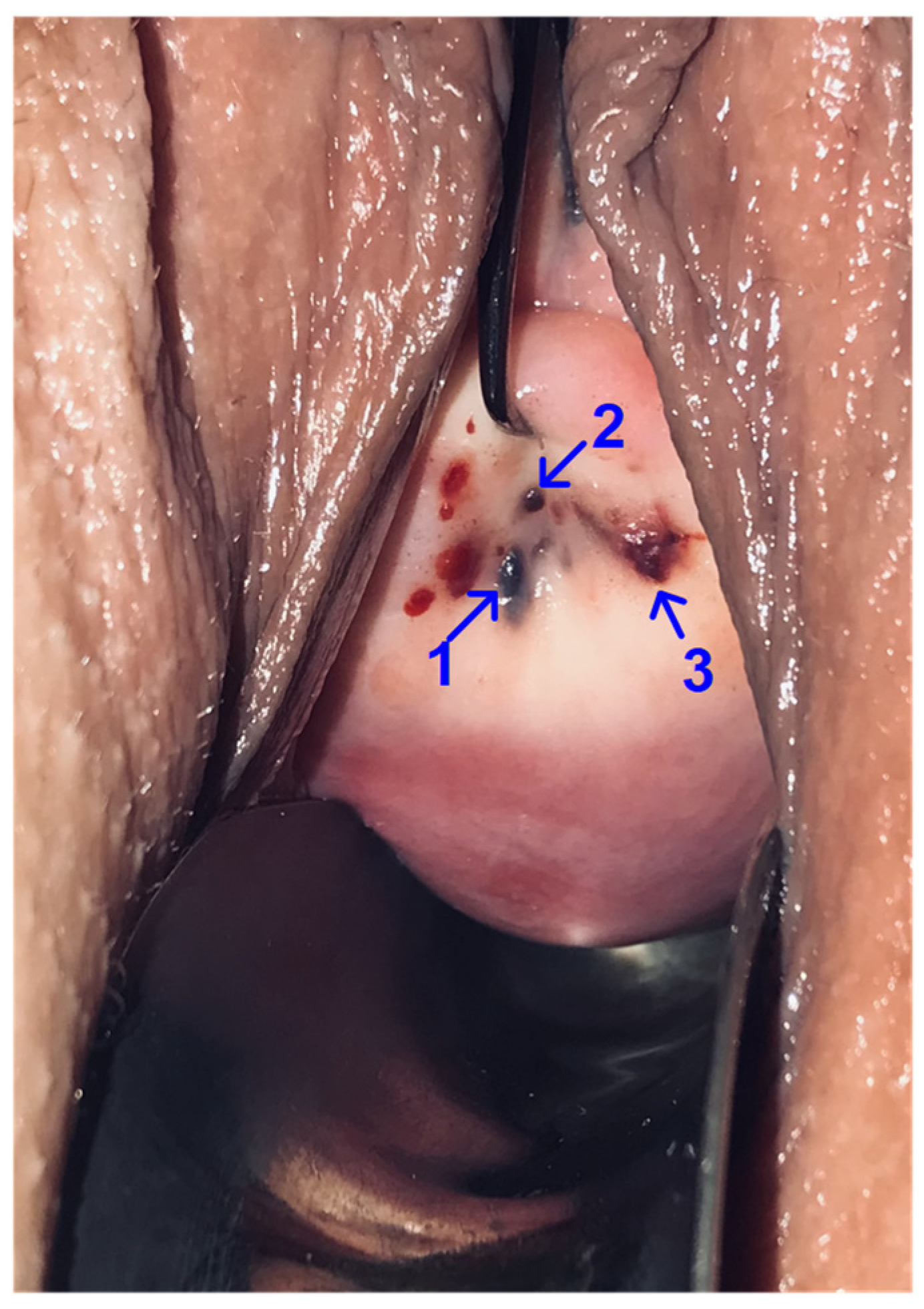
2.1. Clinical Case
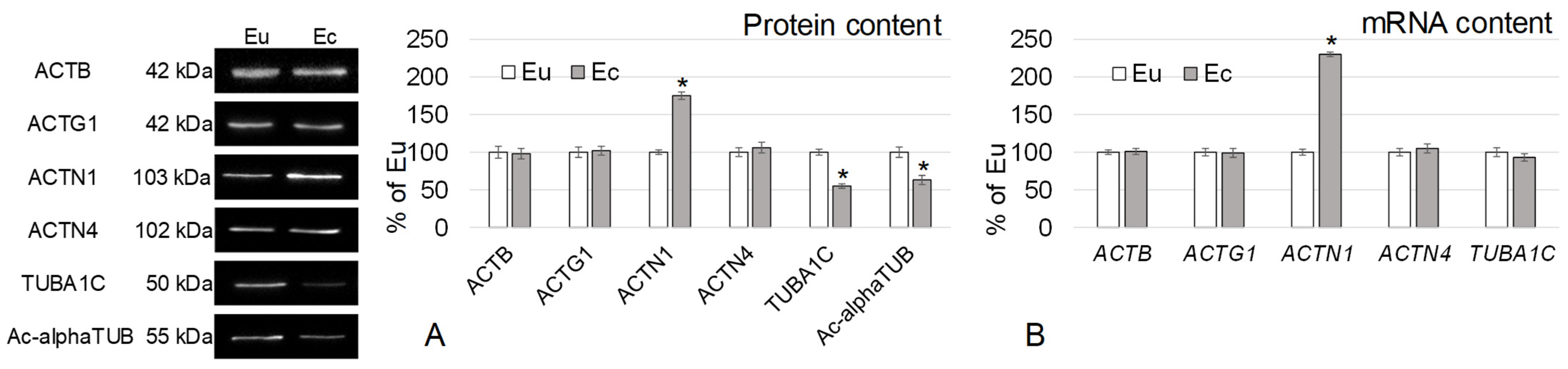
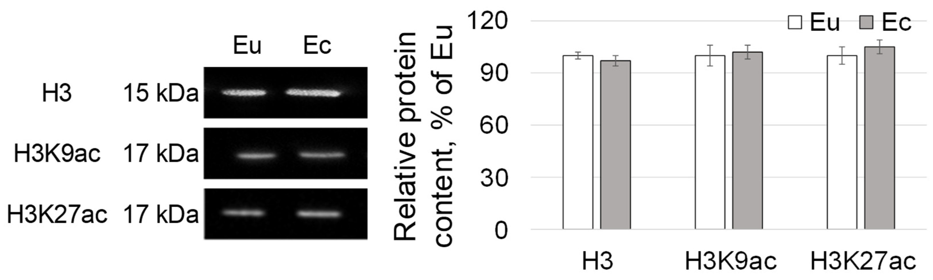
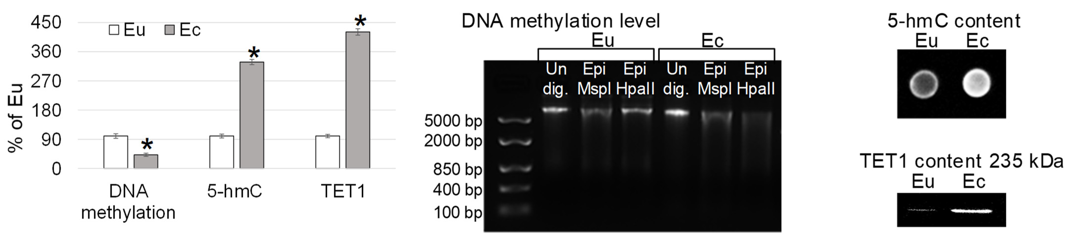
2.2. The relative Content of Cytoskeletal Proteins, Expression of Its Genes and Epigenetic Events in Eutopic and Ectopic Endometrium
3. Discussion
4. Methods
4.1. Western Blotting for the Evaluation of Relative Protein Content
4.2. RT-PCR for the Evaluation of the Relative mRNA Level
4.3. Restriction Analysis (MspI/HpaII) for the Determination of DNA Total Methylation Level
4.4. Dot Blot Method for the Determination of the 5-Hydroxymethylcytosine (5hmC) Content in DNA
4.5. Statistical Analysis
5. Conclusions
Author Contributions
Funding
Institutional Review Board Statement
Informed Consent Statement
Data Availability Statement
Conflicts of Interest
References
- Giudice, L.C.; Kao, L.C. Endometriosis. Lancet 2004, 364, 1789–1799. [Google Scholar] [CrossRef] [PubMed]
- Bulun, S.E. Endometriosis. N. Engl. J. Med. 2009, 360, 268–279. [Google Scholar] [CrossRef] [PubMed]
- Yilmaz, B.D.; Bulun, S.E. Endometriosis and nuclear receptors. Hum. Reprod Update 2019, 25, 473–485. [Google Scholar] [CrossRef] [PubMed]
- Li, J.J.; Duan, H.; Wang, S.; Sun, F.Q.; Gan, L.; Tang, Y.Q.; Xu, Q.; Li, T.C. Expression Pattern of G-Protein-Coupled Estrogen Receptor in Myometrium of Uteri with and without Adenomyosis. Biomed. Res. Int. 2017, 2017, 5974693. [Google Scholar] [CrossRef]
- Vzquez-Martnez, E.R.; Bello-Alvarez, C.; Hermenegildo-Molina, A.L.; Sols-Paredes, M.; Parra-Hernndez, S.; Cruz-Orozco, O.; Silvestri-Tomassoni, J.R.; Escobar-Ponce, L.F.; Hernndez-Lpez, L.A.; Reyes-Mayoral, C.; et al. Expression of Membrane Progesterone Receptors in Eutopic and Ectopic Endometrium of Women with Endometriosis. Biomed. Res. Int. 2020, 2020, 2196024. [Google Scholar] [CrossRef] [PubMed]
- Doshi, J.; Doshi, S.; Sanusi, F.A.; Padwick, M. Persistent post-coital bleeding due to cervical endometriosis. J. Obstet. Gynaecol. 2004, 24, 468–469. [Google Scholar] [CrossRef]
- Albertsen, H.M.; Ward, K. Genes Linked to Endometriosis by GWAS Are Integral to Cytoskeleton Regulation and Suggests That Mesothelial Barrier Homeostasis Is a Factor in the Pathogenesis of Endometriosis. Reprod. Sci. 2017, 24, 803–811. [Google Scholar] [CrossRef]
- Toniyan, K.A.; Povorova, V.V.; Gorbacheva, E.Y.; Boyarintsev, V.V.; Ogneva, I.V. Organization of the Cytoskeleton in Ectopic Foci of the Endometrium with Rare Localization. Biomedicines 2021, 9, 998. [Google Scholar] [CrossRef]
- Flieder, D.B.; Moran, C.A.; Travis, W.D.; Koss, M.N.; Mark, E.J. Pleuro-pulmonary endometriosis and pulmonary ectopic deciduosis: A clinicopathologic and immunohistochemical study of 10 cases with emphasis on diagnostic pitfalls. Hum. Pathol. 1998, 29, 1495–1503. [Google Scholar] [CrossRef]
- Alzayer, H. Pulmonary endometriosis: A rare cause of hydropneumothorax. Respirol. Case Rep. 2019, 7, e432. [Google Scholar] [CrossRef]
- Oner, A.; Karakucuk, S.; Serin, S. Nasolacrimal endometriosis. A case report. Ophthalmic. Res. 2006, 38, 313–314. [Google Scholar] [CrossRef] [PubMed]
- Trkolu, I.; Trkolu, P.; Kurt, J.; Yildirim, H. Presumed nasolacrimal endometriosis. Ophthalmic. Plast Reconstr. Surg. 2008, 24, 47–48. [Google Scholar] [CrossRef]
- Ata, B.; Ates, U.; Usta, T.; Attar, E. Cervical endometriosis, a case presenting with intractable spotting. MedGenMed 2005, 7, 64. [Google Scholar]
- Kao, A.P.; Wang, K.H.; Long, C.Y.; Chai, C.Y.; Tsai, C.F.; Hsieh, T.H.; Hsu, C.Y.; Chang, C.C.; Lee, J.N.; Tsai, E.M. Interleukin-1 induces cyclooxygenase-2 expression and promotes the invasive ability of human mesenchymal stem cells derived from ovarian endometrioma. Fertil. Steril. 2011, 96, 678–684.e1. [Google Scholar] [CrossRef]
- Lebovic, D.I.; Bentzien, F.; Chao, V.A.; Garrett, E.N.; Meng, Y.G.; Taylor, R.N. Induction of an angiogenic phenotype in endometriotic stromal cell cultures by interleukin-1beta. Mol. Hum. Reprod. 2000, 6, 269–275. [Google Scholar] [CrossRef] [PubMed]
- Lebovic, D.I.; Mueller, M.D.; Taylor, R.N. Immunobiology of endometriosis. Fertil. Steril. 2001, 75, 1–10. [Google Scholar] [CrossRef]
- McLaren, J. Vascular endothelial growth factor and endometriotic angiogenesis. Hum. Reprod. Update 2000, 6, 45–55. [Google Scholar] [CrossRef] [PubMed]
- Ulukus, M.; Ulukus, E.C.; Tavmergen Goker, E.N.; Tavmergen, E.; Zheng, W.; Arici, A. Expression of interleukin-8 and monocyte chemotactic protein 1 in women with endometriosis. Fertil. Steril. 2009, 91, 687–693. [Google Scholar] [CrossRef]
- Sikora, J.; Smycz-Kubaska, M.; Mielczarek-Palacz, A.; Kondera-Anasz, Z. Abnormal peritoneal regulation of chemokine activation-The role of IL-8 in pathogenesis of endometriosis. Am. J. Reprod. Immunol. 2017, 77, e12622. [Google Scholar] [CrossRef]
- Flamini, M.I.; Sanchez, A.M.; Goglia, L.; Tosi, V.; Genazzani, A.R.; Simoncini, T. Differential actions of estrogen and SERMs in regulation of the actin cytoskeleton of endometrial cells. Mol. Hum. Reprod. 2009, 15, 675–685. [Google Scholar] [CrossRef]
- Gentilini, D.; Vigano, P.; Somigliana, E.; Vicentini, L.M.; Vignali, M.; Busacca, M.; Di Blasio, A.M. Endometrial stromal cells from women with endometriosis reveal peculiar migratory behavior in response to ovarian steroids. Fertil. Steril. 2010, 93, 706–715. [Google Scholar] [CrossRef]
- Baron, M.D.; Davison, M.D.; Jones, P.; Critchley, D.R. The structure and function of alpha-actinin. Biochem. Soc. Trans. 1987, 15, 796–798. [Google Scholar] [CrossRef]
- Broderick, M.J.; Winder, S.J. Towards a complete atomic structure of spectrin family proteins. J. Struct. Biol. 2002, 137, 184–193. [Google Scholar] [CrossRef]
- Youssoufian, H.; McAfee, M.; Kwiatkowski, D.J. Cloning and chromosomal localization of the human cytoskeletal alpha-actinin gene reveals linkage to the beta-spectrin gene. Am. J. Hum. Genet. 1990, 47, 62–72. [Google Scholar] [PubMed]
- Waites, G.T.; Graham, I.R.; Jackson, P.; Millake, D.B.; Patel, B.; Blanchard, A.D.; Weller, P.A.; Eperon, I.C.; Critchley, D.R. Mutually exclusive splicing of calcium-binding domain exons in chick alpha-actinin. J. Biol. Chem. 1992, 267, 6263–6271. [Google Scholar] [CrossRef] [PubMed]
- Parr, T.; Waites, G.T.; Patel, B.; Millake, D.B.; Critchley, D.R. A chick skeletal-muscle alpha-actinin gene gives rise to two alternatively spliced isoforms which differ in the EF-hand Ca(2+)-binding domain. Eur. J. Biochem. 1992, 210, 801–809. [Google Scholar] [CrossRef] [PubMed]
- Kunishima, S.; Okuno, Y.; Yoshida, K.; Shiraishi, Y.; Sanada, M.; Muramatsu, H.; Chiba, K.; Tanaka, H.; Miyazaki, K.; Sakai, M.; et al. ACTN1 mutations cause congenital macrothrombocytopenia. Am. J. Hum. Genet. 2013, 92, 431–438. [Google Scholar] [CrossRef] [PubMed]
- O’Sullivan, L.R.; Cahill, M.R.; Young, P.W. The Importance of Alpha-Actinin Proteins in Platelet Formation and Function, and Their Causative Role in Congenital Macrothrombocytopenia. Int. J. Mol. Sci. 2021, 22, 9363. [Google Scholar] [CrossRef] [PubMed]
- Li, C.F.; Wang, J.M.; Kang, H.Y.; Huang, C.K.; Wang, J.W.; Fang, F.M.; Wang, Y.H.; Wu, W.R.; Li, S.H.; Yu, S.C.; et al. Characterization of gene amplification-driven SKP2 overexpression in myxofibrosarcoma: Potential implications in tumor progression and therapeutics. Clin. Cancer Res. 2012, 18, 1598–1610. [Google Scholar] [CrossRef] [PubMed]
- Kovac, B.; Mkel, T.P.; Vallenius, T. Increased -actinin-1 destabilizes E-cadherin-based adhesions and associates with poor prognosis in basal-like breast cancer. PLoS ONE 2018, 13, e0196986. [Google Scholar] [CrossRef] [PubMed]
- Xu, J.; Lv, H.; Zhang, B.; Xu, F.; Zhu, H.; Chen, B.; Zhu, C.; Shen, J. miR-30b-5p acts as a tumor suppressor microRNA in esophageal squamous cell carcinoma. J. Thorac. Dis. 2019, 11, 3015–3029. [Google Scholar] [CrossRef] [PubMed]
- Vercellini, P.; Scarfone, G.; Bolis, G.; Stellato, G.; Carinelli, S.; Crosignani, P.G. Site of origin of epithelial ovarian cancer: The endometriosis connection. BJOG 2000, 107, 1155–1157. [Google Scholar] [CrossRef] [PubMed]
- Matalliotakis, M.; Matalliotaki, C.; Goulielmos, G.N.; Patelarou, E.; Tzardi, M.; Spandidos, D.A.; Arici, A.; Matalliotakis, I. Association between ovarian cancer and advanced endometriosis. Oncol. Lett. 2018, 15, 7689–7692. [Google Scholar] [CrossRef] [PubMed]
- Zhu, H.; Li, W.; Shuai, W.; Liu, Y.; Yang, L.; Tan, Y.; Zheng, T.; Yao, H.; Xu, J.; Zhu, Z.; et al. Discovery of novel N-benzylbenzamide derivatives as tubulin polymerization inhibitors with potent antitumor activities. Eur. J. Med. Chem. 2021, 216, 113316. [Google Scholar] [CrossRef]
- Vicari, H.P.; Lima, K.; Gomes, R.D.C.; Fernandes, D.C.; da Silva, J.C.L.; Rodrigues Junior, M.T.; Barroso de Oliveira, A.S.; Dos Santos, R.N.; Andricopulo, A.D.; Coelho, F.; et al. Synthetic cyclopenta[b]indoles exhibit antineoplastic activity by targeting microtubule dynamics in acute myeloid leukemia cells. Eur. J. Pharmacol. 2021, 894, 173853. [Google Scholar] [CrossRef]
- Szwagierczak, A.; Bultmann, S.; Schmidt, C.S.; Spada, F.; Leonhardt, H. Sensitive enzymatic quantification of 5-hydroxymethylcytosine in genomic DNA. Nucleic. Acids. Res. 2010, 38, e181. [Google Scholar] [CrossRef]
- Ito, S.; Shen, L.; Dai, Q.; Wu, S.C.; Collins, L.B.; Swenberg, J.A.; He, C.; Zhang, Y. Tet proteins can convert 5-methylcytosine to 5-formylcytosine and 5-carboxylcytosine. Science 2011, 333, 1300–1303. [Google Scholar] [CrossRef]
- Bochtler, M.; Kolano, A.; Xu, G.L. DNA demethylation pathways: Additional players and regulators. Bioessays 2017, 39, 1–13. [Google Scholar] [CrossRef]
- Lee, J.Y.; Lee, T.H. Effects of DNA methylation on the structure of nucleosomes. J. Am. Chem. Soc. 2012, 134, 173–175. [Google Scholar] [CrossRef]
- Liu, N.T.; Perng, C.L.; Chou, Y.C.; Ko, P.S.; Lin, Y.J.; Lin, Y.C.; Chang, C.C.; Wang, Y.C.; Shang, H.S.; Chao, T.K. Loss of ten-eleven translocation 1 (TET1) expression as a diagnostic and prognostic biomarker of endometrial carcinoma. PLoS ONE 2021, 16, e0259330. [Google Scholar] [CrossRef]


| Cytokines | Result, pg/mL | Reference, pg/mL |
|---|---|---|
| IL-1β | 7.08 * | <5.00 |
| IL-2 | 1.8 | <10 |
| IL-4 | 1.06 | <25 |
| IL-6 | 5.4 | <7 |
| IL-8 | 66.3 * | <62.0 |
| IL-10 | 2.6 | <9.1 |
| IL-12 | 3.14 | <10 |
| IL-18 | 205.766 | 104–650 |
| IFNα | 0.91 | <5 |
| IFNγ | 7.32 | <189 |
| TNFα | 3.99 | <6 |
| Protein | Manufacturer with Catalog Number, Dilution |
|---|---|
| ACTB (beta-actin, 42 kDa) | Santa Cruz Biotechnology, Inc., Santa Cruz, CA, USA, #sc-81178, 1:300 |
| ACTG1 (gamma-actin, 42 kDa) | Santa Cruz Biotechnology, Inc., Santa Cruz, CA, USA, #sc-65638, 1:100 |
| ACTN1 (alpha-actinin1, 103 kDa) | Santa Cruz Biotechnology, Inc., Santa Cruz, CA, USA, #sc-17829, 1:500 |
| ACTN4 (alpha-actinin4, 102 kDa) | Santa Cruz Biotechnology, Inc., Santa Cruz, CA, USA, #sc-393495, 1:100 |
| TUBA1C (alpha-tubulin, 50 kDa) | Abcam, Cambridge, UK, #ab52866, 1:1000–1:50,000 |
| Ac-alphaTUB (acetylated alpha-tubulin, 55 kDa) | Santa Cruz Biotechnology, Inc., USA, #sc-23950, 1:300 |
| H3 (histone H3, 15 kDa) | Abcam, Cambridge, UK, #ab10799, 1 mkg/mL |
| H3K9ac (histone H3 acetylLys10, 17 kDa) | Abcam, Cambridge, UK, #ab4441, 1:10,000 |
| H3K27ac (histone H3 acetylLys28, 17 kDa) | Abcam, Cambridge, UK, #ab4729, 1 mkg/mL |
| TET1 (tet methylcytosine dioxygenase 1, 235 kDa) | Abcam, Cambridge, UK, #ab191698, 2 mkg/mL |
| Gene | Primer Sequence, Forward/Reverse (5′…3′) | Product Size, bp |
|---|---|---|
| ACTB | CTCGCCTTTGCCGATCC/TCTCCATGTCGTCCCAGTTG | 298 |
| ACTG1 | GTTTCTCTGCCGGTCGCAAT/CCGACGATGGAAGGAAACA | 126 |
| ACTN1 | GTGTCCGCCTAGTTCAGTGT/ATTGACCGCCAACACTTTGC | 251 |
| ACTN4 | AATCCAATGAGCACCTCCGC/TGGTGTGCTTGTTGTCGAAG | 243 |
| TUBA1C | CCGGCCACCCTTTCACTACT/CTCATCGTCTCCTTCAGCACT | 76 |
| H3F3A | AATCGACCGGTGGTAAAGCA/GACGCTGGAAGGGAAGTTTG | 183 |
Disclaimer/Publisher’s Note: The statements, opinions and data contained in all publications are solely those of the individual author(s) and contributor(s) and not of MDPI and/or the editor(s). MDPI and/or the editor(s) disclaim responsibility for any injury to people or property resulting from any ideas, methods, instructions or products referred to in the content. |
© 2023 by the authors. Licensee MDPI, Basel, Switzerland. This article is an open access article distributed under the terms and conditions of the Creative Commons Attribution (CC BY) license (https://creativecommons.org/licenses/by/4.0/).
Share and Cite
Toniyan, K.A.; Gorbacheva, E.Y.; Boyarintsev, V.V.; Ogneva, I.V. Endometriosis of the Cervix: A Rare Clinical Case with the Possibility of Comparing the Eutopic and Ectopic Endometrium at the Cellular Level. Int. J. Mol. Sci. 2023, 24, 2184. https://doi.org/10.3390/ijms24032184
Toniyan KA, Gorbacheva EY, Boyarintsev VV, Ogneva IV. Endometriosis of the Cervix: A Rare Clinical Case with the Possibility of Comparing the Eutopic and Ectopic Endometrium at the Cellular Level. International Journal of Molecular Sciences. 2023; 24(3):2184. https://doi.org/10.3390/ijms24032184
Chicago/Turabian StyleToniyan, Konstantin A., Elena Yu. Gorbacheva, Valery V. Boyarintsev, and Irina V. Ogneva. 2023. "Endometriosis of the Cervix: A Rare Clinical Case with the Possibility of Comparing the Eutopic and Ectopic Endometrium at the Cellular Level" International Journal of Molecular Sciences 24, no. 3: 2184. https://doi.org/10.3390/ijms24032184
APA StyleToniyan, K. A., Gorbacheva, E. Y., Boyarintsev, V. V., & Ogneva, I. V. (2023). Endometriosis of the Cervix: A Rare Clinical Case with the Possibility of Comparing the Eutopic and Ectopic Endometrium at the Cellular Level. International Journal of Molecular Sciences, 24(3), 2184. https://doi.org/10.3390/ijms24032184