
Medication-Related Osteonecrosis of the Jaw: A Systematic Review and a Bioinformatic Analysis


Abstract
:1. Introduction
2. Materials and Methods
2.1. Study Selection
2.1.1. Inclusion Criteria
2.1.2. Exclusion Criteria
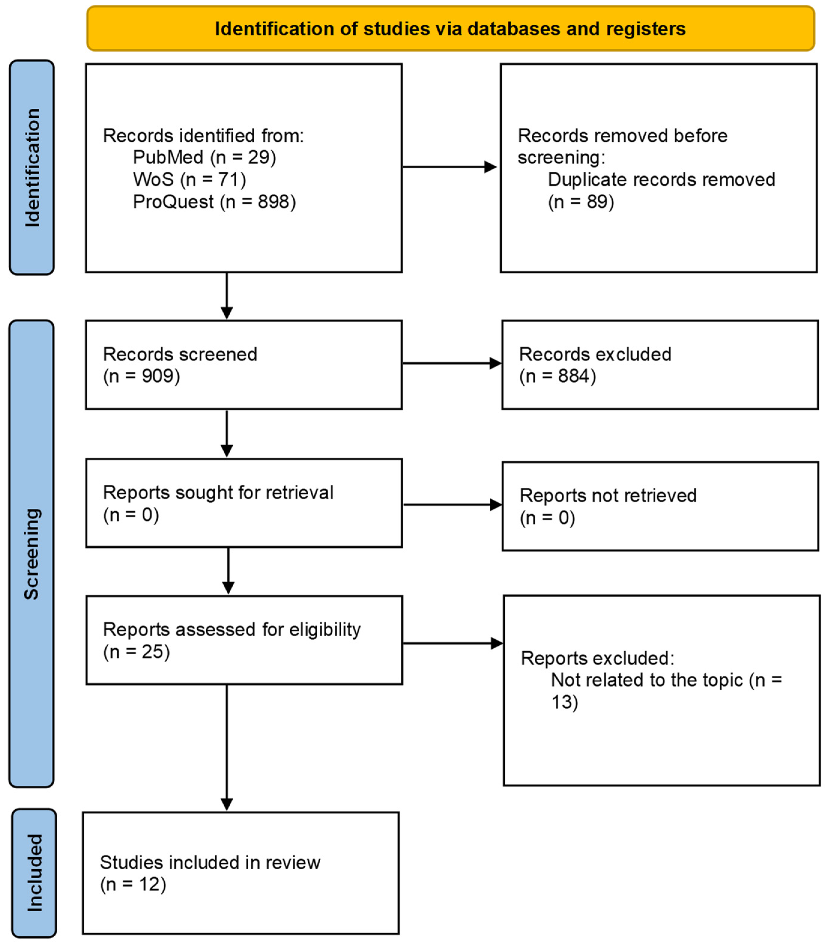
2.1.3. Screening Process
2.2. Data Analysis
2.2.1. Gene Ontology Enrichment Analysis
2.2.2. Protein–Protein Interaction Network and Module Analysis
2.2.3. Pathway Enrichment Analysis
2.2.4. Multi-Omics Network
3. Results
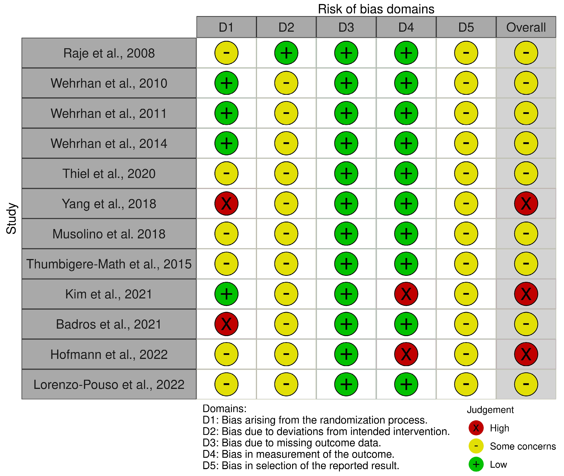
3.1. Systematic Review of Screening for MRONJ
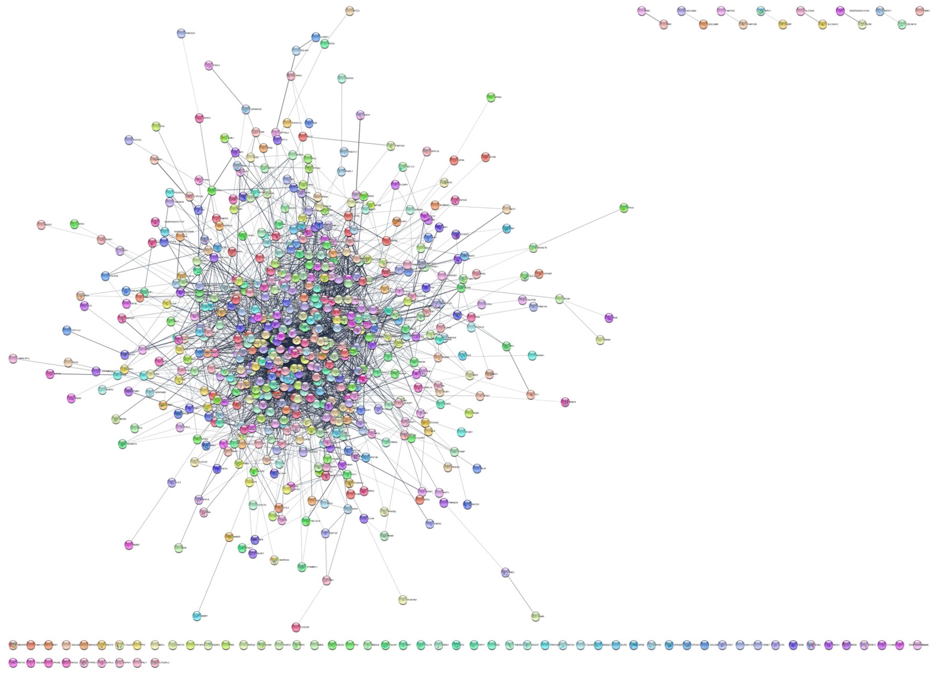
3.2. Network Analysis of Protein Interaction Data
3.3. GO Enrichment Analysis
3.4. Multiomics Networks in MRONJ
3.5. Pathway Enrichment Analysis
4. Discussion
5. Conclusions
Author Contributions
Funding
Data Availability Statement
Acknowledgments
Conflicts of Interest
References
- Marx, R.E. Pamidronate (Aredia) and Zoledronate (Zometa) Induced Avascular Necrosis of the Jaws: A Growing Epidemic. J. Oral Maxillofac. Surg. 2003, 61, 1115–1117. [Google Scholar] [CrossRef] [PubMed]
- Aghaloo, T.L.; Felsenfeld, A.L.; Tetradis, S. Osteonecrosis of the Jaw in a Patient on Denosumab. J. Oral Maxillofac. Surg. 2010, 68, 959–963. [Google Scholar] [CrossRef] [PubMed]
- Taylor, K.H.; Middlefell, L.S.; Mizen, K.D. Osteonecrosis of the Jaws Induced by Anti-RANK Ligand Therapy. Br. J. Oral Maxillofac. Surg. 2010, 48, 221–223. [Google Scholar] [CrossRef] [PubMed]
- Shibahara, T. Antiresorptive Agent-Related Osteonecrosis of the Jaw (Aronj): A Twist of Fate in the Bone. Tohoku J. Exp. Med. 2019, 247, 75–86. [Google Scholar] [CrossRef] [PubMed]
- Lombard, T.; Neirinckx, V.; Rogister, B.; Gilon, Y.; Wislet, S. Medication-Related Osteonecrosis of the Jaw: New Insights into Molecular Mechanisms and Cellular Therapeutic Approaches. Stem Cells Int. 2016, 2016, 8768162. [Google Scholar] [CrossRef] [PubMed]
- Ruggiero, S.L.; Dodson, T.B.; Fantasia, J.; Goodday, R.; Aghaloo, T.; Mehrotra, B.; O’Ryan, F. American Association of Oral and Maxillofacial Surgeons Position Paper on Medication-Related Osteonecrosis of the Jaw—2014 Update. J. Oral Maxillofac. Surg. 2014, 72, 1938–1956. [Google Scholar] [CrossRef]
- Rosella, D.; Papi, P.; Giardino, R.; Cicalini, E.; Piccoli, L.; Pompa, G. Medication-Related Osteonecrosis of the Jaw: Clinical and Practical Guidelines. J. Int. Soc. Prevent. Communit. Dent. 2016, 6, 97. [Google Scholar]
- Brijs, K.; Miclotte, I.; Vermeire, S.; Darche, V.; Politis, C. Osteonecrosis of the Jaw in Patients with Inflammatory Bowel Disease Treated with Tumour Necrosis Factor Alpha Inhibitors. Int. J. Oral. Max. Surg. 2020, 49, 317–324. [Google Scholar] [CrossRef]
- Diz, P.; López-Cedrún, J.L.; Arenaz, J.; Scully, C. Denosumab-Related Osteonecrosis of the Jaw. J. Am. Dent. Assoc. 2012, 143, 981–984. [Google Scholar] [CrossRef]
- Drake, M.T.; Clarke, B.L.; Khosla, S. Bisphosphonates: Mechanism of Action and Role in Clinical Practice. Mayo Clin. Proc. 2008, 83, 1032–1045. [Google Scholar] [CrossRef]
- Pimolbutr, K.; Porter, S.; Fedele, S. Osteonecrosis of the Jaw Associated with Antiangiogenics in Antiresorptive-Naïve Patient: A Comprehensive Review of the Literature. Biomed. Res. Int. 2018, 2018, 8071579. [Google Scholar] [CrossRef]
- Murphy, J.; Mannion, C.J. Medication-Related Osteonecrosis of the Jaws and Quality of Life: Review and Structured Analysis. Br. J. Oral Maxillofac. Surg. 2020, 58, 619–624. [Google Scholar] [CrossRef]
- Schwech, N.; Nilsson, J.; Gabre, P. Incidence and Risk Factors for Medication-related Osteonecrosis after Tooth Extraction in Cancer Patients—A Systematic Review. Clin. Exp. Dent. Res. 2023, 9, 55–65. [Google Scholar] [CrossRef]
- Dalle Carbonare, L.; Mottes, M.; Valenti, M.T. Medication-Related Osteonecrosis of the Jaw (Mronj): Are Antiresorptive Drugs the Main Culprits or Only Accomplices? The Triggering Role of Vitamin d Deficiency. Nutrients 2021, 13, 561. [Google Scholar] [CrossRef]
- Ruggiero, S.L.; Dodson, T.B.; Aghaloo, T.; Carlson, E.R.; Ward, B.B.; Kademani, D. American Association of Oral and Maxillofacial Surgeons’ Position Paper on Medication-Related Osteonecrosis of the Jaws—2022 Update. J. Oral Maxillofac. Surg. 2022, 80, 920–943. [Google Scholar] [CrossRef]
- He, L.; Sun, X.; Liu, Z.; Qiu, Y.; Niu, Y. Pathogenesis and Multidisciplinary Management of Medication-Related Osteonecrosis of the Jaw. Int. J. Oral Sci. 2020, 12, 30. [Google Scholar] [CrossRef] [PubMed]
- Zhang, W.; Gao, L.; Ren, W.; Li, S.; Zheng, J.; Li, S.; Jiang, C.; Yang, S.; Zhi, K. The Role of the Immune Response in the Development of Medication-Related Osteonecrosis of the Jaw. Front. Immunol. 2021, 12, 606043. [Google Scholar] [CrossRef] [PubMed]
- Chang, J.; Hakam, A.E.; McCauley, L.K. Current Understanding of the Pathophysiology of Osteonecrosis of the Jaw. Curr. Osteoporos. Rep. 2018, 16, 584–595. [Google Scholar] [CrossRef]
- Reid, I.R.; Bolland, M.J.; Grey, A.B. Is Bisphosphonate-Associated Osteonecrosis of the Jaw Caused by Soft Tissue Toxicity? Bone 2007, 41, 318–320. [Google Scholar] [CrossRef] [PubMed]
- Peer, A.; Khamaisi, M. Diabetes as a Risk Factor for Medication-Related Osteonecrosis of the Jaw. J. Dent. Res. 2015, 94, 252–260. [Google Scholar] [CrossRef]
- Riccardi, G.; Bellizzi, M.G.; Fatuzzo, I.; Zoccali, F.; Cavalcanti, L.; Greco, A.; Vincentiis, M.D.; Ralli, M.; Fiore, M.; Petrella, C.; et al. Salivary Biomarkers in Oral Squamous Cell Carcinoma: A Proteomic Overview. Proteomes 2022, 10, 37. [Google Scholar] [CrossRef]
- Page, M.J.; McKenzie, J.E.; Bossuyt, P.M.; Boutron, I.; Hoffmann, T.C.; Mulrow, C.D.; Shamseer, L.; Tetzlaff, J.M.; Akl, E.A.; Brennan, S.E.; et al. The PRISMA 2020 Statement: An Updated Guideline for Reporting Systematic Reviews. BMJ 2021, 372, n71. [Google Scholar] [CrossRef] [PubMed]
- The UniProt Consortium; Bateman, A.; Martin, M.-J.; Orchard, S.; Magrane, M.; Ahmad, S.; Alpi, E.; Bowler-Barnett, E.H.; Britto, R.; Bye-A-Jee, H.; et al. Uniprot: The Universal Protein Knowledgebase in 2023. Nucleic Acids Res. 2023, 51, D523–D531. [Google Scholar] [CrossRef]
- Shannon, P.; Markiel, A.; Ozier, O.; Baliga, N.S.; Wang, J.T.; Ramage, D.; Amin, N.; Schwikowski, B.; Ideker, T. Cytoscape: A Software Environment for Integrated Models of Biomolecular Interaction Networks. Genome Res. 2003, 13, 2498–2504. [Google Scholar] [CrossRef]
- Maere, S.; Heymans, K.; Kuiper, M. Bingo: A Cytoscape Plugin to Assess Overrepresentation of Gene Ontology Categories in Biological Networks. Bioinformatics 2005, 21, 3448–3449. [Google Scholar] [CrossRef] [PubMed]
- Doncheva, N.T.; Morris, J.H.; Gorodkin, J.; Jensen, L.J. Cytoscape Stringapp: Network Analysis and Visualization of Proteomics Data. J. Proteome Res. 2019, 18, 623–632. [Google Scholar] [CrossRef] [PubMed]
- Chin, C.-H.; Chen, S.-H.; Wu, H.-H.; Ho, C.-W.; Ko, M.-T.; Lin, C.-Y. Cytohubba: Identifying Hub Objects and Sub-Networks from Complex Interactome. BMC Syst. Biol. 2014, 8 (Suppl. 4), S11. [Google Scholar] [CrossRef] [PubMed]
- Bader, G.D.; Hogue, C.W. An Automated Method for Finding Molecular Complexes in Large Protein Interaction Networks. BMC Bioinform. 2003, 4, 2. [Google Scholar] [CrossRef]
- Wu, G.; Feng, X.; Stein, L. A Human Functional Protein Interaction Network and Its Application to Cancer Data Analysis. Genome Biol. 2010, 11, R53. [Google Scholar] [CrossRef]
- Zhou, G.; Pang, Z.; Lu, Y.; Ewald, J.; Xia, J. OmicsNet 2.0: A Web-Based Platform for Multi-Omics Integration and Network Visual Analytics. Nucleic Acids Res. 2022, 50, W527–W533. [Google Scholar] [CrossRef]
- Chang, L.; Xia, J. Microrna Regulatory Network Analysis Using Mirnet 2.0. In Transcription Factor Regulatory Networks; Song, Q., Tao, Z., Eds.; Springer: New York, NY, USA, 2023; Volume 2594, pp. 185–204. [Google Scholar]
- Raje, N.; Woo, S.-B.; Hande, K.; Yap, J.T.; Richardson, P.G.; Vallet, S.; Treister, N.; Hideshima, T.; Sheehy, N.; Chhetri, S.; et al. Clinical, Radiographic, and Biochemical Characterization of Multiple Myeloma Patients with Osteonecrosis of the Jaw. Clin. Cancer Res. 2008, 14, 2387–2395. [Google Scholar] [CrossRef]
- Wehrhan, F.; Hyckel, P.; Ries, J.; Stockmann, P.; Nkenke, E.; Schlegel, K.A.; Neukam, F.W.; Amann, K. Expression of Msx-1 Is Suppressed in Bisphosphonate Associated Osteonecrosis Related Jaw Tissue-Etiopathology Considerations Respecting Jaw Developmental Biology-Related Unique Features. J. Transl. Med. 2010, 8, 96. [Google Scholar] [CrossRef]
- Wehrhan, F.; Amann, K.; Möbius, P.; Weber, M.; Preidl, R.; Ries, J.; Stockmann, P. BRONJ-Related Jaw Bone Is Associated with Increased Dlx-5 and Suppressed Osteopontin—Implication in the Site-Specific Alteration of Angiogenesis and Bone Turnover by Bisphosphonates. Clin. Oral Investig. 2015, 19, 1289–1298. [Google Scholar] [CrossRef] [PubMed]
- Wehrhan, F.; Hyckel, P.; Amann, K.; Ries, J.; Stockmann, P.; Schlegel, K.; Neukam, F.; Nkenke, E. Msx-1 Is Suppressed in Bisphosphonate-Exposed Jaw Bone Analysis of Bone Turnover-Related Cell Signalling after Bisphosphonate Treatment: ONJ and Msx-1 Expression. Oral Dis. 2011, 17, 433–442. [Google Scholar] [CrossRef]
- Thiel, Y.; Ghayor, C.; Lindhorst, D.; Essig, H.; Weber, F.; Rücker, M.; Schumann, P. Antimicrobial Peptide Gene Expression in Medication-Related Osteonecrosis of the Jaw. Pathol. Res. Pract. 2020, 216, 153245. [Google Scholar] [CrossRef] [PubMed]
- Yang, R.; Tao, Y.; Wang, C.; Shuai, Y.; Jin, L. Circulating microRNA Panel as a Novel Biomarker to Diagnose Bisphosphonate-Related Osteonecrosis of the Jaw. Int. J. Med. Sci. 2018, 15, 1694–1701. [Google Scholar] [CrossRef]
- Musolino, C.; Oteri, G.; Allegra, A.; Mania, M.; D’Ascola, A.; Avenoso, A.; Innao, V.; Allegra, A.G.; Campo, S. Altered microRNA Expression Profile in the Peripheral Lymphoid Compartment of Multiple Myeloma Patients with Bisphosphonate-Induced Osteonecrosis of the Jaw. Ann. Hematol. 2018, 97, 1259–1269. [Google Scholar] [CrossRef]
- Thumbigere-Math, V.; Michalowicz, B.; Jong, E.; Griffin, T.; Basi, D.; Hughes, P.; Tsai, M.; Swenson, K.; Rockwell, L.; Gopalakrishnan, R. Salivary Proteomics in Bisphosphonate-related Osteonecrosis of the Jaw. Oral Dis. 2015, 21, 46–56. [Google Scholar] [CrossRef] [PubMed]
- Kim, J.; Yeon, A.; Parker, S.J.; Shahid, M.; Thiombane, A.; Cho, E.; You, S.; Emam, H.; Kim, D.-G.; Kim, M. Alendronate-Induced Perturbation of the Bone Proteome and Microenvironmental Pathophysiology. Int. J. Med. Sci. 2021, 18, 3261–3270. [Google Scholar] [CrossRef]
- Badros, A.Z.; Meddeb, M.; Weikel, D.; Philip, S.; Milliron, T.; Lapidus, R.; Hester, L.; Goloubeva, O.; Meiller, T.F.; Mongodin, E.F. Prospective Observational Study of Bisphosphonate-Related Osteonecrosis of the Jaw in Multiple Myeloma: Microbiota Profiling and Cytokine Expression. Front. Oncol. 2021, 11, 704722. [Google Scholar] [CrossRef]
- Hofmann, E.; Eggers, B.; Heim, N.; Kramer, F.-J.; Nokhbehsaim, M.; Götz, W. Bevacizumab and Sunitinib Mediate Osteogenic and Pro-Inflammatory Molecular Changes in Primary Human Alveolar Osteoblasts in Vitro. Odontology 2022, 110, 634–647. [Google Scholar] [CrossRef] [PubMed]
- Lorenzo-Pouso, A.I.; Bravo, S.B.; Carballo, J.; Chantada-Vázquez, M.D.P.; Bagán, J.; Bagán, L.; Chamorro-Petronacci, C.M.; Conde-Amboage, M.; López-López, R.; García-García, A.; et al. Quantitative Proteomics in Medication-related Osteonecrosis of the Jaw: A Proof-of-concept Study. Oral Dis. 2023, 29, 2117–2129. [Google Scholar] [CrossRef] [PubMed]
- Ferneini, E.M. Medication-Related Osteonecrosis of the Jaw (Mronj). J. Oral. Maxillofac. Surg. 2021, 79, 1801–1802. [Google Scholar] [CrossRef] [PubMed]
- Bansal, H. Medication-Related Osteonecrosis of the Jaw: An Update. Natl. J. Maxillofac. Surg. 2022, 13, 5. [Google Scholar] [CrossRef] [PubMed]
- Aghaloo, T.; Hazboun, R.; Tetradis, S. Pathophysiology of Osteonecrosis of the Jaws. Oral Maxillofac. Surg. Clin. 2015, 27, 489–496. [Google Scholar] [CrossRef] [PubMed]
- Kuroshima, S.; Al-Omari, F.A.; Sasaki, M.; Sawase, T. Medication-related Osteonecrosis of the Jaw: A Literature Review and Update. Genesis 2022, 60, e23500. [Google Scholar] [CrossRef] [PubMed]
- Reimand, J.; Isserlin, R.; Voisin, V.; Kucera, M.; Tannus-Lopes, C.; Rostamianfar, A.; Wadi, L.; Meyer, M.; Wong, J.; Xu, C.; et al. Pathway Enrichment Analysis and Visualization of Omics Data Using: Profiler, GSEA, Cytoscape and EnrichmentMap. Nat. Protoc. 2019, 14, 482–517. [Google Scholar] [CrossRef]
- Ma, M.; Tan, Z.; Li, W.; Zhang, H.; Liu, Y.; Yue, C. Osteoimmunology and Osteonecrosis of the Femoral Head. Bone Jt. Res. 2022, 11, 26–28. [Google Scholar] [CrossRef]
- Zheng, J.; Yao, Z.; Xue, L.; Wang, D.; Tan, Z. The Role of Immune Cells in Modulating Chronic Inflammation and Osteonecrosis. Front. Immunol. 2022, 13, 1064245. [Google Scholar] [CrossRef]
- Li, Q.; Pu, Y.; Lu, H.; Zhao, N.; Wang, Y.; Guo, Y.; Guo, C. Porphyromonas, Treponema, and Mogibacterium Promote Il8/Ifnγ/Tnfα-Based pro-Inflammation in Patients with Medication-Related Osteonecrosis of the Jaw. J. Oral Microbiol. 2021, 13, 1851112. [Google Scholar] [CrossRef]
- Liu, H.; Li, D.; Zhang, Y.; Li, M. Inflammation, Mesenchymal Stem Cells and Bone Regeneration. Histochem. Cell Biol. 2018, 149, 393–404. [Google Scholar] [CrossRef]
- Zhou, F.; Zhang, G.; Wu, Y.; Xiong, Y. Inflammasome Complexes: Crucial Mediators in Osteoimmunology and Bone Diseases. Int. Immunopharmacol. 2022, 110, 109072. [Google Scholar] [CrossRef] [PubMed]
- Amarasekara, D.S.; Kim, S.; Rho, J. Regulation of Osteoblast Differentiation by Cytokine Networks. Int. J. Mol. Sci. 2021, 22, 2851. [Google Scholar] [CrossRef] [PubMed]
- Amarasekara, D.S.; Yun, H.; Kim, S.; Lee, N.; Kim, H.; Rho, J. Regulation of Osteoclast Differentiation by Cytokine Networks. Immune Netw. 2018, 18, e8. [Google Scholar] [CrossRef] [PubMed]
- Weitzmann, M.N. T-Cells and B-Cells in Osteoporosis. Curr. Opin. Endocrinol. Diabetes. Obes. 2014, 21, 461–467. [Google Scholar] [CrossRef] [PubMed]
- Kalyan, S.; Quabius, E.S.; Wiltfang, J.; Mönig, H.; Kabelitz, D. Can Peripheral Blood γδ T Cells Predict Osteonecrosis of the Jaw? An Immunological Perspective on the Adverse Drug Effects of Aminobisphosphonate Therapy. J. Bone Miner. Res. 2013, 28, 728–735. [Google Scholar] [CrossRef] [PubMed]
- Weitzmann, M.N. Do Γδ T Cells Predict Osteonecrosis of the Jaw? J. Bone Miner. Res. 2013, 28, 723–727. [Google Scholar] [CrossRef]
- Ribot, J.C.; Lopes, N.; Silva-Santos, B. Γδ T Cells in Tissue Physiology and Surveillance. Nat. Rev. Immunol. 2021, 21, 221–232. [Google Scholar] [CrossRef]
- Qu, X.; Wang, Z.; Zhou, T.; Shan, L. Determination of the Molecular Mechanism by Which Macrophages and γδ-T Cells Contribute to ZOL-Induced ONJ. Aging 2020, 12, 20743–20752. [Google Scholar] [CrossRef]
- Wang, S.; Wu, R.; Lu, J.; Jiang, Y.; Huang, T.; Cai, Y. Protein-protein Interaction Networks as Miners of Biological Discovery. Proteomics 2022, 22, 2100190. [Google Scholar] [CrossRef]
- Tiwari, A.; Mukherjee, B.; Dixit, M. Microrna Key to Angiogenesis Regulation: Mirna Biology and Therapy. Curr. Cancer Drug Tar. 2018, 18, 266–277. [Google Scholar] [CrossRef] [PubMed]
- Bastos, P.; Patel, V.; Festy, F.; Hosny, N.; Cook, R.J. In-Vivo Imaging of the Microvasculature of the Soft Tissue Margins of Osteonecrotic Jaw Lesions. Brit. Dent. J. 2017, 223, 699–705. [Google Scholar] [CrossRef] [PubMed]
- Ohlrich, E.J.; Coates, D.E.; Cullinan, M.P.; Milne, T.J.; Zafar, S.; Zhao, Y.; Duncan, W.D.; Seymour, G.J. The Bisphosphonate Zoledronic Acid Regulates Key Angiogenesis-Related Genes in Primary Human Gingival Fibroblasts. Arch. Oral. Biol. 2016, 63, 7–14. [Google Scholar] [CrossRef] [PubMed]
- Zhao, Z.; Sun, W.; Guo, Z.; Zhang, J.; Yu, H.; Liu, B. Mechanisms of lncRNA/microRNA Interactions in Angiogenesis. Life Sci. 2020, 254, 116900. [Google Scholar] [CrossRef]
- He, B.; Zhao, Z.; Cai, Q.; Zhang, Y.; Zhang, P.; Shi, S.; Xie, H.; Peng, X.; Yin, W.; Tao, Y.; et al. Mirna-Based Biomarkers, Therapies, and Resistance in Cancer. Int. J. Biol. Sci. 2020, 16, 2628–2647. [Google Scholar] [CrossRef]
- Bhatti, G.K.; Khullar, N.; Sidhu, I.S.; Navik, U.S.; Reddy, A.P.; Reddy, P.H.; Bhatti, J.S. Emerging Role of Non-coding RNA in Health and Disease. Metab. Brain Dis. 2021, 36, 1119–1134. [Google Scholar] [CrossRef] [PubMed]
- Sotiropoulou, G.; Pampalakis, G.; Lianidou, E.; Mourelatos, Z. Emerging Roles of microRNAs as Molecular Switches in the Integrated Circuit of the Cancer Cell. RNA 2009, 15, 1443–1461. [Google Scholar] [CrossRef]
- Huang, Y.; Du, K.L.; Guo, P.Y.; Zhao, R.M.; Wang, B.; Zhao, X.L.; Zhang, C.Q. IL-16 Regulates Macrophage Polarization as a Target Gene of Mir-145-3p. Mol. Immunol. 2019, 107, 1–9. [Google Scholar] [CrossRef]
- Tian, Z.J.; Liu, B.Y.; Zhang, Y.T.; Chen, X.Z.; Qiao, G.Y.; Wang, S.; Ma, Z.L. MiR-145 Silencing Promotes Steroid-Induced Avascular Necrosis of the Femoral Head Repair via Upregulating VEGF. Eur. Rev. Med. Pharmacol. Sci. 2017, 21, 3763–3769. [Google Scholar]
- Ma, H.; Zhang, W.; Shi, J. Differentially Expressed Genes Reveal the Biomarkers and Molecular Mechanism of Osteonecrosis. J. Healthc. Eng. 2022, 2022, 8684137. [Google Scholar] [CrossRef]
- Li, T.; Li, H.; Wang, Y.; Li, T.; Fan, J.; Xiao, K.; Zhao, R.C.; Weng, X. Microrna-23a Inhibits Osteogenic Differentiation of Human Bone Marrow-Derived Mesenchymal Stem Cells by Targeting Lrp5. Int. J. Biochem. Cell B 2016, 72, 55–62. [Google Scholar] [CrossRef] [PubMed]
- Dong, Y.; Li, T.; Li, Y.; Ren, S.; Fan, J.; Weng, X. MicroRNA-23a-3p Inhibitor Decreases Osteonecrosis Incidence in a Rat Model. Mol. Med. Rep. 2017, 16, 9331–9336. [Google Scholar] [CrossRef] [PubMed]


| mRNA | ||
|---|---|---|
| Reference | Sample Type | Method |
| Raje et al., 2008, 10.1158/1078-0432.CCR-07-1430 [32] | Peripheral blood mononuclear cells. Patients: MM patients with ONJ (n = 8). Controls: MM patients without ONJ (n = 10), healthy volunteers (n = 5). | Affymetrix U133Plus 2.0 Gene Chip (Affymetrix, Santa Clara, CA, USA). |
| Wehrhan et al., 2010, 10.1186/1479-5876-8-96 [33] | Periodontal samples. Patients: patients with BRONJ (n = 20). Controls: non-BP exposed periodontal samples (n = 20). | Microfluid Lab-on-a-Chip technology (Agilent RNA 6000 Pico Kit and the Agilent 2100 Bioanalyzer, Agilent, Waldbronn, Germany). The cDNAs from total RNA were synthesized using the High-Capacity cDNA Archive Kit (Cat. 4322171; Applied Biosystems, Foster City, CA, USA). Real-time RT qPCR (QuantiTect Primer Assay; Qiagen, Hilden, Germany). |
| Wehrhan et al., 2011, 10.1111/j.1601-0825.2010.01778.x [34] | Periodontal samples. Patients: patients with BRONJ (n = 20). Controls: non-BP exposed periodontal samples (n = 20). | Microfluid Lab-on-a-Chip technology (Agilent RNA 6000 Pico Kit and the Agilent 2100 Bioanalyzer, Agilent, Waldbronn, Germany). The cDNAs from total RNA were synthesized using the High-Capacity cDNA Archive Kit (Cat. 4322171; Applied Biosystems, Foster City, CA, USA). Real-time RT qPCR (QuantiTect Primer Assay; Qiagen, Hilden, Germany). |
| Wehrhan et al., 2014 10.1007/s00784-014-1354-7 [35] | Jawbone samples. Patients: patients with BRONJ (n = 15). Controls: non-BP exposed samples (n = 20). | Total RNA extraction (RNeasy Kit, Qiagen, Hilden, Germany). Microfluid Lab-on-a-Chip technology (Agilent RNA 6000 Pico Kit and the Agilent 2100 Bioanalyzer, Agilent, Waldbronn, Germany). High-capacity cDNA Archive Kit (Cat. No. 4322171; Applied Biosystem, Foster City, CA, USA). Real-time RT quantitative PCR analyses: Hs_SPP1_1_SGQuantiTect Primer Assay (200) on the ABI Prism 7300 Sequence Detection System (Applied Biosystems, Waltham, MA, USA). PCR amplification: the QuantiTect TM SYBR® green PCR kit (Cat. No. 204143; Qiagen, Hilden, Germany). |
| Thiel et al., 2020 10.1016/j.prp.2020.153245 [36] | Jawbone samples. Patients: diagnosed with MRONJ (n = 12). Controls: subjects without MRONJ (n = 6). | RNA extraction kit (miRNeasy Mini Kit; Qiagen, Hilden, Germany). The total RNA was reverse transcribed into cDNA using the iScript™ cDNA Synthesis Kit (Bio-Rad, Hercules, CA, USA). PCR amplification: SsoAdvanced™ Universal SYBR® Green Supermix (Bio-Rad, Hercules, CA, USA) Amplification was conducted on the CFX Connect Real-Time PCR System (Bio-Rad, Hercules, CA, USA). |
| miRNA | ||
| Reference | Sample Type | Method |
| Raje et al., 2008, 10.1158/1078-0432.CCR-07-1430. [32] | Peripheral blood mononuclear cells. Patients: MM patients with ONJ (n = 8). Controls: MM patients without ONJ (n = 10), healthy volunteers (n = 5). | Affymetrix U133Plus 2.0 Gene Chip (Affymetrix, Santa Clara, California, USA). |
| Yang et al., 2018, 10.7150/ijms.27593 [37] | Serum. Patients: patients with BRONJ (n = 6). Controls: non-BP healthy individuals (n = 11). | RNA extraction: mirVana Paris Kit (Ambion, Huntingdon, Cambridgeshire, United Kingdom). The microRNAs were reversed to cDNA using the miScript II RT Kit (Qiagen, Hilden, Germany). Q-RT-PCR analysis was conducted using the miScript SYBR Green PCR Kit (Qiagen, Hilden, Germany) with a 7500 Real-Time PCR System (Applied Biosystems, Foster City, CA, USA). |
| Musolino et al. 2018, 10.1007/s00277-018-3296-7 [38] | Peripheral blood. Patients: MM patients with ONJ (n = 5). Controls: healthy volunteers (n = 5). | RNA extraction: the Total Purification Plus Kit (Norgen Biotek Corporation, Thorold, ON, Canada). Total RNA was transcribed into cDNA through an All-in-One miRNA first-strand cDNA synthesis kit (GeneCopoeia Inc., Rockville, MD, USA). Real-Time qPCR employed a 7500 Real-Time PCR System (Applied Biosystems, Foster City, CA, USA). |
| Proteins | ||
| Reference | Sample Type | Method |
| Thumbigere-Math et al., 2015, 10.1111/odi.12204 [39] | Saliva. Patients: BRONJ (n = 20), high- and low-infusion groups. Controls: non-BRONJ patients (n = 20). | iTRAQ labeling was followed by fractionation using strong cation exchange chromatography, and fractions were analyzed by reversed-phase microcapillary LC-S (LTQ-Orbitrap). |
| Kim et al., 2021, 10.7150/ijms.61552 [40] | MG-63, SCC-9, SCC-15, and HUVEC cells. ALN-treated and non-ALN control groups. | 2D-DIGE, followed by MALDI TOF/TOF MS (4800 Plus, Applied Biosystems, Foster City, CA, Life Sciences, USA). |
| Badros et al., 2021, 10.3389/fonc.2021.704722 [41] | Saliva, serum. Patients: MM patients who underwent intravenous BP therapy and developed BRONJ (n = 14). Controls: non-BRONJ MM patients (n = 96). | Luminex™ technology (EMD Millipore, Burlington, MA, USA). |
| Hofmann et al., 2022, 10.1007/s10266-022-00691-y [42] | HAOB cells. BEV/SUN-treated and non-BEV/SUN control groups. | ELISA |
| Lorenzo-Pouso et al., 2022, 10.1111/odi.14201 [43] | Saliva. Patients: Group 1—MRONJ cases (n = 18). Controls: Group 2—individuals undergoing treatment with BMAs for more than 24 months without MRONJ (n = 10). Group 3—healthy volunteers (n = 10). | SDS-PAGE, shotgun DDA by micro-flow LC-MS/MS, a quadrupole-TOF mass spectrometer (Triple TOF 6600 [SCIEX, Framingham, MA, USA]) working in ESI + performed DDA analysis. |
| Topological Parameters | Values |
|---|---|
| The average number of neighbors | 12.755 |
| Clustering coefficient | 0.264 |
| Characteristic path length | 3.294 |
| Network diameter | 9 |
| Number of edges | 3993 |
| Number of nodes | 701 |
| MCC | DMN | MNC | Degree | FPC | Bottleneck | EcCentricity | Closeness | Radiality | Betweenness | Stress | Clustering Coefficient |
|---|---|---|---|---|---|---|---|---|---|---|---|
| ALB | A1BG | ALB | ALB | ALB | ALB | ARF1 | ALB | ALB | ALB | ALB | A2ML1 |
| ANXA5 | AGER | ANXA5 | ANXA5 | ANXA5 | CAT | ARHGDIA | ANXA5 | ANXA5 | ATM | ATM | BANK1 |
| CCL2 | ANGPT1 | B2M | CCL2 | CCL2 | CD44 | ARRB1 | ATM | ATM | CD44 | CD44 | CFHR5 |
| CD44 | CD83 | CCL2 | CD44 | CD44 | CXCL8 | BCL2L11 | CCL2 | CASP8 | CLTC | CXCL8 | DPT |
| CSF3 | CXCL1 | CD44 | CXCL8 | CXCL8 | EEF1A1 | BTK | CD44 | CD44 | EEF2 | EEF2 | ENSP00000330898 |
| CXCL8 | CXCL2 | CXCL8 | CXCR4 | CXCR4 | EGF | CD83 | CXCL8 | CXCL8 | EGF | EGF | ENSP00000377747 |
| CXCR4 | EEF1B2 | CXCR4 | EEF2 | EGF | FUS | COL1A1 | CXCR4 | CXCR4 | GART | GART | FAM213A |
| EGF | EIF2S3 | EGF | EGF | HSPA4 | GART | CYCS | EGF | CYCS | HIST1H4F | HSP90AB1 | GTF3C4 |
| IGF1 | EIF5A2 | HSP90AB1 | HSP90AB1 | IGF1 | HIST1H4F | DNAJB1 | HSP90AB1 | EGF | HSP90AB1 | HSPA4 | CHI3L1 |
| IL1B | CHI3L1 | HSPA4 | HSPA4 | IL1B | HSP90AB1 | FAS | HSPA4 | HSP90AB1 | HSPA4 | IL1B | IL36A |
| IL6 | LRG1 | IGF1 | IGF1 | IL6 | HSPA4 | FCGR3A | IGF1 | HSPA4 | IL6 | IL6 | KRT76 |
| JUN | MMP1 | IL1B | IL1B | ITGB1 | IGF1 | GART | IL1B | IGF1 | JUN | JUN | LMF1 |
| KDR | ORM1 | IL6 | IL6 | JUN | JUN | IL6 | IL6 | IL1B | LMNA | LMNA | ME1 |
| MMP9 | ORM2 | ITGB1 | ITGB1 | MMP9 | KRT14 | ITGB1 | ITGB1 | IL6 | PTPRC | PTPRC | NOV |
| PTGS2 | PSMC1 | JUN | JUN | PTGS2 | LMNA | KRT19 | JUN | JUN | RAB5A | RAB5A | POLR2J3 |
| PTPRC | RPLP1 | MMP9 | MMP9 | PTPRC | PTPRC | NR4A2 | MMP9 | MMP9 | RHOA | RHOA | SEL1L3 |
| SPP1 | RPLP2 | PTPRC | PTPRC | RHOA | RAB5A | PPP2CB | PTPRC | PTPRC | SRSF1 | TFRC | SELM |
| TGFB1 | SAA4 | RHOA | RHOA | SPP1 | RHOA | SAA4 | RHOA | RHOA | TFRC | TNF | SERPIND1 |
| TNF | SERPIND1 | TNF | TNF | TNF | SRSF1 | TXN | TNF | TNF | TNF | VEGFA | TNN |
| VEGFA | TNFRSF11B | VEGFA | VEGFA | VEGFA | TNF | VEGFA | VEGFA | VEGFA | VEGFA | YWHAZ | VPS36 |
| Cluster | Score (Density * Nodes) | Nodes | Edges | Node IDs |
|---|---|---|---|---|
| 1 | 24.794 | 64 | 781 | BGLAP, BMP2, CAT, CCL4, CCT2, CD44, COL1A1, CSF3, CXCL1, CXCL2, CXCL8, CXCR4, CYCS, EEF1A1, EEF1B2, EEF1D, EEF1G, EEF2, EGF, EIF2S3, EIF5A, EIF5A2, FGG, IGF1, IL1B, IL6, ITGB1, JUN, KDR, MARS, MMP1, MMP8, MMP9, NT5E, PSMC1, PTGS2, PTPRC, RHOA, RPL10, RPL12, RPL27A, RPL4, RPLP1, RPLP2, RPS10, RPS12, RPS16, RPS23, RPS25, RPSA, RUNX2, SERPINA1, SERPINC1, SOD2, SPP1, TGFB1, TNF *, TNFRSF11B, TNFSF11, TPT1, VEGFA |
| 2 | 13.429 | 43 | 282 | A1BG *, A2M, AGER, AMBP, ANXA5, APOA2, APOB, APOH, ATM, AZGP1, BCL2L11, C3, C4B, CASP8, CCL2, CP, CREB1, FAS, FCGR3A, FOXO1, GART, GIG25, HP, HPX, HSP90AB1, HSPA4, HSPB1, ITIH2, ITIH4, JAK1, KLRK1, LCK, LCN2, LRG1, MCL1, NFATC1, ORM1, ORM2, PDGFB, TF, TFRC, TTR, TXN |
| 3 | 10.133 | 16 | 76 | DSG1, DSP, IVL, KRT14, KRT15, KRT16, KRT17, KRT4, KRT5, KRT6B, KRT6C, SCEL, SPRR1A *, SPRR1B, SPRR3, TGM1 |
| 4 | 6.933 | 31 | 104 | ACTG2, ALAS2, ATRX, CA2, CBFB, DDX3X, ETS1, GATA2, H2AFJ, HBA1, HBA2, HBB *, HBD, HBG1, HBG2, HIST1H1B, HIST1H1E, HIST1H2AB, HIST1H2AC, HIST1H3J, KMT2A, MYL12A, MYL6, SLC25A37, SLC4A1, SRSF1, SUPT16H, TAL1, TPM2, TPM3, TPM4 |
| 5 | 6 | 6 | 16 | CELF1, FUS, HNRNPK *, MBNL1, SRSF10, SRSF3 |
| GO-ID | Description | p-Value | Corr p-Value | x | n | X | N |
|---|---|---|---|---|---|---|---|
| Biological Process | |||||||
| 2376 | immune system process | 2.9469 × 10⁻¹⁵ | 1.0179 × 10⁻¹¹ | 97 | 947 | 631 | 14,265 |
| 6950 | response to stress | 1.7284 × 10⁻¹³ | 2.9850 × 10⁻¹⁰ | 143 | 1771 | 631 | 14,265 |
| 9611 | response to wounding | 4.6100 × 10⁻¹³ | 5.3076 × 10⁻¹⁰ | 64 | 541 | 631 | 14,265 |
| 6955 | immune response | 2.5317 × 10⁻¹² | 2.1861 × 10⁻⁹ | 68 | 618 | 631 | 14,265 |
| 6952 | defense response | 8.4706 × 10⁻¹² | 5.1466 × 10⁻⁹ | 67 | 620 | 631 | 14,265 |
| 42221 | response to a chemical stimulus | 8.9402 × 10⁻¹² | 5.1466 × 10⁻⁹ | 120 | 1462 | 631 | 14,265 |
| 48513 | organ development | 1.7962 × 10⁻¹¹ | 8.8627 × 10⁻⁹ | 138 | 1792 | 631 | 14,265 |
| 48583 | regulation of response to stimulus | 3.1299 × 10⁻¹¹ | 1.3514 × 10⁻⁸ | 59 | 524 | 631 | 14,265 |
| 9888 | tissue development | 1.3146 × 10⁻¹⁰ | 4.9607 × 10⁻⁸ | 73 | 750 | 631 | 14265 |
| 6954 | inflammatory response | 1.4362 × 10⁻¹⁰ | 4.9607 × 10⁻⁸ | 42 | 315 | 631 | 14,265 |
| Molecular Function | |||||||
| 5515 | protein binding | 2.9969 × 10⁻¹⁹ | 2.6493 × 10⁻¹⁶ | 462 | 8106 | 667 | 15,404 |
| 5198 | structural molecule activity | 2.5854 × 10⁻¹³ | 1.1427 × 10⁻¹⁰ | 68 | 600 | 667 | 15,404 |
| 5488 | binding | 4.2458 × 10⁻¹¹ | 1.2511 × 10⁻⁸ | 596 | 12,340 | 667 | 15,404 |
| 5200 | structural constituent of the cytoskeleton | 8.2347 × 10⁻⁸ | 1.8199 × 10⁻⁵ | 16 | 74 | 667 | 15,404 |
| 3823 | antigen binding | 1.5501 × 10⁻⁷ | 2.3636 × 10⁻⁵ | 14 | 59 | 667 | 15,404 |
| 4857 | enzyme inhibitor activity | 1.6043 × 10⁻⁷ | 2.3636 × 10⁻⁵ | 33 | 279 | 667 | 15,404 |
| 3746 | translation elongation factor activity | 9.3982 × 10⁻⁷ | 1.1869 × 10⁻⁴ | 8 | 20 | 667 | 15,404 |
| 4866 | endopeptidase inhibitor activity | 1.2753 × 10⁻⁶ | 1.4031 × 10⁻⁴ | 21 | 146 | 667 | 15,404 |
| 61135 | endopeptidase regulator activity | 1.4285 × 10⁻⁶ | 1.4031 × 10⁻⁴ | 21 | 147 | 667 | 15,404 |
| 30414 | peptidase inhibitor activity | 3.4059 × 10⁻⁶ | 3.0108 × 10⁻⁴ | 21 | 155 | 667 | 15,404 |
| Cell Component | |||||||
| 5615 | extracellular space | 4.5890 × 10⁻¹⁴ | 2.1385 × 10⁻¹¹ | 78 | 748 | 680 | 16,336 |
| 5576 | extracellular region | 1.9191 × 10⁻¹³ | 4.4715 × 10⁻¹¹ | 151 | 2022 | 680 | 16,336 |
| 44421 | extracellular region part | 2.5524 × 10⁻¹² | 3.9647 × 10⁻¹⁰ | 89 | 985 | 680 | 16,336 |
| 43228 | non-membrane-bounded organelle | 6.4518 × 10⁻¹⁰ | 6.0131 × 10⁻⁸ | 160 | 2425 | 680 | 16,336 |
| 43232 | intracellular non-membrane-bounded organelle | 6.4518 × 10⁻¹⁰ | 6.0131 × 10⁻⁸ | 160 | 2425 | 680 | 16,336 |
| 5737 | cytoplasm | 2.2730 × 10⁻⁹ | 1.7654 × 10⁻⁷ | 393 | 7634 | 680 | 16,336 |
| 5856 | cytoskeleton | 3.1182 × 10⁻⁹ | 2.0758 × 10⁻⁷ | 104 | 1399 | 680 | 16,336 |
| 1533 | cornified envelope | 1.0235 × 10⁻⁸ | 5.9620 × 10⁻⁷ | 10 | 23 | 680 | 16,336 |
| 31983 | vesicle lumen | 2.4577 × 10⁻⁸ | 1.2725 × 10⁻⁶ | 12 | 38 | 680 | 16,336 |
| 31093 | platelet alpha granule lumen | 1.0035 × 10⁻⁷ | 4.6761 × 10⁻⁶ | 11 | 35 | 680 | 16,336 |
| GO-ID | Description | p-Value | Corr p-Value | x | n | X | N |
|---|---|---|---|---|---|---|---|
| Biological Process | |||||||
| 1932 | regulation of protein amino acid phosphorylation | 5.3257 × 10⁻¹¹ | 1.6700 × 10⁻⁸ | 8 | 217 | 17 | 14,306 |
| 42325 | regulation of phosphorylation | 5.5034 × 10⁻¹¹ | 1.6700 × 10⁻⁸ | 10 | 518 | 17 | 14,306 |
| 42327 | positive regulation of phosphorylation | 8.2778 × 10⁻¹¹ | 1.6700 × 10⁻⁸ | 7 | 131 | 17 | 14,306 |
| 19220 | regulation of the phosphate metabolic process | 8.5947 × 10⁻¹¹ | 1.6700 × 10⁻⁸ | 10 | 542 | 17 | 14,306 |
| 51174 | regulation of the phosphorus metabolic process | 8.5947 × 10⁻¹¹ | 1.6700 × 10⁻⁸ | 10 | 542 | 17 | 14,306 |
| 10562 | positive regulation of the phosphorus metabolic process | 9.7175 × 10⁻¹¹ | 1.6700 × 10⁻⁸ | 7 | 134 | 17 | 14,306 |
| 45937 | positive regulation of the phosphate metabolic process | 9.7175 × 10⁻¹¹ | 1.6700 × 10⁻⁸ | 7 | 134 | 17 | 14,306 |
| 35468 | positive regulation of the signaling pathway | 1.2071 × 10⁻¹⁰ | 1.8152 × 10⁻⁸ | 9 | 380 | 17 | 14,306 |
| 48661 | positive regulation of smooth muscle cell proliferation | 1.4481 × 10⁻¹⁰ | 1.9356 × 10⁻⁸ | 5 | 29 | 17 | 14,306 |
| 10647 | positive regulation of cell communication | 2.5305 × 10⁻¹⁰ | 3.0442 × 10⁻⁸ | 9 | 413 | 17 | 14,306 |
| Molecular Function | |||||||
| 5126 | cytokine receptor binding | 3.1215 × 10⁻⁸ | 3.3196 × 10⁻⁶ | 6 | 186 | 17 | 15,443 |
| 5125 | cytokine activity | 4.3968 × 10⁻⁸ | 3.3196 × 10⁻⁶ | 6 | 197 | 17 | 15,443 |
| 8083 | growth factor activity | 6.2752 × 10⁻⁷ | 3.1585 × 10⁻⁵ | 5 | 160 | 17 | 15,443 |
| 70851 | growth factor receptor binding | 1.6672 × 10⁻⁶ | 6.2937 × 10⁻⁵ | 4 | 82 | 17 | 15,443 |
| 5102 | receptor binding | 2.3472 x× 10−6 | 7.0887 × 10⁻⁵ | 8 | 922 | 17 | 15,443 |
| 5515 | protein binding | 1.7881 × 10⁻⁵ | 4.4999 × 10⁻⁴ | 17 | 8122 | 17 | 15,443 |
| 17022 | myosin binding | 2.3660 × 10⁻⁴ | 5.1037 × 10⁻³ | 2 | 21 | 17 | 15,443 |
| 5518 | collagen binding | 7.8337 × 10⁻⁴ | 1.4786 × 10⁻² | 2 | 38 | 17 | 15,443 |
| 8009 | chemokine activity | 1.1976 × 10⁻³ | 2.0093 × 10⁻² | 2 | 47 | 17 | 15,443 |
| 42379 | chemokine receptor binding | 1.4643 × 10⁻³ | 2.1505 × 10⁻² | 2 | 52 | 17 | 15,443 |
| Cell Component | |||||||
| 5615 | extracellular space | 5.3421 × 10⁻¹⁰ | 4.8078 × 10⁻⁸ | 10 | 747 | 17 | 16,377 |
| 44421 | extracellular region part | 7.8189 × 10⁻⁹ | 3.5185 × 10⁻⁷ | 10 | 985 | 17 | 16,377 |
| 31093 | platelet alpha granule lumen | 4.0780 × 10⁻⁸ | 1.0316 × 10⁻⁶ | 4 | 35 | 17 | 16,377 |
| 60205 | cytoplasmic membrane-bounded vesicle lumen | 4.5849 × 10⁻⁸ | 1.0316 × 10⁻⁶ | 4 | 36 | 17 | 16,377 |
| 31983 | vesicle lumen | 5.7381 × 10⁻⁸ | 1.0329 × 10⁻⁶ | 4 | 38 | 17 | 16,377 |
| 31091 | platelet alpha granule | 1.9266 × 10⁻⁷ | 2.8900 × 10⁻⁶ | 4 | 51 | 17 | 16,377 |
| 9986 | cell surface | 7.8170 × 10⁻⁷ | 1.0050 × 10⁻⁵ | 6 | 340 | 17 | 16,377 |
| 30141 | stored secretory granule | 9.9195 × 10⁻⁷ | 1.1159 × 10⁻⁵ | 5 | 186 | 17 | 16,377 |
| 16023 | cytoplasmic membrane-bounded vesicle | 2.0006 × 10⁻⁶ | 2.0006 × 10⁻⁵ | 7 | 647 | 17 | 16,377 |
| 31988 | membrane-bounded vesicle | 2.4025 × 10⁻⁶ | 2.1623 × 10⁻⁵ | 7 | 665 | 17 | 16,377 |
| Reactome Pathway ID | Name | FDR | p-Value | Number of Proteins in Pathway | Proteins from Gene Set |
|---|---|---|---|---|---|
| R-HSA-168249 | Innate immune system | 3.90 × 10⁻¹³ | 3.33 × 10⁻¹⁶ | 1155 | 120 |
| R-HSA-6798695 | Neutrophil degranulation | 7.95 × 10⁻⁸ | 1.82 × 10⁻¹⁰ | 479 | 58 |
| R-HSA-977606 | Regulation of complement cascade | 7.95 × 10⁻⁸ | 2.62 × 10⁻¹⁰ | 127 | 27 |
| R-HSA-2168880 | Scavenging of heme from plasma | 7.95 × 10⁻⁸ | 2.72 × 10⁻¹⁰ | 92 | 23 |
| R-HSA-2173782 | Binding and uptake of ligands by scavenger receptors | 1.25 × 10⁻⁷ | 5.35 × 10⁻¹⁰ | 122 | 26 |
| R-HSA-114608 | Platelet degranulation | 2.54 × 10⁻⁷ | 1.43 × 10⁻⁹ | 128 | 26 |
| R-HSA-166658 | Complement cascade | 2.54 × 10⁻⁷ | 1.52 × 10⁻⁹ | 138 | 27 |
| R-HSA-5690714 | CD22-mediated BCR regulation | 3.74 × 10⁻⁷ | 2.64 × 10⁻⁹ | 70 | 19 |
| R-HSA-76005 | Response to elevated platelet cytosolic Ca2+ | 3.74 × 10⁻⁷ | 3.12 × 10⁻⁹ | 133 | 26 |
| R-HSA-2029482 | Regulation of actin dynamics for phagocytic cup formation | 3.74 × 10⁻⁷ | 3.20 × 10⁻⁹ | 143 | 27 |
| Reactome Pathway ID | Name | FDR | p-Value | Number of Proteins in Pathway | Proteins from Gene Set |
|---|---|---|---|---|---|
| R-HSA-6785807 | Interleukin-4 and interleukin-13 signaling | 4.49 × 10⁻⁸ | 1.83 × 10⁻¹⁰ | 112 | 7 |
| R-HSA-449147 | Signaling by interleukins | 6.39 × 10⁻⁷ | 6.95 × 10⁻⁹ | 466 | 9 |
| R-HSA-6783783 | Interleukin-10 signaling | 6.39 × 10⁻⁷ | 7.88 × 10⁻⁹ | 47 | 5 |
| R-HSA-1280215 | Cytokine signaling in the immune system | 1.12 × 10⁻⁶ | 1.83 × 10⁻⁸ | 730 | 10 |
| R-HSA-76002 | Platelet activation, signaling, and aggregation | 1.47 × 10⁻³ | 3.37 × 10⁻⁵ | 260 | 5 |
| Reactome Pathway ID | Name | Merged p-Value | Merged FDR | Term Genes | miRNAs | Direct Target Genes |
|---|---|---|---|---|---|---|
| R-HSA-6785807 | Interleukin-4 and interleukin-13 signaling | 5.9652 × 10⁻³³ | 1.2229 × 10⁻³⁰ | 122 | hsa-miR-21-5p | IL1B, VEGFA |
| hsa-miR-23a-3p | CXCL8 | |||||
| hsa-miR-145-5p | VEGFA | |||||
| hsa-miR-186-5p | VEGFA | |||||
| hsa-miR-16-1-3p | VEGFA | |||||
| R-HSA-449147 | Signaling by interleukins | 7.6257 × 10⁻²⁴ | 7.8163 × 10⁻²² | 512 | hsa-miR-21-5p | IL1B, VEGFA |
| hsa-miR-23a-3p | CXCL8 | |||||
| hsa-miR-145-5p | VEGFA | |||||
| hsa-miR-186-5p | VEGFA | |||||
| hsa-miR-16-1-3p | VEGFA | |||||
| R-HSA-1643685 | Diseases | 2.1882 × 10⁻¹⁵ | 6.4083 × 10⁻¹⁴ | 1819 | hsa-miR-21-5p | IL1B, VEGFA |
| hsa-miR-145-5p | VEGFA | |||||
| R-HSA-1280215 | Cytokine signaling in the immune system | 1.3377 × 10⁻¹³ | 2.1094 × 10⁻¹² | 10501 | hsa-miR-21-5p | IL1B, VEGFA |
| hsa-miR-145-5p | CD44, VEGFA | |||||
| hsa-miR-16-1-3p | VEGFA | |||||
| R-HSA-74160 | Gene expression (transcription) | 2.3944 × 10⁻¹³ | 3.2723 × 10⁻¹² | 1661 | hsa-miR-21-5p | VEGFA |
| R-HSA-9006934 | Signaling by receptor tyrosine kinases | 4.79563 × 10⁻¹³ | 6.14441 × 10⁻¹² | 528 | hsa-miR-21-5p | VEGFA |
| hsa-miR-145-5p | VEGFA | |||||
| R-HSA-212436 | Generic transcription pathway | 8.76149 × 10⁻¹³ | 1.05653 × 10⁻¹¹ | 1372 | hsa-miR-21-5p | VEGFA |
| hsa-miR-145-5p | VEGFA | |||||
| R-HSA-195258 | RHO GTPase effectors | 3.07483 × 10⁻⁷ | 1.40076 × 10⁻⁶ | 333 | hsa-miR-186-3p | ITGB1 |
| R-HSA-8866910 | TFAP2 (AP-2) family regulates the transcription of growth factors and their receptors | 2.98627 × 10⁻⁶ | 1.11306 × 10⁻⁵ | 15 | hsa-miR-21-5p | VEGFA |
| hsa-miR-145-5p | VEGFA | |||||
| R-HSA-168256 | Immune system | 1.30146 × 10⁻⁵ | 3.75774 × 10⁻⁵ | 2755 | hsa-miR-21-5p | IL1B, VEGFA |
| R-HSA-162582 | Signal transduction | 1.37359 × 10⁻⁵ | 3.91092 × 10⁻⁵ | 3138 | hsa-miR-21-5p | VEGFA |
| hsa-miR-145-5p | VEGFA | |||||
| R-HSA-8864260 | Transcriptional regulation by the AP-2 (TFAP2) family of transcription factors | 2.69119 × 10⁻⁵ | 6.89618 × 10⁻⁵ | 38 | hsa-miR-145-5p | VEGFA |
| R-HSA-446652 | Interleukin-1 family signaling | 4.46946 × 10⁻⁴ | 5.51562 × 10⁻⁴ | 165 | hsa-miR-21-5p | IL1B |
| R-HSA-6783783 | Interleukin-10 signaling | 4.49321 × 10⁻⁴ | 5.51562 × 10⁻⁴ | 59 | hsa-miR-21-5p | IL1B |
| R-HSA-1474244 | Extracellular matrix organization | 8.44467 × 10⁻⁴ | 9.15957 × 10⁻⁴ | 318 | hsa-miR-145-5p | CD44 |
| R-HSA-5660668 | CLEC7A/inflammasome pathway | 1.724928 × 10⁻³ | 1.724928 × 10⁻³ | 6 | hsa-miR-21-5p | IL1B |
Disclaimer/Publisher’s Note: The statements, opinions and data contained in all publications are solely those of the individual author(s) and contributor(s) and not of MDPI and/or the editor(s). MDPI and/or the editor(s) disclaim responsibility for any injury to people or property resulting from any ideas, methods, instructions or products referred to in the content. |
© 2023 by the authors. Licensee MDPI, Basel, Switzerland. This article is an open access article distributed under the terms and conditions of the Creative Commons Attribution (CC BY) license (https://creativecommons.org/licenses/by/4.0/).
Share and Cite
Laputková, G.; Talian, I.; Schwartzová, V. Medication-Related Osteonecrosis of the Jaw: A Systematic Review and a Bioinformatic Analysis. Int. J. Mol. Sci. 2023, 24, 16745. https://doi.org/10.3390/ijms242316745
Laputková G, Talian I, Schwartzová V. Medication-Related Osteonecrosis of the Jaw: A Systematic Review and a Bioinformatic Analysis. International Journal of Molecular Sciences. 2023; 24(23):16745. https://doi.org/10.3390/ijms242316745
Chicago/Turabian StyleLaputková, Galina, Ivan Talian, and Vladimíra Schwartzová. 2023. "Medication-Related Osteonecrosis of the Jaw: A Systematic Review and a Bioinformatic Analysis" International Journal of Molecular Sciences 24, no. 23: 16745. https://doi.org/10.3390/ijms242316745
APA StyleLaputková, G., Talian, I., & Schwartzová, V. (2023). Medication-Related Osteonecrosis of the Jaw: A Systematic Review and a Bioinformatic Analysis. International Journal of Molecular Sciences, 24(23), 16745. https://doi.org/10.3390/ijms242316745