
The Role of Carbon Nanoparticles as Lymph Node Tracers in Colorectal Cancer: A Systematic Review and Meta-Analysis
 , and
, and
Abstract
:1. Introduction
2. Results
3. Discussion
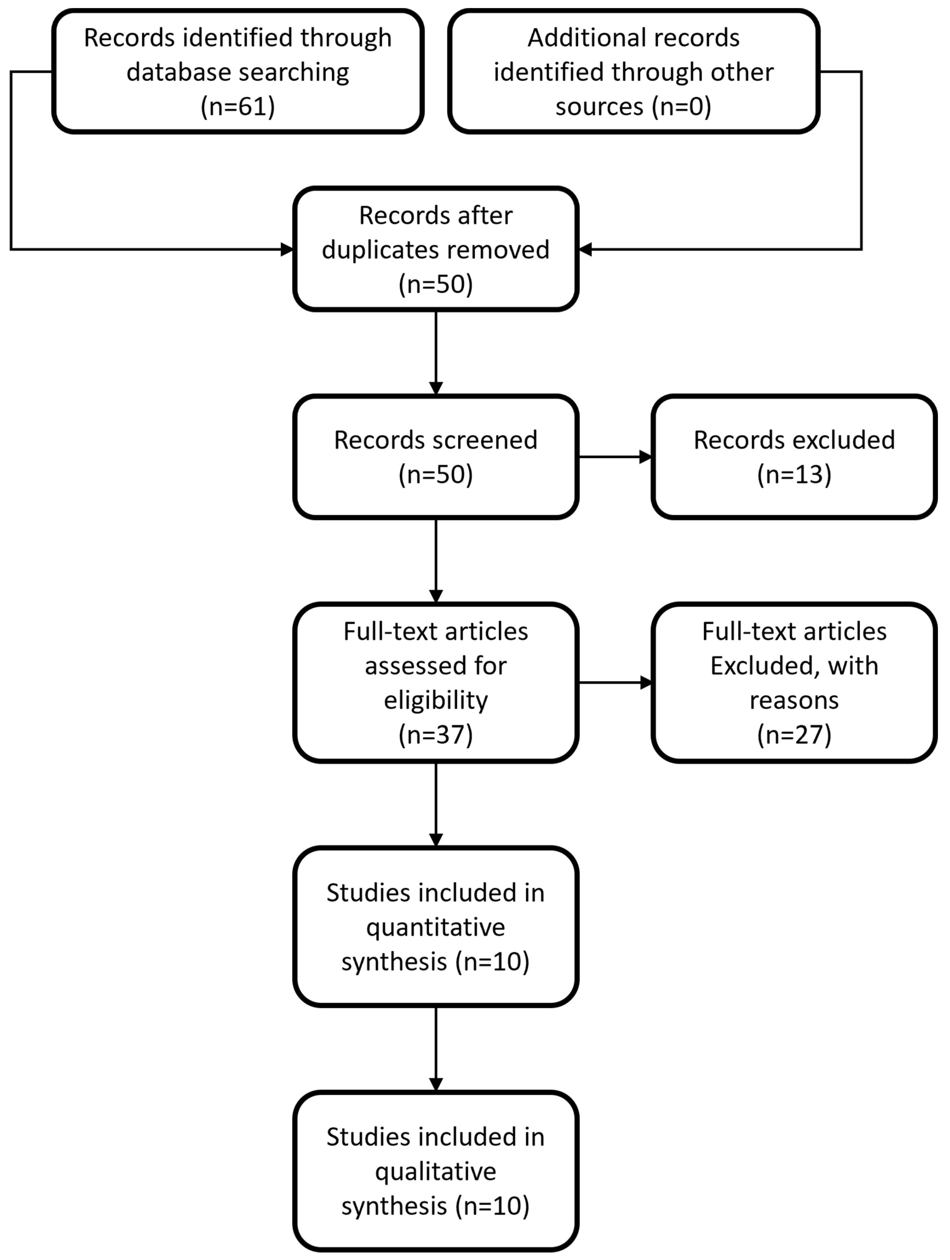
4. Materials and Methods
5. Conclusions
Author Contributions
Funding
Data Availability Statement
Conflicts of Interest
References
- Baidoun, F.; Elshiwy, K.; Elkeraie, Y.; Merjaneh, Z.; Khoudari, G.; Sarmini, M.T.; Gad, M.; Al-Husseini, M.; Saad, A. Colorectal Cancer Epidemiology: Recent Trends and Impact on Outcomes. Curr. Drug Targets 2021, 22, 998–1009. [Google Scholar] [CrossRef] [PubMed]
- Dekker, E.; Tanis, P.J.; Vleugels, J.L.A.; Kasi, P.M.; Wallace, M.B. Colorectal cancer. Lancet 2019, 394, 1467–1480. [Google Scholar] [CrossRef] [PubMed]
- Mattiuzzi, C.; Sanchis-Gomar, F.; Lippi, G. Concise update on colorectal cancer epidemiology. Ann. Transl. Med. 2019, 7, 609. [Google Scholar] [CrossRef] [PubMed]
- Liu, Z.; Xu, Y.; Xu, G.; Baklaushev, V.P.; Chekhonin, V.P.; Peltzer, K.; Ma, W.; Wang, X.; Wang, G.; Zhang, C. Nomogram for predicting overall survival in colorectal cancer with distant metastasis. BMC Gastroenterol. 2021, 21, 103. [Google Scholar] [CrossRef] [PubMed]
- De Divitiis, C.; Nasti, G.; Montano, M.; Fisichella, R.; Iaffaioli, R.V.; Berretta, M. Prognostic and predictive response factors in colorectal cancer patients: Between hope and reality. World J. Gastroenterol. WJG 2014, 20, 15049–15059. [Google Scholar] [CrossRef]
- Jin, M.; Frankel, W.L. Lymph Node Metastasis in Colorectal Cancer. Surg. Oncol. Clin. N. Am. 2018, 27, 401–412. [Google Scholar] [CrossRef]
- Resch, A.; Langner, C. Lymph node staging in colorectal cancer: Old controversies and recent advances. World J. Gastroenterol. WJG 2013, 19, 8515–8526. [Google Scholar] [CrossRef]
- Huh, J.W.; Kim, C.H.; Kim, H.R.; Kim, Y.J. Factors predicting oncologic outcomes in patients with fewer than 12 lymph nodes retrieved after curative resection for colon cancer. J. Surg. Oncol. 2012, 105, 125–129. [Google Scholar] [CrossRef]
- Märkl, B.; Schaller, T.; Krammer, I.; Cacchi, C.; Arnholdt, H.M.; Schenkirsch, G.; Kretsinger, H.; Anthuber, M.; Spatz, H. Methylene blue-assisted lymph node dissection technique is not associated with an increased detection of lymph node metastases in colorectal cancer. Mod. Pathol. Off. J. US Can. Acad. Pathol. Inc. 2013, 26, 1246–1254. [Google Scholar] [CrossRef]
- Basten, O.; Bandorski, D.; Bismarck, C.; Neumann, K.; Fisseler-Eckhoff, A. Acetone compression. A fast, standardized method to investigate gastrointestinal lymph nodes. Pathologe 2010, 31, 218–224. [Google Scholar] [CrossRef]
- Lisik, K.; Krokosz, A. Application of Carbon Nanoparticles in Oncology and Regenerative Medicine. Int. J. Mol. Sci. 2021, 22, 8341. [Google Scholar] [CrossRef] [PubMed]
- Koimtzis, G.; Stefanopoulos, L.; Alexandrou, V.; Tteralli, N.; Brooker, V.; Alawad, A.A.; Carrington-Windo, E.; Karakasis, N.; Geropoulos, G.; Papavramidis, T. The Role of Carbon Nanoparticles in Lymph Node Dissection and Parathyroid Gland Preservation during Surgery for Thyroid Cancer: A Systematic Review and Meta-Analysis. Cancers 2022, 14, 4016. [Google Scholar] [CrossRef] [PubMed]
- Tao, S.; Zhang, Z.; Li, L.; Yuan, X.; Chen, H.; Zhang, Y.; Fu, C. Characteristics of systematic lymph node dissection and influencing factors of sentinel lymph node biopsy using carbon nanoparticles in endometrial carcinoma: A single-center study. World J. Surg. Oncol. 2023, 21, 39. [Google Scholar] [CrossRef] [PubMed]
- Li, J.; Jia, S.; Wang, Y.; Zhang, Y.; Kong, L.; Cao, Y.; Liu, Y.; Chen, B. Long-term tracing and staining of carbon nanoparticles for axillary lymph nodes in patients with locally advanced breast cancer treated with neoadjuvant chemotherapy. Asian J. Surg. 2022, 45, 89–96. [Google Scholar] [CrossRef]
- Wang, H.; Chen, M.M.; Zhu, G.S.; Ma, M.G.; Du, H.S.; Long, Y.P. Lymph node mapping with carbon nanoparticles and the risk factors of lymph node metastasis in gastric cancer. J. Huazhong Univ. Sci. Technol. Med. Sci. 2016, 36, 865–870. [Google Scholar] [CrossRef]
- Liu, P.; Tan, J.; Tan, Q.; Xu, L.; He, T.; Lv, Q. Application of Carbon Nanoparticles in Tracing Lymph Nodes and Locating Tumors in Colorectal Cancer: A Concise Review. Int. J. Nanomed. 2020, 15, 9671–9681. [Google Scholar] [CrossRef]
- Li, J.; Deng, X.; Wang, L.; Liu, J.; Xu, K. Clinical application of carbon nanoparticles in lymphatic mapping during colorectal cancer surgeries: A systematic review and meta-analysis. Dig. Liver Dis. 2020, 52, 1445–1454. [Google Scholar] [CrossRef]
- Zhang, R.J.; Chen, Y.L.; Deng, X.; Yang, H. Carbon Nanoparticles for Thyroidectomy and Central Lymph Node Dissection for Thyroid Cancer. Am. Surg. 2022, 89, 2227–2236. [Google Scholar] [CrossRef]
- Cai, H.K.; He, H.F.; Tian, W.; Zhou, M.Q.; Hu, Y.; Deng, Y.C. Colorectal cancer lymph node staining by activated carbon nanoparticles suspension in vivo or methylene blue in vitro. World J. Gastroenterol. 2012, 18, 6148–6154. [Google Scholar] [CrossRef]
- Wang, Y.; Deng, H.; Chen, H.; Liu, H.; Xue, Q.; Yan, J.; Li, G. Preoperative Submucosal Injection of Carbon Nanoparticles Improves Lymph Node Staging Accuracy in Rectal Cancer after Neoadjuvant Chemoradiotherapy. J. Am. Coll. Surg. 2015, 221, 923–930. [Google Scholar] [CrossRef]
- Wang, Q.; Chen, E.; Cai, Y.; Chen, C.; Jin, W.; Zheng, Z.; Jin, Y.; Chen, Y.; Zhang, X.; Li, Q. Preoperative endoscopic localization of colorectal cancer and tracing lymph nodes by using carbon nanoparticles in laparoscopy. World J. Surg. Oncol. 2016, 14, 231. [Google Scholar] [CrossRef] [PubMed]
- Zhang, X.M.; Liang, J.W.; Wang, Z.; Kou, J.T.; Zhou, Z.X. Effect of preoperative injection of carbon nanoparticle suspension on the outcomes of selected patients with mid-low rectal cancer. Chin. J. Cancer 2016, 35, 33. [Google Scholar] [CrossRef] [PubMed]
- Wang, L.Y.; Li, J.H.; Zhou, X.; Zheng, Q.C.; Cheng, X. Clinical application of carbon nanoparticles in curative resection for colorectal carcinoma. OncoTargets Ther. 2017, 10, 5585–5589. [Google Scholar] [CrossRef] [PubMed]
- Pan, L.; Ye, F.; Liu, J.J.; Ba, X.Q.; Sheng, Q.S. A study of using carbon nanoparticles to improve lymph nodes staging for laparoscopic-assisted radical right hemicolectomy in colon cancer. Int. J. Color. Dis. 2018, 33, 1131–1134. [Google Scholar] [CrossRef] [PubMed]
- Li, K.; Chen, D.; Chen, W.; Liu, Z.; Jiang, W.; Liu, X.; Cui, Z.; Wei, Z.; Li, Z.; Yan, J. A case–control study of using carbon nanoparticles to trace decision-making lymph nodes around inferior mesenteric artery in rectal cancer. Surg. Endosc. 2019, 33, 904–910. [Google Scholar] [CrossRef] [PubMed]
- Tang, L.; Sun, L.; Zhao, P.; Kong, D. Effect of activated carbon nanoparticles on lymph node harvest in patients with colorectal cancer. Color. Dis. 2019, 21, 427–431. [Google Scholar] [CrossRef]
- Wang, R.; Mo, S.; Liu, Q.; Zhang, W.; Zhang, Z.; He, Y.; Cai, G.; Li, X. The safety and effectiveness of carbon nanoparticles suspension in tracking lymph node metastases of colorectal cancer: A prospective randomized controlled trial. Jpn. J. Clin. Oncol. 2020, 50, 535–542. [Google Scholar] [CrossRef]
- Ge, W.; Li, Q.; Liu, W.J.; Zhang, X.-Q.; Fan, X.-S.; Shao, L.-H.; Tao, L.; Guan, W.-X.; Chen, G. Carbon nanoparticle suspension could help get a more accurate nodal staging for patient with rectal cancer. Sci. Rep. 2021, 11, 9933. [Google Scholar] [CrossRef]
- Mauro, N.; Utzeri, M.A.; Varvarà, P.; Cavallaro, G. Functionalization of Metal and Carbon Nanoparticles with Potential in Cancer Theranostics. Molecules 2021, 26, 3085. [Google Scholar] [CrossRef]
- Hosseini, S.M.; Mohammadnejad, J.; Najafi-Taher, R.; Zadeh, Z.B.; Tanhaei, M.; Ramakrishna, S. Multifunctional Carbon-Based Nanoparticles: Theranostic Applications in Cancer Therapy and Diagnosis. ACS Appl. Bio Mater. 2023, 6, 1323–1338. [Google Scholar] [CrossRef]
- Fiorito, S.; Serafino, A.; Andreola, F.; Togna, A.; Togna, G. Toxicity and Biocompatibility of Carbon Nanoparticles. J. Nanosci. Nanotechnol. 2006, 6, 591–599. [Google Scholar] [CrossRef] [PubMed]
- Xu, S.; Li, Z.; Xu, M.; Peng, H. The role of carbon nanoparticle in lymph node detection and parathyroid gland protection during thyroidectomy for non-anaplastic thyroid carcinoma- a meta-analysis. PLoS ONE 2020, 15, e0223627. [Google Scholar] [CrossRef] [PubMed]
- Xiao, J.; Shen, Y.; Yang, X.; Wei, M.; Meng, W.; Wang, Z. Methylene blue can increase the number of lymph nodes harvested in colorectal cancer: A meta-analysis. Int. J. Color. Dis. 2023, 38, 50. [Google Scholar] [CrossRef] [PubMed]
- Iguchi, K.; Watanabe, J.; Suwa, Y.; Chida, K.; Atsumi, Y.; Numata, M.; Sato, T.; Takeda, K.; Kunisaki, C. The effect of preoperative endoscopic tattooing using India ink on lymph node yield in laparoscopic colectomy for stage I right-sided colon cancer. Int. J. Color. Dis. 2023, 38, 77. [Google Scholar] [CrossRef] [PubMed]
- Safiejko, K.; Tarkowski, R.; Kozlowski, T.P.; Koselak, M.; Jachimiuk, M.; Tarasik, A.; Pruc, M.; Smereka, J.; Szarpak, L. Safety and Efficacy of Indocyanine Green in Colorectal Cancer Surgery: A Systematic Review and Meta-Analysis of 11,047 Patients. Cancers 2022, 14, 1036. [Google Scholar] [CrossRef] [PubMed]
- Villegas-Tovar, E.; Jimenez-Lillo, J.; Jimenez-Valerio, V.; Diaz-Giron-Gidi, A.; Faes-Petersen, R.; Otero-Piñeiro, A.; De Lacy, F.B.; Martinez-Portilla, R.J.; Lacy, A.M. Performance of Indocyanine green for sentinel lymph node mapping and lymph node metastasis in colorectal cancer: A diagnostic test accuracy meta-analysis. Surg. Endosc. 2020, 34, 1035–1047. [Google Scholar] [CrossRef]
- Ahmad, N.Z.; Azam, M.; Fraser, C.N.; Coffey, J.C. A systematic review and meta-analysis of the use of methylene blue to improve the lymph node harvest in rectal cancer surgery. Technol. Coloproctol. 2023, 27, 361–371. [Google Scholar] [CrossRef]
- Zhu, J.; Tian, W.; Xu, Z.; Jiang, K.; Sun, H.; Wang, P.; Huang, T.; Guo, Z.; Zhang, H.; Liu, S.; et al. Expert consensus statement on parathyroid protection in thyroidectomy. Ann. Transl. Med. 2015, 3, 230. [Google Scholar] [CrossRef]
- Patel, H.P.; Chadwick, D.R.; Harrison, B.J.; Balasubramanian, S.P. Systematic review of intravenous methylene blue in parathyroid surgery. Br. J. Surg. 2012, 99, 1345–1351. [Google Scholar] [CrossRef]
- Kang, J.; Park, H.S.; Kim, I.K.; Song, Y.; Baik, S.H.; Sohn, S.-K.; Lee, K.Y. Effect of preoperative colonoscopic tattooing on lymph node harvest in T1 colorectal cancer. Int. J. Color. Dis. 2015, 30, 1349–1355. [Google Scholar] [CrossRef]
- Emile, S.H.; Elfeki, H.; Shalaby, M.; Sakr, A.; Sileri, P.; Laurberg, S.; Wexner, S.D. Sensitivity and specificity of indocyanine green near-infrared fluorescence imaging in detection of metastatic lymph nodes in colorectal cancer: Systematic review and meta-analysis. J. Surg. Oncol. 2017, 116, 730–740. [Google Scholar] [CrossRef]
- Liu, D.; Liang, L.; Liu, L.; Zhu, Z. Does intraoperative indocyanine green fluorescence angiography decrease the incidence of anastomotic leakage in colorectal surgery? A systematic review and meta-analysis. Int. J. Color. Dis. 2021, 36, 57–66. [Google Scholar] [CrossRef] [PubMed]
- Zhang, W.; Che, X. Effect of indocyanine green fluorescence angiography on preventing anastomotic leakage after colorectal surgery: A meta-analysis. Surg. Today 2021, 51, 1415–1428. [Google Scholar] [CrossRef] [PubMed]
- Chan, D.K.H.; Lee, S.K.F.; Ang, J.J. Indocyanine green fluorescence angiography decreases the risk of colorectal anastomotic leakage: Systematic review and meta-analysis. Surgery 2020, 168, 1128–1137. [Google Scholar] [CrossRef] [PubMed]
- Blanco-Colino, R.; Espin-Basany, E. Intraoperative use of ICG fluorescence imaging to reduce the risk of anastomotic leakage in colorectal surgery: A systematic review and meta-analysis. Technol. Coloproctol. 2018, 22, 15–23. [Google Scholar] [CrossRef]
- Pang, H.Y.; Chen, X.L.; Song, X.H.; Galiullin, D.; Zhao, L.-Y.; Liu, K.; Zhang, W.-H.; Yang, K.; Chen, X.-Z.; Hu, J.-K. Indocyanine green fluorescence angiography prevents anastomotic leakage in rectal cancer surgery: A systematic review and meta-analysis. Langenbecks Arch. Surg. 2021, 406, 261–271. [Google Scholar] [CrossRef]
- Zhang, X.; Li, J.G.; Zhang, S.Z.; Chen, G. Comparison of indocyanine green and carbon nanoparticles in endoscopic techniques for central lymph nodes dissection in patients with papillary thyroid cancer. Surg. Endosc. 2020, 34, 5354–5359. [Google Scholar] [CrossRef]
- He, M.; Liang, S.; Deng, H.; Zhang, G.; Wang, Z.; Yang, X.; Wang, Y.; Li, Y.; Sun, X.; Wang, J. Comparing carbon nanoparticles and indocyanine green for sentinel lymph node mapping in endometrial cancer: A randomized-controlled single-center trial. J. Surg. Oncol. 2023, 128, 332–343. [Google Scholar] [CrossRef]
- Qin, X.; Yang, M.; Zheng, X. Comparative study of indocyanine green combined with blue dye with methylene blue only and carbon nanoparticles only for sentinel lymph node biopsy in breast cancer. Ann. Surg. Treat. Res. 2019, 97, 1–6. [Google Scholar] [CrossRef]
- Tian, Y.; Pang, Y.; Yang, P.; Guo, H.; Liu, Y.; Zhang, Z.; Ding, P.; Zheng, T.; Li, Y.; Fan, L.; et al. The safety and efficacy of carbon nanoparticle suspension injection versus indocyanine green tracer-guided lymph node dissection during radical gastrectomy (FUTURE-01): A single-center randomized controlled trial protocol. Front. Oncol. 2023, 12, 1044854. [Google Scholar] [CrossRef]

| Study | Experimental Group (n, Age) | Control Group (n, Age) | Sex (Male/Female) | Cancer Type |
|---|---|---|---|---|
| [19] | 20, 57.5 ± 11.5 | 20, 64.9 ± 7.4 | 20/20 | Colorectal |
| [20] | 45, 53.1 ± 12.0 | 107, 54.0 ± 11.6 | 113/39 | Rectal |
| [21] | 27, 62.81 ± 11.29 | 27, 64.63 ± 10.05 | 33/21 | Colorectal |
| [22] | 35, 60.0 ± 10.7 | 52, 58.9 ± 9.4 | 40/47 | Rectal |
| [23] | 344, 58.6 ± 12.4 | 126, 59.1 ± 12.2 | 261/209 | Colorectal |
| [24] | 52, not mentioned | 47, not mentioned | Not mentioned | Colon |
| [25] | 33, 57.2 ± 9.4 | 33, 57.8 ± 9.7 | 52/14 | Rectal |
| [26] | 40, 57.9 ± 11.8 | 39, 59.3 ± 10.7 | 31/48 | Colon |
| [27] | 123, 58.98 ± 9.836 | 116, 56.78 ± 10.824 | 146/93 | Colorectal |
| [28] | 60, 61.0 ± 11.7 | 72, 64.2 ± 10.4 | 81/51 | Colorectal |
| Study | Experimental | Control | Mean Diff. | Weight | Forest Plot | ||||
|---|---|---|---|---|---|---|---|---|---|
| n | Mean | sd | n | Mean | sd | IV, Random, 95% CI | (%) | ||
| [19] | 20 | 26.80 | 8.40 | 20 | 12.20 | 3.20 | 14.60 [10.66, 18.54] | 8.34 |  |
| [20] | 45 | 21.10 | 9.60 | 107 | 8.00 | 4.60 | 13.10 [10.16, 16.04] | 9.72 | |
| [21] | 27 | 14.41 | 3.32 | 27 | 8.96 | 2.90 | 5.45 [3.79, 7.11] | 11.31 | |
| [22] | 35 | 28.20 | 9.40 | 52 | 22.70 | 7.30 | 5.50 [1.81, 9.19] | 8.67 | |
| [23] | 344 | 9.13 | 6.30 | 126 | 8.89 | 6.11 | 0.24 [−1.02, 1.50] | 11.7 | |
| [24] | 52 | 20.90 | 0.70 | 47 | 17.60 | 0.90 | 3.30 [2.98, 3.62] | 12.23 | |
| [25] | 33 | 24.06 | 13.20 | 33 | 16.21 | 9.09 | 7.85 [2.38, 13.32] | 6.43 | |
| [26] | 40 | 31.30 | 8.10 | 39 | 21.90 | 5.30 | 9.40 [6.39, 12.41] | 9.62 | |
| [27] | 123 | 19.84 | 6.43 | 116 | 17.41 | 7.23 | 2.43 [0.69, 4.17] | 11.23 | |
| [28] | 60 | 19.30 | 6.70 | 72 | 15.10 | 5.70 | 4.20 [2.05, 6.35] | 10.76 | |
| Total 95% CI | 779 | 639 | 6.15 [4.14, 8.16] | ||||||
| Study | Experimental | Control | Log Odds-Ratio | Weight | Forest Plot | ||
|---|---|---|---|---|---|---|---|
| Events | Total | Events | Total | IV, Random, 95% CI | (%) | ||
| [19] | 20 | 20 | 10 | 20 | 3.71 [0.78,6.65] | 11.68 | 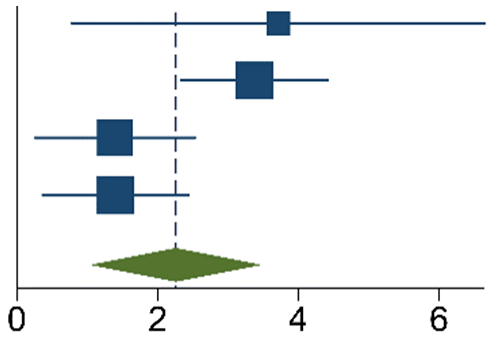 |
| [20] | 40 | 45 | 23 | 107 | 3.37 [2.34,4.41] | 29.84 | |
| [21] | 19 | 27 | 10 | 27 | 1.40 [0.26,2.53] | 28.57 | |
| [27] | 118 | 123 | 99 | 116 | 1.40 [0.37,2.43] | 29.91 | |
| Total | 2.26 [1.06,3.46] | ||||||
| Study | Experimental | Control | Log Odds-Ratio | Weight | Forest Plot | ||
|---|---|---|---|---|---|---|---|
| Events | Total | Events | Total | IV, Random, 95% CI | (%) | ||
| [19] | 50 | 535 | 32 | 223 | −0.49 [−0.96, −0.01] | 17.66 | 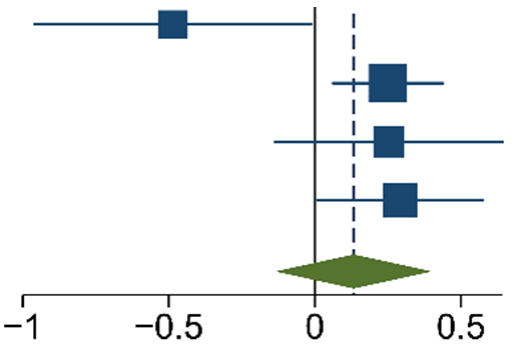 |
| [23] | 574 | 3143 | 166 | 1120 | 0.25 [0.06, 0.44] | 33.23 | |
| [24] | 72 | 1088 | 43 | 825 | 0.25 [−0.14, 0.64] | 21.59 | |
| [26] | 156 | 1252 | 82 | 854 | 0.29 [0.01, 0.58] | 27.52 | |
| Total | 0.13 [−0.13,0.40] | ||||||
Disclaimer/Publisher’s Note: The statements, opinions and data contained in all publications are solely those of the individual author(s) and contributor(s) and not of MDPI and/or the editor(s). MDPI and/or the editor(s) disclaim responsibility for any injury to people or property resulting from any ideas, methods, instructions or products referred to in the content. |
© 2023 by the authors. Licensee MDPI, Basel, Switzerland. This article is an open access article distributed under the terms and conditions of the Creative Commons Attribution (CC BY) license (https://creativecommons.org/licenses/by/4.0/).
Share and Cite
Koimtzis, G.; Geropoulos, G.; Stefanopoulos, L.; Chalklin, C.G.; Karniadakis, I.; Alexandrou, V.; Tteralli, N.; Carrington-Windo, E.; Papacharalampous, A.; Psarras, K. The Role of Carbon Nanoparticles as Lymph Node Tracers in Colorectal Cancer: A Systematic Review and Meta-Analysis. Int. J. Mol. Sci. 2023, 24, 15293. https://doi.org/10.3390/ijms242015293
Koimtzis G, Geropoulos G, Stefanopoulos L, Chalklin CG, Karniadakis I, Alexandrou V, Tteralli N, Carrington-Windo E, Papacharalampous A, Psarras K. The Role of Carbon Nanoparticles as Lymph Node Tracers in Colorectal Cancer: A Systematic Review and Meta-Analysis. International Journal of Molecular Sciences. 2023; 24(20):15293. https://doi.org/10.3390/ijms242015293
Chicago/Turabian StyleKoimtzis, Georgios, Georgios Geropoulos, Leandros Stefanopoulos, Christopher Gwydion Chalklin, Ioannis Karniadakis, Vyron Alexandrou, Nikos Tteralli, Eliot Carrington-Windo, Andreas Papacharalampous, and Kyriakos Psarras. 2023. "The Role of Carbon Nanoparticles as Lymph Node Tracers in Colorectal Cancer: A Systematic Review and Meta-Analysis" International Journal of Molecular Sciences 24, no. 20: 15293. https://doi.org/10.3390/ijms242015293
APA StyleKoimtzis, G., Geropoulos, G., Stefanopoulos, L., Chalklin, C. G., Karniadakis, I., Alexandrou, V., Tteralli, N., Carrington-Windo, E., Papacharalampous, A., & Psarras, K. (2023). The Role of Carbon Nanoparticles as Lymph Node Tracers in Colorectal Cancer: A Systematic Review and Meta-Analysis. International Journal of Molecular Sciences, 24(20), 15293. https://doi.org/10.3390/ijms242015293