
Determination of Thrombogenicity Levels of Various Antiphospholipid Antibodies by a Modified Thrombin Generation Assay in Patients with Suspected Antiphospholipid Syndrome

 , ,
, ,
Abstract
:1. Introduction
1.1. Antiphospholipid Antibodies
1.1.1. Seronegative APS
1.1.2. APS Criteria Antibodies
1.1.3. APS Non-Criteria Antibodies
2. Results
3. Discussion
4. Methods and Materials
4.1. Study Design and Population
4.2. Sample Collection
4.3. Coagulation Tests for Lupus Anticoagulans
4.4. Chemiluminiscence Immunoassay for aCL, Anti-β2GPI including Structural Variation Anti-DI
4.5. Enzyme Linked Imunosorbent Assay for Anti-PS/PT and Anti-Annexin
4.6. Thrombin Generation Assay Modified by APC
4.7. Statistics
5. Conclusions
Author Contributions
Funding
Institutional Review Board Statement
Informed Consent Statement
Data Availability Statement
Conflicts of Interest
References
- Hughes, G.R. Thrombosis, abortion, cerebral disease, and the lupus anticoagulant. Br. Med. J. (Clin. Res. Ed.) 1983, 287, 1088–1089. [Google Scholar] [CrossRef] [PubMed]
- Lopez-Pedrera, C.; Barbarroja, N.; Patiño-Trives, A.M.; Collantes, E.; Aguirre, M.A.; Perez-Sanchez, C. New Biomarkers for Atherothrombosis in Antiphospholipid Syndrome: Genomics and Epigenetics Approaches. Front. Immunol. 2019, 10, 764. [Google Scholar] [CrossRef] [PubMed]
- Radic, M.; Pattanaik, D. Cellular and Molecular Mechanisms of Anti-Phospholipid Syndrome. Front. Immunol. 2018, 9, 969. [Google Scholar] [CrossRef]
- Schreiber, K.; Sciascia, S.; de Groot, P.G.; Devreese, K.; Jacobsen, S.; Ruiz-Irastorza, G.; Salmon, J.E.; Shoenfeld, Y.; Shovman, O.; Hunt, B.J. Antiphospholipid syndrome. Nat. Rev. Dis. Primers 2018, 4, 17103. [Google Scholar] [CrossRef] [PubMed]
- Cervera, R. Antiphospholipid syndrome. Thromb. Res. 2017, 151 (Suppl. S1), S43–S47. [Google Scholar] [CrossRef]
- Liu, L.; Sun, D. Pregnancy outcomes in patients with primary antiphospholipid syndrome: A systematic review and meta-analysis. Medicine 2019, 98, e15733. [Google Scholar] [CrossRef] [PubMed]
- Whitaker, K.L. Antiphospholipid antibody syndrome: The difficulties of diagnosis. JAAPA Off. J. Am. Acad. Physician Assist. 2017, 30, 10–14. [Google Scholar] [CrossRef] [PubMed]
- Miyakis, S.; Lockshin, M.D.; Atsumi, T.; Branch, D.W.; Brey, R.L.; Cervera, R.; Derksen, R.H.; DE Grrot, P.G.; Koike, T.; Meroni, P.L.; et al. International consensus statement on an update of the classification criteria for definite antiphospholipid syndrome (APS). J. Thromb. Haemost. JTH 2006, 4, 295–306. [Google Scholar] [CrossRef]
- Devreese, K.M.J.; Ortel, T.L.; Pengo, V.; de Laat, B. Laboratory criteria for antiphospholipid syndrome: Communication from the SSC of the ISTH. J. Thromb. Haemost. JTH 2018, 16, 809–813. [Google Scholar] [CrossRef]
- Pengo, V.; Banzato, A.; Bison, E.; Bracco, A.; Denas, G.; Ruffatti, A. What have we learned about antiphospholipid syndrome from patients and antiphospholipid carrier cohorts? Semin. Thromb. Hemost. 2012, 38, 322–327. [Google Scholar] [CrossRef]
- Pengo, V.; Ruffatti, A.; Del Ross, T.; Tonello, M.; Cuffaro, S.; Hoxha, A.; Banzato, A.; Bison, E.; Denas, G.; Bracco, A.; et al. Confirmation of initial antiphospholipid antibody positivity depends on the antiphospholipid antibody profile. J. Thromb. Haemost. JTH 2013, 11, 1527–1531. [Google Scholar] [CrossRef]
- Pengo, V.; Ruffatti, A.; Legnani, C.; Gresele, P.; Barcellona, D.; Erba, N.; Testa, S.; Marongiu, F.; Bison, E.; Denas, G.; et al. Clinical course of high-risk patients diagnosed with antiphospholipid syndrome. J. Thromb. Haemost. JTH 2010, 8, 237–242. [Google Scholar] [CrossRef]
- Sciascia, S.; Murru, V.; Sanna, G.; Roccatello, D.; Khamashta, M.A.; Bertolaccini, M.L. Clinical accuracy for diagnosis of antiphospholipid syndrome in systemic lupus erythematosus: Evaluation of 23 possible combinations of antiphospholipid antibody specificities. J. Thromb. Haemost. JTH 2012, 10, 2512–2518. [Google Scholar] [CrossRef]
- De Groot, P.G.; Derksen, R.H. Pathophysiology of the antiphospholipid syndrome. J. Thromb. Haemost. JTH 2005, 3, 1854–1860. [Google Scholar] [CrossRef]
- Douxfils, J.; Morimont, L.; Delvigne, A.S.; Devel, P.; Masereel, B.; Haguet, H.; Bouvy, C.; Dogné, J.M. Validation and standardization of the ETP-based activated protein C resistance test for the clinical investigation of steroid contraceptives in women: An unmet clinical and regulatory need. Clin. Chem. Lab. Med. 2020, 58, 294–305. [Google Scholar] [CrossRef]
- Albay, A.; Artim-Esen, B.; Pericleous, C.; Wincup, C.; Giles, I.; Rahman, A.; McDonnell, T. Domain I of β2GPI is capable of blocking serum IgA antiphospholipid antibodies binding in vitro: An effect enhanced by PEGylation. Lupus 2019, 28, 893–897. [Google Scholar] [CrossRef]
- Cervera, R.; Conti, F.; Doria, A.; Iaccarino, L.; Valesini, G. Does seronegative antiphospholipid syndrome really exist? Autoimmun. Rev. 2012, 11, 581–584. [Google Scholar] [CrossRef]
- Conti, F.; Capozzi, A.; Truglia, S.; Lococo, E.; Longo, A.; Misasi, R.; Alessandri, C.; Valesini, G.; Sorice, M. The mosaic of “seronegative” antiphospholipid syndrome. J. Immunol. Res. 2014, 2014, 389601. [Google Scholar] [CrossRef]
- Salle, V. Seronegative antiphospholipid syndrome: Myth or reality? Rev. Med. Interne 2020, 41, 265–274. [Google Scholar] [CrossRef]
- Conti, F.; Andreoli, L.; Crisafulli, F.; Mancuso, S.; Truglia, S.; Tektonidou, M.G. Does seronegative obstetric APS exist? “pro” and “cons”. Autoimmun. Rev. 2019, 18, 102407. [Google Scholar] [CrossRef]
- Ortona, E.; Capozzi, A.; Colasanti, T.; Conti, F.; Alessandri, C.; Longo, A.; Garofalo, T.; Margutti, P.; Misasi, R.; Khamashta, M.A.; et al. Vimentin/cardiolipin complex as a new antigenic target of the antiphospholipid syndrome. Blood 2010, 116, 2960–2967. [Google Scholar] [CrossRef] [PubMed]
- Misasi, R.; Capozzi, A.; Longo, A.; Recalchi, S.; Lococo, E.; Alessandri, C.; Conti, F.; Valesini, G.; Sorice, M. “New” antigenic targets and methodological approaches for refining laboratory diagnosis of antiphospholipid syndrome. J. Immunol. Res. 2015, 2015, 858542. [Google Scholar] [CrossRef] [PubMed]
- Tripodi, A. Laboratory testing for lupus anticoagulants: A review of issues affecting results. Clin. Chem. 2007, 53, 1629–1635. [Google Scholar] [CrossRef] [PubMed]
- Chaturvedi, S.; McCrae, K.R. Diagnosis and management of the antiphospholipid syndrome. Blood Rev. 2017, 31, 406–417. [Google Scholar] [CrossRef]
- Meroni, P.L.; Tincani, A.; Harris, E.N.; Valesini, G.; Hughes, G.R.; Balestrieri, G. The pathophysiology of anti-phospholipid antibodies. Clin. Exp. Rheumatol. 1989, 7 (Suppl. S3), S81–S84. [Google Scholar]
- Liu, T.; Gu, J.; Wan, L.; Hu, Q.; Teng, J.; Liu, H.; Cheng, X.; Ye, J.; Su, Y.; Sun, Y.; et al. “Non-criteria” antiphospholipid antibodies add value to antiphospholipid syndrome diagnoses in a large Chinese cohort. Arthritis Res. Ther. 2020, 22, 33. [Google Scholar] [CrossRef]
- De Groot, P.G.; Meijers, J.C. β(2)-Glycoprotein I: Evolution, structure and function. J. Thromb. Haemost. JTH 2011, 9, 1275–1284. [Google Scholar] [CrossRef]
- Perdan-Pirkmajer, K.; Žigon, P.; Boc, A.; Podovsovnik, E.; Cucnik, S.; Mavri, A.; Rotar, Ž.; Ambrozic, A. The Predictive Value of the aCL and Anti-β2GPI at the Time of Acute Deep Vein Thrombosis—A Two-Year Prospective Study. Biomedicines 2021, 9, 901. [Google Scholar] [CrossRef]
- Chayoua, W.; Kelchtermans, H.; Moore, G.W.; Musiał, J.; Wahl, D.; de Laat, B.; Devreese, K.M.J. Identification of high thrombotic risk triple-positive antiphospholipid syndrome patients is dependent on anti-cardiolipin and anti-β2glycoprotein I antibody detection assays. J. Thromb. Haemost. JTH 2018, 16, 2016–2023. [Google Scholar] [CrossRef]
- Decavele, A.S.; Schouwers, S.; Devreese, K.M. Evaluation of three commercial ELISA kits for anticardiolipin and anti-beta2-glycoprotein I antibodies in the laboratory diagnosis of the antiphospholipid syndrome. Int. J. Lab. Hematol. 2011, 33, 97–108. [Google Scholar] [CrossRef]
- Gebhart, J.; Posch, F.; Koder, S.; Quehenberger, P.; Perkmann, T.; Kuessel, L.; Ay, C.; Pabinger, I. High risk of adverse pregnancy outcomes in women with a persistent lupus anticoagulant. Blood Adv. 2019, 3, 769–776. [Google Scholar] [CrossRef]
- Pierangeli, S.S.; Favaloro, E.J.; Lakos, G.; Meroni, P.L.; Tincani, A.; Wong, R.C.; Harris, E.N. Standards and reference materials for the anticardiolipin and anti-β2glycoprotein I assays: A report of recommendations from the APL Task Force at the 13th International Congress on Antiphospholipid Antibodies. Clin. Chim. Acta Int. J. Clin. Chem. 2012, 413, 358–360. [Google Scholar] [CrossRef]
- Willis, R.; Lakos, G.; Harris, E.N. Standardization of antiphospholipid antibody testing--historical perspectives and ongoing initiatives. Semin. Thromb. Hemost. 2014, 40, 172–177. [Google Scholar] [CrossRef]
- Ahluwalia, J.; Sreedharanunni, S. The Laboratory Diagnosis of the Antiphospholipid Syndrome. Indian J. Hematol. Blood Transfus. Off. J. Indian Soc. Hematol. Blood Transfus. 2017, 33, 8–14. [Google Scholar] [CrossRef]
- Janek, D.; Slavik, L.; Ulehlova, J.; Krcova, V.; Hlusi, A.; Prochazkova, J. Validation of a New Panel of Automated Chemiluminescence Assays for Anticardiolipin Antibodies in the Screening for Antiphospholipid Syndrome. Clin. Lab. 2016, 62, 1309–1315. [Google Scholar] [CrossRef]
- Bizzaro, N.; Ghirardello, A.; Zampieri, S.; Iaccarino, L.; Tozzoli, R.; Ruffatti, A.; Villalta, D.; Tonutti, E.; Doria, A. Anti-prothrombin antibodies predict thrombosis in patients with systemic lupus erythematosus: A 15-year longitudinal study. J. Thromb. Haemost. JTH 2007, 5, 1158–1164. [Google Scholar] [CrossRef]
- Cifù, A.; Domenis, R.; Pistis, C.; Curcio, F.; Fabris, M. Anti-β2-glycoprotein I and anti-phosphatidylserine/prothrombin antibodies exert similar pro-thrombotic effects in peripheral blood monocytes and endothelial cells. Auto-Immun. Highlights 2019, 10, 3. [Google Scholar] [CrossRef]
- Sciascia, S.; Khamashta, M.A.; Bertolaccini, M.L. New tests to detect antiphospholipid antibodies: Antiprothrombin (aPT) and anti-phosphatidylserine/prothrombin (aPS/PT) antibodies. Curr. Rheumatol. Rep. 2014, 16, 415. [Google Scholar] [CrossRef]
- Sciascia, S.; Radin, M.; Sanna, G.; Cecchi, I.; Roccatello, D.; Bertolaccini, M.L. Clinical utility of the global anti-phospholipid syndrome score for risk stratification: A pooled analysis. Rheumatology 2018, 57, 661–665. [Google Scholar] [CrossRef]
- Conti, F.; Alessandri, C.; Spinelli, F.R.; Capozzi, A.; Martinelli, F.; Recalchi, S.; Misasi, R.; Valesini, G.; Sorice, M. TLC immunostaining for detection of “antiphospholipid” antibodies. Methods Mol. Biol. 2014, 1134, 95–101. [Google Scholar] [CrossRef]
- Yin, D.; de Laat, B.; Devreese, K.M.J.; Kelchtermans, H. The clinical value of assays detecting antibodies against domain I of β2-glycoprotein I in the antiphospholipid syndrome. Autoimmun. Rev. 2018, 17, 1210–1218. [Google Scholar] [CrossRef]
- Rand, J.H. Antiphospholipid antibody-mediated disruption of the annexin-V antithrombotic shield: A thrombogenic mechanism for the antiphospholipid syndrome. J. Autoimmun. 2000, 15, 107–111. [Google Scholar] [CrossRef]
- Bertolaccini, M.L.; Amengual, O.; Atsumi, T.; Binder, W.L.; de Laat, B.; Forastiero, R.; Kutteh, W.H.; Lambert, M.; Matsubayashi, H.; Murthy, V.; et al. ‘Non-criteria’ aPL tests: Report of a task force and preconference workshop at the 13th International Congress on Antiphospholipid Antibodies, Galveston, TX, USA, April 2010. Lupus 2011, 20, 191–205. [Google Scholar] [CrossRef]
- Chandler, W.L.; Roshal, M. Optimization of plasma fluorogenic thrombin-generation assays. Am. J. Clin. Pathol. 2009, 132, 169–179. [Google Scholar] [CrossRef]
- Duarte, R.C.F.; Ferreira, C.N.; Rios, D.R.A.; Reis, H.J.D.; Carvalho, M.D.G. Thrombin generation assays for global evaluation of the hemostatic system: Perspectives and limitations. Rev. Bras. Hematol. Hemoter. 2017, 39, 259–265. [Google Scholar] [CrossRef]
- Hron, G.; Kollars, M.; Binder, B.R.; Eichinger, S.; Kyrle, P.A. Identification of patients at low risk for recurrent venous thromboembolism by measuring thrombin generation. JAMA 2006, 296, 397–402. [Google Scholar] [CrossRef]
- Billoir, P.; Miranda, S.; Levesque, H.; Benhamou, Y.; Le Cam Duchez, V. Hypercoagulability Evaluation in Antiphospholipid Syndrome without Anticoagulation Treatment with Thrombin Generation Assay: A Preliminary Study. J. Clin. Med. 2021, 10, 2728. [Google Scholar] [CrossRef]
- Heit, J.A.; Sobell, J.L.; Li, H.; Sommer, S.S. The incidence of venous thromboembolism among Factor V Leiden carriers: A community-based cohort study. J. Thromb. Haemost. JTH 2005, 3, 305–311. [Google Scholar] [CrossRef]
- Liestøl, S.; Sandset, P.M.; Mowinckel, M.C.; Wisløff, F. Activated protein C resistance determined with a thrombin generation-based test is associated with thrombotic events in patients with lupus anticoagulants. J. Thromb. Haemost. JTH 2007, 5, 2204–2210. [Google Scholar] [CrossRef]
- Jiang, D.; Lim, W.; Crowther, M.; Garcia, D. A systematic review of the association between anti-β-2 glycoprotein I antibodies and APS manifestations. Blood Adv. 2021, 5, 3931–3936. [Google Scholar] [CrossRef] [PubMed]
- Pastori, D.; Bucci, T.; Triggiani, M.; Ames, P.R.J.; Parrotto, S.; Violi, F.; Pignatelli, P.; Farcomeni, A. Immunoglobulin G (IgG) anticardiolipin antibodies and recurrent cardiovascular events. A systematic review and Bayesian meta-regression analysis. Autoimmun. Rev. 2019, 18, 519–525. [Google Scholar] [CrossRef] [PubMed]
- Pires Da Rosa, G.; Rodríguez-Pintó, I.; Cervera, R.; Espinosa, G. Management of patients with antiphospholipid antibodies: What to do in laboratory scenarios that do not fit the guidelines. Expert Rev. Hematol. 2021, 14, 457–466. [Google Scholar] [CrossRef] [PubMed]
- Truglia, S.; Mancuso, S.; Capozzi, A.; Recalchi, S.; Riitano, G.; Longo, A.; De Carolis, S.; Spinelli, F.R.; Alessandri, C.; Ceccarelli, F.; et al. ‘Non-criteria antiphospholipid antibodies’: Bridging the gap between seropositive and seronegative antiphospholipid syndrome. Rheumatology 2022, 61, 826–833. [Google Scholar] [CrossRef] [PubMed]
- Zhu, R.; Cheng, C.Y.; Yang, Y.; Denas, G.; Pengo, V. Prevalence of aPhosphatidylserine/prothrombin antibodies and association with antiphospholipid antibody profiles in patients with antiphospholipid syndrome: A systematic review and meta-analysis. Thromb. Res. 2022, 214, 106–114. [Google Scholar] [CrossRef]
- Egri, N.; Bentow, C.; Rubio, L.; Norman, G.L.; López-Sañudo, S.; Mahler, M.; Pérez-Isidro, A.; Cervera, R.; Viñas, O.; Espinosa, G.; et al. Anti-Phosphatidylserine/Prothrombin Antibodies at Two Points: Correlation With Lupus Anticoagulant and Thrombotic Risk. Front. Immunol. 2021, 12, 754469. [Google Scholar] [CrossRef]
- Slavik, L.J.D.; Ulehlova, J.; Krcova, V.; Hlusi, A. Detection of Anti-Domain I β-2 Glycoprotein I Antibodies as New Potential Target in Antiphospholipid Syndrome Diagnosis. J. Hematol. Thromboembolic Dis. 2017, 5, 276. [Google Scholar] [CrossRef]
- Guo, H.; Zhang, Y.; Li, A.; Wang, C.; Yang, S.; Zhang, Y.; Zhang, J.; Qiao, R. Anti-domain 1 of beta2-glycoprotein I aids risk stratification in lupus anticoagulant-positive patients. Clin. Exp. Med. 2019, 19, 339–345. [Google Scholar] [CrossRef]
- Slavik, L.; Jacova, J.; Friedecky, D.; Ulehlova, J.; Tauber, Z.; Prochazkova, J.; Hlusi, A.; Palova, M. Evaluation of the DOAC-Stop Procedure by LC-MS/MS Assays for Determining the Residual Activity of Dabigatran, Rivaroxaban, and Apixaban. Off. J. Int. Acad. Clin. Appl. Thromb. Hemost. 2019, 25. [Google Scholar] [CrossRef]
- Pengo, V.; Tripodi, A.; Reber, G.; Rand, J.H.; Ortel, T.L.; Galli, M.; De Groot, P.G. Update of the guidelines for lupus anticoagulant detection. Subcommittee on Lupus Anticoagulant/Antiphospholipid Antibody of the Scientific and Standardisation Committee of the International Society on Thrombosis and Haemostasis. J. Thromb. Haemost. JTH 2009, 7, 1737–1740. [Google Scholar] [CrossRef]
- McDonnell, T.C.R.; Willis, R.; Pericleous, C.; Ripoll, V.M.; Giles, I.P.; Isenberg, D.A.; Brasier, A.R.; Gonzalez, E.B.; Papalardo, E.; Romay-Penabad, Z.; et al. PEGylated Domain I of Beta-2-Glycoprotein I Inhibits the Binding, Coagulopathic, and Thrombogenic Properties of IgG From Patients With the Antiphospholipid Syndrome. Front. Immunol. 2018, 9, 2413. [Google Scholar] [CrossRef]
- Linnemann, B. Antiphospholipid syndrome-an update. VASA Z. Fur Gefasskrankh. 2018, 47, 451–464. [Google Scholar] [CrossRef]
- Devreese, K.M.J. How to Interpret Antiphospholipid Laboratory Tests. Curr. Rheumatol. Rep. 2020, 22, 38. [Google Scholar] [CrossRef]
- Nakamura, H.; Oku, K.; Amengual, O.; Ohmura, K.; Fujieda, Y.; Kato, M.; Bohgaki, T.; Yasuda, S.; Atsumi, T. First-Line, Non-Criterial Antiphospholipid Antibody Testing for the Diagnosis of Antiphospholipid Syndrome in Clinical Practice: A Combination of Anti-β(2)-Glycoprotein I Domain I and Anti-Phosphatidylserine/Prothrombin Complex Antibodies Tests. Arthritis Care Res. 2018, 70, 627–634. [Google Scholar] [CrossRef]
- Zhou, J.; Hou, X.; Zhang, H.; Wang, T.; Cui, L. The Clinical Performance of a New Chemiluminescent Immunoassay in Measuring Anti-β2 Glycoprotein 1 and Anti-Cardiolipin Antibodies. Med. Sci. Monit. Int. Med. J. Exp. Clin. Res. 2018, 24, 6816–6822. [Google Scholar] [CrossRef]
- Aydin, S. A short history, principles, and types of ELISA, and our laboratory experience with peptide/protein analyses using ELISA. Peptides 2015, 72, 4–15. [Google Scholar] [CrossRef]
- Kohl, T.O.; Ascoli, C.A. Immunometric Double-Antibody Sandwich Enzyme-Linked Immunosorbent Assay. Cold Spring Harb. Protoc. 2017, 2017, pdb.prot093724. [Google Scholar] [CrossRef]
- Kohl, T.O.; Ascoli, C.A. Indirect Immunometric ELISA. Cold Spring Harb. Protoc. 2017, 2017, 396–401. [Google Scholar] [CrossRef]
- Tebo, A.E.; Jaskowski, T.D.; Phansalkar, A.R.; Litwin, C.M.; Branch, D.W.; Hill, H.R. Diagnostic performance of phospholipid-specific assays for the evaluation of antiphospholipid syndrome. Am. J. Clin. Pathol. 2008, 129, 870–875. [Google Scholar] [CrossRef]
- Tripodi, A. Thrombin Generation Assay and Its Application in the Clinical Laboratory. Clin. Chem. 2016, 62, 699–707. [Google Scholar] [CrossRef]
- Depasse, F.; Binder, N.B.; Mueller, J.; Wissel, T.; Schwers, S.; Germer, M.; Hermes, B.; Turecek, P.L. Thrombin generation assays are versatile tools in blood coagulation analysis: A review of technical features, and applications from research to laboratory routine. J. Thromb. Haemost. JTH 2021, 19, 2907–2917. [Google Scholar] [CrossRef]
- Binder, N.B.; Depasse, F.; Mueller, J.; Wissel, T.; Schwers, S.; Germer, M.; Hermes, B.; Turecek, P.L. Clinical use of thrombin generation assays. J. Thromb. Haemost. JTH 2021, 19, 2918–2929. [Google Scholar] [CrossRef]
- Devreese, K.M.J.; Zuily, S.; Meroni, P.L. Role of antiphospholipid antibodies in the diagnosis of antiphospholipid syndrome. J. Transl. Autoimmun. 2021, 4, 100134. [Google Scholar] [CrossRef]
- Fabris, M.; Giacomello, R.; Poz, A.; Pantarotto, L.; Tanzi, N.; Curcio, F.; Tonutti, E. The introduction of anti-phosphatidylserine/prothrombin autoantibodies in the laboratory diagnostic process of anti-phospholipid antibody syndrome: 6 months of observation. Auto-Immun. Highlights 2014, 5, 63–67. [Google Scholar] [CrossRef]
- Meroni, P.L.; Chighizola, C.B.; Rovelli, F.; Gerosa, M. Antiphospholipid syndrome in 2014: More clinical manifestations, novel pathogenic players and emerging biomarkers. Arthritis Res. Ther. 2014, 16, 209. [Google Scholar] [CrossRef]
- Radin, M.; Barinotti, A.; Foddai, S.G.; Cecchi, I.; Rubini, E.; Roccatello, D.; Menegatti, E.; Sciascia, S. Cerebrovascular events in patients with isolated anti-phosphatidyl-serine/prothrombin antibodies. Immunol. Res. 2021, 69, 372–377. [Google Scholar] [CrossRef]
- Shi, H.; Zheng, H.; Yin, Y.F.; Hu, Q.Y.; Teng, J.L.; Sun, Y.; Liu, H.L.; Cheng, X.B.; Ye, J.N.; Su, Y.T.; et al. Antiphosphatidylserine/prothrombin antibodies (aPS/PT) as potential diagnostic markers and risk predictors of venous thrombosis and obstetric complications in antiphospholipid syndrome. Clin. Chem. Lab. Med. 2018, 56, 614–624. [Google Scholar] [CrossRef]

| APS | SN-APS | Healthy Volunteers | FV Leiden Heterozygous | |
|---|---|---|---|---|
| N | 139 | 36 | 47 | 21 |
| Sex (women/men) | 70/69 | 14/22 | 34/13 | 8/13 |
| APS | SN-APS | |
|---|---|---|
| Single positivity | 108/139 | |
| Double positivity | 27/139 | |
| Triple positivity | 4/139 | |
| Single positivity LA | 61/139 | |
| Single positivity aCL | 37/139 | |
| Single positivity anti-β2GPI | 10/139 | |
| anti-DI | 6/139 | |
| Anti-PS/PT | 9/36 | |
| Anti-annexin V | 27/36 |
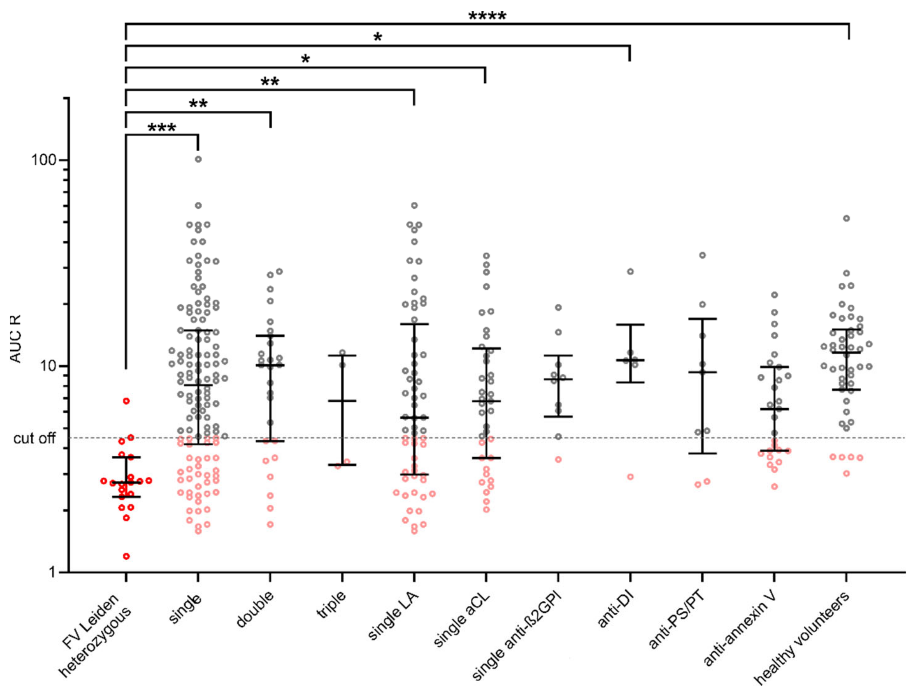
| Cut-Off ≤ 4.5 | ||
|---|---|---|
| % (N) | p Value | |
| Single positivity | 35.2 (38) | <0.001 |
| Double positivity | 29.6 (8) | <0.01 |
| Triple positivity | 50.0 (2) | |
| Single positivity LA | 41.0 (25) | <0.01 |
| Single positivity aCL | 32.4 (12) | <0.05 |
| Single positivity anti-β2GPI | 10.0 (1) | |
| Anti-annexin V | 40.7 (11) | |
| Anti-PS/PT | 22.2 (2) | |
| anti-DI | 16.7 (1) | <0.05 |
| Healthy volunteers | 10.6 (5) | <0.0001 |
Publisher’s Note: MDPI stays neutral with regard to jurisdictional claims in published maps and institutional affiliations. |
© 2022 by the authors. Licensee MDPI, Basel, Switzerland. This article is an open access article distributed under the terms and conditions of the Creative Commons Attribution (CC BY) license (https://creativecommons.org/licenses/by/4.0/).
Share and Cite
Bradáčová, P.; Slavík, L.; Skoumalová, A.; Úlehlová, J.; Kriegová, E.; Manukyan, G.; Friedecký, D.; Piskláková, B.; Ullrychová, J.; Procházková, J.; et al. Determination of Thrombogenicity Levels of Various Antiphospholipid Antibodies by a Modified Thrombin Generation Assay in Patients with Suspected Antiphospholipid Syndrome. Int. J. Mol. Sci. 2022, 23, 8973. https://doi.org/10.3390/ijms23168973
Bradáčová P, Slavík L, Skoumalová A, Úlehlová J, Kriegová E, Manukyan G, Friedecký D, Piskláková B, Ullrychová J, Procházková J, et al. Determination of Thrombogenicity Levels of Various Antiphospholipid Antibodies by a Modified Thrombin Generation Assay in Patients with Suspected Antiphospholipid Syndrome. International Journal of Molecular Sciences. 2022; 23(16):8973. https://doi.org/10.3390/ijms23168973
Chicago/Turabian StyleBradáčová, Pavla, Luděk Slavík, Adéla Skoumalová, Jana Úlehlová, Eva Kriegová, Gayane Manukyan, David Friedecký, Barbora Piskláková, Jana Ullrychová, Jana Procházková, and et al. 2022. "Determination of Thrombogenicity Levels of Various Antiphospholipid Antibodies by a Modified Thrombin Generation Assay in Patients with Suspected Antiphospholipid Syndrome" International Journal of Molecular Sciences 23, no. 16: 8973. https://doi.org/10.3390/ijms23168973
APA StyleBradáčová, P., Slavík, L., Skoumalová, A., Úlehlová, J., Kriegová, E., Manukyan, G., Friedecký, D., Piskláková, B., Ullrychová, J., Procházková, J., & Hluší, A. (2022). Determination of Thrombogenicity Levels of Various Antiphospholipid Antibodies by a Modified Thrombin Generation Assay in Patients with Suspected Antiphospholipid Syndrome. International Journal of Molecular Sciences, 23(16), 8973. https://doi.org/10.3390/ijms23168973