Article

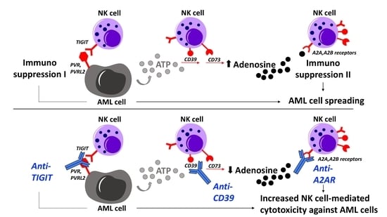
Combined Blockade of TIGIT and CD39 or A2AR Enhances NK-92 Cell-Mediated Cytotoxicity in AML

1
Department of Oncology, Hematology and Bone Marrow Transplantation with Section Pneumology, Hubertus Wald University Cancer Center, University Medical Center Hamburg-Eppendorf, 20251 Hamburg, Germany
2
Mildred Scheel Cancer Career Center HaTriCS4, University Medical Center Hamburg-Eppendorf, 20251 Hamburg, Germany
3
Infectious Diseases Unit, I. Department of Medicine, University Medical Center Hamburg-Eppendorf, 20251 Hamburg, Germany
4
Department of Neurology, University Medical Center Hamburg-Eppendorf, 20251 Hamburg, Germany
5
Institute of Immunology, University Medical Center Hamburg-Eppendorf, 20251 Hamburg, Germany
*
Author to whom correspondence should be addressed.
F.B. and E.S.—shared first authorship.
S.M. and W.F.—shared last authorship.
Int. J. Mol. Sci. 2021, 22(23), 12919; https://doi.org/10.3390/ijms222312919
Submission received: 22 October 2021 / Revised: 26 November 2021 / Accepted: 26 November 2021 / Published: 29 November 2021
(This article belongs to the Special Issue Immunological Approaches in Tumor Therapy)

Abstract

:
This study aimed to characterize different natural killer (NK) cell phenotypes on bone marrow and peripheral blood cells from acute myeloid leukemia (AML) patients and healthy donors (HDs). Our data show that CD56dimCD16 and CD56brightCD16 NK cells represent the predominant NK cell subpopulations in AML, while the CD56dimCD16+ NK cells are significantly reduced compared to HDs. Moreover, TIGIT+ and PVRIG+ cells cluster on the CD56dimCD16+ subset whereas CD39+ and CD38+ cells do so on CD56brightCD16 NK cells in AML. Furthermore, functional effects of (co-)blockade of TIGIT and CD39 or A2AR on NK cell functionality were analyzed. These experiments revealed that the single blockade of the TIGIT receptor results in an increased NK-92 cell-mediated killing of AML cells in vitro. Combined targeting of CD39 or A2AR significantly augments the anti-TIGIT-mediated lysis of AML cells. Our data indicate that distinct NK cell subsets in AML exhibit different immunosuppressive patterns (via the TIGIT/PVRIG receptors and the purinergic pathway). In summary, we conclude that TIGIT, CD39, and A2AR constitute relevant inhibitory checkpoints of NK cells in AML patients. A combinatorial blockade synergistically strengthens NK-92 cell-mediated cytotoxicity. As inhibitors of TIGIT, CD39, and A2AR are clinically available, studies on their combined use could be conducted in the near future.

Graphical Abstract

1. Introduction

Human natural killer (NK) cells belong to the key effectors of cancer immune surveillance [1]. As innate lymphoid cells, they possess an intrinsic selectivity and capacity to kill cancer cells without requiring prior sensitization, which is distinct from the effector T cells of the adaptive immune system. They establish a robust cytotoxic immune response much more rapidly than T cells and have the ability to recruit other adaptive responders [2,3]. These features make NK cells promising candidates for immunotherapeutic strategies in the treatment of cancer [3]. NK cells are primarily classified according to their phenotype, based on the surface expression of CD56 and CD16 [4]. The CD3CD56dimCD16+ NK cell population has been defined as cytotoxic, mainly detected in the peripheral blood from healthy individuals [5]. The CD3CD56dimCD16 and CD3CD56brightCD16 NK cells have regulatory functions by secretion of different cytokines, such as interferon-gamma (IFN-y), upon exposure to the microenvironment [6,7]. The CD56CD16+ NK cells are defined as an unconventional NK cell subset increased in chronic infections and cancer and with significantly decreased effector functions [8,9,10].
In contrast to the single dominant T cell receptor (TCR) on T cells, NK cells express a broad array of activating and inhibitory receptors on their cell surface regulating the functional activity in response to target cells and the microenvironment [11]. A well-characterized model of NK cell activation is the ‘missing-self’ mechanism [12]. Killer cell immunoglobulin-like receptors (KIRs) bind to human leukocyte antigen (HLA) molecules to transduce inhibitory signals, thereby inhibiting NK cell activation, whereas the reduction or loss of major histocompatibility complex (MHC) or HLA expression leads to NK cell activation in a missing-self manner [2,13]. Another recognition mechanism is the antibody-dependent cellular cytotoxicity (ADCC). The NK cell receptors FcγRIIIA/CD16a and/or FcγRIIC/CD32c bind to Fc portions of antibodies that are bound to target cells, leading to NK cell activation [2,14]. Similarly to T cells, NK cells are known to downregulate their responses upon interactions if a signal persists by upregulation of inhibitory receptors [15]. Besides KIRs, NK cells can express further multiple inhibitory receptors, such as CD94/NKG2A, hepatitis A virus cellular receptor (TIM-3), and the T cell immunoreceptor with Ig and ITIM domains (TIGIT) [1,16]. Even if their functional potential has been much less studied compared to that in T cells [3], in acute myeloid leukemia (AML), it was recently found that a higher frequency of TIGIT+ NK cells in the blood was associated with a poorer prognosis [17]. TIGIT and the poliovirus receptor-related immunoglobulin domain-containing protein (PVRIG, CD112R) are inhibitory receptors that compete with DNAX accessory molecule-1 (DNAM-1, CD226), an activating receptor that is downregulated on NK cells in AML [18], for their ligands poliovirus receptor (PVR, CD155) and poliovirus receptor-related 2 (PVRL2, CD112). Both ligands are highly expressed by AML cells and also associated with a poor prognosis [19]. The inhibitory receptor programmed cell death-1 (PD-1) mediates functional defects in cytotoxic NK cells in various cancers [20,21]. Lymphocyte activation gene 3 (LAG-3) is expressed on activated NK cells and has been described as a negative regulator of NK cell cytokine production during chronic stimulation [22].
In addition, NK cell function is also regulated by metabolic signaling including the purinergic pathway [23]. Increased proliferation of the tumor cells and deprivation of oxygen increases utilization of adenosine triphosphate (ATP) and activation of the cancer-associated ectoenzymes ectonucleoside triphosphate diphosphohydrolase-1 (CD39) and ecto-5′-nucleotidase (CD73), which catalyze sequential dephosphorylation of ATP to adenosine monophosphate (AMP) and extracellular adenosine [24,25], whereas cyclic ADP-ribose hydrolase (CD38) catalyzes the hydrolysis of nicotinamide adenine dinucleotide (NAD) [26]. Accumulation of extracellular adenosine interacts with adenosine receptors expressed on NK cells, mediating suppressive signals most strongly via the adenosine A2A receptor (A2AR) [27,28].
NK cell-based immunotherapies have recently been proposed for various types of cancer since impaired NK cell effector functions have been described. This impaired NK cell functionality correlates with poor prognosis [29,30,31,32]. There are additional functional advantages of employing NK cells in contrast to T cells [33]. In AML, impaired NK cell function was associated with AML progress [17,34]. One of the advantages of developing allogeneic ready-to-use chimeric antigen receptor (CAR)-NK cells compared with CAR-T cells is their greater safety [35,36] because of their independence of HLA matching [13]. Results from current preclinical and early clinical studies in relapsed and refractory AML showed promising results from numerous NK cell-based immunotherapies [37,38], which can be augmented by the blockade of checkpoint receptors.
Hypothesizing that NK cells are dysfunctional in AML [10,34], in this study, we aimed to characterize “exhaustion” patterns and functionality of NK cells derived from the peripheral blood (PB) and bone marrow (BM) of patients with AML. In contrast to the well-defined T cell exhaustion, a consensus on the characteristic features of NK cell “exhaustion” is missing [15,39]. In this study, we focused on the (co-)expression of the inhibitory receptors TIGIT, PVRIG, PD-1, and LAG-3 as well as the ectonucleotidases CD39, CD73, and CD38 and we investigated whether TIGIT blockade together with blockade of the purinergic signaling can reinvigorate NK cell-mediated killing of AML blasts. Thereby, we aimed to identify suitable targets for antibody-based immunotherapeutic approaches to overcome NK cell dysfunction.

2. Results

2.1. Bone Marrow-Derived NK Cells of AML Patients Show a Shift towards CD56brightCD16 and CD56dimCD16 Cells and Are Associated with a Reduced CD56dimCD16+ Population

Expression of the receptors CD56 and CD16 was assessed on NK cells derived from the peripheral blood (PB, n = 15) and bone marrow (BM, n = 25) aspirates from patients with untreated newly diagnosed AML and compared to PB specimens of healthy donors (HD, n = 12) (for the gating strategy, see Supplementary Materials Figure S1: Gating strategy). Of note, the study included only AML patients positive for at least one of the surface receptors CD117, CD34, and CD33. This selection enabled us to differentiate AML cells from NK cells. Based on their CD56 and CD16 expression, NK cells were differentiated into the following subsets (Figure 1A): the CD56dimCD16, CD56brightCD16, CD56dimCD16+, and CD56CD16+ NK cells.
The phenotypic characterization of the mononuclear cells revealed that the expression of CD56 and CD16 was reduced in the PB and BM specimens from patients with AML in comparison to that in the PB of HDs (for CD56: p < 0.0001, p = 0.01 and for CD16: p < 0.0001, p = 0.0015; Figure 1B). As illustrated in the t-distributed stochastic neighbor embedding (tSNE) analyses, the distribution of NK cell subpopulations differed between patients with AML and HDs (Figure 1C). Relative to HDs, the frequency of CD56dimCD16 and CD56brightCD16 NK cells was increased in the BM from AML patients (p = 0.0027, p = 0.05; Figure 1D), whereas the frequency of CD56dimCD16+ NK cells was significantly decreased in the BM and the PB from AML patients (p = 0.0078, p = 0.008; Figure 1D). In addition, a low frequency of BM-derived CD56dimCD16+ NK cells correlated with higher frequencies of CD56brightCD16 and CD56dimCD16 NK cells (r = −0.6, p = 0.06 and r = −0.7, p < 0.0001; Figure 1E). The same tendency was seen when correlating the AML PB-derived NK cell subpopulations (Figure 1E). Taken together, in contrast to HDs, our data indicate a shift from cytotoxic to regulatory NK cells in AML.

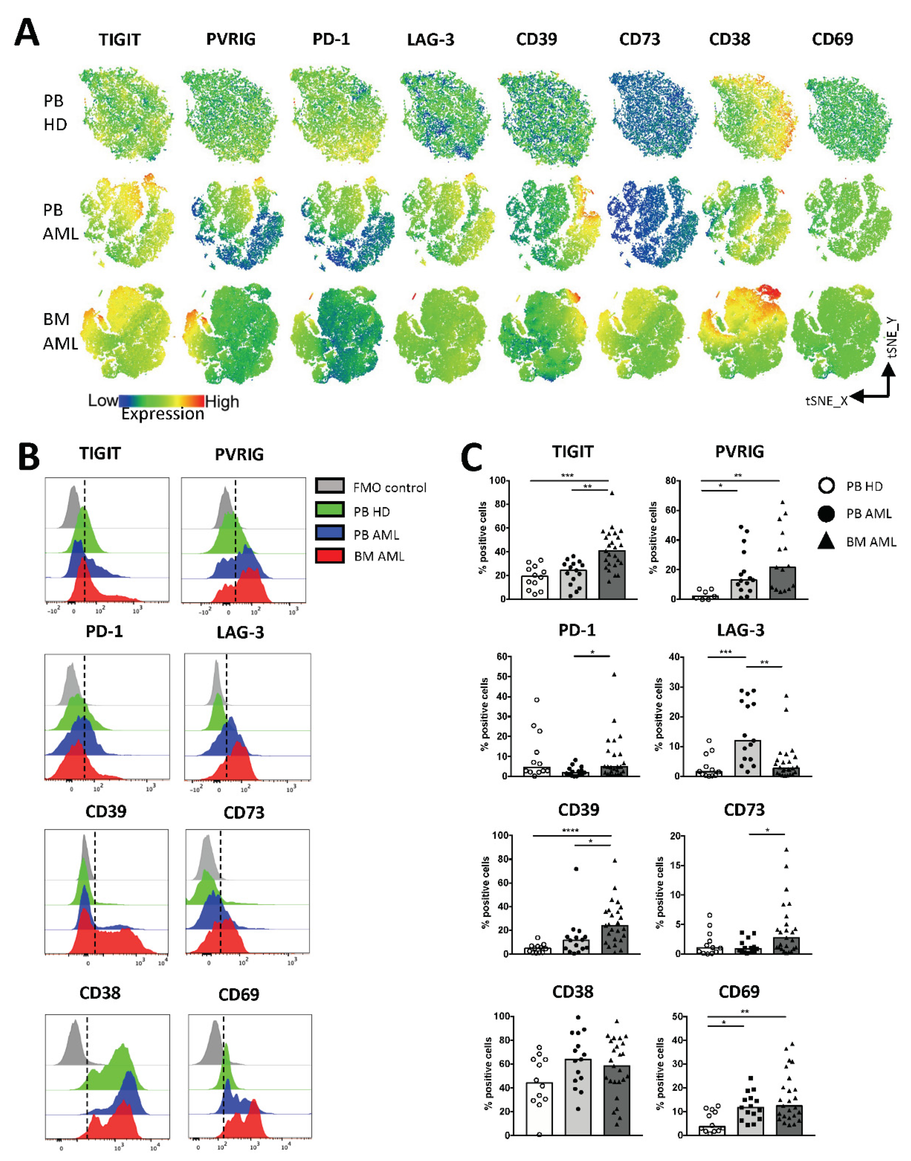
2.2. NK Cells of Patients with AML Express TIGIT, PVRIG, CD39, and CD69

The (co-)expression of inhibitory receptors has been identified as characteristic feature of altered T cell functionality in cancer including in AML. Less is known about immune checkpoint molecules on NK cells as potential targets for AML immunotherapy. Thus, we assessed the surface expression of the inhibitory receptors TIGIT, PVRIG, PD-1, and LAG-3, of the ectonucleotidases CD39, CD73, and CD38 and of the activation marker CD69 on NK cells derived from the BM (n = 25, except for PVRIG n = 15) and PB (n = 15) from AML patients in contrast to HDs (n = 12, for PVRIG n = 7). As depicted in the tSNE plots and summary analyses (Figure 2A–C), the frequency of TIGIT+, PVRIG+, CD39+, and CD69+ NK cells was significantly increased in the BM from patients with AML in comparison to PB mononuclear cells derived from HDs (p = 0.0002, p = 0.0024, p < 0.0001, p = 0.0022). In contrast, in the PB from AML patients, TIGIT was not increased (Figure 2C). Our studies of the PB mononuclear cells show an enrichment of PVRIG+, LAG-3+, and CD69+ NK cells in AML in comparison to the HDs (p = 0.02, p = 0.0006, p = 0.0036; Figure 2C). As previously reported in the literature for some cancer types [40,41], the co-inhibitory receptor PD-1 and the ectonucleotidase CD73 were expressed by NK cells but at low levels without a significant difference between the AML and HD cohorts (Figure 2C). Analyses of the purinergic ectoenzyme CD38 revealed high numbers of positive NK cells in all three cohorts but without a significant difference (Figure 2C). Furthermore, our comparison of paired PB and BM specimens from AML patients revealed an increased frequency of TIGIT+, PVRIG+, and CD39+ NK cells in the BM of AML patients compared to the PB (Supplementary Materials Figure S2: Checkpoint expression on NK cells in corresponding PB- and BM-derived aspirates of AML patients). In summary, our data show an aberrant NK cell population in the PB and BM from patients with AML in comparison to HDs characterized by increased frequencies of TIGIT+, PVRIG+, CD39+, and CD69+ NK cells.

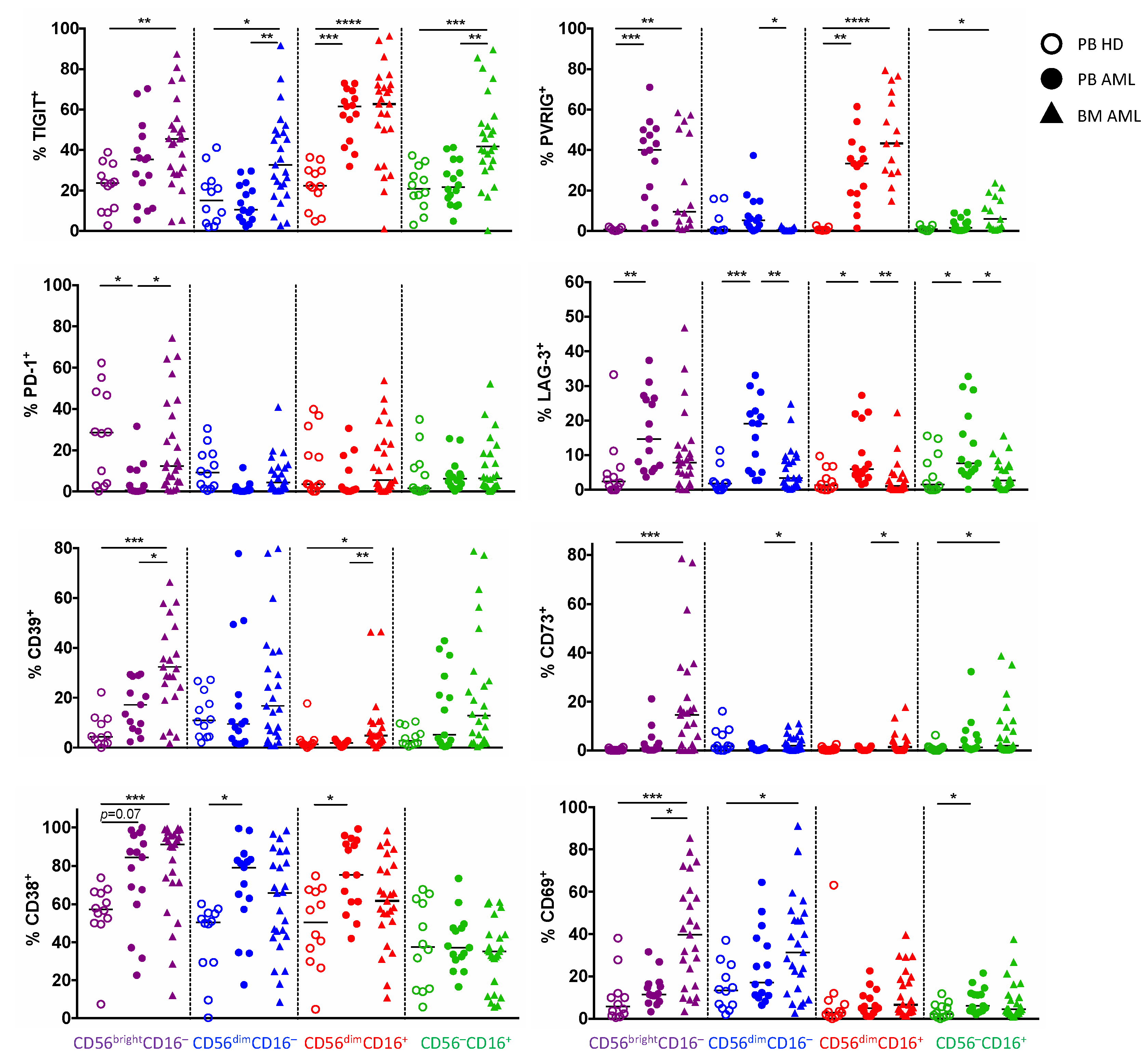
2.3. TIGIT, PVRIG, CD39, and CD38 Expression Is Related to the CD56brightCD16 and the CD56dimCD16+ NK Cell Population in AML

Since our analyses revealed that in AML, NK cells show a characteristic shift towards the CD56brightCD16 and CD56dimCD16 cells, we compared the four NK cell subpopulations regarding their expression of the inhibitory receptors, the ectonucleotidases, and the activation status in the PB and the BM. The further data refer only to results with a significant difference in the molecule expression in BM and PB aspirates from AML patients in comparison to PB from HDs. TIGIT and PVRIG were increasingly expressed by the CD56dimCD16+ subpopulation in the PB and BM from patients with AML (for TIGIT: p = 0.0004, p < 0.0001; and for PVRIG: p = 0.0078, p < 0.0001; Figure 3), whereas the expression of CD39 and CD38 was significantly upregulated on the CD56brightCD16 cells in AML-derived PB and BM in comparison to the HD-derived PB (for CD39: p = 0.08, p < 0.0001; and for CD38: p = 0.07, p = 0.0030; Figure 3). The remaining molecules showed no clustering or differences in expression within the single NK cell subsets during the comparison of AML and HDs. Together, these subgroup analyses show an increased amount of TIGIT+ and PVRIG+ cytotoxic CD56dimCD16+ NK cells, whereas the regulatory CD56brightCD16 population expresses higher levels of CD39 and CD38 in AML in comparison to HDs.

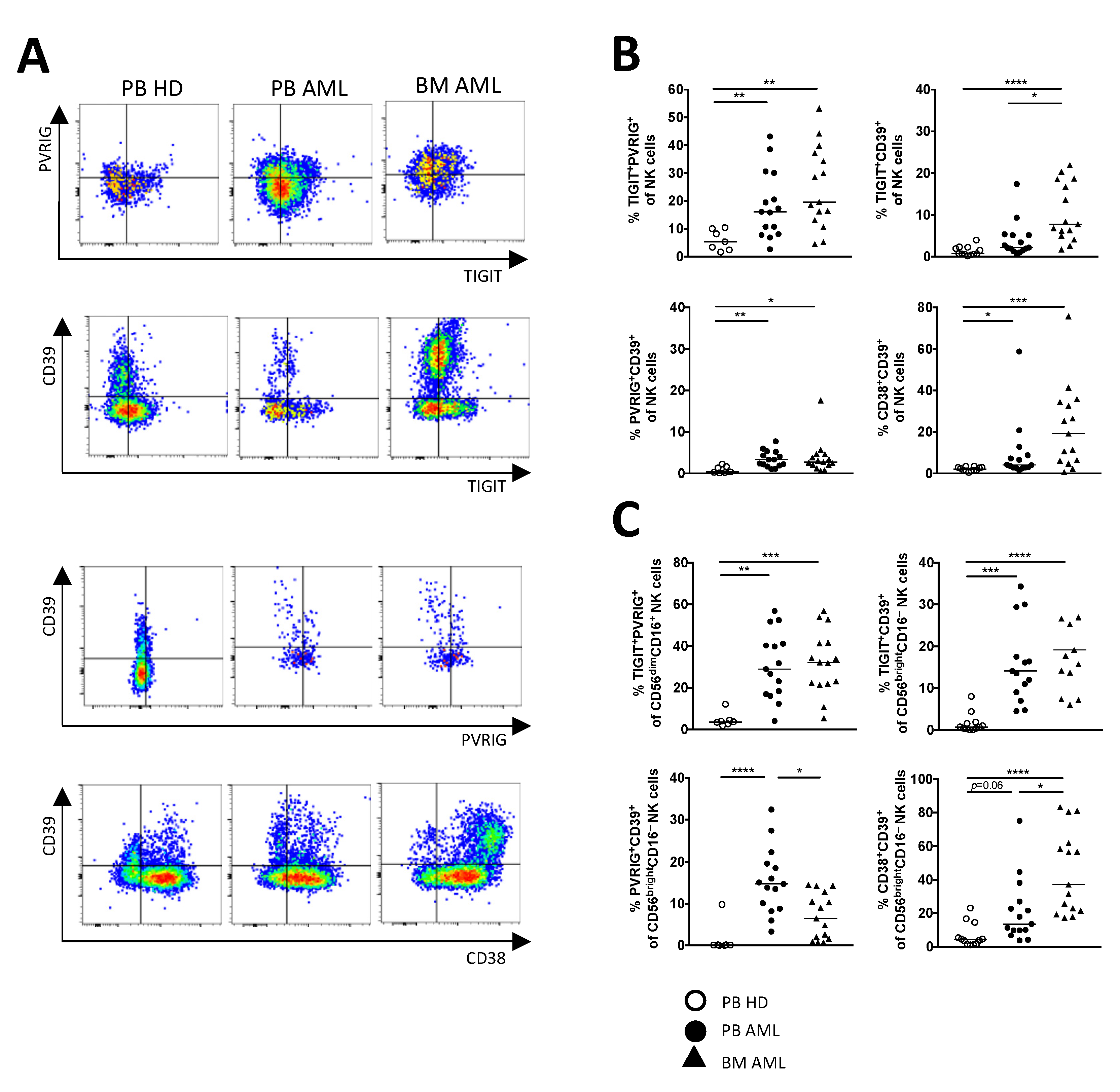
2.4. PVRIG and CD39 Are Co-Expressed with TIGIT on CD56dimCD16+ and CD56brightCD16 NK Cells in AML

Since the expression of TIGIT, PVRIG, CD39, and CD38 on NK cell subpopulations was altered in AML in comparison to HDs, we further investigated the co-expression of TIGIT, PVRIG, PD-1, LAG-3, CD39, CD73, CD38, and CD69 on the total NK cells as well as within the different NK cell subpopulations. The following data refer only to the significant results identified in both AML-derived BM and PB aspirates in comparison to HDs. As depicted in Figure 4A, our co-expression data and the analysis by tSNE (Figure 2A) discovered that the expression of CD39 and PVRIG largely overlapped with the TIGIT+ NK cells in AML (Figure 4B). Moreover, the highest frequencies of TIGIT+PVRIG+ cells were observed within the CD56dimCD16+ subpopulation (PB AML vs. PB HD p = 0.0019, BM AML vs. PB HD p = 0.0005; Figure 4C), whereas TIGIT+CD39+ cells were found mainly in the CD56brightCD16 NK cell population in AML (PB AML vs. PB HD p = 0.0003, BM AML vs. PB HD p < 0.0001; Figure 4C). Additionally, we found a significant enrichment of NK cells co-expressing CD39 together with CD38 (Figure 4B). This cluster of co-expression was also located in the CD56brightCD16 cells in AML (PB AML vs. PB HD p = 0.06, BM AML vs. PB HD p < 0.0001; Figure 4C). In conclusion, molecules of the TIGIT pathway and the purinergic signaling appear to be the most aberrant expressed checkpoint molecules on NK cells in AML. Moreover, in contrast to HDs, our analyses show that TIGIT and PVRIG are significantly co-expressed on CD56dimCD16+ cells in AML, whereas CD39 is mainly found on CD56brightCD16 NK cells, which also co-express CD38 or TIGIT.

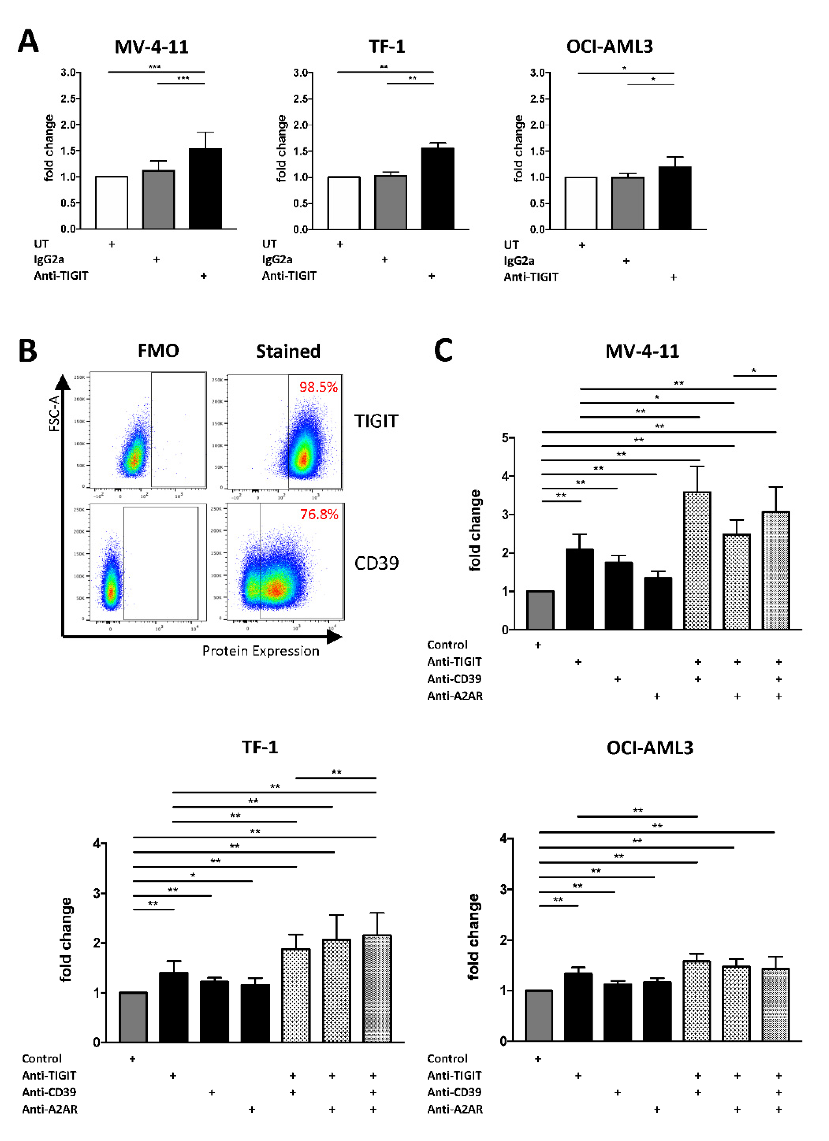
2.5. Single or Combined Checkpoint Blockade Increases NK-92 Cell-Mediated Cytotoxicity In Vitro

In our immunophenotypic analyses, we detected that the inhibitory receptor TIGIT as well as molecules of the purinergic signaling are the most aberrant checkpoint targets on NK cells in AML. We have previously demonstrated that the ligands PVR and PVRL2 are highly expressed by AML cell lines (including MV-4-11, TF-1, and OCI-AML3) and primary AML cells [19]. Next, we analyzed the therapeutic potential of blocking TIGIT and the purinergic signaling via anti-CD39 blockade or adenosine A2A receptor (A2AR) antagonism on natural killer cells to restore the function of exhausted NK cells and improve their cytotoxic activity against AML cells. TIGIT as well as CD39 are highly expressed on the NK-92 cell line, which was used for these assays (frequency of TIGIT+ and CD39+ NK-92 cells: 98.5% and 76.8%, respectively, Figure 5B).
The single blockade of the TIGIT receptor resulted in an increased NK cell-mediated killing of AML cells after 24 h in all 3 AML cell lines (anti-TIGIT vs. IgG2a for MV-4-11 p = 0.001, for TF-1 p = 0.0078 and for OCI-AML3 p = 0.0156; Figure 5A). In addition, targeting of the purinergic pathway via a blockade of CD39 or A2AR augmented the NK cell-mediated lysis of the AML cells (anti-CD39 vs. control for MV-4-11 p = 0.0039, for TF-1 p = 0.0039 and for OCI-AML3 p = 0.0039 and anti-A2AR vs. control for MV-4-11 p = 0.0039, for TF-1 p = 0.0195 and for OCI-AML3 p = 0.0039; Figure 5C).
Due to these promising results, we tested the effects of the TIGIT blockade in combination with an inhibition of the purinergic pathway on NK cell-mediated killing. Dual blockade of either CD39 or A2AR together with TIGIT significantly increased AML cell lysis in 2/3 cell lines in comparison to a single blockade or control treatment (Figure 5C). Moreover, triple blockade augmented NK cell-mediated lysis of MV-4-11 cells in comparison to a dual blockade of TIGIT in combination with A2AR (Figure 5C). Triple blockade also augmented NK cell-mediated killing of the TF-1 AML cells compared to the dual inhibition of TIGIT and CD39 (Figure 5C). Further information about the significance is depicted in the Supplementary Materials Table S1: p-values for the differences in the fold change of 7-AAD+ AML cells. All effects of blockades are also analyzed and plotted as frequencies in the Supplementary Materials Figure S3: Frequencies of 7-AAD+ AML cells after NK cell-mediated lysis and Table S2 p-values for the differences in the frequency of 7-AAD+ AML cells.

3. Discussion

This study aimed to characterize NK cells in AML patients in comparison to HDs and to determine whether (co-)blockade of TIGIT and CD39 or A2AR can restore the functionality of NK cells in AML. We observed reduced cytotoxic CD56dimCD16+ NK cells in the BM and PB from AML patients compared to HDs, whereas CD56dimCD16 and CD56brightCD16 NK cells represent the two dominant NK cell subpopulations in AML. The presence of CD56dimCD16 and CD56brightCD16 NK cells was associated with an increased expression of TIGIT, PVRIG, CD39, CD38, and CD69 in AML. Interestingly, regarding the single expression of TIGIT and CD39, we observed no significant differences in the frequency of TIGIT+ and CD39+ NK cells when we compared the PB from AML patients with that of HDs, whereas the expression of LAG-3 was only increased in the PB of AML patients. However, regarding co-expression clusters unique to AML, we observed significantly increased frequencies of TIGIT+PVRIG+ NK cells located in the CD56dimCD16+ population in the PB as well as in the BM from AML patients. CD39 and CD38, on the other hand, were more frequently expressed by CD56brightCD16 cells in AML-derived PB and BM aspirates in comparison to HDs. Due to these data, we hypothesized that distinct NK cell subsets in AML exhibited different immunosuppressive strategies (inhibitory signaling via TIGIT/PVRIG expression and immunosuppression via the adenosine-A2AR signaling). Further functional evaluation of the TIGIT axis and the purinergic signaling revealed that the single blockade of the TIGIT receptor resulted in an increased NK cell-mediated killing of AML cells after 24 h in all 3 AML cell lines. In addition, targeting of the purinergic pathway via the blockade of CD39 or A2AR augmented the NK cell-mediated lysis of AML cells. Furthermore, dual blockade of either CD39 or A2AR together with TIGIT significantly increased AML cell lysis in 2/3 cell lines in comparison to both a single blockade respectively and the control treatment. These findings implicate a synergistic effect of the combinatorial blockade of the purinergic pathway together with the TIGIT axis in AML.
In line with data in the literature, the majority of the analyzed NK cells derived from PB of HDs were further delineated as mature CD56dimCD16+ NK cells [42]. Contrary to HDs, in AML, we found a significantly altered distribution of NK cells with increased frequencies of PB- and BM-derived CD56brightCD16 and CD56dimCD16 NK cell populations. Overall, we observed a loss of CD56 expression on NK cells, which has also been observed by Chretien et al. [10]. In addition, a reduction of cytotoxic NK cells was recently described in different hematological malignancies (including AML) and solid tumors, especially at the tumor sites [43]. Functionally, tumor-derived NK cells were characterized by the production of the vascular endothelial growth factor (VEGF; indicating a pro-angiogenetic potential) and reduced cytotoxic effector functions [43]. Furthermore, Salomé et al. recently showed that AML-derived CD56+CD16 NK cells produce reduced levels of granulysin, leading to an impairment in the degranulation process [44]. In light of this, we propose that aberrant NK cell populations with reduced cytotoxic functions might represent a feature of immune evasion in cancer.
NK cell function strongly depends on complex interactions between signals transmitted by activating and inhibitory receptors. Similar to T cells, NK cell-mediated anti-tumor responses were previously described to be governed by PD-1 and LAG-3 receptor signaling [21,22]. More recently, the novel inhibitory receptors TIGIT and PVRIG have also been discovered to control NK cell activation and effector function [45].
Our studies revealed that PD-1 and LAG-3 expression of NK cells was only slightly dysregulated in AML, whereas TIGIT and PVRIG were expressed on the majority of AML-derived NK cells, in particular on the cytotoxic CD56dimCD16+ NK cells. Interestingly, we observed a significant co-expression of TIGIT and PVRIG on the largest part of CD56dimCD16+ NK cells, suggesting that this NK cell subset represents the terminally activated and exhausted NK cell population in AML. Increased expression of TIGIT has previously been described for several cancer entities including AML and myelodysplastic syndrome [16,46]. Regarding the axis of CD226 and TIGIT/CD96/PVRIG, in which shared ligands and receptor-ligand affinities regulate the immune response, it was recently reported that the stimulatory receptor CD226 is downregulated on NK cells from AML patients, whereas in line with our data, an increased expression of TIGIT on T cells and NK cells was observed [19,46]. Moreover, in a cohort of 36 AML patients, the frequency of CD226TIGIT+CD96+ NK cells was associated with poor prognosis of the patients [46]. Additionally, NK cell-mediated lysis of leukemias was dependent on CD226 [47]. Liu et al. also recently illustrated that TIGIT+ NK cells exhibit lower anti-leukemia effects, manifested by reduced cytokine production, degranulation, and cytotoxicity [17]. We confirmed these effects and extended their observations, showing that TIGIT blockade in combination with targeting the purinergic signaling could further enhance the anti-leukemic function of NK cells [17]. Our results are also in line with data from solid cancer, where blockade of TIGIT could boost functional responsiveness of NK cell subsets against ovarian cancer cell lines [48].
PVRIG, identified in 2016 [49], interacts as an inhibitory receptor with PVRL2 but not PVR. PVRL2 is highly expressed by AML cells [19] but also solid tumor cells [50], thereby adding more complexity in the TIGIT/CD226 signaling. Our data showed the expression of PVRIG+ NK cells (particularly within the CD56dimCD16+ subset) in the PB and BM of patients with AML. In contrast to our findings, Li et al. recently found unaltered PVRIG expression on NK cells in AML patients’ BM [51]. However, Li et al. observed significantly increased NK cell killing of PVRL2+ AML cell lines upon PVRIG blockade [51]. Together, these data support the relevance of TIGIT/PVRIG signaling in the suppression of anti-leukemic NK cell responses.
CD39 and CD73-generated adenosine followed by engagement of the adenosine receptor A2A additionally represents a potent immunosuppressive pathway, leading to the progression of solid tumors and hematological malignancies [28]. Concerning tumor-infiltrating NK cells, extracellular adenosine signaling via the A2AR has been shown to regulate proliferation, maturation, and cytotoxic function [52]. Here, we performed the phenotypical characterization of NK cells in AML in comparison to HDs and found an aberrant subset of CD56brightCD16 NK cells that can be further defined by an increased frequency of cells co-expressing CD39 and CD38. Consistent with our findings, CD39 expression has been reported to be upregulated on NK cells in chronic infection (e.g., HIV infection) and solid cancer, thereby associating with a poor prognosis [53,54,55]. Moreover, Zhang et al. showed that CD39 promotes tumor progression and lung metastases by suppressing NK cells and IFN-y function in different melanoma models [55]. In addition, in CD39-deficient mice or wild-type mice treated with an inhibitor of ecto-NTPDases (POM-1), reduced formation of metastases and increased NK cell cytotoxicity were observed [55,56]. NK cell cytotoxicity could be further augmented by a combinatorial checkpoint blockade of CD39 together with A2AR or PD-1 [56].
The ectoenzyme CD38 is predominantly expressed by NK cells [57] and stimulation of CD38 with agonistic antibodies led to Ca++ flux, Zeta-chain-associated protein kinase 70 (ZAP70) phosphorylation, mitogen-activated protein kinase (MAPK) activation, and subsequent secretion of IFN-y [58]. Interestingly, we observed an increased frequency of CD38+CD56brightCD16 NK cells in AML, which confirmed previous findings for solid cancers, where CD38-mediated adenosine production by CD56brightCD16 NK cells accounts for a significant CD4+ T cell inhibition [59]. Moreover, clinical administration of the monoclonal antibody Daratumumab for myeloma patients demonstrated a depletion of CD38high NK cells, leading to enhanced CD38low/– NK-cell subsets with increased NK cell-dependent cytotoxicity against multiple myeloma cells [60,61].
Adenosine receptors including the A2A receptor are expressed by NK cells. Collective reports demonstrated that exogenous adenosine reduces NK cell cytotoxicity and cytokine production, particularly via the A2AR engagement. A2AR–/– NK cells exert increased anti-tumor immunity and reduced tumor growth in mouse tumor models [62]. Moreover, A2AR small-molecule inhibition alone or together with a PD-L1 blockade led to tumor reduction and improved T cell and NK cell cytotoxicity [62,63].
Comparing the checkpoint expression on NK cells derived from the paired PB and BM aspirates, we observed that our data from PB were quite discrepant from the BM findings. This could be explained by a different microenvironment in the BM, which might more accurately reflect the immune milieu at the tumor site, as has been demonstrated for solid cancer studies. Williams et al. recently demonstrated similar discrepancies when comparing T cells derived from the PB with that derived from BM of AML patients [64].
Recently, increased focus has been placed on the combination of checkpoint inhibitors to achieve synergistic anti-cancer immunity [65]. Dual blockade of TIGIT and PD-1 has demonstrated enhanced anti-cancer immunity in several cancers [66,67]. Recently, it has also been described in mouse tumor models that a combined blockade of the A2AR and PD-L1 led to decreased tumor volume correlating with T cell response [63]. Though most of these studies reflected the efficacy of combinatorial blockade by exploring T cell-mediated cytotoxicity, there is growing evidence that efficacy depends on NK cells as well [68]. Up to now, in AML, the question remains as to how NK cell-mediated cytotoxicity can be improved. Here, we found that combined blockade of TIGIT together with CD39 or A2AR could significantly improve the cytotoxicity of NK cells in vitro. Because of the differential expression of the TIGIT/PVRIG axis and CD39 on different NK cell populations, the combined blockade of these pathways might augment the cytotoxic functionality of different NK cell populations in vivo. Interestingly, we observed no differences when either performing blockade of CD39 or A2AR in addition to TIGIT. This might indicate that inhibition of CD39 has no evident advantages over A2AR blockade to increase NK cell-mediated cytotoxicity. To our knowledge, ours is the first study evaluating the combinatorial effect of these two immunosuppressive pathways.
Our study has several limitations. The observations made in this study must be interpreted with caution due to the relatively small sample size of 25 patients newly diagnosed with AML and should be validated in a larger cohort. Considering the predominant expression of TIGIT and CD39 on the NK-92 cell line, further research with primary NK cells from AML patients is needed to unravel the role of TIGIT together with the purinergic signaling in AML in more detail. Moreover, the different effects of blocking CD39 or A2AR should be further investigated in more complex settings including primary cells and mouse experiments; thus, contrasting the dual impact of a CD39 blockade on increased proinflammatory ATP levels together with a reduction of extracellular adenosine with a single reduction of adenosine accumulation mediated by an A2AR inhibition.
Together, our data support the hypothesis that the inhibitory TIGIT/PVRIG receptor signaling, as well as the metabolic purinergic signaling, constitute relevant pathways for NK cell dysfunction in AML.

4. Materials and Methods

4.1. Clinical Cohorts

Peripheral blood specimens (PB, n = 15) and bone marrow-derived aspirates (BM, n = 25) were collected from patients with newly diagnosed acute myeloid leukemia (AML) before the start of intensive chemotherapeutic treatment as well as PB from age-matched healthy donors (HD, n = 12) after written informed consent in accordance with the Declaration of Helsinki and approval of the local ethics board of the Ärztekammer Hamburg (PV3469). All PB and BM samples were taken from patients with non-acute promyelocytic leukemia (non-APL). Paired PB and BM samples from the same patient were available for 15 of 25 AML patients. The median age of the AML patient cohort was 66 years (range 25–84), and the median age of the healthy donors was 59 years (range 41–70) (Supplementary Materials Table S3 Patient characteristics).

4.2. Multiparameter Flow Cytometry and Surface Staining

For multiparameter flow cytometry (MFC) analyses, cryopreserved PB and BM mononuclear cells (PBMCs, BMMCs) from patients with CD117+/CD34+/CD33+ AML and PBMCs of HDs were thawed in a 37 °C water bath and counted. Then, 1 × 10⁶ PBMCs or BMMCs of each specimen were washed with PBS (phosphate-buffered saline) (Dulbecco’s Phosphate Buffered Saline, Gibco Thermo Fisher Scientific, Waltham, MA, USA). After FcR blocking (FcR Blocking Reagent, human, Miltenyi Biotec, Bergisch Gladbach, Germany) for 5 min in the dark, PBMCs and BMMCs were stained with the Zombie NIR Fixable Viability Kit (BioLegend, San Diego, CA, USA) for exclusion of dead cells according to the manufacturer’s instructions. This step was followed by another washing step with PBS. For surface staining, cells were incubated with appropriate fluorochrome-conjugated antibodies including anti-CD3 (OKT3), anti-CD4 (RPA-T4), anti-CD8 (RPA-T8), anti-CD56 (5.1H11), anti-CD16 (3G8), anti-CD117 (104D2), anti-CD33 (P67.6), anti-CD34 (8G12), anti-CD19 (HIB19), anti-CD14 (63D3), anti-TIGIT (A15153G), anti-PD-1 (EH12.2H7), anti-LAG-3 (11C3C65), anti-CD39 (A1 and TU66), anti-CD73 (AD2), anti-CD38 (HIT2), anti-CD69 (FN50), anti-CD112R (W16216D), anti-CD47 (CC2C6), anti-CD138 (MI15), and anti-CD25 (M-A251) for 20 min in the dark. Antibodies were obtained from BioLegend (San Diego, CA, USA) and BD Biosciences (Franklin Lakes, NJ, USA). To wash out unbound antibodies, the cells were washed again with PBS. In the last step, the stained PBMCs and BMMCs were fixed with 0.5% paraformaldehyde (Thermo Fisher Scientific, Waltham, MA, USA) and after an incubation time of 15 min, washed again with PBS. For measuring compensation controls, single-stained BDCompBeads (Anti-Mouse Ig, k/Negative Control Compensation Particle Set, BD Biosciences, Franklin Lakes, NJ, USA) were used. For live/dead (Zombie) compensation, compensation beads stained with anti-CD19 (APC Cy-7, BioLegend) were used. All samples were acquired on a BD FACSymphony A3 with BD FACSDiva software version 8 (BD Biosciences, Franklin Lakes, NJ, USA).

4.3. NK Cell-Mediated Cytotoxicity Assay

Allogeneic natural killer cell-mediated cytotoxicity assays were performed by using the NK-92 cell line as effector cells and the MV-4-11, TF-1, or OCI-AML3 cell lines as target AML cells. NK cells were co-cultured with AML cells, which were labeled with CellTracker (CT) green CMFDA (Invitrogen Thermo Fisher, Waltham, MA, USA) according to the manufacturer’s instructions in a 6:1 effector:target ratio. Co-culture was plated in a 96-well plate (1 × 10⁶ cells/mL) in NK-92 culture medium (see Section 4.4. Cell lines) and incubated with different blocking antibodies for 24 h at 37 °C and 5% CO2. Each well of the 96-well plate contained a 200 µL volume in total, including the blocking antibodies. For experiments with single blockade of TIGIT, co-cultured cells were incubated with 50 µg/mL anti-TIGIT antibody (Ultra-LEAF Purified anti-human TIGIT antibody clone A15153G, BioLegend, San Diego, CA, USA) or mouse IgG2a isotype control (Ultra-LEAF Purified Mouse IgG2a, K Isotype Ctrl antibody, clone MG2a-53, BioLegend, San Diego, CA, USA) or left untreated. Experiments were performed in triplicates and repeated four to five times (MV-4-11 n = 5, TF-1 n = 4, OCI-AML3 n = 4, respectively) with the same results. Bars display the mean ± SD. For experiments with single, double, or triple blockade of TIGIT together with CD39 and of the adenosine A2A receptor, cells were co-cultured in the following conditions: control wells contained 50 µg/mL mouse IgG2a isotype control, 100 µg/mL control nanobody (the target non-binding nanobody L-10e was used in the same format as the CD39-specific nanobody [69,70,71]), and 0.01% DMSO (Sigma-Aldrich, St. Louis, MO, USA) as the A2AR antagonist was dissolved in DMSO. For the single or combinatorial checkpoint-blockade, 50µg/mL anti-TIGIT antibody (BioLegend, San Diego, CA, USA) and/or 100 µg/mL of an inhibitory anti-CD39 nanobody (the CD39-specific nanobody SB24 was generated from an immunized alpaca and reformatted into a human IgG1 heavy-chain antibody format carrying the LALAPG mutations using established protocols [69,70,71]) and/or 10 µM adenosine A2A receptor antagonist (AZD4635, kindly provided by Astra Zeneca R&D Boston, Waltham, MA, USA) were administered. In cases of a single blockade, test cultures were supplemented with the two non-corresponding controls, whereas in cases of a double blockade, test cultures were supplemented with the one non-corresponding control to make it possible to compare the results with the control wells. After 24 h of incubation, the cells of each well were collected and washed with PBS and FcR blocking was performed. After a second PBS washing step, cells were stained with 7-Aminoactinomycin D (7-AAD) Viability Staining Solution (BioLegend, San Diego, CA, USA) according to the manufacturer’s protocol. After an incubation time of 10 min at room temperature, the samples were subsequently analyzed on a BD FACSymphony A3 with BD FACSDiva software version 8 (BD Biosciences, Franklin Lakes, NJ, USA).
The frequency of AML cell lysis was determined via positivity of 7-AAD and CT green using flow cytometry. Mean values ± SD of the triplicates were calculated and plotted.

4.4. Cell Lines

The AML cell lines TF-1 and OCI-AML3 as well as the NK-92 were purchased from the DSMZ (Deutsche Sammlung von Mikroorganismen und Zellkulturen GmbH, Braunschweig, Germany), and the MV-4-11 cell line was purchased from ATCC (American Type Culture Collection, Manassas, VA, USA). MV-4-11 cells were cultured in RPMI 1640 (Gibco, Thermo Fisher Scientific, Waltham, MA, USA) and supplemented with 10% fetal bovine serum (FBS superior, Sigma-Aldrich, St. Louis, MO, USA). TF-1 cells were cultured in RPMI 1640 supplemented with 20% FBS and 5ng/mL human granulocyte-macrophage colony-stimulating factor (GM-CSF, PeproTech GmbH, Hamburg, Germany). OCI-AML3 cells were cultured in MEM-Alpha medium (Gibco, Thermo Fisher Scientific, Waltham, MA, USA) supplemented with 20% FBS. NK-92 cells were cultured in MEM-alpha medium supplemented with 12.5% FBS, 12.5% horse serum (Gibco, Thermo Fisher Scientific, Waltham, MA, USA), 100 U/mL Interleukin 2 (Recombinant human Interleukin-2, PeproTech GmbH, Hamburg, Germany), and 0.1 mM 2-Mercaptoethanol (Gibco, Thermo Fisher Scientific, Waltham, MA, USA). The NK-92 cells express TIGIT and CD39, which were regularly assessed by MFC. Cell cultures were incubated at 37 °C and 5% CO2.

4.5. Statistical Analysis

All flow cytometric data were analyzed using FlowJo version 10.4 and 10.5.2 software (BD Life Sciences, FlowJo, LCC, Ashland, OR, USA). Statistical analyses were carried out using Prism 8.0 (GraphPad Software, San Diego, CA, USA). All groups were tested for normal distribution with the Kolmogorov–Smirnov test. Non-normally distributed data was analyzed by the Mann–Whitney test for two unpaired groups, the Wilcoxon test for two paired groups, and the Kruskal–Wallis or Friedmann tests for more than two groups, respectively. Pearson’s correlation and Spearman’s rank correlation coefficient were applied for bivariate correlation analysis. The NK cell-mediated cytotoxicity assays were tested by the Wilcoxon test for two paired groups. Frequencies in the text are described as medians unless stated otherwise (as indicated in the figure legend). p-values smaller than 0.05 were considered significant, where *, **, ***, and **** indicate p-values between 0.01 and 0.05, 0.001 and 0.1, 0.0001 and 0.001, and <0.0001 respectively.

5. Conclusions

This study showed a reduced frequency of cytotoxic CD56dimCD16+ NK cells, whereas CD56dimCD16 and CD56brightCD16 NK cells were significantly increased in AML patients compared to HDs. Co-expression of TIGIT and PVRIG was found on the CD56dimCD16+ population and of CD39 and CD38 on CD56brightCD16 cells in AML but not in HDs. In addition, the single blockade of the TIGIT receptor resulted in an increased NK cell-mediated killing of AML cells that can be further augmented by targeting of the purinergic pathway via a combinatorial blockade of TIGIT together with CD39 or A2AR. In summary, we conclude that TIGIT, CD39, and A2AR constitute relevant checkpoints for AML-derived NK cells. Regarding their differential expression on CD56dimCD16+ and CD56brightCD16 NK cells, a combinatorial blockade seemed to synergistically improve anti-AML cytotoxicity. Future experiments analyzing the functional relevance of TIGIT, A2AR, and CD39 in NK cell cytotoxicity are ongoing involving primary AML cells and mouse experiments. Co-blocking TIGIT and CD39/A2AR could be a promising immunotherapeutic strategy for AML patients. Additionally, with respect to CAR-NK technologies, NK cells could be alternatively engineered to be deficient in TIGIT, CD39, or A2AR, thereby improving their antitumor effector functions. Particularly in the context that anti-TIGIT and anti-CD39/A2AR antibodies as well as small molecules are available, further (clinical) studies on their combined use need to be initiated.

Supplementary Materials

The following are available online at https://www.mdpi.com/article/10.3390/ijms222312919/s1.

Author Contributions

F.B. and E.S. designed the research study, performed the experiments, analyzed the data, and wrote the manuscript. J.W., J.S.z.W., Y.D., T.M., C.B. reviewed the manuscript. F.K.-N., S.M. reviewed the manuscript and provided the CD39 inhibitors. W.F. designed the research study and wrote the manuscript. All authors have read and agreed to the published version of the manuscript.

Funding

This work was supported by the MSNZ, University Medical Center Hamburg-Eppendorf, supported by the Deutsche Krebshilfe (to F.B. and S.M.), SFB1328 (project Z02, to F.K.-N.). E.S. was supported by the E.W. Kuhlmann Stiftung. J.S.z.W. was supported by the German Research Agency (SFB841 project A6).

Institutional Review Board Statement

The study was approved by the local ethics board of the Ärztekammer Hamburg (PV3469, 17 June 2010). Informed written consent was obtained from all patients.

Informed Consent Statement

Not applicable.

Data Availability Statement

The datasets used and/or analyzed during the current study are available from the corresponding authors on reasonable request ([email protected]).

Acknowledgments

We thank all our patients for their trust, understanding and willingness to provide their blood and bone marrow samples to our research. We would like to thank Alwin Schuller from AstraZeneca R&D Boston, Waltham, MA, USA for providing the A2AR inhibitor, and Jana Muschhammer, Gabi Vohwinkel, Dorte Wendt and Birte Albrecht for excellent technical assistance. Julian Schulze zur Wiesch was supported by the DFG (SFB 1328 A12, and SFB841 A6).

Conflicts of Interest

FB: Travel grant Daiichi Sankyo, Servier, Novartis; advisory board by Jazz. GmbH, Daiichi Sankyo. WF: Membership on an entity’s board of directors or advisory Amgen, ARIAD/Incucyte, Pfizer, Novartis, Jazz Pharmaceuticals, Morphosys, Abbvie, Celgene; patents and royalities: Amgen; other support for meeting attendance Amgen, Gilead, Jazz Pharmaceuticals, Servier, Daiichi Sankyo; research funding Amgen, Pfizer. Travel grant, advisory board and research funding by Amgen Inc., travel grant and advisory board by TEVA GmbH, the advisory board: Ariad/Incucyte Inc., travel grant by Gilead Inc and Jazz. GmbH, research funding by Pfizer Inc. CB: Travel grant: Astra Zeneca, Bayer Healthcare, Berlin Chemie, Bristol Myers Squipp, Jansen Cilag, Merck Serono, Merck Sharp Dohme, Novartis, Roche Pharma, Sanofi Aventis; advisory board: Astra Zeneca, Bayer Healthcare, Berlin Chemie, Bristol Myers Squipp, Jansen Cilag, Merck Serono, Merck Sharp Dohme, Novartis, Roche Pharma, Sanofi Aventis; invited speaker: AOK Germany, med update, Merck Serono; Honoraria: AOK Germany, Astra Zeneca, Bayer Healthcare, Berlin Chemie, GSO Research Organisation, Jansen- Cilag, med update, Merck Serono, Merck Sharp Dohme, Novartis, Roche Pharma, Sanofi Aventis. The sponsors had no role in the design, execution, interpretation, or writing of the study. The remaining authors declare that they have no conflict of interest.

Abbreviations

7-AAD: 7-Aminoactinomycin D; A2AR: Adenosine A2A receptor; ADCC: Antibody-dependent cellular cytotoxicity; AML: Acute myeloid leukemia; AMP: Adenosine monophosphate; ATP: Adenosine triphosphate; BM: Bone marrow; BMMCs: Bone marrow mononuclear cells; CD38: Cyclic ADP-ribose hydrolase; CD39: Ectonucleoside triphosphate diphosphohydrolase-1; CD73: Ecto-5′-nucleotidase; CAR: Chimeric antigen receptor; CT: Cell tracker; DNAM-1: DNAX accessory molecule-1 (CD226); DNMT3A: DNA (cytosine-5)-methyltransferase 3A; ELN: European LeukemiaNet classification; FAB: French-American-British classification; FBS: Fetal bovine serum; FLT3 ITD: FMS-like tyrosine kinase 3 internal tandem duplication; FLT3 TKD: FMS-like tyrosine kinase 3 tyrosine kinase domain; FMO: Fluorescence minus one; GM-CSF: Granulocyte-macrophage colony-stimulating factor; GvHD: Graft versus host disease; HD: Healthy donor; HLA: Human leukocyte antigen; IFN-y: Interferon-gamma; KIRs: Killer-cell immunoglobulin-like receptors; LAG-3: Lymphocyte activation gene 3; MAPK: Mitogen-activated protein kinase; MFC: Multiparameter flow cytometry; MFI: Median fluorescence intensity; MHC: Major histocompatibility complex; NAD: Nicotinamide adenine dinucleotide; NK: Natural killer; non-APL: Non-acute promyelocytic leukemia; NPM1: Nucleophosmin-1; PB: Peripheral blood; PBMCs: Peripheral blood mononuclear cells; PBS: Phosphate buffered saline; PD-1: Programmed cell death-1; PD-L1: Programmed cell death-ligand 1; POM-1: Inhibitor of ecto-NTPDases; PVR: Poliovirus receptor (CD155); PVRIG: Poliovirus receptor-related immunoglobulin domain containing protein (CD112R); PVRL2: Poliovirus receptor-related 2 (CD112); TCR: T cell receptor; TIGIT: T cell immunoreceptor with Ig and ITIM domains TIM-3: Hepatitis A virus cellular receptor; TNF-a: Tumor necrosis factor alpha; tSNE: t-distributed stochastic neighbor embedding; VEGF: Vascular endothelial growth factor; ZAP70: Zeta-chain-associated protein kinase 70.

References

  1. Cao, Y.; Wang, X.; Jin, T.; Tian, Y.; Dai, C.; Widarma, C.; Song, R.; Xu, F. Immune checkpoint molecules in natural killer cells as potential targets for cancer immunotherapy. Signal Transduct. Target. Ther. 2020, 5, 250. [Google Scholar] [CrossRef]
  2. Rosenberg, J.; Huang, J. CD8+ T cells and NK cells: Parallel and complementary soldiers of immunotherapy. Curr. Opin. Chem. Eng. 2018, 19, 9–20. [Google Scholar] [CrossRef]
  3. Kwon, H.-J.; Kim, N.; Kim, H.S. Molecular checkpoints controlling natural killer cell activation and their modulation for cancer immunotherapy. Exp. Mol. Med. 2017, 49, 1–11. [Google Scholar] [CrossRef]
  4. Mandal, A.; Viswanathan, C. Natural killer cells: In health and disease. Hematol. Stem Cell Ther. 2015, 8, 47–55. [Google Scholar] [CrossRef] [Green Version]
  5. Poli, A.; Michel, T.; Thérésine, M.; Andrès, E.; Hentges, F.; Zimmer, J. CD56bright natural killer (NK) cells: An important NK cell subset. Immunology 2009, 126, 458–465. [Google Scholar] [CrossRef]
  6. Amand, M.; Iserentant, G.; Poli, A.; Sleiman, M.; Fievez, V.; Sanchez, I.P.; Sauvageot, N.; Michel, T.; Aouali, N.; Janji, B.; et al. Human CD56dimCD16dim Cells as an Individualized Natural Killer Cell Subset. Front. Immunol. 2017, 8, 699. [Google Scholar] [CrossRef]
  7. Stabile, H.; Fionda, C.; Gismondi, A.; Santoni, A. Role of Distinct Natural Killer Cell Subsets in Anticancer Response. Front. Immunol. 2017, 8, 293. Available online: https://www.frontiersin.org/article/10.3389/fimmu.2017.00293 (accessed on 21 October 2021). [CrossRef] [Green Version]
  8. Mavilio, D.; Lombardo, G.; Benjamin, J.; Kim, D.; Follman, D.; Marcenaro, E.; O’Shea, A.M.; Kinter, A.; Kovacs, C.; Moretta, A. Characterization of CD56-/CD16+ natural killer (NK) cells: A highly dysfunctional NK subset expanded in HIV-infected viremic individuals. Proc. Natl. Acad. Sci. USA 2005, 102, 2886–2891. [Google Scholar] [CrossRef] [Green Version]
  9. Milush, J.M.; López-Vergès, S.A.; York, V.; Deeks, S.G.; Martin, J.N.; Hecht, F.M.; Lanier, L.L.; Nixon, D.F. CD56negCD16+NK cells are activated mature NK cells with impaired effector function during HIV-1 infection. Retrovirology 2013, 10, 158. [Google Scholar] [CrossRef] [Green Version]
  10. Chretien, A.-S.; Devillier, R.; Granjeaud, S.; Cordier, C.; Demerle, C.; Salem, N.; Wlosik, J.; Orlanducci, F.; Gorvel, L.; Fattori, S.; et al. High-dimensional mass cytometry analysis of NK cell alterations in AML identifies a subgroup with adverse clinical outcome. Proc. Natl. Acad. Sci. USA 2021, 118, e2020459118. [Google Scholar] [CrossRef]
  11. Pegram, H.J.; Andrews, D.M.; Smyth, M.J.; Darcy, P.K.; Kershaw, M.H. Activating and inhibitory receptors of natural killer cells. Immunol. Cell Biol. 2011, 89, 216–224. [Google Scholar] [CrossRef]
  12. Jaeger, B.; Vivier, E. Natural Killer Cell Tolerance: Control by self or self-control? Cold Spring Harb. Perspect. Biol. 2012, 4, a007229. [Google Scholar]
  13. Paul, S.; Lal, G. The Molecular Mechanism of Natural Killer Cells Function and Its Importance in Cancer Immunotherapy. Front. Immunol. 2017, 8, 1124. [Google Scholar] [CrossRef] [Green Version]
  14. Wang, W.; Erbe, A.K.; Hank, J.A.; Morris, Z.S.; Sondel, P.M. NK cell-mediated antibody-dependent cellular cytotoxicity in cancer immunotherapy. Front. Immunol. 2015, 6, 368. [Google Scholar] [CrossRef] [Green Version]
  15. Judge, S.; Murphy, W.J.; Canter, R.J. Characterizing the Dysfunctional NK Cell: Assessing the Clinical Relevance of Exhaustion, Anergy, and Senescence. Front. Cell. Infect. Microbiol. 2020, 10, 49. Available online: https://www.frontiersin.org/article/10.3389/fcimb.2020.00049 (accessed on 15 November 2021). [CrossRef] [Green Version]
  16. Meng, F.; Li, L.; Lu, F.; Yue, J.; Liu, Z.; Zhang, W.; Fu, R. Overexpression of TIGIT in NK and T Cells Contributes to Tumor Immune Escape in Myelodysplastic Syndromes. Front. Oncol. 2020, 10, 1–13. [Google Scholar] [CrossRef]
  17. Liu, G.; Zhang, Q.; Yang, J.; Li, X.; Xian, L.; Li, W.; Lin, T.; Cheng, J.; Lin, Q.; Xu, X.; et al. Increased TIGIT expressing NK cells with dysfunctional phenotype in AML patients correlated with poor prognosis. Cancer Immunol. Immunother. 2021, 21, 2978. [Google Scholar] [CrossRef]
  18. Sanchez-Correa, B.; Gayoso, I.; Bergua, J.M.; Casado, J.G.; Morgado, S.; Solana, R.; Tarazona, R. Decreased expression of DNAM-1 on NK cells from acute myeloid leukemia patients. Immunol. Cell Biol. 2011, 90, 109–115. [Google Scholar] [CrossRef]
  19. Stamm, H.; Klingler, F.; Grossjohann, E.-M.; Muschhammer, J.; Vettorazzi, E.; Heuser, M.; Mock, U.; Thol, F.; Vohwinkel, G.; Latuske, E.; et al. Immune checkpoints PVR and PVRL2 are prognostic markers in AML and their blockade represents a new therapeutic option. Oncogene 2018, 37, 5269–5280. Available online: http://www.ncbi.nlm.nih.gov/pubmed/29855615 (accessed on 15 November 2021). [CrossRef] [Green Version]
  20. Quatrini, L.; Mariotti, F.R.; Munari, E.; Tumino, N.; Vacca, P.; Moretta, L. The immune checkpoint PD-1 in natural killer cells: Expression, function and targeting in tumour immunotherapy. Cancers 2020, 12, 3285. [Google Scholar] [CrossRef]
  21. Niu, C.; Li, M.; Zhu, S.; Chen, Y.; Zhou, L.; Xu, D.; Xu, J.; Li, Z.; Li, W.; Cui, J. PD-1-positive Natural Killer Cells have a weaker antitumor function than that of PD-1-negative Natural Killer Cells in Lung Cancer. Int. J. Med. Sci. 2020, 17, 1964–1973. [Google Scholar] [CrossRef] [PubMed]
  22. Narayanan, S.; Ahl, P.J.; Bijin, V.A.; Kaliaperumal, N.; Lim, S.G.; Wang, C.I.; Fairhurst, A.M.; Connolly, J.E. LAG3 is a central regulator of NK cell cytokine production. bioRxiv 2020. [Google Scholar] [CrossRef]
  23. Allard, B.; Allard, D.; Buisseret, L.; Stagg, J. The adenosine pathway in immuno-oncology. Nat. Rev. Clin. Oncol. 2020, 17, 611–629. [Google Scholar] [CrossRef]
  24. Ohta, A. A metabolic immune checkpoint: Adenosine in Tumor Microenvironment. Front. Immunol. 2016, 7, 1–11. [Google Scholar] [CrossRef] [Green Version]
  25. Allard, B.; Longhi, M.S.; Robson, S.C.; Stagg, J. The ectonucleotidases CD39 and CD73: Novel checkpoint inhibitor targets. Immunol. Rev. 2017, 276, 121–144. [Google Scholar] [CrossRef] [Green Version]
  26. Hogan, K.A.; Chini, C.C.S.; Chini, E.N. The Multi-faceted Ecto-enzyme CD38: Roles in Immunomodulation, Cancer, Aging, and Metabolic Diseases. Front. Immunol. 2019, 10, 1187. Available online: https://www.frontiersin.org/article/10.3389/fimmu.2019.01187 (accessed on 21 October 2021). [CrossRef] [Green Version]
  27. Leone, R.D.; Emens, L.A. Targeting adenosine for cancer immunotherapy. J. Immunother. Cancer 2018, 6, 1–9. [Google Scholar] [CrossRef] [Green Version]
  28. Chambers, A.M.; Wang, J.; Lupo, K.B.; Yu, H.; Lanman, N.M.A.; Matosevic, S. Adenosinergic Signaling Alters Natural Killer Cell Functional Responses. Front. Immunol. 2018, 9, 2533. [Google Scholar] [CrossRef]
  29. Du, Y.; Wei, Y. Therapeutic Potential of Natural Killer Cells in Gastric Cancer. Front. Immunol. 2019, 9, 1–12. [Google Scholar] [CrossRef] [Green Version]
  30. Zheng, Y.; Li, Y.; Lian, J.; Yang, H.; Li, F.; Zhao, S.; Qi, Y.; Zhang, Y.; Huang, L. TNF-α-induced Tim-3 expression marks the dysfunction of infiltrating natural killer cells in human esophageal cancer. J. Transl. Med. 2019, 17, 165. [Google Scholar] [CrossRef] [Green Version]
  31. Charap, A.J.; Enokida, T.; Brody, R.; Sfakianos, J.; Miles, B.; Bhardwaj, N.; Horowitz, A. Landscape of natural killer cell activity in head and neck squamous cell carcinoma. J. Immunother. Cancer 2020, 8, e001523. Available online: http://jitc.bmj.com/content/8/2/e001523.abstract (accessed on 21 October 2021). [CrossRef]
  32. Lee, H.; Da Silva, I.; Palendira, U.; Scolyer, R.; Long, G.; Wilmott, J. Targeting NK Cells to Enhance Melanoma Response to Immunotherapies. Cancers 2021, 13, 1363. [Google Scholar] [CrossRef] [PubMed]
  33. Hu, W.; Wang, G.; Huang, D.; Sui, M.; Xu, Y. Cancer Immunotherapy Based on Natural Killer Cells: Current Progress and New Opportunities. Front. Immunol. 2019, 10, 1205. Available online: https://www.frontiersin.org/article/10.3389/fimmu.2019.01205 (accessed on 21 October 2021). [CrossRef] [PubMed]
  34. Carlsten, M.; Järås, M. Natural Killer Cells in Myeloid Malignancies: Immune Surveillance, NK Cell Dysfunction, and Pharmacological Opportunities to Bolster the Endogenous NK Cells. Front. Immunol. 2019, 10, 1–18. [Google Scholar] [CrossRef] [Green Version]
  35. Olson, J.A.; Leveson-Gower, D.B.; Gill, S.; Baker, J.; Beilhack, A.; Negrin, R.S. NK cells mediate reduction of GVHD by inhibiting activated, alloreactive T cells while retaining GVT effects. Blood 2010, 115, 4293–4301. [Google Scholar] [CrossRef] [Green Version]
  36. Caldwell, K.J.; Gottschalk, S.; Talleur, A.C. Allogeneic CAR Cell Therapy—More Than a Pipe Dream. Front. Immunol. 2021, 11, 3466. Available online: https://www.frontiersin.org/article/10.3389/fimmu.2020.618427 (accessed on 21 October 2021). [CrossRef] [PubMed]
  37. Xu, J.; Niu, T. Natural killer cell-based immunotherapy for acute myeloid leukemia. J. Hematol. Oncol. 2020, 13, 167. [Google Scholar] [CrossRef] [PubMed]
  38. Kaweme, N.M.; Zhou, F. Optimizing NK Cell-Based Immunotherapy in Myeloid Leukemia: Abrogating an Immunosuppressive Microenvironment. Front. Immunol. 2021, 12, 2348. Available online: https://www.frontiersin.org/article/10.3389/fimmu.2021.683381 (accessed on 21 October 2021). [CrossRef]
  39. Bi, J.; Tian, Z. NK Cell Exhaustion. Front. Immunol. 2017, 8, 760. [Google Scholar] [CrossRef] [PubMed]
  40. JJudge, S.J.; Dunai, C.; Aguilar, E.G.; Vick, S.C.; Sturgill, I.R.; Khuat, L.T.; Stoffel, K.M.; Van Dyke, J.; Longo, D.L.; Darrow, M.A.; et al. Minimal PD-1 expression in mouse and human NK cells under diverse conditions. J. Clin. Investig. 2020, 130, 3051–3068. [Google Scholar] [CrossRef] [PubMed] [Green Version]
  41. Neo, S.Y.; Yang, Y.; Record, J.; Ma, R.; Chen, X.; Chen, Z.; Tobin, N.; Blake, E.; Seitz, C.; Thomas, R.; et al. CD73 immune checkpoint defines regulatory NK cells within the tumor microenvironment. J. Clin. Investig. 2020, 130, 1185–1198. [Google Scholar] [CrossRef] [Green Version]
  42. Dogra, P.; Rancan, C.; Ma, W.; Toth, M.; Senda, T.; Carpenter, D.J.; Kubota, M.; Matsumoto, R.; Thapa, P.; Szabo, P.A.; et al. Tissue Determinants of Human NK Cell Development, Function, and Residence. Cell 2020, 180, 749–763.e13. [Google Scholar] [CrossRef] [Green Version]
  43. Molgora, M.; Cortez, V.; Colonna, M. Killing the Invaders: NK Cell Impact in Tumors and Anti-Tumor Therapy. Cancers 2021, 13, 595. [Google Scholar] [CrossRef]
  44. Salomé, B.; Gomez-Cadena, A.; Loyon, R.; Suffiotti, M.; Salvestrini, V.; Wyss, T.; Vanoni, G.; Ruan, D.F.; Rossi, M.; Tozzo, A.; et al. CD56 as a marker of an ILC1-like population with NK cell properties that is functionally impaired in AML. Blood Adv. 2019, 3, 3674–3687. [Google Scholar] [CrossRef]
  45. Sanchez-Correa, B.; Valhondo, I.; Hassouneh, F.; Lopez-Sejas, N.; Pera, A.; Bergua, J.M.; Arcos, M.J.; Bañas, H.; Casas-Avilés, I.; Durán, E.; et al. DNAM-1 and the TIGIT/PVRIG/TACTILE axis: Novel immune checkpoints for natural killer cell-based cancer immunotherapy. Cancers 2019, 11, 877. [Google Scholar] [CrossRef] [Green Version]
  46. Valhondo, I.; Hassouneh, F.; Lopez-Sejas, N.; Pera, A.; Sanchez-Correa, B.; Guerrero, B.; Bergua, J.M.; Arcos, M.J.; Bañas, H.; Casas-Avilés, I.; et al. Characterization of the DNAM-1, TIGIT and TACTILE Axis on Circulating NK, NKT-Like and T Cell Subsets in Patients with Acute Myeloid Leukemia. Cancers 2020, 12, 2171. [Google Scholar] [CrossRef]
  47. Pende, D.; Spaggiari, G.M.; Marcenaro, S.; Martini, S.; Rivera, P.; Capobianco, A.; Falco, M.; Lanino, E.; Pierri, I.; Zambello, R.; et al. Analysis of the receptor-ligand interactions in the natural killer–mediated lysis of freshly isolated myeloid or lymphoblastic leukemias: Evidence for the involvement of the Poliovirus receptor (CD155) and Nectin-2 (CD112). Blood 2005, 105, 2066–2073. [Google Scholar] [CrossRef]
  48. Maas, R.J.; Evert, J.S.H.-V.; Van der Meer, J.M.; Mekers, V.; Rezaeifard, S.; Korman, A.J.; de Jonge, P.K.; Cany, J.; Woestenenk, R.; Schaap, N.P.; et al. TIGIT blockade enhances functionality of peritoneal NK cells with altered expression of DNAM-1/TIGIT/CD96 checkpoint molecules in ovarian cancer. Oncoimmunology 2020, 9, 1843247. [Google Scholar] [CrossRef]
  49. Zhu, Y.; Paniccia, A.; Schulick, A.C.; Chen, W.; Koenig, M.R.; Byers, J.T.; Yao, S.; Bevers, S.; Edil, B.H. Identification of CD112R as a novel checkpoint for human T cells. J. Exp. Med. 2016, 213, 167–176. [Google Scholar] [CrossRef]
  50. Li, Y.; Zhang, Y.; Cao, G.; Zheng, X.; Sun, C.; Wei, H.; Tian, Z.; Xiao, W.; Sun, R.; Sun, H. Blockade of checkpoint receptor PVRIG unleashes anti-tumor immunity of NK cells in murine and human solid tumors. J. Hematol. Oncol. 2021, 14, 100. [Google Scholar] [CrossRef]
  51. Li, J.; Whelan, S.; Kotturi, M.F.; Meyran, D.; D’Souza, C.; Hansen, K.; Liang, S.; Hunter, J.; Trapani, J.A.; Neeson, P.J. PVRIG is a novel NK cell immune checkpoint receptor in acute myeloid leukemia. Haematologica 2020, 25, 8547. [Google Scholar] [CrossRef]
  52. Cekic, C.; Linden, J. Purinergic regulation of the immune system. Nat. Rev. Immunol. 2016, 16, 177–192. [Google Scholar] [CrossRef]
  53. Dierks, P.; Wroblewski, R.; Eberhard, J.M.; Martrus, G.; Degen, O.; Hertling, S.; Schmiedel, S.; Lunemann, S.; Hüfner, A.; Lohse, A.W.; et al. Brief Report: Increased Frequency of CD39+ CD56bright Natural Killer Cells in HIV-1 Infection Correlates With Immune Activation and Disease Progression. JAIDS J. Acquir. Immune Defic. Syndr. 2017, 74, 467–472. [Google Scholar] [CrossRef]
  54. Zheng, Y.; Li, Y.; Tang, B.; Zhao, Q.; Wang, D.; Liu, Y.; Guo, M.; Zhao, S.; Qi, Y.; Zhang, Y.; et al. IL-6-induced CD39 expression on tumor-infiltrating NK cells predicts poor prognosis in esophageal squamous cell carcinoma. Cancer Immunol. Immunother. 2020, 69, 2371–2380. [Google Scholar] [CrossRef]
  55. Zhang, H.; Vijayan, D.; Li, X.-Y.; Robson, S.C.; Geetha, N.; Teng, M.W.L.; Smyth, M.J. The role of NK cells and CD39 in the immunological control of tumor metastases. Oncoimmunology 2019, 8, e1593809. [Google Scholar] [CrossRef] [PubMed] [Green Version]
  56. Yan, J.; Li, X.-Y.; Aguilera, A.R.; Xiao, C.; Jacoberger-Foissac, C.; Nowlan, B.; Robson, S.C.; Beers, C.; Moesta, A.K.; Geetha, N.; et al. Control of Metastases via Myeloid CD39 and NK Cell Effector Function. Cancer Immunol. Res. 2020, 8, 356–367. [Google Scholar] [CrossRef] [Green Version]
  57. Abel, A.M.; Yang, C.; Thakar, M.S.; Malarkannan, S. Natural Killer Cells: Development, Maturation, and Clinical Utilization. Front. Immunol. 2018, 9, 1869. [Google Scholar] [CrossRef] [Green Version]
  58. Sconocchia, G.; Titus, J.A.; Mazzoni, A.; Visintin, A.; Pericle, F.; Hicks, S.W.; Malavasi, F.; Segal, D.M. CD38 triggers cytotoxic responses in activated human natural killer cells. Blood 1999, 94, 3864–3871. [Google Scholar] [CrossRef]
  59. Morandi, F.; Horenstein, A.L.; Chillemi, A.; Quarona, V.; Chiesa, S.; Imperatori, A.; Zanellato, S.; Mortara, L.; Gattorno, M.; Pistoia, V.; et al. CD56 bright CD16 − NK Cells Produce Adenosine through a CD38-Mediated Pathway and Act as Regulatory Cells Inhibiting Autologous CD4 + T Cell Proliferation. J Immunol. 2015, 195, 965–972. [Google Scholar] [CrossRef] [Green Version]
  60. Wang, Y.; Zhang, Y.; Hughes, T.; Zhang, J.; Caligiuri, M.A.; Benson, D.M.; Yu, J. Fratricide of NK Cells in Daratumumab Therapy for Multiple Myeloma Overcome by Ex Vivo–Expanded Autologous NK Cells. Clin. Cancer Res. 2018, 24, 4006–4017. [Google Scholar] [CrossRef] [Green Version]
  61. Mahaweni, N.M.; Bos, G.M.J.; Mitsiades, C.S.; Tilanus, M.G.J.; Wieten, L. Daratumumab augments alloreactive natural killer cell cytotoxicity towards CD38+ multiple myeloma cell lines in a biochemical context mimicking tumour microenvironment conditions. Cancer Immunol. Immunother. 2018, 67, 861–872. [Google Scholar] [CrossRef] [Green Version]
  62. Young, A.; Ngiow, S.F.; Gao, Y.; Patch, A.-M.; Barkauskas, D.S.; Messaoudene, M.; Lin, G.; Coudert, J.D.; Stannard, K.A.; Zitvogel, L.; et al. A2AR Adenosine Signaling Suppresses Natural Killer Cell Maturation in the Tumor Microenvironment. Cancer Res. 2018, 78, 1003–1016. [Google Scholar] [CrossRef] [PubMed] [Green Version]
  63. Borodovsky, A.; Barbon, C.M.; Wang, Y.; Ye, M.; Prickett, L.; Chandra, D.; Shaw, J.; Deng, N.; Sachsenmeier, K.; Clarke, J.D.; et al. Small molecule AZD4635 inhibitor of A2AR signaling rescues immune cell function including CD103+ dendritic cells enhancing anti-tumor immunity. J. Immunother. Cancer 2020, 8, e000417. [Google Scholar] [CrossRef]
  64. Williams, P.; Basu, S.; Garcia-Manero, G.; Hourigan, C.S.; Oetjen, K.A.; Cortes, J.E.; Ravandi, F.; Jabbour, E.J.; Al-Hamal, Z.; Konopleva, M.; et al. The distribution of T-cell subsets and the expression of immune checkpoint receptors and ligands in patients with newly diagnosed and relapsed acute myeloid leukemia. Cancer 2019, 125, 1470–1481. Available online: http://www.ncbi.nlm.nih.gov/pubmed/30500073 (accessed on 15 November 2021). [CrossRef] [Green Version]
  65. Dixon, K.O.; Schorer, M.; Nevin, J.; Etminan, Y.; Amoozgar, Z.; Kondo, T.; Kurtulus, S.; Kassam, N.; Sobel, R.A.; Fukumura, D.; et al. Functional Anti-TIGIT Antibodies Regulate Development of Autoimmunity and Antitumor Immunity. J. Immunol. 2018, 200, 3000–3007. [Google Scholar] [CrossRef]
  66. Chauvin, J.-M.; Pagliano, O.; Fourcade, J.; Sun, Z.; Wang, H.; Sander, C.; Kirkwood, J.M.; Chen, T.-H.T.; Maurer, M.; Korman, A.J.; et al. TIGIT and PD-1 impair tumor antigen–specific CD8+ T cells in melanoma patients. J. Clin. Investig. 2015, 125, 2046–2058. [Google Scholar] [CrossRef]
  67. Hung, A.L.; Maxwell, R.; Theodros, D.; Belcaid, Z.; Mathios, D.; Luksik, A.S.; Kim, E.; Wu, A.; Xia, Y.; Garzon-Muvdi, T.; et al. TIGIT and PD-1 dual checkpoint blockade enhances antitumor immunity and survival in GBM. OncoImmunology 2018, 7, e1466769. [Google Scholar] [CrossRef]
  68. Zhang, Q.; Bi, J.; Zheng, X.; Chen, Y.; Wang, H.; Wu, W.; Wang, Z.; Wu, Q.; Peng, H.; Wei, H.; et al. Blockade of the checkpoint receptor TIGIT prevents NK cell exhaustion and elicits potent anti-tumor immunity. Nat. Immunol. 2018, 19, 723–732. [Google Scholar] [CrossRef]
  69. KKoch-Nolte, F.; Reyelt, J.; Schößow, B.; Schwarz, N.; Scheuplein, F.; Rothenburg, S.; Haag, F.; Alzogaray, V.; Cauerhff, A.; Goldbaum, F.A. Single domain antibodies from llama effectively and specifically block T cell ecto-ADP-ribosyltransferase ART2.2in vivo. FASEB J. 2007, 21, 3490–3498. [Google Scholar] [CrossRef]
  70. Eden, T.; Menzel, S.; Wesolowski, J.; Bergmann, P.; Nissen, M.; Dubberke, G.; Seyfried, F.; Albrecht, B.; Haag, F.; Koch-Nolte, F. A cDNA Immunization Strategy to Generate Nanobodies against Membrane Proteins in Native Conformation. Front. Immunol. 2018, 8, 1989. Available online: https://www.frontiersin.org/article/10.3389/fimmu.2017.01989 (accessed on 21 October 2021). [CrossRef] [PubMed] [Green Version]
  71. Baum, N.; Fliegert, R.; Bauche, A.; Hambach, J.; Menzel, S.; Haag, F.; Bannas, P.; Koch-Nolte, F. Daratumumab and Nanobody-Based Heavy Chain Antibodies Inhibit the ADPR Cyclase but not the NAD+ Hydrolase Activity of CD38-Expressing Multiple Myeloma Cells. Cancers 2020, 13, 76. [Google Scholar] [CrossRef]
Figure 1. Increased frequencies of CD56brightCD16 and CD56dimCD16 cells are associated with a reduced CD56dimCD16+ population in AML. Flow cytometric analysis of the expression of CD56 and CD16 on mononuclear CD3 cells was performed for peripheral blood (PB, n = 15) specimens and bone marrow (BM, n = 25) aspirates from patients with acute myeloid leukemia (AML) and PB-derived CD3 cells from healthy donors (HDs, n = 12). (A) The gating strategy and representative flow cytometry plots show the classification of the natural killer (NK) cell subpopulations. (B) Summary data illustrating the median fluorescence intensity (MFI) of CD56 and CD16. (C) The distribution of CD56 and CD16 is depicted in t-distributed stochastic neighbor embedding (tSNE) heat maps from 4 PB samples of HDs (upper graphs), 4 PB samples of AML patients (middle graphs), and 4 BM aspirates of AML patients (lower graphs). (D) Summary data show the frequency of CD56dimCD16, CD56dimCD16+, CD56brightCD16, CD56CD16+ NK cells from PB and BM aspirates of AML patients and PB of HDs. (E) Correlative analysis of the distribution of NK cell subpopulations was performed for BM- and PB-derived aspirates of the AML patients. p values were obtained by the ANOVA and Kruskal–Wallis test. * p < 0.05, ** p < 0.01, **** p < 0.0001. Pearson’s test was used to test for correlations.
Ijms 22 12919 g001
Figure 2. NK cells in AML highly express TIGIT, PVRIG, CD39, and CD69. The expression of TIGIT, PVRIG, PD-1, LAG-3, CD39, CD73, CD38, and CD69 was analyzed in the peripheral blood (PB, n = 15) and bone marrow (BM, n = 25, except for PVRIG n = 15 due to a converted multiparameter flow cytometry (MFC) panel) aspirates from patients with AML and compared with the PB (n = 12, for PVRIG n = 7) from healthy donors (HDs). (A) The distribution of TIGIT, PVRIG, PD-1, LAG-3, CD39, CD73, CD38, and CD69 on NK cells is depicted in tSNE heat maps analyzed for PB specimens of 4 HDs (upper graphs), 4 PB specimens of AML patients (middle graphs), and 4 BM aspirates of AML patients (lower graphs). (B) Representative flow cytometry plots showing the surface expression of the checkpoint molecules on NK cells from HDs (green histogram), PB-derived NK cells from AML patients (blue histogram), and BM-derived NK cells from AML patients (red histogram) versus the fluorescence minus one control (FMO, gray histogram). (C) Summary data illustrating the frequency of TIGIT+, PVRIG+, PD-1+, LAG-3+, CD39+, CD73+, CD38+, and CD69+ NK cells from the PB of HDs in comparison to the PB and BM aspirates of AML patients. p values were obtained by the ANOVA and Kruskal–Wallis test. * p < 0.05, ** p < 0.01, *** p < 0.001, **** p < 0.0001.
Ijms 22 12919 g002
Figure 3. Expression of TIGIT, PVRIG, CD39, and CD38 is related to distinct NK cell populations. The expression of TIGIT, PVRIG, PD-1, LAG-3, CD39, CD73, CD38, and CD69 was compared between the different NK cell subpopulations (CD56brightCD16, CD56dimCD16, CD56dimCD16+, and CD56CD16+) in the peripheral blood (PB, n = 15) and bone marrow (BM, n = 25, except for PVRIG n = 15 due to a converted MFC panel) from patients with AML and the PB from healthy donors (HDs, n = 12, for PVRIG n = 7). Summary data of the expression analyses on NK cell subpopulations are depicted comparing HDs vs. patients with AML. p values were obtained by ANOVA and Kruskal–Wallis test. * p < 0.05, ** p < 0.01, *** p < 0.001, **** p < 0.0001.
Ijms 22 12919 g003
Figure 4. PVRIG and CD39 are co-expressed more frequently with TIGIT on NK cells in AML. Dual co-expression of the checkpoint molecules was analyzed in the peripheral blood (PB, n = 15) and bone marrow (BM, n = 25, except for PVRIG n = 15 due to a converted MFC panel) from AML patients and was compared with the PB from HDs (n = 12, for PVRIG n = 7). (A) Representative flow cytometry plots illustrate the co-expression of the checkpoint molecules. (B) Summary data show the co-expression of TIGIT, PVRIG, CD39, and CD38 on the total NK cell fraction. (C) Relevant co-expression of TIGIT, PVRIG, CD39, and CD38 is displayed on CD56brightCD16 and CD56dimCD16+ NK cells. p values were obtained by the ANOVA and Kruskal–Wallis test. * p < 0.05, ** p < 0.01, *** p < 0.001, **** p < 0.0001.
Ijms 22 12919 g004
Figure 5. NK-92 cell-mediated cytotoxicity can be augmented by a combined blockade of TIGIT together with CD39 or A2AR. Blockade of TIGIT, CD39, and A2AR was analyzed for NK-92 cells co-cultured with AML cells over 24h in vitro. (A) Effects of the TIGIT blockade on NK cell-mediated lysis were analyzed by detecting the frequency of lysed AML cells that was defined by double positivity of CT green and 7-AAD measured by MFC analysis. Summary data are depicted as the mean ± SD fold changes, relative to the untreated condition for the AML cell lines MV-4-11 (n = 5), TF-1 (n = 4), OCI-AML3 (n = 4). (B) Representative flow cytometry plots showing the expression of TIGIT and CD39 on NK-92 cells. (C) NK cell-mediated lysis with the single blockade of TIGIT was compared with the additional administration of CD39 and A2AR blocking. Results are depicted as the mean ± SD fold changes of dead target cells, relative to the control containing the isotype controls and DMSO. Again, NK cell cytotoxicity was tested for the AML cell lines MV-4-11 (n = 3), TF-1 (n = 3), OCI-AML3 (n = 3). All measurements were performed in technical triplicates. p values were obtained by the Wilcoxon test. * p < 0.05, ** p < 0.01, *** p < 0.001.
Ijms 22 12919 g005
Publisher’s Note: MDPI stays neutral with regard to jurisdictional claims in published maps and institutional affiliations.

