Mass Spectrometry Imaging as a Potential Tool to Investigate Human Osteoarthritis at the Tissue Level

 ,
,  , ,
, ,
Abstract
1. Introduction
2. OA Disease-Specific Biomarkers
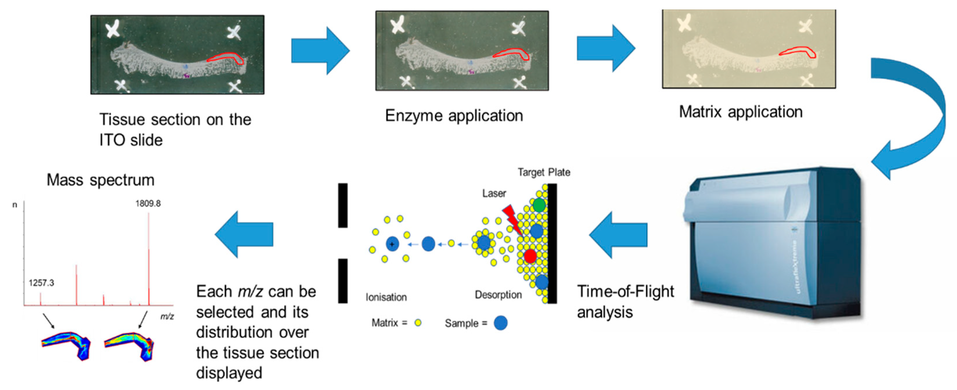
3. Proteomic-Based Imaging Approaches in OA Joint Tissue Studies
3.1. MALDI-MSI Analysis of Articular Cartilage
3.2. MALDI-MSI Analysis of Synovial Membrane
3.3. MALDI-MSI Analysis of Subchondral Bone
4. Challenges and Future Directions for Biomarkers in OA Joint-Derived Samples
5. Conclusions
Author Contributions
Funding
Acknowledgments
Conflicts of Interest
Abbreviations
| OA | osteoarthritis |
| ECM | extracellular matrix |
| MSI | mass spectrometry imaging |
| PTMs | post-translational modifications |
| MALDI-MSI | matrix-assisted laser desorption/ionisation mass spectrometry imaging |
| FF | fresh-frozen |
| FFPE | formalin-fixed paraffin-embedded |
| 2D | two-dimensional |
| LC | liquid chromatography |
| CTX-II | C-telopeptide fragments of type II collagen |
| Helix-II | type II collagen fragment |
| C2C | cartilage type II collagen |
| Coll2-1 | oxidative-related type II collagen |
| Coll2-1 NO2 | oxidative-related type II collagen in nitrated form |
| TIINE | type II collagen neoepitope |
| PIIANP | N-propeptide of collagen IIA |
| PIICP | carboxyl terminus propeptide of type II procollagen |
| COMP | cartilage oligomeric matrix protein |
| MRI | magnetic resonance imaging |
| BMI | body mass index |
| HA | hyaluronan |
| NTX-I | N-telopeptide fragments of collagen type-I |
| CTX-I | C-telopeptide fragments of collagen type-I |
| ITO | indium tin oxide |
| LC-MS/MS | liquid chromatography with tandem mass spectrometry |
| IHC | immunohistochemistry |
| ITO | indium tin oxide |
| RA | rheumatoid arthritis |
| LC-ESI-MS/MS | liquid chromatography/electrospray ionisation tandem mass spectrometry |
References
- GBD 2017 Disease and Injury Incidence and Prevalence Collaborators. Global, regional, and national incidence, prevalence, and years lived with disability for 354 diseases and injuries for 195 countries and territories, 1990-2017: A systematic analysis for the Global Burden of Disease Study 2017. Lancet 2018, 392, 1789–1858. [Google Scholar] [CrossRef]
- King, L.K.; March, L.; Anandacoomarasamy, A. Obesity & osteoarthritis. Indian J. Med. Res. 2013, 138, 185–193. [Google Scholar] [PubMed]
- Mueller, M.B.; Tuan, R.S. Anabolic/Catabolic balance in pathogenesis of osteoarthritis: Identifying molecular targets. PM&R 2011, 3, S3–S11. [Google Scholar]
- Loeser, R.F.; Goldring, S.R.; Scanzello, C.R.; Goldring, M.B. Osteoarthritis: A disease of the joint as an organ. Arthritis Rheum. 2012, 64, 1697–1707. [Google Scholar] [CrossRef]
- Carr, A.J.; Robertsson, O.; Graves, S.; Price, A.J.; Arden, N.K.; Judge, A.; Beard, D.J. Knee replacement. Lancet 2012, 379, 1331–1340. [Google Scholar] [CrossRef]
- Braun, H.J.; Gold, G.E. Diagnosis of osteoarthritis: Imaging. Bone 2012, 51, 278–288. [Google Scholar] [CrossRef]
- Chen, D.; Shen, J.; Zhao, W.; Wang, T.; Han, L.; Hamilton, J.L.; Im, H.-J. Osteoarthritis: Toward a comprehensive understanding of pathological mechanism. Bone Res. 2017, 5, 16044. [Google Scholar] [CrossRef]
- Chughtai, K.; Heeren, R.M.A. Mass Spectrometric Imaging for Biomedical Tissue Analysis. Chem. Rev. 2010, 110, 3237–3277. [Google Scholar] [CrossRef]
- Zhang, Y.; Fonslow, B.R.; Shan, B.; Baek, M.-C.; Yates, J.R. Protein Analysis by Shotgun/Bottom-up Proteomics. Chem. Rev. 2013, 113, 2343–2394. [Google Scholar] [CrossRef]
- Ly, A.; Longuespee, R.; Casadonte, R.; Wandernoth, P.; Schwamborn, K.; Bollwein, C.; Marsching, C.; Kriegsmann, K.; Hopf, C.; Weichert, W.; et al. Site-to-Site Reproducibility and Spatial Resolution in MALDI-MSI of Peptides from Formalin-Fixed Paraffin-Embedded Samples. Proteom. Clin. Appl. 2019, 13, e1800029. [Google Scholar] [CrossRef]
- Nguyen, L.T.; Sharma, A.R.; Chakraborty, C.; Saibaba, B.; Ahn, M.E.; Lee, S.S. Review of Prospects of Biological Fluid Biomarkers in Osteoarthritis. Int. J. Mol. Sci. 2017, 18, 601. [Google Scholar] [CrossRef] [PubMed]
- Lotz, M.; Martel-Pelletier, J.; Christiansen, C.; Brandi, M.L.; Bruyere, O.; Chapurlat, R.; Collette, J.; Cooper, C.; Giacovelli, G.; Kanis, J.A.; et al. Value of biomarkers in osteoarthritis: Current status and perspectives. Ann. Rheum. Dis. 2013, 72, 1756–1763. [Google Scholar] [CrossRef] [PubMed]
- Kraus, V.B.; Burnett, B.; Coindreau, J.; Cottrell, S.; Eyre, D.; Gendreau, M.; Gardiner, J.; Garnero, P.; Hardin, J.; Henrotin, Y.; et al. Application of biomarkers in the development of drugs intended for the treatment of osteoarthritis. Osteoarthr. Cartil. 2011, 19, 515–542. [Google Scholar] [CrossRef] [PubMed]
- Tanishi, N.; Yamagiwa, H.; Hayami, T.; Mera, H.; Koga, Y.; Omori, G.; Endo, N. Relationship between radiological knee osteoarthritis and biochemical markers of cartilage and bone degradation (urine CTX-II and NTX-I): The Matsudai Knee Osteoarthritis Survey. J. Bone Miner. Metab. 2009, 27, 605–612. [Google Scholar] [CrossRef] [PubMed]
- Huang, M.; Zhao, J.; Huang, Y.; Dai, L.; Zhang, X. Meta-analysis of urinary C-terminal telopeptide of type II collagen as a biomarker in osteoarthritis diagnosis. J. Orthop. Translat. 2017, 13, 50–57. [Google Scholar] [CrossRef]
- Zhai, G.; Eshghi, E.A. Biomarkers for osteoarthritis: Investigation, identification, and prognosis. Curr. Biomark Find 2012, 2, 19–28. [Google Scholar] [CrossRef]
- Hunter, D.J.; Li, J.; LaValley, M.; Bauer, D.C.; Nevitt, M.; DeGroot, J.; Poole, R.; Eyre, D.; Guermazi, A.; Gale, D.; et al. Cartilage markers and their association with cartilage loss on magnetic resonance imaging in knee osteoarthritis: The Boston Osteoarthritis Knee Study. Arthritis Res. Ther. 2007, 9, R108. [Google Scholar] [CrossRef]
- Berry, P.A.; Maciewicz, R.A.; Cicuttini, F.M.; Jones, M.D.; Hellawell, C.J.; Wluka, A.E. Markers of bone formation and resorption identify subgroups of patients with clinical knee osteoarthritis who have reduced rates of cartilage loss. J. Rheumatol. 2010, 37, 1252–1259. [Google Scholar] [CrossRef]
- Bettica, P.; Cline, G.; Hart, D.J.; Meyer, J.; Spector, T.D. Evidence for increased bone resorption in patients with progressive knee osteoarthritis: Longitudinal results from the Chingford study. Arthritis Rheum. 2002, 46, 3178–3184. [Google Scholar] [CrossRef]
- Sugiyama, S. Procollagen II C propeptide level in the synovial fluid as a predictor of radiographic progression in early knee osteoarthritis. Ann. Rheum. Dis. 2003, 62, 27–32. [Google Scholar] [CrossRef]
- Kraus, V.B.; Collins, J.E.; Hargrove, D.; Losina, E.; Nevitt, M.; Katz, J.N.; Wang, S.X.; Sandell, L.J.; Hoffmann, S.C.; Hunter, D.J. Predictive validity of biochemical biomarkers in knee osteoarthritis: Data from the FNIH OA Biomarkers Consortium. Ann. Rheum. Dis. 2017, 76, 186–195. [Google Scholar] [CrossRef] [PubMed]
- Rotterud, J.H.; Reinholt, F.P.; Beckstrom, K.J.; Risberg, M.A.; Aroen, A. Relationship between CTX-II and patient characteristics, patient-reported outcome, muscle strength, and rehabilitation in patients with a focal cartilage lesion of the knee: A prospective exploratory cohort study of 48 patients. BMC Musculoskelet. Disord. 2014, 15, 99. [Google Scholar] [CrossRef] [PubMed]
- Sowers, M.F.; Karvonen-Gutierrez, C.A.; Yosef, M.; Jannausch, M.; Jiang, Y.; Garnero, P.; Jacobson, J. Longitudinal changes of serum COMP and urinary CTX-II predict X-ray defined knee osteoarthritis severity and stiffness in women. Osteoarthr. Cartil. 2009, 17, 1609–1614. [Google Scholar] [CrossRef] [PubMed]
- Lohmander, L.S.; Atley, L.M.; Pietka, T.A.; Eyre, D.R. The release of crosslinked peptides from type II collagen into human synovial fluid is increased soon after joint injury and in osteoarthritis. Arthritis Rheum. 2003, 48, 3130–3139. [Google Scholar] [CrossRef] [PubMed]
- Kumahashi, N.; Sward, P.; Larsson, S.; Lohmander, L.S.; Frobell, R.; Struglics, A. Type II collagen C2C epitope in human synovial fluid and serum after knee injury--associations with molecular and structural markers of injury. Osteoarthr. Cartil. 2015, 23, 1506–1512. [Google Scholar] [CrossRef]
- He, G.; Chen, X.; Zhang, G.; Lin, H.; Li, R.; Wu, X. Detection of urine C2C and trace element level in patients with knee osteoarthritis. Cell Biochem. Biophys. 2014, 70, 475–479. [Google Scholar] [CrossRef]
- Bay-Jensen, A.C.; Liu, Q.; Byrjalsen, I.; Li, Y.; Wang, J.; Pedersen, C.; Leeming, D.J.; Dam, E.B.; Zheng, Q.; Qvist, P.; et al. Enzyme-linked immunosorbent assay (ELISAs) for metalloproteinase derived type II collagen neoepitope, CIIM—Increased serum CIIM in subjects with severe radiographic osteoarthritis. Clin. Biochem. 2011, 44, 423–429. [Google Scholar] [CrossRef]
- Charni, N.; Juillet, F.; Garnero, P. Urinary type II collagen helical peptide (HELIX-II) as a new biochemical marker of cartilage degradation in patients with osteoarthritis and rheumatoid arthritis. Arthritis Rheum. 2005, 52, 1081–1090. [Google Scholar] [CrossRef]
- He, Y.; Siebuhr, A.S.; Brandt-Hansen, N.U.; Wang, J.; Su, D.; Zheng, Q.; Simonsen, O.; Petersen, K.K.; Arendt-Nielsen, L.; Eskehave, T.; et al. Type X collagen levels are elevated in serum from human osteoarthritis patients and associated with biomarkers of cartilage degradation and inflammation. BMC Musculoskelet. Disord. 2014, 15, 309. [Google Scholar] [CrossRef]
- Ma, T.; Zhang, Z.; Song, X.; Bai, H.; Li, Y.; Li, X.; Zhao, J.; Ma, Y.; Gao, L. Combined detection of COMP and CS846 biomarkers in experimental rat osteoarthritis: A potential approach for assessment and diagnosis of osteoarthritis. J. Orthop. Surg. Res. 2018, 13, 230. [Google Scholar] [CrossRef]
- Germaschewski, F.M.; Matheny, C.J.; Larkin, J.; Liu, F.; Thomas, L.R.; Saunders, J.S.; Sully, K.; Whittall, C.; Boyle, Y.; Peters, G.; et al. Quantitation OF ARGS aggrecan fragments in synovial fluid, serum and urine from osteoarthritis patients. Osteoarthr. Cartil. 2014, 22, 690–697. [Google Scholar] [CrossRef] [PubMed]
- Verma, P.; Dalal, K. Serum cartilage oligomeric matrix protein (COMP) in knee osteoarthritis: A novel diagnostic and prognostic biomarker. J. Orthop. Res. 2013, 31, 999–1006. [Google Scholar] [CrossRef] [PubMed]
- Senolt, L.; Braun, M.; Olejarova, M.; Forejtova, S.; Gatterova, J.; Pavelka, K. Increased pentosidine, an advanced glycation end product, in serum and synovial fluid from patients with knee osteoarthritis and its relation with cartilage oligomeric matrix protein. Ann. Rheum. Dis. 2005, 64, 886–890. [Google Scholar] [CrossRef] [PubMed]
- Wang, Y.; Li, D.; Xu, N.; Tao, W.; Zhu, R.; Sun, R.; Fan, W.; Zhang, P.; Dong, T.; Yu, L. Follistatin-like protein 1: A serum biochemical marker reflecting the severity of joint damage in patients with osteoarthritis. Arthritis Res. Ther. 2011, 13, R193. [Google Scholar] [CrossRef] [PubMed]
- Henrotin, Y.; Gharbi, M.; Mazzucchelli, G.; Dubuc, J.E.; De Pauw, E.; Deberg, M. Fibulin 3 peptides Fib3-1 and Fib3-2 are potential biomarkers of osteoarthritis. Arthritis Rheum. 2012, 64, 2260–2267. [Google Scholar] [CrossRef]
- Li, H.; Li, L.; Min, J.; Yang, H.; Xu, X.; Yuan, Y.; Wang, D. Levels of metalloproteinase (MMP-3, MMP-9), NF-kappaB ligand (RANKL), and nitric oxide (NO) in peripheral blood of osteoarthritis (OA) patients. Clin. Lab. 2012, 58, 755–762. [Google Scholar]
- Ozler, K.; Aktas, E.; Atay, C.; Yilmaz, B.; Arikan, M.; Gungor, S. Serum and knee synovial fluid matrixmetalloproteinase-13 and tumor necrosis factor-alpha levels in patients with late stage osteoarthritis. Acta Orthop. Traumatol. Turc. 2016, 50, 670–673. [Google Scholar] [CrossRef]
- Rubenhagen, R.; Schuttrumpf, J.P.; Sturmer, K.M.; Frosch, K.H. Interleukin-7 levels in synovial fluid increase with age and MMP-1 levels decrease with progression of osteoarthritis. Acta Orthop. 2012, 83, 59–64. [Google Scholar] [CrossRef]
- Li, W.; Du, C.; Wang, H.; Zhang, C. Increased serum ADAMTS-4 in knee osteoarthritis: A potential indicator for the diagnosis of osteoarthritis in early stages. Genet. Mol. Res. 2014, 13, 9642–9649. [Google Scholar] [CrossRef]
- Hegemann, N.; Wondimu, A.; Ullrich, K.; Schmidt, M.F.G. Synovial MMP-3 and TIMP-1 levels and their correlation with cytokine expression in canine rheumatoid arthritis. Vet. Immunol. Immunopathol. 2003, 91, 199–204. [Google Scholar] [CrossRef]
- Sasaki, E.; Tsuda, E.; Yamamoto, Y.; Iwasaki, K.; Inoue, R.; Takahashi, I.; Sawada, K.; Fujita, H.; Umeda, T.; Nakaji, S.; et al. Serum hyaluronan levels increase with the total number of osteoarthritic joints and are strongly associated with the presence of knee and finger osteoarthritis. Int. Orthop. 2013, 37, 925–930. [Google Scholar] [CrossRef] [PubMed]
- Guan, J.; Liu, Z.; Li, F.; Feng, J.S.; Wang, H.J.; Chu, J.G.; Song, Y.Z.; Xie, L.; Ding, L.B. Increased Synovial Fluid YKL-40 Levels are Linked with Symptomatic Severity in Knee Osteoarthritis Patients. Clin. Lab. 2015, 61, 991–997. [Google Scholar] [CrossRef] [PubMed]
- Jordan, K.M.; Syddall, H.E.; Garnero, P.; Gineyts, E.; Dennison, E.M.; Sayer, A.A.; Delmas, P.D.; Cooper, C.; Arden, N.K. Urinary CTX-II and glucosyl-galactosyl-pyridinoline are associated with the presence and severity of radiographic knee osteoarthritis in men. Ann. Rheum. Dis. 2006, 65, 871–877. [Google Scholar] [CrossRef] [PubMed]
- Kumm, J.; Tamm, A.; Lintrop, M.; Tamm, A. Diagnostic and prognostic value of bone biomarkers in progressive knee osteoarthritis: A 6-year follow-up study in middle-aged subjects. Osteoarthr. Cartil. 2013, 21, 815–822. [Google Scholar] [CrossRef] [PubMed]
- Nikahval, B.; Nazifi, S.; Heidari, F.; Khafi, M.S.A. Evaluation of the changes of P1NP and CTX in synovial fluid and blood serum of dogs with experimental osteoarthritis. Comp. Clin. Pathol. 2016, 25, 559–563. [Google Scholar] [CrossRef]
- Ok, S.-M.; Lee, S.-M.; Park, H.R.; Jeong, S.-H.; Ko, C.-C.; Kim, Y.-I. Concentrations of CTX I, CTX II, DPD, and PYD in the urine as a biomarker for the diagnosis of temporomandibular joint osteoarthritis: A preliminary study. Cranio 2018, 36, 366–372. [Google Scholar] [CrossRef]
- Harkey, M.S.; Luc, B.A.; Golightly, Y.M.; Thomas, A.C.; Driban, J.B.; Hackney, A.C.; Pietrosimone, B. Osteoarthritis-related biomarkers following anterior cruciate ligament injury and reconstruction: A systematic review. Osteoarthr. Cartil. 2015, 23, 1–12. [Google Scholar] [CrossRef]
- Groseclose, M.R.; Andersson, M.; Hardesty, W.M.; Caprioli, R.M. Identification of proteins directly from tissue: In situ tryptic digestions coupled with imaging mass spectrometry. J. Mass Spectrom. 2007, 42, 254–262. [Google Scholar] [CrossRef]
- Casadonte, R.; Kriegsmann, M.; Zweynert, F.; Friedrich, K.; Baretton, G.; Otto, M.; Deininger, S.O.; Paape, R.; Belau, E.; Suckau, D.; et al. Imaging mass spectrometry to discriminate breast from pancreatic cancer metastasis in formalin-fixed paraffin-embedded tissues. Proteomics 2014, 14, 956–964. [Google Scholar] [CrossRef]
- Briggs, M.T.; Ho, Y.Y.; Kaur, G.; Oehler, M.K.; Everest-Dass, A.V.; Packer, N.H.; Hoffmann, P. N-glycan matrix-assisted laser desorption/ionization mass spectrometry imaging protocol for formalin-fixed paraffin-embedded tissues. Rapid Commun. Mass Spectrom. 2017, 31, 825–841. [Google Scholar] [CrossRef]
- Cillero-Pastor, B.; Eijkel, G.B.; Kiss, A.; Blanco, F.J.; Heeren, R.M. Matrix-assisted laser desorption ionization-imaging mass spectrometry: A new methodology to study human osteoarthritic cartilage. Arthritis Rheum. 2013, 65, 710–720. [Google Scholar] [CrossRef] [PubMed]
- Peffers, M.J.; Cillero-Pastor, B.; Eijkel, G.B.; Clegg, P.D.; Heeren, R.M. Matrix assisted laser desorption ionization mass spectrometry imaging identifies markers of ageing and osteoarthritic cartilage. Arthritis Res. Ther. 2014, 16, R110. [Google Scholar] [CrossRef] [PubMed]
- Kriegsmann, M.; Seeley, E.H.; Schwarting, A.; Kriegsmann, J.; Otto, M.; Thabe, H.; Dierkes, B.; Biehl, C.; Sack, U.; Wellmann, A.; et al. MALDI MS imaging as a powerful tool for investigating synovial tissue. Scand. J. Rheumatol. 2012, 41, 305–309. [Google Scholar] [CrossRef] [PubMed]
- Cillero-Pastor, B.; Eijkel, G.B.; Blanco, F.J.; Heeren, R.M. Protein classification and distribution in osteoarthritic human synovial tissue by matrix-assisted laser desorption ionization mass spectrometry imaging. Anal. Bioanal. Chem. 2015, 407, 2213–2222. [Google Scholar] [CrossRef]
- Briggs, M.T.; Kuliwaba, J.S.; Muratovic, D.; Everest-Dass, A.V.; Packer, N.H.; Findlay, D.M.; Hoffmann, P. MALDI mass spectrometry imaging of N-glycans on tibial cartilage and subchondral bone proteins in knee osteoarthritis. Proteomics 2016, 16, 1736–1741. [Google Scholar] [CrossRef]
- Sanchez, C.; Bay-Jensen, A.C.; Pap, T.; Dvir-Ginzberg, M.; Quasnichka, H.; Barrett-Jolley, R.; Mobasheri, A.; Henrotin, Y. Chondrocyte secretome: A source of novel insights and exploratory biomarkers of osteoarthritis. Osteoarthr. Cartil. 2017, 25, 1199–1209. [Google Scholar] [CrossRef]
- Lories, R.J.; Luyten, F.P. The bone-cartilage unit in osteoarthritis. Nat. Rev. Rheumatol. 2011, 7, 43–49. [Google Scholar] [CrossRef]
- Neogi, T. Clinical significance of bone changes in osteoarthritis. Ther. Adv. Musculoskelet. Dis. 2012, 4, 259–267. [Google Scholar] [CrossRef]
- Mareddy, S.; Broadbent, J.; Crawford, R.; Xiao, Y. Proteomic profiling of distinct clonal populations of bone marrow mesenchymal stem cells. J. Cell Biochem. 2009, 106, 776–786. [Google Scholar] [CrossRef]
- Urita, A.; Matsuhashi, T.; Onodera, T.; Nakagawa, H.; Hato, M.; Amano, M.; Seito, N.; Minami, A.; Nishimura, S.-I.; Iwasaki, N. Alterations of high-mannose type N-glycosylation in human and mouse osteoarthritis cartilage. Arthritis Rheum. 2011, 63, 3428–3438. [Google Scholar] [CrossRef]
- Angel, P.M.; Mehta, A.; Norris-Caneda, K.; Drake, R.R. MALDI Imaging Mass Spectrometry of N-glycans and Tryptic Peptides from the Same Formalin-Fixed, Paraffin-Embedded Tissue Section. Methods Mol. Biol. 2018, 1788, 225–241. [Google Scholar] [PubMed]
- Ruiz-Romero, C.; Rego-Perez, I.; Blanco, F.J. What did we learn from ‘omics’ studies in osteoarthritis. Curr. Opin. Rheumatol. 2018, 30, 114–120. [Google Scholar] [CrossRef] [PubMed]
- Turiak, L.; Shao, C.; Meng, L.; Khatri, K.; Leymarie, N.; Wang, Q.; Pantazopoulos, H.; Leon, D.R.; Zaia, J. Workflow for combined proteomics and glycomics profiling from histological tissues. Anal. Chem. 2014, 86, 9670–9678. [Google Scholar] [CrossRef] [PubMed]
- Heijs, B.; Holst, S.; Briaire-de Bruijn, I.H.; van Pelt, G.W.; de Ru, A.H.; van Veelen, P.A.; Drake, R.R.; Mehta, A.S.; Mesker, W.E.; Tollenaar, R.A.; et al. Multimodal Mass Spectrometry Imaging of N-Glycans and Proteins from the Same Tissue Section. Anal. Chem. 2016, 88, 7745–7753. [Google Scholar] [CrossRef] [PubMed]


| OA-Affected Tissue | Sample Type | Molecule Type | Biomarker(s) | References |
|---|---|---|---|---|
| Articular Cartilage | Synovial Fluid | Type II collagen | C-propeptide of collagen type II (PIICP) | Sugiyama et al., 2003 [20] |
| Serum | N-propeptide IIA of type II collagen (PIIANP) | Kraus et al., 2017 [21] | ||
| Urine, Synovial Fluid | C-terminal telopeptide of collagen type II (CTX-II) | Rotterud et al., 2014 [22]; Sowers et al., 2009 [23]; Lohmander et al., 2003 [24] | ||
| Serum, Synovial Fluid, Urine | Type II collagen cleavage product (C2C) | Kumahashi et al., 2015 [25]; He et al., 2014 [26] | ||
| Serum | Matrix metalloproteinase-derived fragment of type II collagen (CIIM) | Bay-Jensen et al., 2011 [27] | ||
| Urine | Helical peptide of type II collagen (HELIX-II) | Charni et al., 2005 [28] | ||
| Serum | Type X collagen | C-terminus of collagen type X (C-Col-10) | He et al., 2014 [29] | |
| Serum | Aggrecan | Aggrecan chondroitin sulfate epitope 846 | Ma et al., 2018 [30] | |
| Synovial Fluid | Aggrecanase-generated aggrecan fragment with the ARGS neoepitope | Germaschewski et al., 2014 [31] | ||
| Serum | Non-collagenous and aggrecan proteins | Cartilage oligomeric matrix protein (COMP and its deaminated form D-COMP) | Ma et al., 2018 [30]; Verma et al., 2013 [32]; Sowers et al., 2009 [23] | |
| Serum, Synovial Fluid | Pentosidine | Senolt et al., 2005 [33] | ||
| Serum | Follistatin-like protein (FSTL-1) | Wang et al., 2011 [34] | ||
| Serum | Fibulin (peptides of fibulin 3, Fib3-1, -2) | Henrotin et al., 2012 [35] | ||
| Serum | Proteolytic enzymes | Matrix metalloproteinases (MMP-3, -9) | Li et al., 2012 [36] | |
| Synovial Fluid | Matrix metalloproteinases (MMP-1, -13) | Ozler et al., 2016 [37]; Rubenhagen et al., 2012 [38] | ||
| Serum | A disintegrin and metalloproteinase with thrombospondin-like motif 4 (ADAMTS-4) | Li et al., 2014 [39] | ||
| Synovial Fluid | Proteolytic enzyme inhibitors | Tissue inhibitor of matrix metalloproteinase (TIMP-1) | Hegemann et al., 2003 [40] | |
| Synovium | Serum | Non-collagenous proteins | Hyaluronan (Hyaluronic acid; HA) | Sasaki et al., 2013 [41] |
| Synovial Fluid | Cartilage glycoprotein 39 (YKL-40) | Guan et al., 2015 [42] | ||
| Urine | Type III collagen | Glucosyl-galactosyl pyridinoline (Glc-Gal-PYD) | Jordan et al., 2006 [43] | |
| Subchondral Bone | Serum | Type I collagen | Aminoterminal propeptide of collagen type I (PINP) | Kumm et al., 2013 [44] |
| Serum, Urine | C-telopeptide fragment of collagen type-I (CTX-I) | Nikahval et al., 2016 [45]; Bettica et al., 2002 [19] | ||
| Urine | N-telopeptide fragment of collagen type-I (NTX-I) | Bettica et al., 2002 [19] | ||
| Urine | Non-isomerised C-telopeptide fragment of collagen type-I (Alpha-CTX-I); Isomerised C-telopeptide fragment of collagen type-I (Beta-CTX-I) | Kraus et al., 2017 [21] | ||
| Urine | Pyridinoline (PYD) | Ok et al., 2018 [46] | ||
| Urine | Deoxypyridinoline (DPD) | Ok et al., 2018 [46] | ||
| Serum | Non-collagenous protein | Osteocalcin (OC) | Kumm et al., 2013 [44] | |
| Urine | Mid-fragments of osteocalcin (Mid OC) | Kumm et al., 2013 [44] |
| OA-Affected Tissue | Origin | Disease | Age in Years | Specific Marker(s) Identified | Validation | Reference |
|---|---|---|---|---|---|---|
| Articular Cartilage | Human | OA (n = 10) vs. HC (n = 10) | OA (51–84 years old), HC (51–91 years old) | Aggrecan core protein (ACAN), Biglycan (BGN), Cartilage intermediate layer protein 1 (CILP-1), Cartilage oligomeric matrix protein (COMP), Collagen alpha-1(II) chain (COL-2A-1), Decorin (DCN), Fibromodulin (FMOD), Fibronectin (FN), Prolargin, Protein embryonic large molecule derived from yolk sac (ELY-S) | IHC | Cillero-Pastor et al., 2013 [51] |
| Horse | OA (n = 3) vs. Young (n = 3) and Old (n = 3) | OA (greater than 15 years old; 52 years in human), Young (4 years old; 14 years in-human), Old (greater-than 15 years old; 52 years in human) | Biglycan (BGN), Cartilage intermediate-layer protein-1 (CILP-1), Cartilage oligomeric-matrix protein (COMP), Collagen alpha-1(II)-chain (COL-2A-1), Collectin-43 (CL-43), Chondroadherin, Fibronectin (FN), Matrilin-3 (MATN-3), Melanoma inhibitory activity-3 (MIA-3) | IHC | Peffers et al., 2014 [52] | |
| Synovial Membrane | Human | OA (n = 3) vs. RA (n = 3) | NA | Calgranulins, Defensins, Thymosins | NA | Kriegsmann et al., 2012 [53] |
| Human | OA (n = 3) vs. HC (n = 3) | OA (69–82 years old), HC (60–78 years old) | Actin aortic smooth muscle (ACTA), Biglycan (BGN), Fibronectin (FN), Haemoglobin subunit-alpha-2 (HBA-2), Haemoglobin subunit beta (HBB) | IHC | Cillero-Pastor et al., 2015 [54] | |
| Subchondral Bone | Human | OA with bone marrow lesions (n = 2) vs. OA without bone marrow lesions (n = 1) | One male aged 52 years, two females aged 68 and 74 years | N-glycans | LC-ESI MS/MS | Briggs et al., 2016 [55] |
© 2020 by the authors. Licensee MDPI, Basel, Switzerland. This article is an open access article distributed under the terms and conditions of the Creative Commons Attribution (CC BY) license (http://creativecommons.org/licenses/by/4.0/).
Share and Cite
Lee, Y.-R.; Briggs, M.T.; Condina, M.R.; Puddy, H.; Anderson, P.H.; Hoffmann, P.; Kuliwaba, J.S. Mass Spectrometry Imaging as a Potential Tool to Investigate Human Osteoarthritis at the Tissue Level. Int. J. Mol. Sci. 2020, 21, 6414. https://doi.org/10.3390/ijms21176414
Lee Y-R, Briggs MT, Condina MR, Puddy H, Anderson PH, Hoffmann P, Kuliwaba JS. Mass Spectrometry Imaging as a Potential Tool to Investigate Human Osteoarthritis at the Tissue Level. International Journal of Molecular Sciences. 2020; 21(17):6414. https://doi.org/10.3390/ijms21176414
Chicago/Turabian StyleLee, Yea-Rin, Matthew T. Briggs, Mark R. Condina, Hamish Puddy, Paul H. Anderson, Peter Hoffmann, and Julia S. Kuliwaba. 2020. "Mass Spectrometry Imaging as a Potential Tool to Investigate Human Osteoarthritis at the Tissue Level" International Journal of Molecular Sciences 21, no. 17: 6414. https://doi.org/10.3390/ijms21176414
APA StyleLee, Y.-R., Briggs, M. T., Condina, M. R., Puddy, H., Anderson, P. H., Hoffmann, P., & Kuliwaba, J. S. (2020). Mass Spectrometry Imaging as a Potential Tool to Investigate Human Osteoarthritis at the Tissue Level. International Journal of Molecular Sciences, 21(17), 6414. https://doi.org/10.3390/ijms21176414