Review

Alterations of Expression of the Serotonin 5-HT4 Receptor in Brain Disorders

1
Department of Molecular, Cellular and Biomedical Sciences, CUNY School of Medicine, New York, NY 10031, USA
2
Ph.D. Programs in Biochemistry and Biology, The Graduate Center, City University of New York, New York, NY 10031, USA
*
Author to whom correspondence should be addressed.
Int. J. Mol. Sci. 2018, 19(11), 3581; https://doi.org/10.3390/ijms19113581
Submission received: 14 October 2018 / Revised: 3 November 2018 / Accepted: 6 November 2018 / Published: 13 November 2018
(This article belongs to the Special Issue Molecular Psychiatry)

Abstract

:
The serotonin 4 receptor, 5-HT4R, represents one of seven different serotonin receptor families and is implicated in a variety of physiological functions and their pathophysiological variants, such as mood and depression or anxiety, food intake and obesity or anorexia, or memory and memory loss in Alzheimer’s disease. Its central nervous system expression pattern in the forebrain, in particular in caudate putamen, the hippocampus and to lesser extent in the cortex, predispose it for a role in executive function and reward-related actions. In rodents, regional overexpression or knockdown in the prefrontal cortex or the nucleus accumbens of 5-HT4R was shown to impact mood and depression-like phenotypes, food intake and hypophagia; however, whether expression changes are causally involved in the etiology of such disorders is not clear. In this context, more data are emerging, especially based on PET technology and the use of ligand tracers that demonstrate altered 5-HT4R expression in brain disorders in humans, confirming data stemming from post-mortem tissue and preclinical animal models. In this review, we would like to present the current knowledge of 5-HT4R expression in brain regions relevant to mood/depression, reward and executive function with a focus on 5-HT4R expression changes in brain disorders or caused by drug treatment, at both the transcript and protein levels.

1. Introduction

5-HT receptors are composed of 7 families (5-HT1–7 receptors), comprising 14 structurally and pharmacologically distinct 5-HT receptor subtypes [1]. All receptors are G-protein-coupled, with the exception of the 5-HT3R that belongs to the superfamily of ligand-gated ion channels. Members of all 7 receptor families are expressed in the brain: 5-HT1 receptors are Gαi/0-coupled and two receptors of this family, 5-HT1aR and 5-HT1bR, have an important function as somatodendritic autoreceptors expressed on neurons of the raphe nuclei that produce 5-HT, but they are also expressed as postsynaptic heteroreceptors in several brain areas [2]. The three members of the Gαq/11-coupled 5-HT2R family have well defined roles in the periphery such as in the vascular system and muscle contraction; however, their function in the brain is not well understood A potential link between a 5-HT2CR allele and vulnerability to affective disorders has been reported, and a number of antipsychotics have inverse agonist activity at 5-HT2C receptors [3]. 5-HT2CR KO mice are highly obese and suffer from epilepsies [4]. 5-HT2aR mediates the hallucinatory and psychotic action of psychedelic drugs such as LSD or psylocibin [5]. Human brain Gαi/o-coupled 5-HT5R expression is localized to the cerebral cortex, hippocampus, cerebellum, and a role in mood and major depression was postulated, using pharmacological tools and knockout mice [6]. 5-HT6Rs are postsynaptic Gαs-coupled receptors strongly expressed in the striatum, nucleus accumbens and cortex, and moderately in the hippocampus, amygdala, and hypothalamus. They control among others central cholinergic function [6]. The Gαs-coupled 5HT7R is mainly expressed in the limbic system, and a potential role in sleep, circadian rhythmic activity and mood has been suggested [6]. Among all 5-HT receptors, the type 3 receptor is the only ligand-gated ion channel receptor triggering rapid depolarization via the opening of non-selective cation channels. 5-HT3R expression in the forebrain is low, but higher levels are present in the hippocampus and amygdala [6].
5-HT4R was initially identified in cultured mouse colliculi cells and guinea pig brain using a functional cAMP stimulation assay [7]. In 1995, its cloning was reported [8]. Two different splicing variants, a short one, found in the striatum and a long one in the whole brain [8] were initially described, while others found the short form also present more universally in the brain [9].
Expression in the brain is greatest in the basal ganglia, the hippocampal formation and the cortex, as shown in human and rat brain [10,11]. 5-HT4R is also widely distributed in the body. Outside the CNS, it is found along the gastrointestinal tract (esophagus, ileum and colon) [12]. It is also present in the bladder, the heart and the adrenal glands. 5-HT4 receptors are well known for their peripheral effects on the gastrointestinal tract, and are targets in the treatment of dyspepsia, gastroesophageal reflux disease, gastroparesis or irritable bowel syndrome [13,14]. Serotonin affects heart contractility through 5-HT4R which is expressed in the human and pig atrium and ventricle, while, interestingly, in the rat it is only expressed in the atrium [15]. 5-HT4R activation leads to the contraction of heart but also to tachycardia and arrhythmia [15]. The cardiac contractile effects of 5-HT4R are restricted to human and pig atria and are absent from a large number of laboratory animals, such as rat, guinea pig, rabbit and frog [16]. 5-HT4R was also shown to be overexpressed in the cortex of the adrenal gland of a subtype of Cushing syndrome patients, a condition caused by cortisone hyper-production [17].
Compared to other serotonin receptors, the gene encoding 5-HT4R (htr4) is large and its architecture is complicated, with 38 exons spaced over 700 kb [18]. As a G-protein coupled receptor, 5-HT4R signals through both G protein-dependent and G protein-independent pathways. The major G protein engaged by 5-HT4R signaling is Gαs, leading to the activation of the cAMP/PKA pathway [19]. The G-protein independent non-canonical pathway activates Src and ERK kinases, leading to pERK1/2 phosphorylation [20].
5-HT4R KO mice develop normally, with no differences in body weight, metabolism, social behavior, or sleep pattern [21]. However, when stressed they exhibit reduced hypophagia [21] and re-expression of 5-HT4R in the medial prefrontal cortex rescues this phenotype [22]. In mice, 5-HT4R was also shown to link appetite and feeding to addiction-related behaviors since 5-HT4R activation in the nucleus accumbens provokes anorexia and hyperactivity, concurrently upregulating a gene induced by cocaine and amphetamine (CART) while knockdown thereof inhibits MDMA-induced hyperactivity [23].
One of the earliest functions attributed to 5-HT4R in rodents is its excitatory effect on acetylcholine release in the frontal cortex and the hippocampus [24,25] which was linked to its role in enhancing memory and cognition [26,27,28,29]. For example, a two-week treatment with 5-HT4R partial agonist RS67333 improved memory in the object recognition test in mice [30]. Olfactory associative learning was enhanced by another partial agonist (SL65.0155) in rats [31]. Other paradigms assessing social memory, autoshaping and spatial and place learning, showed a memory enhancing effect of 5-HT4R stimulation [29,32,33]. Conversely, receptor antagonists induced a consistent deficit in (olfactory) associative memory formation [34,35], and weakened passive avoidance memory [36]. Paralleling these behavioral changes are structural plasticity effects of potentiated learning-induced dendritic spine growth in the hippocampus in mice, an effect which is abolished by 5-HT4R inhibition [37].
5-HT4 receptors have also been found to modulate GABA and dopamine release [18,26]. Serotonin depolarizes globus pallidus neurons, increases their firing rate and alters GABA release in a 5-HT4R-dependent manner involving pre- and postsynaptic mechanisms [38]. In guinea pig, the 5-HT4R agonist BIMU-8 increased GABA release from hippocampus indirectly via cholinergic muscarinic receptors [39]. 5-HT4 receptors exert excitatory control on DA release in the striatum, while a receptor antagonist blocks this effect [40]. In freely moving rats, the 5-HT4 antagonist GR125487 significantly reduced the nigrostriatal haloperidol-induced but not basal DA outflow without affecting the mesoaccumbal DA release, indicating that 5-HT4R exerts facilitatory control under activated conditions [41]. This finding is also important in the context of Parkinson’s disease where the substantia nigra is selectively vulnerable to degeneration compared to the VTA, leading to a depletion in striatal dopamine. In a rat model of PD, the 5-HT4R agonist prucalopride selectively enhanced L-DOPA-stimulated DA release in the rat SNr and the PFC but not in the hippocampus or the striatum [42].
5-HT4R also impacts global serotonergic tone. 5-HT4R KO mice have diminished tissue levels of 5-HT and its main metabolite, 5-HIAA, increased serotonin transporter (SERT) at the protein and transcript levels, as well as decreased 5-HT1AR binding sites [43]. 5-HT4R is a component of a feedback loop projecting from the PFC to the dorsal raphe nuclei (DRN). More specifically, in mice, systemic 5-HT4R stimulation or overexpression of 5-HT4R in the mPFC increased the firing rate of DRN neurons, thus creating a positive feedback PFC-DRN loop involving 5-HT4R activation in cortical projections neurons, glutamate release in the DRN and enhanced DRN firing [44,45,46].
5-HT4R is a major candidate in mediating antidepressant drug action. As early as 1997, a role of 5-HT4R in anxiety-like behavior was described in rats [47]. More recently, this topic has received more interest, possibly due to the need to identify novel, fast acting antidepressant drugs. Indeed, it was described in rodents that subchronic (3 days) treatment with 5-HT4R agonist yields behavioral as well as biochemical responses in the hippocampus (CREB phosphorylation, neurogenesis) that are comparable to responses to treatment with SSRIs over 3 weeks [48], possibly through its action in the above mentioned PFC-DRN feedback loop [44,45,46].
These findings clearly indicate that 5-HT4R is a major regulator of the homeostasis of several neurotransmitter systems, implying a role in brain disorders such as Alzheimer’s, Huntington’s, Parkinson’s diseases or Major Depressive Disorder. Our review aims at summarizing the current knowledge of 5-HT4R expression in the brain. We also want to present knowledge on cell-type specific expression, which has not yet been studied extensively, partly due to the lack of immunohistochemistry-competent antibodies as well as resolution limits of binding experiments in brain slices with radioactive antagonists.

2. Promoter Studies and Transcript Variants

Surprisingly little is known about the transcriptional regulation of the htr4 gene across tissues. The human 5-HT4 receptor gene is located on chromosome 5 (5q31–q33) and contains five exons and eight alternatively spliced cassettes that code for the internal and C-terminal splice variants [16]. Human htr4 mRNA is transcribed from a very complex gene encompassing 38 exons spanning over 700 kb [18], and multiple C-terminal isoforms are expressed in specific tissues in the CNS. To date, it is not known how 5-HT4 receptor expression is regulated in the brain, and so far we have only partial knowledge about the promoter, derived from human atrial tissue and placenta [16,49]. In the heart, the major transcription start site of the htr4 gene is located at −3185 bp upstream of the first start codon [16]. In placenta, the 5′-UTR is even longer, spanning over 5100 bp upstream from the translation start site [49]. The different 5′-UTRs upstream of the translation initiation codon are interesting since they may hold an additional key to understand region and cell-type specific regulation of protein expression.
The human promoter lacks TATA and CAAT canonical motifs, but contains several transcription factors binding sites. Transient transfection assays with human 5-HT4 receptor promoter-luciferase constructs identified an approx. 1.2 kb fragment of 5′-non-transcribed sequence as promoter in human cell lines but not monkey COS-7 cells [16] indicating that there is a tissue-specific expression of yet unknown transcription factors. We found in mouse brain that there is a region-specific negative transcriptional regulation of htr4 exerted by the kinase CK2. Examination of conditional mouse knockouts of CK2 in the hippocampus, striatum and the cortex indicated an upregulation of 5-HT4R mRNA selectively in the cortex [46]. Furthermore, in luciferase assays, using a 4 kb element upstream of the mouse gene fused to luciferase cDNA, expression was promoted when CK2 was inhibited or knocked down in human HeLa cells but not in Hek293 or monkey COS-7 cells, again underlining the importance of tissue-specific transcription factors.
Instead of the TATA box, Maillet et al. described the presence of a sequence in the human gene (TTCACTTT) that can function as a core promoter sequence similarly to the TATA box [16]. For other species, no promoter studies were performed.
There are differential transcription initiation sites in different tissues such as human heart and placenta while for the brain no such data are yet available. While the transcription initiation start site does not affect the protein-coding region, it may alter the transcription efficiency and the expression pattern of 5-HT4R. It is hypothesized that such a long 5′-UTR reduces RNA translation and leads to low levels of expressed transcripts by causing premature initiation at a wrong ATG and preventing the ribosome from reaching the correct start codon [16].
Taken together, in particular in the human brain, there is a lack of data about the 5′-UTR, the promoter and the transcription factors that are active at the promoter for htr4.

3. SNPs in Non-Coding Regions

In addition to the 5′-UTRs, isoforms can also vary in the 3′-UTR. These 3′-UTRs are targets for post-transcriptional regulation by non-coding RNAs such as miRNAs. Within the 3′-UTR of the 5-HT4R (b) and (i) isoforms from the GI tract from humans with irritable bowel syndrome (IBS), a single nucleotide polymorphism, termed 5-HT4R (b_2) was found to be predominantly present in a subtype of IBS patients. This isoform lacks two of the three miRNA binding sites for miR-16 family/miR-103/107 and, compared to the full length 5-HT4R (b) isoform, its expression yielded higher 5-HT4R protein levels. It was further shown that miR-16 and miR-103 are responsible for the downregulation of the transcript in vitro which is impaired in the 5-HT4R (b_2) mutant [50].
Another miRNA, Let-7a, was also postulated to have the potential to regulate 5-HT4R [51].
Several genome wide association studies (GWAS) and meta-analyses have associated twelve intronic SNPs in the non-coding region of human htr4 with pulmonary function [52,53]. The same SNPs have been associated with the clinical phenotypes of airflow obstruction and COPD and asthma [53,54]. A SNP in a non-coding region could affect transcriptional regulation or generate a splicing signal. In this context, the pulmonary function of 5-HT4R KO mice was found to exhibit higher baseline lung resistance, confirming a role of 5-HT4R in airway diseases [55]. No mechanistic studies have yet been performed to understand the impact of the described SNP on transcription and splicing.

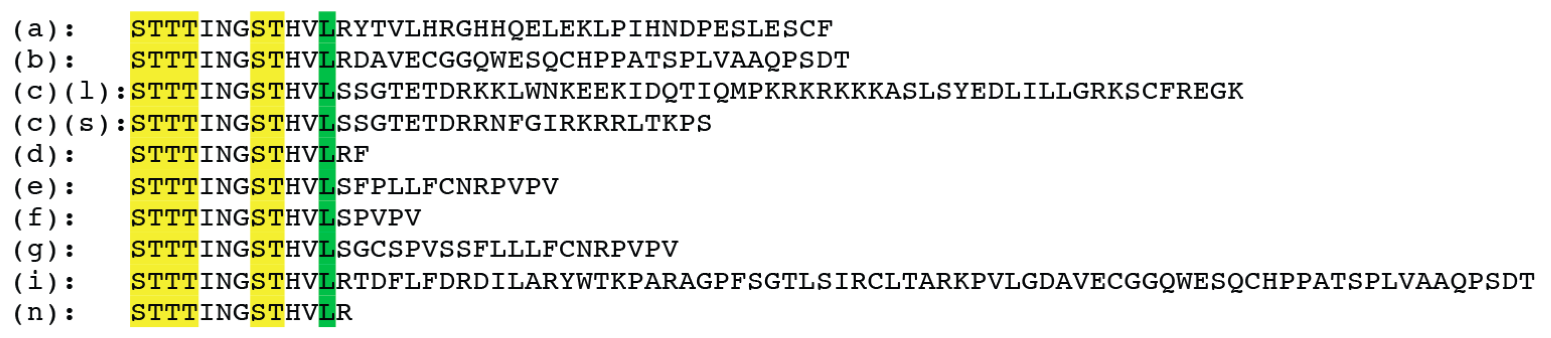
4. Isoforms and Alternative Splicing

In contrast to promoter-dependent transcriptional initiation sites which will still yield the same transcript but alter expression levels, splicing affects the protein sequence.
Since the first publication in 1995 which described a short and a long isoform, several other isoforms were discovered: There are at least 11 human 5-HT4 receptor splice variants (a–i,n) [18,56,57,58,59]. All splice variants differ at the C-terminus with the exception of 5-HT4R (h) which is an internal splice variant with an insert in the 2nd extracellular loop [60], (Figure 1) and the (n) isoform which lacks the C-terminal exon [61].
Human 5-HT4 receptor isoforms (a–i and n) are highly expressed in the central nervous system [18,56,61]. Isoform (b) is the most abundant form in the CNS and periphery, and is expressed in the caudate nucleus, putamen, amygdala, pituitary gland, and small intestine. Isoform (a) is highly expressed in the amygdala, hippocampus, nucleus accumbens, and caudate nucleus and at lower levels in the small intestine, the atrium, and pituitary gland. Isoform (c) is highly expressed in the pituitary gland and small intestine and to a lesser degree in the caudate nucleus, hippocampus, and putamen. Isoform (d) is not present in the CNS but is found in the small intestine [18,61,62]. Isoform (g) seems to be highly expressed in the hypothalamus and cortex [63]. The (n) variant, which lacks the alternatively spliced C-terminal exon, is abundantly expressed in human peripheral tissues and brain regions involved in mood disorders (frontal cortex, hippocampus) [61].
Mice are currently thought to have five [64] and rats four isoforms, with the fourth, (c1) isoform expressed in the gastrointestinal tract [59,63]. In rat brain, no significant difference in expression between the long and short variants has been found by ISH [65]. The C-terminal sequences will determine the baseline activity (with the shorter isoforms being more active) or the ability to recruit binding partners such as β-arrestins and GRKs, sorting nexins or the NHERF PDZ adaptor protein [19,66,67] This will affect internalization kinetics which are different between isoforms [68]. Finally, isoforms can differ in their G protein coupling, since the 5-HT4R (b) isoform can couple via Gαi as well as Gαs [69].
To date, no specific isoform has been linked to a brain disorder; however, an interaction cannot be excluded since such studies have not been performed and would be very challenging. Most human studies using PET technology or radioactive labeling are based on ligands which cannot distinguish between isoforms. Quantitative RT-PCR was used to detect different isoforms and their expression in the rodent brain; however, no studies in disease models have employed this approach. The fact that mice or rats do not express the same isoforms than humans suggests that fine tuning of 5-HT4R signaling through a differential expression of longer or short, more active versus less active isoforms, may occur in different species.

5. Post-Translational Regulation

5.1. Phosphorylation

The amount of membrane-localized and active GPCR is a result of the ratio between receptor endocytosis and recycling. Endocytosis is initiated through (S/T) phosphorylation of GPCRs in their intracellular domains by G protein-coupled receptor kinases (GRKs) and second messenger kinases such as PKA or PKC [70]. Binding of arrestins to GRK-phosphorylated receptors results in receptor desensitization [71] and internalization [72,73,74,75,76].
Fourteen phosphosites in the 3rd intracellular loop and in the C-terminal tail of 5-HT4R that was heterologously expressed in retinal rod cells of the mouse were identified [77]; however, the identity of the kinases has not been determined. Neither has it been tested whether the phosphorylation of these sites is activity dependent.
In Hek293 cells, it was shown that GRK2 phosphorylates and desensitizes 5-HT4R resulting in downregulation of the cAMP/PKA pathway, while GRK5-mediated 5-HT4R phosphorylation resulted in reduced inhibition of ERK phosphorylation [19,78].

5.2. Palmitoylation

Palmitoylation is a lipid modification in which a cysteine SH group undergoes esterification with a palmitoyl group, generating an anchor to the lipid bilayer of the plasma membrane. This modification is readily reversible and, similar to phosphorylation/dephosphorylation, allows for rapid regulation of protein function, affecting GCPR endocytosis, phosphorylation, desensitization and ultimately cellular signaling. Biochemical studies in insect (Sf9) and mammalian cells (Cos7) showed that several 5-HT receptors (5-HT1aR, 5-HT1bR, 5-HT4R and 5-HT7R) are palmitoylated in their C-terminal tails. The mouse 5-HT4R (a) variant is palmitoylated at 3 highly conserved cysteine sites and at a C-terminal cysteine that is variant-specific. Palmitoylation near or close to protein-protein interaction motifs will affect the binding properties of the receptor, impact on constitutive activity or internalization via β-arrestin-2 [79,80].

5.3. Glycosylation

Only one study describes two putative N–linked glycosylation sites that conform to the consensus sequence N–X–S/T (X being any amino acid but proline) for glycosylation. These are located on the extracellular side of 5-HT4R, one at the N-terminus and one in the 2nd extracellular loop [77].
What is clearly missing in our understanding of all described post-translational modifications are data generated from physiologically expressed 5-HT4R such as in mouse brain, a comparison between brain regions and an analysis in response to drug treatment or of brain disease models. Finally, the functionality of each of these modifications should be addressed, in particular on their effect on protein stability and receptor homeostasis.

6. Basal Expression

6.1. Transcript Level

5-HT4R transcript expression in rodents mainly stems from in situ hybridization (ISH] experiments: In rat brain slices, ISH probes showed strong expression in the basal ganglia (caudate putamen, ventral striatum], olfactory tubercle, medial habenula and hippocampal formation while none was detected in globus pallidus and substantia nigra [65]. Similarly, human postmortem brains showed highest levels of 5-HT4 receptor mRNA in caudate nucleus, putamen, nucleus accumbens, and the hippocampal formation but none in globus pallidus and substantia nigra [10].
A brain-wide comprehensive appraisal of cell-specific expression is still warranted; however, some evidence has been published: Dual-label in situ hybridization for 5-HT4R and neuronal markers suggests expression in basal forebrain GABAergic parvalbumin synthesizing and glutamatergic cells and in glutamatergic pyramidal neurons in the medial prefrontal cortex and hippocampus of rat and guinea pig (CA1, CA3) [62,81]. 5-HT4R mRNA is present in 60% of rat PFC pyramidal neurons of the frontal cortex as assessed by single cell mRNA/cDNA profiling [82,83].
In rat hippocampal slices, the 5-HT4R agonist, cisapride, leads to increased hippocampal pyramidal cell activity and serotonin release, indirectly indicating that 5-HT4R is expressed in these cells [84].

6.2. Protein Level

Our knowledge on 5-HT4R protein expression stems to a large degree from radioactive ligand binding studies which for the most part has mirrored results of ISH studies. Indeed, a large number of radioligands exist that are specific to 5-HT4R.
High densities of [3H]-GR 113808 or [125I]-SB 207710 binding sites are present in the ventral and dorsal striatum, substantia nigra, globus pallidus and ventral pallidum, interpeduncular nucleus, islands of Calleja, and olfactory tubercle in guinea pig, mouse and rat brain, lower densities are found in the hippocampus, septal region, neocortex, amygdala and colliculi as well as habenular and several thalamic and hypothalamic nuclei [65,85,86,87]. [125I]-SB 207710 binding in the caudate putamen shows a rostrocaudal and mediolateral increasing gradient of receptor densities, paralleling that observed for mRNA localization [65].
Kainic acid injection into the caudate-putamen of rats to destroy GABAergic striatal projection neurons resulted in a dramatic decrease of radioactive ligand binding, suggesting that 5-HT4R is expressed in these neurons [11]. Similarly, 6-OHDA-lesion of dopaminergic neurons did not lead to a reduction in radioactive ligand binding but only to increased binding in the caudate putamen and globus pallidus. This allows the conclusion that 5-HT4R expression does not occur in DA neurons of the SN [11]. These studies were confirmed by comparing ISH and radioligand labeling data: The presence of mRNA in the rat caudate putamen and its absence in substantia nigra pars compacta and the globus pallidus suggests again that receptors found in binding studies in the caudate putamen and globus pallidus are synthesized by striatonigral and striatopallidal cells [62]. Comparison of mRNA distribution with receptor distribution as visualized with [125I]-SB 207710 further indicates that 5-HT4 receptors are localized somatodendritically (e.g., in caudate putamen) and on axon terminals (e.g., in substantia nigra and globus pallidus) [65,88].
Transgenic Bac-GFP mice where GFP is expressed under the 5-HT4R promoter are enabling a highly detailed look at protein expression in individual cells and confirm moderate to strong expression in the olfactory bulb, cerebral cortex, subicular cortex, hippocampus, striatum, globus pallidus, midbrain, pons medulla, cerebellum and weak expression in the piriform cortex, basal forebrain and the thalamus (Gensat Founder AU103). Dual immunohistochemical analysis showed expression of GFP in GABAergic spiny projection neurons but not in striatal interneurons [89]. Another transgenic mouse line where the β-galactosidase gene was knocked-in at the htr4 gene locus shows LacZ localization in mature but not immature granule cells as suggested by staining with the neural marker, NeuN, and calbindin (mature granule cell marker) [90]. Another study confirmed that 5-HT4 receptors are expressed in efferent GABAergic neurons of the nucleus accumbens projecting to the lateral hypothalamus [23].
Species-specific differences of 5-HT4R protein expression were found between mouse/rat and guinea pig in the globus pallidus, substantia nigra and interpeduncular nucleus [87].
Using [3H]-prucalopride and [3H]-GR116712 or [125I]-SB 207710 in binding studies of human post-mortem brain slices, the highest densities were found in the basal ganglia (caudate nucleus, putamen, nucleus accumbens, globus pallidus, substantia nigra). Moderate to low densities were detected in the hippocampal formation and in the cortical mantle [10]. Additionally, using the labeled antagonist GR 113808, expression in the human amygdala was reported [91]. In the neocortex, the binding showed a distinct lamination pattern with high levels in superficial layers and a band displaying lower levels in deep cortical layers [92]. Membrane binding studies with [3H]-GR 113808 resulted in highest binding in the human caudate nucleus, followed by substantial densities in the lenticular nucleus, the substantia nigra, the hippocampus and frontal cortex, whereas no binding could be detected in the cerebellum [82]
The expression data from all species studied are summarized in Table 1.

7. Changes in Expression in Brain Disorders and Changes Induced by Drug Treatment

In the healthy population there is a baseline difference of 5-HT4R protein expression between sexes. Women show lower 5-HT4R binding (by 13%) in the limbic system and the difference was most pronounced in the amygdala, which is highly involved in the processing and memorizing of emotions [93].
Studies using [3H]-GR 113808 in the rat have revealed that during development, prenatal expression is low, with the exception of the brainstem, indicating that 5-HT4R is largely dispensable in development. Interestingly, the synchronous appearance of 5-HT4 receptors and cholinergic markers validates the notion of 5-HT4R-mediated control over acetylcholine release [94].
With age, 5-HT4R expression goes down as older humans present lower 5-HT4R binding [93].
Table 2 assembles data on expression changes in disease or that are pharmacologically induced.

7.1. Depression and Anxiety

The understanding of the roles that 5-HT4 receptors play in mood disorders mainly comes from preclinical studies. Several rodent models of depression and anxiety, such as bulbectomy, glucocorticoid receptor heterozygous mice, social defeat stress or exposure to prenatal stress, all indicated changes in 5-HT4R expression: In mice, the experience of social defeat led to 5-HT4R mRNA up-regulation in the midbrain raphe nuclei and the VTA, as determined by RNA seq [105]. Similarly, restraint stress induced hypophagia and increased 5-HT4R mRNA levels in the medial prefrontal cortex [22]. In contrast, maternal stress led to a reduction of all mouse 5-HT4R variants on the mRNA level as assessed by qPCR, with the strongest difference observed for the (b) variant, while chemically induced 5-HT depletion in the embryo only affected the expression of the (b) variant in the embryonic telencephalon [106].
After bulbectomy, 5-HT4R protein binding was increased in the rat ventral hippocampus and olfactory tubercles but unchanged in the dorsal hippocampus, frontal and caudal caudate putamen. 5-HT transporter (SERT) binding was unchanged in the hippocampus and caudate putamen and slightly down in lateral septum and globus pallidus [97]. GR(+/−) mice had increased 5-HT4R binding in the caudal caudate putamen and the olfactory tubercles, decreased SERT binding in the frontal caudate putamen but no changes for 5-HT4R and SERT in the hippocampus [97]. In contrast, in the Flinders Sensitive Line, a rat model of depression, 5-HT4R binding was decreased in the dorsal and ventral hippocampus [100].
A 3-week long treatment regimen with the SSRI fluoxetine decreased the density of 5-HT4 receptor binding in the CA1 field of hippocampus as well as in several areas of the striatum in rats [101]. In contrast, 5-HT4R in layer 5 of the cerebral cortex was shown to be selectively upregulated after fluoxetine treatment in p11-GFP bacTRAP mice [107]. Interestingly, when 5-HT4R expression was quantified by qPCR on whole cortical lysate no difference in response to fluoxetine treatment was detected, while a 16-fold upregulation in the deep cortical layers was found after TRAP purification. This study clearly demonstrates that methods of purification and enrichment are necessary to achieve a resolution that is sufficient to characterize the dynamics of 5-HT4R expression. Given that chronic fluoxetine in mice lead to a specific upregulation in layer 5 of the cortex [107], it is clear that research into expression changes needs to be approached with techniques achieving high resolution since global expression changes might be counterweighed by cell-type and subregion-specific compensatory changes.
Data generated in humans with [11C]-SB 207145 brain PET imaging suggest that 5-HT4R is involved in the neurobiological mechanism underlying familial risk for depression, and that lower striatal but not cortical 5-HT4 receptor binding is associated with an increased risk for developing major depressive disorder [108]. However, in the caudate nucleus, the relationship between 5-HT4R and suicide risk was inverse: Postmortem studies found increased 5-HT4 receptor binding in the caudate nucleus and frontal cortex of depressed suicide victims [96]. Polymorphisms of the htr4 gene were found to correlate with major depression and/or bipolar disorders [109].
A PET study showed a global reduction in cerebral 5-HT4R binding in healthy volunteers after a 3 week treatment with fluoxetine [110], pointing towards an inverse correlation of global 5-HT4R binding and synaptic serotonin levels, or an activity-induced downregulation response.
In summary, there is strong evidence regarding the involvement of 5-HT4R in the etiology and expression of depression; however, different preclinical models of depression and anxiety and binding studies in humans show different responses in 5-HT4R expression in different brain regions that need to be further addressed.

7.2. Food Intake and Obesity

High levels of 5-HT4R are observed in obese humans [99] and in overfed rats in the caudate putamen and the nucleus accumbens shell [103]. Injection of 5-HT4R agonist into the nucleus accumbens reduces the drive to eat while injection of 5-HT4R antagonist or knockdown in the nucleus accumbens induces hyperphagia in fed mice [111]. These data suggest that changes in 5-HT4R expression may play a role in eating disorders. Indeed, PET studies showed a correlation between the body mass index and 5-HT4R protein in the nucleus accumbens, ventral pallidum, the orbitofrontal cortex and hippocampus [99]. Furthermore, the density of 5-HT4 receptors was found to be decreased in the temporal cortex of Alzheimer’s disease patients who also suffer from hyperphagia [112].

7.3. Memory and Alzheimer’s Disease

A role for 5-HT4R in Alzheimer’s disease has been described: The receptor was linked to APP processing and β-amyloid generation in rodent models of Alzheimer’s disease. Chronic administration of 5-HT4R agonists reduced β-amyloid pathology through the promotion of non-amyloidogenic cleavage of the precursor of Aβ and the consequent promotion of the neurotrophic protein, sAPPα, thereby alleviating AD pathology as well as reducing plaque load [113,114]. In a transgenic Alzheimer’s mouse model, stimulation of 5-HT4R exerted pro-cognitive effects, which resulted in enhanced learning through increasing acetylcholine levels [24,113,115,116]. This body of work is largely based on the use of 5-HT4R pharmacological tools and shows that 5-HT4R stimulation enhanced performance on memory tasks in rodents while receptor antagonists induced worsening of the performance on these tasks.
During memory consolidation in a food retrieval learning paradigm, 5-HT4 radioligand binding showed an upregulation in olfactory lobule, caudate putamen, fundus striatum, hippocampus (CA2) and several cortical regions of young adult animals. In contrast, some but not all tested regions of older rats (hippocampal CA2 and CA3 areas, and frontal, parietal, and temporal cortex) expressed reduced 5-HT4 receptor density [102] pointing towards age-dependent regulation of 5-HT4R expression.
In humans, PET studies with [11C]-SB207145 as tracer and an episodic memory verbal learning test, resulted in an unexpected negative correlation of 5-HT4R and memory function in healthy young volunteers. Thus, in humans, unlike what was hypothesized based on rodent studies, fewer hippocampal 5-HT4Rs are representative of a better episodic memory function [98]. In newly diagnosed Alzheimer’s disease patients, 5-HT4R binding was positively correlated to β-amyloid burden and negatively to cognitive performance (MMSE score) suggesting that cerebral 5-HT4R is upregulated during preclinical stage, possibly as compensatory effect to decreased levels of interstitial 5-HT [117].
No preclinical studies exist to date that show changes in 5-HT4R expression in mouse models of Alzheimer’s disease. In humans, [3H]-GR 113808 labeling of post mortem brain tissue showed decreased 5-HT4-receptor expression in the hippocampus and prefrontal cortex in patients with Alzheimer’s disease [95]. However, another study contradicts these findings revealing no changes in 5-HT4R density in Alzheimer’s disease in frontal and temporal cortices [118].
Thus, to corroborate the relation between 5-HT4R expression and memory function in humans, in healthy and disease states, further studies are warranted.

7.4. Schizophrenia

Limited evidence indicates that 5-HT4R polymorphisms could predispose to schizophrenia [119] and attention deficit hyperactivity disorder (ADHD) [120].

7.5. Parkinson’s Disease

Expression of 5-HT4R was found to be altered in rodent models of PD. Depletion of dopamine neurons by 6-OHDA leads to increased 5-HT4R receptor binding in the caudal caudate-putamen and globus pallidus (+93%) [11]. In contrast, in 6-OHDA lesioned mice, 5-HT4R mRNA was reduced (4-fold) while L-DOPA treatment doubled the 5-HT4R expression in the D2-SPNs. In D1-SPNs, changes only occurred after L-DOPA treatment (2-fold) [104]. For technical reasons in this study, no comparison of the total expression levels in D1- and D2-SPNs could be made. However, these findings are very interesting since they suggest a potential role for 5-HT4R in L-DOPA induced dyskinesia. In post-mortem studies of PD subjects, 5-HT4R binding in putamen and substantia nigra was found to be unaltered [91]. The small number of patients (N = 6), and the non-discrimination of medication, treatment duration and disease severity does, in our opinion, not allow a conclusive statement.
Future work involving spatially restricted deletions of 5-HT4 receptors or local administration of pharmacological ligands is necessary to more precisely determine the cellular and circuit-based mechanisms by which 5-HT4 receptors influence behavior.

8. Other Proteins Affecting 5-HT4R Signaling

8.1. SERT (5-HTT)

It is not surprising that genetic alteration of the serotonin transporter gene (5-HTT) has implications in mood disorders: For example, mice overexpressing SERT (OE) or with SERT depletion (KO) present anxiolytic-like or more anxious behaviors, respectively, when compared to WT littermates [121,122]. At the molecular level, in the homozygous SERT KO mice, the activity of the 5-HT1A autoreceptor is decreased [123,124] while 5-HT2A receptor function is enhanced [125,126,127]. Protein levels of 5-HT4R are altered in the SERT KO and SERT OE mice. Precisely, autoradiography studies with [3H]-SB 207145 radioligand show increased 5-HT4 receptor binding in the SERT OE mice in all brain regions but the amygdala. Inversely, in the SERT KO mice, 5-HT4R binding is decreased in all regions studied. This is consistent with studies providing evidence that chronic treatment with SSRIs in healthy individuals decreased 5-HT4R binding as seen in PET imaging [110]. Studies in rodents replicate this result of decreased 5-HT4R-dependent activation of adenylate cyclase and reduced electrophysiological activity in the hippocampus [128]. In a similar fashion, mice overexpressing 5-HT4R in the mPFC exhibit stress-induced hypophagia and a corresponding 5-HT4R-dependent downregulation of SERT and 5-HT1A transcripts. Oppositely, siRNA mediated knockdown of 5-HT4R in the mPFC induces hyperphagia [22].
These studies are important because they highlight that altered 5-HT concentration is most likely responsible for changes in 5-HT4R receptor binding as a compensatory mechanism; they also highlight the bi-directionality of this process, since exogenous alterations in 5-HT4R levels induce changes in 5-HT availability, negatively regulating the expression of SERT as well as serotonin receptors.

8.2. Adaptor Protein p11

S100 calcium effector protein p11 (S100A10), a depression marker protein, has been identified in a yeast-based screening system as a binding partner to 5-HT4R, with greater affinity to 5-HT4R than to other serotonin receptors, such as 5-HT1B and 5-HT1D receptors [129]. p11 co-localizes with 5-HT4R in brain regions that play an important role in major depressive disorder like cingulate cortex, hippocampus, amygdala and striatum as seen by in situ hybridization and immunohistochemistry using the transgenic bac-GFP mice where GFP is expressed under the 5-HT4R promoter. p11 KO mice show reduced 5-HT4R protein in radioligand binding assays, are behaviorally less sensitive to antidepressant treatment and do not respond to 5-HT4R agonist. As binding partner of 5-HT4R and adaptor protein for many other GPCRs, p11 recruits 5-HT4R to the site of its action, the plasma membrane [129].

8.3. CK2

CK2 is a constitutively active and ubiquitously expressed kinase. Recently, CK2 has been identified as a negative regulator of the 5-HT4R [46]. Knockdown or inhibition of CK2 in vitro elevates 5-HT4R receptor-dependent cAMP generation and increases receptor localization at the plasma membrane in monkey COS7 cells. Interestingly, in the mouse brain, mRNA upregulation of the 5-HT4R is specific to the PFC. Virally-mediated focal knockdown of CK2 or overexpression of 5-HT4R in the mPFC generates an anti-depressed and anxiolytic-like phenotype that is similar to the phenotypes observed with CK2 knockout in the forebrain driven by Emx1-Cre or Drd1a-Cre. In addition, such conditional CK2 KO mice are more responsive to antidepressant drugs and 5-HT4R agonist (RS 67333) treatment [46].

8.4. Testosterone

Several studies describe the relationship between sex hormones and serotonin in mood-related disorders. The prevalence of major depressive disorder is 1.7 times higher in women than in men [130]. Several studies correlate depressive episodes with hormonal changes especially in the menstrual cycle in women although the exact mechanism by which this happens is not clear [130]. In men, it has been found that plasma testosterone negatively correlates with brain 5-HT4R binding in humans throughout the brain [131]. Higher levels of testosterone lead to increased serotonergic signaling but whether testosterone directly regulates levels through steroid hormone receptors co-localized with 5-HT4R or by an indirect mechanism (e.g., increased of serotonergic tonus through other targets) to decrease expression of 5-HT4R needs to be further examined.

8.5. Nav1.7

Nav1.7 is a voltage-gated sodium channel required for nociceptive neuronal activation. While humans lacking Nav1.7 and genetic KO mice show absence of pain, a pharmacological antagonist of this channel failed to decrease pain sensitivity, indicating that receptor signaling mediated activation of nociceptive neurons might not be the only mechanism involved in pain alleviation. For example, loss of Nav1.7 coincides with upregulation of met-enkephalin, an endogenous opioid peptide in sensory neurons, increasing opioid activity and anti-nociceptive signaling. In addition, Nav 1.7 KO mice present reduced levels of 5-HT4R in dorsal root ganglia [132]. Both effects, i.e., changes in enkephalin and 5-HT4R expression and signaling, take place in peripheral nociceptive neurons and together contribute to the analgesic effect [133].

9. Conclusions

It is clear that changes in 5-HT4R expression correlate with several disease states. In order to clarify whether these changes are also causative or involved in the etiology of disease, the expression needs to be assessed on a cellular level in preclinical models. While 5-HT4R overexpression in rodents, for example, through virus injection, is truly helpful in delineating the role of 5-HT4R in certain brain regions and cell types, these experiments have the disadvantage of introducing the gene under an exogenous promoter thus leading to non-physiological levels of expression and lacking the opportunity to study transcriptional regulation. Thus, it is preferable to study transgenic mice in which a labeled version of the receptor is expressed under its endogenous promoter such as the transgenic mouse line where the β-galactosidase gene is knocked in at the htr4 gene locus [90], enabling unambiguous cell identification or cell-type specific purifications and quantification methods. Human PET or post mortem studies are important to verify hypotheses but may not allow the resolution needed.
Another aspect that has to be taken into consideration is the fact that splicing variants differ between species. The factors responsible for these differences are unknown but may be important in understanding human pathologies. To bridge this knowledge gap, it would be interesting to generate, through streamlining the gene architecture by engineering/deleting of splicing sites, mice which expressing specific (human) variants only and to determine whether this will affect 5-HT4R-dependent phenotypes (e.g., electrophysiological properties, neurotransmitter release, receptor homeostasis, behavior and biochemical signaling cascades). Once this has been established, we will be in a better position to develop more suitable 5-HT4R mouse models to study human disease.

Conflicts of Interest

The authors declare no conflict of interest.

References

  1. Palacios, J.M. Serotonin receptors in brain revisited. Brain Res 1645. [Google Scholar] [CrossRef] [PubMed]
  2. Riad, M.; Garcia, S.; Watkins, K.C.; Jodoin, N.; Doucet, E.; Langlois, X.; el Mestikawy, S.; Hamon, M.; Descarries, L. Somatodendritic localization of 5-ht1a and preterminal axonal localization of 5-ht1b serotonin receptors in adult rat brain. J. Comp. Neurol. 2000, 417, 181–194. [Google Scholar] [CrossRef]
  3. Lerer, B.; Macciardi, F.; Segman, R.H.; Adolfsson, R.; Blackwood, D.; Blairy, S.; Del Favero, J.; Dikeos, D.G.; Kaneva, R.; Lilli, R.; et al. Variability of 5-ht2c receptor cys23ser polymorphism among european populations and vulnerability to affective disorder. Mol. Psychiatry 2001, 6, 579–585. [Google Scholar] [CrossRef] [PubMed]
  4. Tecott, L.H.; Sun, L.M.; Akana, S.F.; Strack, A.M.; Lowenstein, D.H.; Dallman, M.F.; Julius, D. Eating disorder and epilepsy in mice lacking 5-ht2c serotonin receptors. Nature 1995, 374, 542–546. [Google Scholar] [CrossRef] [PubMed]
  5. Vollenweider, F.X.; Kometer, M. The neurobiology of psychedelic drugs: Implications for the treatment of mood disorders. Nat. Rev. Neurosci. 2010, 11, 642–651. [Google Scholar] [CrossRef] [PubMed]
  6. Hannon, J.; Hoyer, D. Molecular biology of 5-ht receptors. Behav. Brain Res. 2008, 195, 198–213. [Google Scholar] [CrossRef] [PubMed]
  7. Dumuis, A.; Bouhelal, R.; Sebben, M.; Cory, R.; Bockaert, J. A nonclassical 5-hydroxytryptamine receptor positively coupled with adenylate cyclase in the central nervous system. Mol. Pharmacol. 1988, 34, 880–887. [Google Scholar] [PubMed]
  8. Gerald, C.; Adham, N.; Kao, H.T.; Olsen, M.A.; Laz, T.M.; Schechter, L.E.; Bard, J.A.; Vaysse, P.J.; Hartig, P.R.; Branchek, T.A.; et al. The 5-ht4 receptor: Molecular cloning and pharmacological characterization of two splice variants. EMBO J. 1995, 14, 2806–2815. [Google Scholar] [CrossRef] [PubMed]
  9. Claeysen, S.; Sebben, M.; Journot, L.; Bockaert, J.; Dumuis, A. Cloning, expression and pharmacology of the mouse 5-ht(4l) receptor. FEBS Lett. 1996, 398, 19–25. [Google Scholar] [CrossRef]
  10. Bonaventure, P.; Hall, H.; Gommeren, W.; Cras, P.; Langlois, X.; Jurzak, M.; Leysen, J.E. Mapping of serotonin 5-ht(4) receptor mrna and ligand binding sites in the post-mortem human brain. Synapse 2000, 36, 35–46. [Google Scholar] [CrossRef]
  11. Compan, V.; Daszuta, A.; Salin, P.; Sebben, M.; Bockaert, J.; Dumuis, A. Lesion study of the distribution of serotonin 5-ht4 receptors in rat basal ganglia and hippocampus. Eur. J. Neurosci. 1996, 8, 2591–2598. [Google Scholar] [CrossRef] [PubMed]
  12. Tonini, M.; Pace, F. Drugs acting on serotonin receptors for the treatment of functional gi disorders. Dig. Dis. 2006, 24, 59–69. [Google Scholar] [CrossRef] [PubMed]
  13. Hegde, S.S.; Wong, A.G.; Perry, M.R.; Ku, P.; Moy, T.M.; Loeb, M.; Eglen, R.M. 5-ht4 receptor mediated stimulation of gastric emptying in rats. Naunyn-Schmiedebergs Arch. Pharmacol. 1995, 351, 589–595. [Google Scholar] [CrossRef] [PubMed]
  14. Callahan, M.J. Irritable bowel syndrome neuropharmacology. A review of approved and investigational compounds. J. Clin. Gastroenterol. 2002, 35, S58–S67. [Google Scholar] [CrossRef] [PubMed]
  15. Qvigstad, E.; Brattelid, T.; Sjaastad, I.; Andressen, K.W.; Krobert, K.A.; Birkeland, J.A.; Sejersted, O.M.; Kaumann, A.J.; Skomedal, T.; Osnes, J.B.; et al. Appearance of a ventricular 5-ht4 receptor-mediated inotropic response to serotonin in heart failure. Cardiovasc. Res. 2005, 65, 869–878. [Google Scholar] [CrossRef] [PubMed]
  16. Maillet, M.; Gastineau, M.; Bochet, P.; Asselin-Labat, M.L.; Morel, E.; Laverriere, J.N.; Lompre, A.M.; Fischmeister, R.; Lezoualc’h, F. Functional studies of the 5′-untranslated region of human 5-ht4 receptor mrna. Biochem. J. 2005, 387, 463–471. [Google Scholar] [CrossRef] [PubMed]
  17. Cartier, D.; Lihrmann, I.; Parmentier, F.; Bastard, C.; Bertherat, J.; Caron, P.; Kuhn, J.M.; Lacroix, A.; Tabarin, A.; Young, J.; et al. Overexpression of serotonin4 receptors in cisapride-responsive adrenocorticotropin-independent bilateral macronodular adrenal hyperplasia causing cushing’s syndrome. J. Clin. Endocrinol. Metab. 2003, 88, 248–254. [Google Scholar] [CrossRef] [PubMed]
  18. Bockaert, J.; Claeysen, S.; Compan, V.; Dumuis, A. 5-ht4 receptors. Curr. Drug Targets CNS Neurol. Disord. 2004, 3, 39–51. [Google Scholar] [CrossRef] [PubMed]
  19. Barthet, G.; Gaven, F.; Framery, B.; Shinjo, K.; Nakamura, T.; Claeysen, S.; Bockaert, J.; Dumuis, A. Uncoupling and endocytosis of 5-hydroxytryptamine 4 receptors. Distinct molecular events with different grk2 requirements. J. Biol. Chem. 2005, 280, 27924–27934. [Google Scholar] [CrossRef] [PubMed]
  20. Barthet, G.; Framery, B.; Gaven, F.; Pellissier, L.; Reiter, E.; Claeysen, S.; Bockaert, J.; Dumuis, A. 5-hydroxytryptamine 4 receptor activation of the extracellular signal-regulated kinase pathway depends on src activation but not on g protein or beta-arrestin signaling. Mol. Biol. Cell 2007, 18, 1979–1991. [Google Scholar] [CrossRef] [PubMed]
  21. Compan, V.; Zhou, M.; Grailhe, R.; Gazzara, R.A.; Martin, R.; Gingrich, J.; Dumuis, A.; Brunner, D.; Bockaert, J.; Hen, R. Attenuated response to stress and novelty and hypersensitivity to seizures in 5-ht4 receptor knock-out mice. J. Neurosci. 2004, 24, 412–419. [Google Scholar] [CrossRef] [PubMed]
  22. Jean, A.; Laurent, L.; Delaunay, S.; Doly, S.; Dusticier, N.; Linden, D.; Neve, R.; Maroteaux, L.; Nieoullon, A.; Compan, V. Adaptive control of dorsal raphe by 5-ht4 in the prefrontal cortex prevents persistent hypophagia following stress. Cell Rep. 2017, 21, 901–909. [Google Scholar] [CrossRef] [PubMed]
  23. Jean, A.; Laurent, L.; Bockaert, J.; Charnay, Y.; Dusticier, N.; Nieoullon, A.; Barrot, M.; Neve, R.; Compan, V. The nucleus accumbens 5-htr(4)-cart pathway ties anorexia to hyperactivity. Transl. Psychiatry 2012, 2, e203. [Google Scholar] [CrossRef] [PubMed]
  24. Consolo, S.; Arnaboldi, S.; Giorgi, S.; Russi, G.; Ladinsky, H. 5-ht4 receptor stimulation facilitates acetylcholine release in rat frontal cortex. Neuroreport 1994, 5, 1230–1232. [Google Scholar] [CrossRef] [PubMed]
  25. Siniscalchi, A.; Badini, I.; Beani, L.; Bianchi, C. 5-ht4 receptor modulation of acetylcholine outflow in guinea pig brain slices. Neuroreport 1999, 10, 547–551. [Google Scholar] [CrossRef] [PubMed]
  26. Bijak, M.; Misgeld, U. Effects of serotonin through serotonin1a and serotonin4 receptors on inhibition in the guinea-pig dentate gyrus in vitro. Neuroscience 1997, 78, 1017–1026. [Google Scholar] [CrossRef]
  27. Meneses, A.; Hong, E. Effects of 5-ht4 receptor agonists and antagonists in learning. Pharmacol. Biochem. Behav. 1997, 56, 347–351. [Google Scholar] [CrossRef]
  28. Fontana, D.J.; Daniels, S.E.; Wong, E.H.; Clark, R.D.; Eglen, R.M. The effects of novel, selective 5-hydroxytryptamine (5-ht)4 receptor ligands in rat spatial navigation. Neuropharmacology 1997, 36, 689–696. [Google Scholar] [CrossRef]
  29. Mohler, E.G.; Shacham, S.; Noiman, S.; Lezoualc’h, F.; Robert, S.; Gastineau, M.; Rutkowski, J.; Marantz, Y.; Dumuis, A.; Bockaert, J.; et al. Vrx-03011, a novel 5-ht4 agonist, enhances memory and hippocampal acetylcholine efflux. Neuropharmacology 2007, 53, 563–573. [Google Scholar] [CrossRef] [PubMed]
  30. Quiedeville, A.; Boulouard, M.; Hamidouche, K.; Da Silva Costa-Aze, V.; Nee, G.; Rochais, C.; Dallemagne, P.; Fabis, F.; Freret, T.; Bouet, V. Chronic activation of 5-ht4 receptors or blockade of 5-ht6 receptors improve memory performances. Behav. Brain Res. 2015, 293, 10–17. [Google Scholar] [CrossRef] [PubMed]
  31. Marchetti, E.; Jacquet, M.; Escoffier, G.; Miglioratti, M.; Dumuis, A.; Bockaert, J.; Roman, F.S. Enhancement of reference memory in aged rats by specific activation of 5-ht(4) receptors using an olfactory associative discrimination task. Brain Res. 2011, 1405, 49–56. [Google Scholar] [CrossRef] [PubMed]
  32. Letty, S.; Child, R.; Dumuis, A.; Pantaloni, A.; Bockaert, J.; Rondouin, G. 5-ht4 receptors improve social olfactory memory in the rat. Neuropharmacology 1997, 36, 681–687. [Google Scholar] [CrossRef]
  33. Lelong, V.; Dauphin, F.; Boulouard, M. Rs 67333 and d-cycloserine accelerate learning acquisition in the rat. Neuropharmacology 2001, 41, 517–522. [Google Scholar] [CrossRef]
  34. Lamirault, L.; Simon, H. Enhancement of place and object recognition memory in young adult and old rats by rs 67333, a partial agonist of 5-ht4 receptors. Neuropharmacology 2001, 41, 844–853. [Google Scholar] [CrossRef]
  35. Marchetti, E.; Dumuis, A.; Bockaert, J.; Soumireu-Mourat, B.; Roman, F.S. Differential modulation of the 5-ht(4) receptor agonists and antagonist on rat learning and memory. Neuropharmacology 2000, 39, 2017–2027. [Google Scholar] [CrossRef]
  36. Galeotti, N.; Ghelardini, C.; Bartolini, A. Role of 5-ht4 receptors in the mouse passive avoidance test. J. Pharmacol. Exp. Ther. 1998, 286, 1115–1121. [Google Scholar] [PubMed]
  37. Restivo, L.; Roman, F.; Dumuis, A.; Bockaert, J.; Marchetti, E.; Ammassari-Teule, M. The promnesic effect of g-protein-coupled 5-ht4 receptors activation is mediated by a potentiation of learning-induced spine growth in the mouse hippocampus. Neuropsychopharmacology 2008, 33, 2427–2434. [Google Scholar] [CrossRef] [PubMed]
  38. Chen, L.; Yung, K.K.; Chan, Y.S.; Yung, W.H. 5-ht excites globus pallidus neurons by multiple receptor mechanisms. Neuroscience 2008, 151, 439–451. [Google Scholar] [CrossRef] [PubMed]
  39. Bianchi, C.; Rodi, D.; Marino, S.; Beani, L.; Siniscalchi, A. Dual effects of 5-ht4 receptor activation on gaba release from guinea pig hippocampal slices. Neuroreport 2002, 13, 2177–2180. [Google Scholar] [CrossRef] [PubMed]
  40. Bonhomme, N.; De Deurwaerdere, P.; Le Moal, M.; Spampinato, U. Evidence for 5-ht4 receptor subtype involvement in the enhancement of striatal dopamine release induced by serotonin: A microdialysis study in the halothane-anesthetized rat. Neuropharmacology 1995, 34, 269–279. [Google Scholar] [CrossRef]
  41. Lucas, G.; Di Matteo, V.; De Deurwaerdere, P.; Porras, G.; Martin-Ruiz, R.; Artigas, F.; Esposito, E.; Spampinato, U. Neurochemical and electrophysiological evidence that 5-ht4 receptors exert a state-dependent facilitatory control in vivo on nigrostriatal, but not mesoaccumbal, dopaminergic function. Eur. J. Neurosci. 2001, 13, 889–898. [Google Scholar] [CrossRef] [PubMed]
  42. Navailles, S.; Di Giovanni, G.; De Deurwaerdere, P. The 5-ht4 agonist prucalopride stimulates l-dopa-induced dopamine release in restricted brain regions of the hemiparkinsonian rat in vivo. CNS Neurosci. Ther. 2015, 21, 745–747. [Google Scholar] [CrossRef] [PubMed]
  43. Conductier, G.; Dusticier, N.; Lucas, G.; Cote, F.; Debonnel, G.; Daszuta, A.; Dumuis, A.; Nieoullon, A.; Hen, R.; Bockaert, J.; et al. Adaptive changes in serotonin neurons of the raphe nuclei in 5-ht(4) receptor knock-out mouse. Eur. J. Neurosci. 2006, 24, 1053–1062. [Google Scholar] [CrossRef] [PubMed]
  44. Lucas, G.; Compan, V.; Charnay, Y.; Neve, R.L.; Nestler, E.J.; Bockaert, J.; Barrot, M.; Debonnel, G. Frontocortical 5-ht4 receptors exert positive feedback on serotonergic activity: Viral transfections, subacute and chronic treatments with 5-ht4 agonists. Biol. Psychiatry 2005, 57, 918–925. [Google Scholar] [CrossRef] [PubMed]
  45. Covington, H.E., 3rd; Lobo, M.K.; Maze, I.; Vialou, V.; Hyman, J.M.; Zaman, S.; LaPlant, Q.; Mouzon, E.; Ghose, S.; Tamminga, C.A.; et al. Antidepressant effect of optogenetic stimulation of the medial prefrontal cortex. J. Neurosci. 2010, 30, 16082–16090. [Google Scholar] [CrossRef] [PubMed]
  46. Castello, J.; LeFrancois, B.; Flajolet, M.; Greengard, P.; Friedman, E.; Rebholz, H. Ck2 regulates 5-ht4 receptor signaling and modulates depressive-like behavior. Mol. Psychiatry 2018, 23, 872–882. [Google Scholar] [CrossRef] [PubMed]
  47. Kennett, G.A.; Bright, F.; Trail, B.; Blackburn, T.P.; Sanger, G.J. Anxiolytic-like actions of the selective 5-ht4 receptor antagonists sb 204070a and sb 207266a in rats. Neuropharmacology 1997, 36, 707–712. [Google Scholar] [CrossRef]
  48. Lucas, G.; Rymar, V.V.; Du, J.; Mnie-Filali, O.; Bisgaard, C.; Manta, S.; Lambas-Senas, L.; Wiborg, O.; Haddjeri, N.; Pineyro, G.; et al. Serotonin(4) (5-ht(4)) receptor agonists are putative antidepressants with a rapid onset of action. Neuron 2007, 55, 712–725. [Google Scholar] [CrossRef] [PubMed]
  49. Hiroi, T.; Hayashi-Kobayashi, N.; Nagumo, S.; Ino, M.; Okawa, Y.; Aoba, A.; Matsui, H. Identification and characterization of the human serotonin-4 receptor gene promoter. Biochem. Biophys. Res. Commun. 2001, 289, 337–344. [Google Scholar] [CrossRef] [PubMed]
  50. Wohlfarth, C.; Schmitteckert, S.; Hartle, J.D.; Houghton, L.A.; Dweep, H.; Fortea, M.; Assadi, G.; Braun, A.; Mederer, T.; Pohner, S.; et al. Mir-16 and mir-103 impact 5-ht4 receptor signalling and correlate with symptom profile in irritable bowel syndrome. Sci. Rep. 2017, 7, 14680. [Google Scholar] [CrossRef] [PubMed]
  51. Bai, M.; Zhu, X.Z.; Zhang, Y.; Zhang, S.; Zhang, L.; Xue, L.; Zhong, M.; Zhang, X. Anhedonia was associated with the dysregulation of hippocampal htr4 and microrna let-7a in rats. Physiol. Behav. 2014, 129, 135–141. [Google Scholar] [CrossRef] [PubMed]
  52. Hancock, D.B.; Eijgelsheim, M.; Wilk, J.B.; Gharib, S.A.; Loehr, L.R.; Marciante, K.D.; Franceschini, N.; van Durme, Y.M.; Chen, T.H.; Barr, R.G.; et al. Meta-analyses of genome-wide association studies identify multiple loci associated with pulmonary function. Nat. Genet. 2010, 42, 45–52. [Google Scholar] [CrossRef] [PubMed]
  53. Wilk, J.B.; Shrine, N.R.; Loehr, L.R.; Zhao, J.H.; Manichaikul, A.; Lopez, L.M.; Smith, A.V.; Heckbert, S.R.; Smolonska, J.; Tang, W.; et al. Genome-wide association studies identify chrna5/3 and htr4 in the development of airflow obstruction. Am. J. Respir. Crit. Care Med. 2012, 186, 622–632. [Google Scholar] [CrossRef] [PubMed]
  54. Soler Artigas, M.; Loth, D.W.; Wain, L.V.; Gharib, S.A.; Obeidat, M.; Tang, W.; Zhai, G.; Zhao, J.H.; Smith, A.V.; Huffman, J.E.; et al. Genome-wide association and large-scale follow up identifies 16 new loci influencing lung function. Nat. Genet. 2011, 43, 1082–1090. [Google Scholar] [CrossRef] [PubMed] [Green Version]
  55. House, J.S.; Li, H.; DeGraff, L.M.; Flake, G.; Zeldin, D.C.; London, S.J. Genetic variation in htr4 and lung function: Gwas follow-up in mouse. FASEB J. 2015, 29, 323–335. [Google Scholar] [CrossRef] [PubMed]
  56. Blondel, O.; Gastineau, M.; Langlois, M.; Fischmeister, R. The 5-ht4 receptor antagonist ml10375 inhibits the constitutive activity of human 5-ht4(c) receptor. Br. J. Pharmacol. 1998, 125, 595–597. [Google Scholar] [CrossRef] [PubMed]
  57. Brattelid, T.; Kvingedal, A.M.; Krobert, K.A.; Andressen, K.W.; Bach, T.; Hystad, M.E.; Kaumann, A.J.; Levy, F.O. Cloning, pharmacological characterisation and tissue distribution of a novel 5-ht4 receptor splice variant, 5-ht4(i). Naunyn-Schmiedebergs Arch. Pharmacol. 2004, 369, 616–628. [Google Scholar] [CrossRef] [PubMed]
  58. Coupar, I.M.; Desmond, P.V.; Irving, H.R. Human 5-ht(4) and 5-ht(7) receptor splice variants: Are they important? Curr. Neuropharmacol. 2007, 5, 224–231. [Google Scholar] [CrossRef] [PubMed]
  59. Ray, A.M.; Kelsell, R.E.; Houp, J.A.; Kelly, F.M.; Medhurst, A.D.; Cox, H.M.; Calver, A.R. Identification of a novel 5-ht(4) receptor splice variant (r5-ht(4c1)) and preliminary characterisation of specific 5-ht(4a) and 5-ht(4b) receptor antibodies. Eur. J. Pharmacol. 2009, 604, 1–11. [Google Scholar] [CrossRef] [PubMed]
  60. Bender, E.; Pindon, A.; van Oers, I.; Zhang, Y.B.; Gommeren, W.; Verhasselt, P.; Jurzak, M.; Leysen, J.; Luyten, W. Structure of the human serotonin 5-ht4 receptor gene and cloning of a novel 5-ht4 splice variant. J. Neurochem. 2000, 74, 478–489. [Google Scholar] [CrossRef] [PubMed]
  61. Vilaro, M.T.; Domenech, T.; Palacios, J.M.; Mengod, G. Cloning and characterization of a novel human 5-ht4 receptor variant that lacks the alternatively spliced carboxy terminal exon. Rt-pcr distribution in human brain and periphery of multiple 5-ht4 receptor variants. Neuropharmacology 2002, 42, 60–73. [Google Scholar] [CrossRef]
  62. Vilaro, M.T.; Cortes, R.; Mengod, G. Serotonin 5-ht4 receptors and their mrnas in rat and guinea pig brain: Distribution and effects of neurotoxic lesions. J. Comp. Neurol. 2005, 484, 418–439. [Google Scholar] [CrossRef] [PubMed]
  63. Claeysen, S.; Faye, P.; Sebben, M.; Taviaux, S.; Bockaert, J.; Dumuis, A. 5-ht4 receptors: Cloning and expression of new splice variants. Ann. N. Y. Acad. Sci. 1998, 861, 49–56. [Google Scholar] [CrossRef] [PubMed]
  64. Kaizuka, T.; Hayashi, T. Comparative analysis of palmitoylation sites of serotonin (5-ht) receptors in vertebrates. Neuropsychopharmacol. Rep. 2018, 38, 75–85. [Google Scholar] [CrossRef] [PubMed]
  65. Vilaro, M.T.; Cortes, R.; Gerald, C.; Branchek, T.A.; Palacios, J.M.; Mengod, G. Localization of 5-ht4 receptor mrna in rat brain by in situ hybridization histochemistry. Mol. Brain Res. 1996, 43, 356–360. [Google Scholar] [CrossRef]
  66. Claeysen, S.; Sebben, M.; Becamel, C.; Bockaert, J.; Dumuis, A. Novel brain-specific 5-ht4 receptor splice variants show marked constitutive activity: Role of the c-terminal intracellular domain. Mol. Pharmacol. 1999, 55, 910–920. [Google Scholar] [PubMed]
  67. Joubert, L.; Hanson, B.; Barthet, G.; Sebben, M.; Claeysen, S.; Hong, W.; Marin, P.; Dumuis, A.; Bockaert, J. New sorting nexin (snx27) and nherf specifically interact with the 5-ht4a receptor splice variant: Roles in receptor targeting. J. Cell Sci. 2004, 117, 5367–5379. [Google Scholar] [CrossRef] [PubMed]
  68. Pindon, A.; Van Hecke, G.; Josson, K.; Van Gompel, P.; Lesage, A.; Leysen, J.E.; Jurzak, M. Internalization of human 5-ht4a and 5-ht4b receptors is splice variant dependent. Biosci. Rep. 2004, 24, 215–223. [Google Scholar] [CrossRef] [PubMed]
  69. Pindon, A.; van Hecke, G.; van Gompel, P.; Lesage, A.S.; Leysen, J.E.; Jurzak, M. Differences in signal transduction of two 5-ht4 receptor splice variants: Compound specificity and dual coupling with galphas- and galphai/o-proteins. Mol. Pharmacol. 2002, 61, 85–96. [Google Scholar] [CrossRef] [PubMed]
  70. Benovic, J.L.; Strasser, R.H.; Caron, M.G.; Lefkowitz, R.J. Beta-adrenergic receptor kinase: Identification of a novel protein kinase that phosphorylates the agonist-occupied form of the receptor. Proc. Natl. Acad. Sci. USA 1986, 83, 2797–2801. [Google Scholar] [CrossRef] [PubMed]
  71. Lohse, M.J.; Benovic, J.L.; Codina, J.; Caron, M.G.; Lefkowitz, R.J. Beta-arrestin: A protein that regulates beta-adrenergic receptor function. Science 1990, 248, 1547–1550. [Google Scholar] [CrossRef] [PubMed]
  72. Krupnick, J.G.; Benovic, J.L. The role of receptor kinases and arrestins in g protein-coupled receptor regulation. Annu. Rev. Pharmacol. Toxicol. 1998, 38, 289–319. [Google Scholar] [CrossRef] [PubMed]
  73. Pitcher, J.A.; Freedman, N.J.; Lefkowitz, R.J. G protein-coupled receptor kinases. Annu. Rev. Biochem. 1998, 67, 653–692. [Google Scholar] [CrossRef] [PubMed]
  74. Stadel, J.M.; Strulovici, B.; Nambi, P.; Lavin, T.N.; Briggs, M.M.; Caron, M.G.; Lefkowitz, R.J. Desensitization of the beta-adrenergic receptor of frog erythrocytes. Recovery and characterization of the down-regulated receptors in sequestered vesicles. J. Biol. Chem. 1983, 258, 3032–3038. [Google Scholar] [PubMed]
  75. Ferguson, S.S. Evolving concepts in g protein-coupled receptor endocytosis: The role in receptor desensitization and signaling. Pharmacol. Rev. 2001, 53, 1–24. [Google Scholar] [PubMed]
  76. Willets, J.M.; Mistry, R.; Nahorski, S.R.; Challiss, R.A. Specificity of g protein-coupled receptor kinase 6-mediated phosphorylation and regulation of single-cell m3 muscarinic acetylcholine receptor signaling. Mol. Pharmacol. 2003, 64, 1059–1068. [Google Scholar] [CrossRef] [PubMed]
  77. Salom, D.; Wang, B.; Dong, Z.; Sun, W.; Padayatti, P.; Jordan, S.; Salon, J.A.; Palczewski, K. Post-translational modifications of the serotonin type 4 receptor heterologously expressed in mouse rod cells. Biochemistry 2012, 51, 214–224. [Google Scholar] [CrossRef] [PubMed]
  78. Barthet, G.; Carrat, G.; Cassier, E.; Barker, B.; Gaven, F.; Pillot, M.; Framery, B.; Pellissier, L.P.; Augier, J.; Kang, D.S.; et al. Beta-arrestin1 phosphorylation by grk5 regulates g protein-independent 5-ht4 receptor signalling. EMBO J. 2009, 28, 2706–2718. [Google Scholar] [CrossRef] [PubMed]
  79. Ponimaskin, E.; Dumuis, A.; Gaven, F.; Barthet, G.; Heine, M.; Glebov, K.; Richter, D.W.; Oppermann, M. Palmitoylation of the 5-hydroxytryptamine4a receptor regulates receptor phosphorylation, desensitization, and beta-arrestin-mediated endocytosis. Mol. Pharmacol. 2005, 67, 1434–1443. [Google Scholar] [CrossRef] [PubMed]
  80. Ponimaskin, E.G.; Heine, M.; Joubert, L.; Sebben, M.; Bickmeyer, U.; Richter, D.W.; Dumuis, A. The 5-hydroxytryptamine(4a) receptor is palmitoylated at two different sites, and acylation is critically involved in regulation of receptor constitutive activity. J. Biol. Chem. 2002, 277, 2534–2546. [Google Scholar] [CrossRef] [PubMed]
  81. Penas-Cazorla, R.; Vilaro, M.T. Serotonin 5-ht4 receptors and forebrain cholinergic system: Receptor expression in identified cell populations. Brain Struct. Funct. 2015, 220, 3413–3434. [Google Scholar] [CrossRef] [PubMed]
  82. Domenech, T.; Beleta, J.; Fernandez, A.G.; Gristwood, R.W.; Cruz Sanchez, F.; Tolosa, E.; Palacios, J.M. Identification and characterization of serotonin 5-ht4 receptor binding sites in human brain: Comparison with other mammalian species. Mol. Brain Res. 1994, 21, 176–180. [Google Scholar] [CrossRef]
  83. Feng, J.; Cai, X.; Zhao, J.; Yan, Z. Serotonin receptors modulate gaba(a) receptor channels through activation of anchored protein kinase c in prefrontal cortical neurons. J. Neurosci. 2001, 21, 6502–6511. [Google Scholar] [CrossRef] [PubMed]
  84. Roychowdhury, S.; Haas, H.; Anderson, E.G. 5-ht1a and 5-ht4 receptor colocalization on hippocampal pyramidal cells. Neuropharmacology 1994, 33, 551–557. [Google Scholar] [CrossRef]
  85. Grossman, C.J.; Kilpatrick, G.J.; Bunce, K.T. Development of a radioligand binding assay for 5-ht4 receptors in guinea-pig and rat brain. Br. J. Pharmacol. 1993, 109, 618–624. [Google Scholar] [CrossRef] [PubMed]
  86. Waeber, C.; Sebben, M.; Grossman, C.; Javoy-Agid, F.; Bockaert, J.; Dumuis, A. [3h]-gr113808 labels 5-ht4 receptors in the human and guinea-pig brain. Neuroreport 1993, 4, 1239–1242. [Google Scholar] [CrossRef] [PubMed]
  87. Waeber, C.; Sebben, M.; Nieoullon, A.; Bockaert, J.; Dumuis, A. Regional distribution and ontogeny of 5-ht4 binding sites in rodent brain. Neuropharmacology 1994, 33, 527–541. [Google Scholar] [CrossRef]
  88. Mengod, G.; Vilaro, M.T.; Raurich, A.; Lopez-Gimenez, J.F.; Cortes, R.; Palacios, J.M. 5-ht receptors in mammalian brain: Receptor autoradiography and in situ hybridization studies of new ligands and newly identified receptors. Histochem. J. 1996, 28, 747–758. [Google Scholar] [CrossRef] [PubMed]
  89. Egeland, M.; Warner-Schmidt, J.; Greengard, P.; Svenningsson, P. Co-expression of serotonin 5-ht(1b) and 5-ht(4) receptors in p11 containing cells in cerebral cortex, hippocampus, caudate-putamen and cerebellum. Neuropharmacology 2011, 61, 442–450. [Google Scholar] [CrossRef] [PubMed]
  90. Imoto, Y.; Kira, T.; Sukeno, M.; Nishitani, N.; Nagayasu, K.; Nakagawa, T.; Kaneko, S.; Kobayashi, K.; Segi-Nishida, E. Role of the 5-ht4 receptor in chronic fluoxetine treatment-induced neurogenic activity and granule cell dematuration in the dentate gyrus. Mol. Brain 2015, 8, 29. [Google Scholar] [CrossRef] [PubMed] [Green Version]
  91. Wong, E.H.; Reynolds, G.P.; Bonhaus, D.W.; Hsu, S.; Eglen, R.M. Characterization of [3h]gr 113808 binding to 5-ht4 receptors in brain tissues from patients with neurodegenerative disorders. Behav. Brain Res. 1996, 73, 249–252. [Google Scholar] [CrossRef]
  92. Varnas, K.; Halldin, C.; Pike, V.W.; Hall, H. Distribution of 5-ht4 receptors in the postmortem human brain--an autoradiographic study using [125i]sb 207710. Eur. Neuropsychopharmacol. 2003, 13, 228–234. [Google Scholar] [CrossRef]
  93. Madsen, K.; Haahr, M.T.; Marner, L.; Keller, S.H.; Baare, W.F.; Svarer, C.; Hasselbalch, S.G.; Knudsen, G.M. Age and sex effects on 5-ht(4) receptors in the human brain: A [(11)c]sb207145 pet study. J. Cereb. Blood Flow Metab. 2011, 31, 1475–1481. [Google Scholar] [CrossRef] [PubMed]
  94. Waeber, C.; Sebben, M.; Bockaert, J.; Dumuis, A. Regional distribution and ontogeny of 5-ht4 binding sites in rat brain. Behav. Brain Res. 1995, 73, 259–262. [Google Scholar] [CrossRef]
  95. Reynolds, G.P.; Mason, S.L.; Meldrum, A.; De Keczer, S.; Parnes, H.; Eglen, R.M.; Wong, E.H. 5-hydroxytryptamine (5-ht)4 receptors in post mortem human brain tissue: Distribution, pharmacology and effects of neurodegenerative diseases. Br. J. Pharmacol. 1995, 114, 993–998. [Google Scholar] [CrossRef] [PubMed]
  96. Rosel, P.; Arranz, B.; Urretavizcaya, M.; Oros, M.; San, L.; Navarro, M.A. Altered 5-ht2a and 5-ht4 postsynaptic receptors and their intracellular signalling systems ip3 and camp in brains from depressed violent suicide victims. Neuropsychobiology 2004, 49, 189–195. [Google Scholar] [CrossRef] [PubMed]
  97. Licht, C.L.; Kirkegaard, L.; Zueger, M.; Chourbaji, S.; Gass, P.; Aznar, S.; Knudsen, G.M. Changes in 5-ht4 receptor and 5-ht transporter binding in olfactory bulbectomized and glucocorticoid receptor heterozygous mice. Neurochem. Int. 2010, 56, 603–610. [Google Scholar] [CrossRef] [PubMed]
  98. Haahr, M.E.; Fisher, P.; Holst, K.; Madsen, K.; Jensen, C.G.; Marner, L.; Lehel, S.; Baare, W.; Knudsen, G.; Hasselbalch, S. The 5-ht4 receptor levels in hippocampus correlates inversely with memory test performance in humans. Hum. Brain Mapp. 2013, 34, 3066–3074. [Google Scholar] [CrossRef] [PubMed]
  99. Haahr, M.E.; Rasmussen, P.M.; Madsen, K.; Marner, L.; Ratner, C.; Gillings, N.; Baare, W.F.; Knudsen, G.M. Obesity is associated with high serotonin 4 receptor availability in the brain reward circuitry. Neuroimage 2012, 61, 884–888. [Google Scholar] [CrossRef] [PubMed]
  100. Licht, C.L.; Marcussen, A.B.; Wegener, G.; Overstreet, D.H.; Aznar, S.; Knudsen, G.M. The brain 5-ht4 receptor binding is down-regulated in the flinders sensitive line depression model and in response to paroxetine administration. J. Neurochem. 2009, 109, 1363–1374. [Google Scholar] [CrossRef] [PubMed]
  101. Vidal, R.; Valdizan, E.M.; Mostany, R.; Pazos, A.; Castro, E. Long-term treatment with fluoxetine induces desensitization of 5-ht4 receptor-dependent signalling and functionality in rat brain. J. Neurochem. 2009, 110, 1120–1127. [Google Scholar] [CrossRef] [PubMed] [Green Version]
  102. Manuel-Apolinar, L.; Rocha, L.; Pascoe, D.; Castillo, E.; Castillo, C.; Meneses, A. Modifications of 5-ht4 receptor expression in rat brain during memory consolidation. Brain Res. 2005, 1042, 73–81. [Google Scholar] [CrossRef] [PubMed]
  103. Ratner, C.; Ettrup, A.; Bueter, M.; Haahr, M.E.; Compan, V.; le Roux, C.W.; Levin, B.; Hansen, H.H.; Knudsen, G.M. Cerebral markers of the serotonergic system in rat models of obesity and after roux-en-y gastric bypass. Obesity 2012, 20, 2133–2141. [Google Scholar] [CrossRef] [PubMed]
  104. Heiman, M.; Heilbut, A.; Francardo, V.; Kulicke, R.; Fenster, R.J.; Kolaczyk, E.D.; Mesirov, J.P.; Surmeier, D.J.; Cenci, M.A.; Greengard, P. Molecular adaptations of striatal spiny projection neurons during levodopa-induced dyskinesia. Proc. Natl. Acad. Sci. USA 2014, 111, 4578–4583. [Google Scholar] [CrossRef] [PubMed] [Green Version]
  105. Kudryavtseva, N.N.; Smagin, D.A.; Kovalenko, I.L.; Galyamina, A.G.; Vishnivetskaya, G.B.; Babenko, V.N.; Orlov, Y.L. serotonergic genes in the development of anxiety/depression-like state and pathology of aggressive behavior in male mice: Rna-seq data. Mol. Biol 2017, 51, 288–300. [Google Scholar] [CrossRef]
  106. Chen, A.; Kelley, L.D.; Janusonis, S. Effects of prenatal stress and monoaminergic perturbations on the expression of serotonin 5-ht(4) and adrenergic beta(2) receptors in the embryonic mouse telencephalon. Brain Res. 2012, 1459, 27–34. [Google Scholar] [CrossRef] [PubMed]
  107. Schmidt, E.F.; Warner-Schmidt, J.L.; Otopalik, B.G.; Pickett, S.B.; Greengard, P.; Heintz, N. Identification of the cortical neurons that mediate antidepressant responses. Cell 2012, 149, 1152–1163. [Google Scholar] [CrossRef] [PubMed]
  108. Madsen, K.; Torstensen, E.; Holst, K.K.; Haahr, M.E.; Knorr, U.; Frokjaer, V.G.; Brandt-Larsen, M.; Iversen, P.; Fisher, P.M.; Knudsen, G.M. Familial risk for major depression is associated with lower striatal 5-ht(4) receptor binding. Int. J. Neuropsychopharmacol. 2014, 18. [Google Scholar] [CrossRef]
  109. Ohtsuki, T.; Ishiguro, H.; Detera-Wadleigh, S.D.; Toyota, T.; Shimizu, H.; Yamada, K.; Yoshitsugu, K.; Hattori, E.; Yoshikawa, T.; Arinami, T. Association between serotonin 4 receptor gene polymorphisms and bipolar disorder in japanese case-control samples and the nimh genetics initiative bipolar pedigrees. Mol. Psychiatry 2002, 7, 954–961. [Google Scholar] [CrossRef] [PubMed]
  110. Haahr, M.E.; Fisher, P.M.; Jensen, C.G.; Frokjaer, V.G.; Mahon, B.M.; Madsen, K.; Baare, W.F.; Lehel, S.; Norremolle, A.; Rabiner, E.A.; et al. Central 5-ht4 receptor binding as biomarker of serotonergic tonus in humans: A [11c]sb207145 pet study. Mol. Psychiatry 2014, 19, 427–432. [Google Scholar] [CrossRef] [PubMed]
  111. Jean, A.; Conductier, G.; Manrique, C.; Bouras, C.; Berta, P.; Hen, R.; Charnay, Y.; Bockaert, J.; Compan, V. Anorexia induced by activation of serotonin 5-ht4 receptors is mediated by increases in cart in the nucleus accumbens. Proc. Natl. Acad. Sci. USA 2007, 104, 16335–16340. [Google Scholar] [CrossRef] [PubMed]
  112. Tsang, S.W.; Keene, J.; Hope, T.; Spence, I.; Francis, P.T.; Wong, P.T.; Chen, C.P.; Lai, M.K. A serotoninergic basis for hyperphagic eating changes in Alzheimer’s disease. J. Neurol. Sci. 2010, 288, 151–155. [Google Scholar] [CrossRef] [PubMed]
  113. Baranger, K.; Giannoni, P.; Girard, S.D.; Girot, S.; Gaven, F.; Stephan, D.; Migliorati, M.; Khrestchatisky, M.; Bockaert, J.; Marchetti-Gauthier, E.; et al. Chronic treatments with a 5-ht4 receptor agonist decrease amyloid pathology in the entorhinal cortex and learning and memory deficits in the 5xfad mouse model of Alzheimer’s disease. Neuropharmacology 2017, 126, 128–141. [Google Scholar] [CrossRef] [PubMed]
  114. Giannoni, P.; Gaven, F.; de Bundel, D.; Baranger, K.; Marchetti-Gauthier, E.; Roman, F.S.; Valjent, E.; Marin, P.; Bockaert, J.; Rivera, S.; et al. Early administration of rs 67333, a specific 5-ht4 receptor agonist, prevents amyloidogenesis and behavioral deficits in the 5xfad mouse model of Alzheimer’s disease. Front. Aging Neurosci. 2013, 5, 96. [Google Scholar] [CrossRef] [PubMed]
  115. Bockaert, J.; Claeysen, S.; Compan, V.; Dumuis, A. 5-ht(4) receptors, a place in the sun: Act two. Curr. Opin. Pharmacol. 2011, 11, 87–93. [Google Scholar] [CrossRef] [PubMed]
  116. Brodney, M.A.; Johnson, D.E.; Sawant-Basak, A.; Coffman, K.J.; Drummond, E.M.; Hudson, E.L.; Fisher, K.E.; Noguchi, H.; Waizumi, N.; McDowell, L.L.; et al. Identification of multiple 5-ht(4) partial agonist clinical candidates for the treatment of Alzheimer’s disease. J. Med. Chem. 2012, 55, 9240–9254. [Google Scholar] [CrossRef] [PubMed]
  117. Madsen, K.; Neumann, W.J.; Holst, K.; Marner, L.; Haahr, M.T.; Lehel, S.; Knudsen, G.M.; Hasselbalch, S.G. Cerebral serotonin 4 receptors and amyloid-beta in early Alzheimer’s disease. J. Alzheimers Dis. 2011, 26, 457–466. [Google Scholar] [CrossRef] [PubMed]
  118. Lai, M.K.; Tsang, S.W.; Francis, P.T.; Esiri, M.M.; Hope, T.; Lai, O.F.; Spence, I.; Chen, C.P. [3h]gr113808 binding to serotonin 5-ht(4) receptors in the postmortem neocortex of alzheimer disease: A clinicopathological study. J. Neural Transm. 2003, 110, 779–788. [Google Scholar] [PubMed]
  119. Suzuki, T.; Iwata, N.; Kitamura, Y.; Kitajima, T.; Yamanouchi, Y.; Ikeda, M.; Nishiyama, T.; Kamatani, N.; Ozaki, N. Association of a haplotype in the serotonin 5-ht4 receptor gene (htr4) with japanese schizophrenia. Am. J. Med. Genet. B Neuropsychiatr. Genet. 2003, 121B, 7–13. [Google Scholar] [CrossRef] [PubMed]
  120. Li, J.; Wang, Y.; Zhou, R.; Wang, B.; Zhang, H.; Yang, L.; Faraone, S.V. Association of attention-deficit/hyperactivity disorder with serotonin 4 receptor gene polymorphisms in han chinese subjects. Neurosci. Lett. 2006, 401, 6–9. [Google Scholar] [CrossRef] [PubMed]
  121. Holmes, A.; Murphy, D.L.; Crawley, J.N. Reduced aggression in mice lacking the serotonin transporter. Psychopharmacology 2002, 161, 160–167. [Google Scholar] [CrossRef] [PubMed]
  122. Jennings, K.A.; Loder, M.K.; Sheward, W.J.; Pei, Q.; Deacon, R.M.; Benson, M.A.; Olverman, H.J.; Hastie, N.D.; Harmar, A.J.; Shen, S.; et al. Increased expression of the 5-ht transporter confers a low-anxiety phenotype linked to decreased 5-ht transmission. J. Neurosci. 2006, 26, 8955–8964. [Google Scholar] [CrossRef] [PubMed]
  123. Mannoury la Cour, C.; Boni, C.; Hanoun, N.; Lesch, K.P.; Hamon, M.; Lanfumey, L. Functional consequences of 5-ht transporter gene disruption on 5-ht(1a) receptor-mediated regulation of dorsal raphe and hippocampal cell activity. J. Neurosci. 2001, 21, 2178–2185. [Google Scholar] [CrossRef] [PubMed]
  124. Gobbi, G.; Murphy, D.L.; Lesch, K.; Blier, P. Modifications of the serotonergic system in mice lacking serotonin transporters: An in vivo electrophysiological study. J. Pharmacol. Exp. Ther. 2001, 296, 987–995. [Google Scholar] [PubMed]
  125. Basselin, M.; Fox, M.A.; Chang, L.; Bell, J.M.; Greenstein, D.; Chen, M.; Murphy, D.L.; Rapoport, S.I. Imaging elevated brain arachidonic acid signaling in unanesthetized serotonin transporter (5-htt)-deficient mice. Neuropsychopharmacology 2009, 34, 1695–1709. [Google Scholar] [CrossRef] [PubMed]
  126. Qu, Y.; Villacreses, N.; Murphy, D.L.; Rapoport, S.I. 5-ht2a/2c receptor signaling via phospholipase a2 and arachidonic acid is attenuated in mice lacking the serotonin reuptake transporter. Psychopharmacology 2005, 180, 12–20. [Google Scholar] [CrossRef] [PubMed]
  127. Jennings, K.A.; Sheward, W.J.; Harmar, A.J.; Sharp, T. Evidence that genetic variation in 5-ht transporter expression is linked to changes in 5-ht2a receptor function. Neuropharmacology 2008, 54, 776–783. [Google Scholar] [CrossRef] [PubMed]
  128. Vidal, R.; Valdizan, E.M.; Vilaro, M.T.; Pazos, A.; Castro, E. Reduced signal transduction by 5-ht4 receptors after long-term venlafaxine treatment in rats. Br. J. Pharmacol. 2010, 161, 695–706. [Google Scholar] [CrossRef] [PubMed] [Green Version]
  129. Warner-Schmidt, J.L.; Flajolet, M.; Maller, A.; Chen, E.Y.; Qi, H.; Svenningsson, P.; Greengard, P. Role of p11 in cellular and behavioral effects of 5-ht4 receptor stimulation. J. Neurosci. 2009, 29, 1937–1946. [Google Scholar] [CrossRef] [PubMed]
  130. Albert, P.R. Why is depression more prevalent in women? J. Psychiatry Neurosci. 2015, 40, 219–221. [Google Scholar] [CrossRef] [PubMed]
  131. Perfalk, E.; Cunha-Bang, S.D.; Holst, K.K.; Keller, S.; Svarer, C.; Knudsen, G.M.; Frokjaer, V.G. Testosterone levels in healthy men correlate negatively with serotonin 4 receptor binding. Psychoneuroendocrinology 2017, 81, 22–28. [Google Scholar] [CrossRef] [PubMed]
  132. Minett, M.S.; Pereira, V.; Sikandar, S.; Matsuyama, A.; Lolignier, S.; Kanellopoulos, A.H.; Mancini, F.; Iannetti, G.D.; Bogdanov, Y.D.; Santana-Varela, S.; et al. Endogenous opioids contribute to insensitivity to pain in humans and mice lacking sodium channel nav1.7. Nat. Commun. 2015, 6, 8967. [Google Scholar] [CrossRef] [PubMed]
  133. Isensee, J.; Krahe, L.; Moeller, K.; Pereira, V.; Sexton, J.E.; Sun, X.; Emery, E.; Wood, J.N.; Hucho, T. Synergistic regulation of serotonin and opioid signaling contributes to pain insensitivity in nav1.7 knockout mice. Sci. Signal. 2017, 10. [Google Scholar] [CrossRef] [PubMed]
Figure 1. Alignment of C-termini of isoforms found in human tissue: green: leucine 358, the last amino acid common to all variants. For the c isoform, a short and a long one were described. Yellow: S/T cluster necessary for b-arrestin dependent receptor endocytosis.
Ijms 19 03581 g001
Table 1. Compilation of studies of expression of the 5-HT4 receptor in human, mouse and rat brain in the basal/healthy state.
speciesTissueCell TypeTranscript/ProteinMethodReference
humancaudate nucleus, putamen, nucleus accumbens, globus pallidus, substantia nigra. Lesser densities in hippocampus and cortex protein [3H]-R116712 and [3H]-pruclapride bindingBonaventure et al., 2000, [10]
caudate nucleus, putamen, nucleus accumbens, and in the hippocampal formation but not in globus pallidus and substantia nigra mRNAin situ hybridizationBonaventure et al., 2000, [10]
caudate nucleus, putamen, nucleus accumbens, globus pallidus, substantia nigra, hippocampus (CA1, subiculum), neocortex protein[125I-]SB 20771 bindingVarnas et al., 2003, [92],
human, calf, guinea pig caudate nucleus, lenticular nucleus, substantia nigra, hippocampus, frontal cortex protein[3H]-GR 113808 binding to membrane preparations Domenech et al., 1994, [82]
caudate nucleus, lateral pallidum, putamen, medial pallidum, temporal cortex, hippocampus, amygdala, frontal cortex, cerebellar cortex protein[3H]-GR 113808 bindingReynolds et al., 1995, [95]
rat islands of Calleja, olfactory tubercle, ventral pallidum, fundus striati, amygdala, habenula and septo-hippocampal system. striatum, substantia nigra (lateralis), interpeduncular nucleus, superior colliculus protein[3H]-GR 113808 bindingWaeber et al., 1996, [94]
caudate putamen, ventral striatum, medial habenula and hippocampus mRNAin situ hybridizationVilaró et al., 1996, [65]
prefrontal cortex60% of pryamidal glutamatergic neuronsmRNA, proteinindirect through stimulation Feng et al., 2001, [80]
basal forebrain, hippocampus, cortexGABAergic, glutamatergic and parvalbumin-containing neurons, hippocampal and cortical glutamatergic neurons mRNAin situ hybridizationPenas-Cazorla et al., 2015, [78]
rat, guinea pigstriatum, globus pallidaus, hippocampus, substantia nigra, olfactory tubercle protein[3H]-GR 113808 bindingGrossman et al., 1993, [82]
striatum, hippocampusstriatal GABAergic projection neurons, projection from dentate granule cells to field CA3, habenulo-interpeduncular pathway, somatodendritically and axonallymRNA, protein [125I]-SB 207710 binding, in situ hybridizationVilaro et al., 2005, [62]
mousestriatumGABAergic projection neurons but not dopaminergic neurons proteinKainic acid lesions and [3H]-GR 113808 bindingCompan et al., 1996, [11]
striatumGABAergic projection neurons proteinimmunohistochemistryEgeland et al., 2011, [89]
dentate gyrus mature granule cellsproteinLacZ-IR stainingImoto et al., 2015, [90]
Table 2. Compilation of studies of expression of the 5-HT4 receptor in human, mouse and rat brain in the disease state, in disease models or after drug treatment.
SpeciesTissueCondition/TreatmentDirection of ChangeTranscript/ProteinMethodReference
humanfrontal cortex, caudate nucleussuicide victimsupproteinantagonist bindingRosel et al., 2004, [96]
association with bipolar disorder SNPs sequencing of PCR productsOhtsuki et al.,2002, [97]
hippocampus, frontal cortexAlzheimer’s diseasedownprotein[3H]-GR113808 bindingReynolds et al.,1995, [95]
putamenHuntington’s diseasedownprotein[3H]-GR113808 binding Reynolds et al., 1995, [95]
hippocampuscognition, episodic memory, recall negative correlation proteinPET, [11C]-SB207145Haahr et al., 2013, [98]
nucleus accumbens, ventral pallidum, orbitofrontal cortex,hippocampusbody mass index, obesitypositive correlationproteinPET, [11C]-SB207145Haahr et al., 2012, [99]
ratstriatum, subthalamic nucleus, hippocampuslesion of serotonergic nucleiup in rostral dorsal, ventral striatum, substantia nigra, hippocampusprotein[3H]-GR113808 bindingCompan et al., 1996, [11]
striatum (caudate putamen, globus palliduslesion of DA neurons up protein[3H]-GR113808 bindingCompan et al., 1996, [11]
hippocampus, lateral globus pallidusFlinders sensitive line (depression model)downprotein[3H]-SB207145 bindingLicht et al., 2009, [100]
hippocampus, hypothalamus, caudate putamen, nucleus accumbens, Globus pallidus21 days paroxetine (SSRI)down after SSRIprotein[3H]-SB207145 bindingLicht et al., 2009, [100]
hippocampus, hypothalamus4 days of 5-HT depletionup after 5-HT depletionprotein[3H]-SB207145 bindingLicht et al., 2009, [100]
hippocampus (CA1), striatum21 days of fluoxetine (SSRI)down [3H]-GR113808 bindingVidal et al., 2009, [101]
15 regions incl.hippocampuslearning: autoshaping test for food retrieval upregulated in most regionsprotein[3H]-GR113808 bindingManuel-Apolinar et al., 2005, [102]
caudate putamen, nucleus accumbens rat models of obesityupprotein[3H]SB207145 bindingRatner et al., 2012, [103]
hippocampusmaternal deprivation, unpredictable stressdown mRNA, protein qPCR and Western bottingBai et al., 2014, [51]
mousestriatum6-OHDA lesion model of Parkinson’s disease down in D2 MSNsmRNAAffymetrix GeneChip microarrayHeiman et al., 2014, [104]
ventral hippocampusbulbectomyup in ventral hippocampus, down in olfactory tuberclesprotein[3H]-SB207145 binding Licht et al., 2010, [97]
caudal putamenGR (+/−) mice upprotein[3H]-SB207145 binding Licht et al., 2010, [97]
midbrain raphe nuclei and VTAsocial defeat up after defeatmRNARNA seqKudryavtseva et al., 2017, [105]
prefrontal cortexrestraint stressup after restraint mRNAqPCRJean et al., 2017, [22]

