Abstract
In this research, we studied the synthesis and characterization of a novel amidine derivative of benzoperimidine derived from 1-aminoanthraquinone, focusing on its emission properties and potential applications in confocal laser scanning microscopy. The synthesized compound exhibited pronounced solvatochromic behavior in various solvents. Spectroscopic analysis, including 1H-, 13C-, and mass spectrometry, confirmed the chemical structure. The structure of three compounds was also determined using X-ray diffraction analysis; this study revealed the structural features of these substances in the solid state. The compound’s antimicrobial activity was evaluated using the agar diffusion method with the bacterium Bacillus subtilis subsp. Spizizenii. Furthermore, the study introduces a dye designed for imaging of the parasitic flatworm Opisthorchis felineus, demonstrating its potential in visualizing biological specimens.
1. Introduction
Functionalized benzanthrone (7H-benzo[de]anthracen-7-one) dyes of donor-π-acceptor (D-π-A) architecture have been the subject of considerable study over the past few decades. Such donor-donating functional groups include, but are not limited to, amino [1,2], amido [3,4], amidino [5], aminophosphono [6,7], imino [8,9], alkynyl [10], alkoxy or aryloxy [11], and thioether [12] substituents. The distinctive photophysical properties—photostability, high quantum yields, large Stokes shifts, and high absorption coefficients—of these benzanthrone derivatives make them valuable for applications in sensing and dyeing. A recent review compiled and analyzed the progress of the past three decades in terms of the direct synthesis of functionalized benzanthrone compounds, modification of the benzanthrone core, their luminescent properties, and related applications [13].
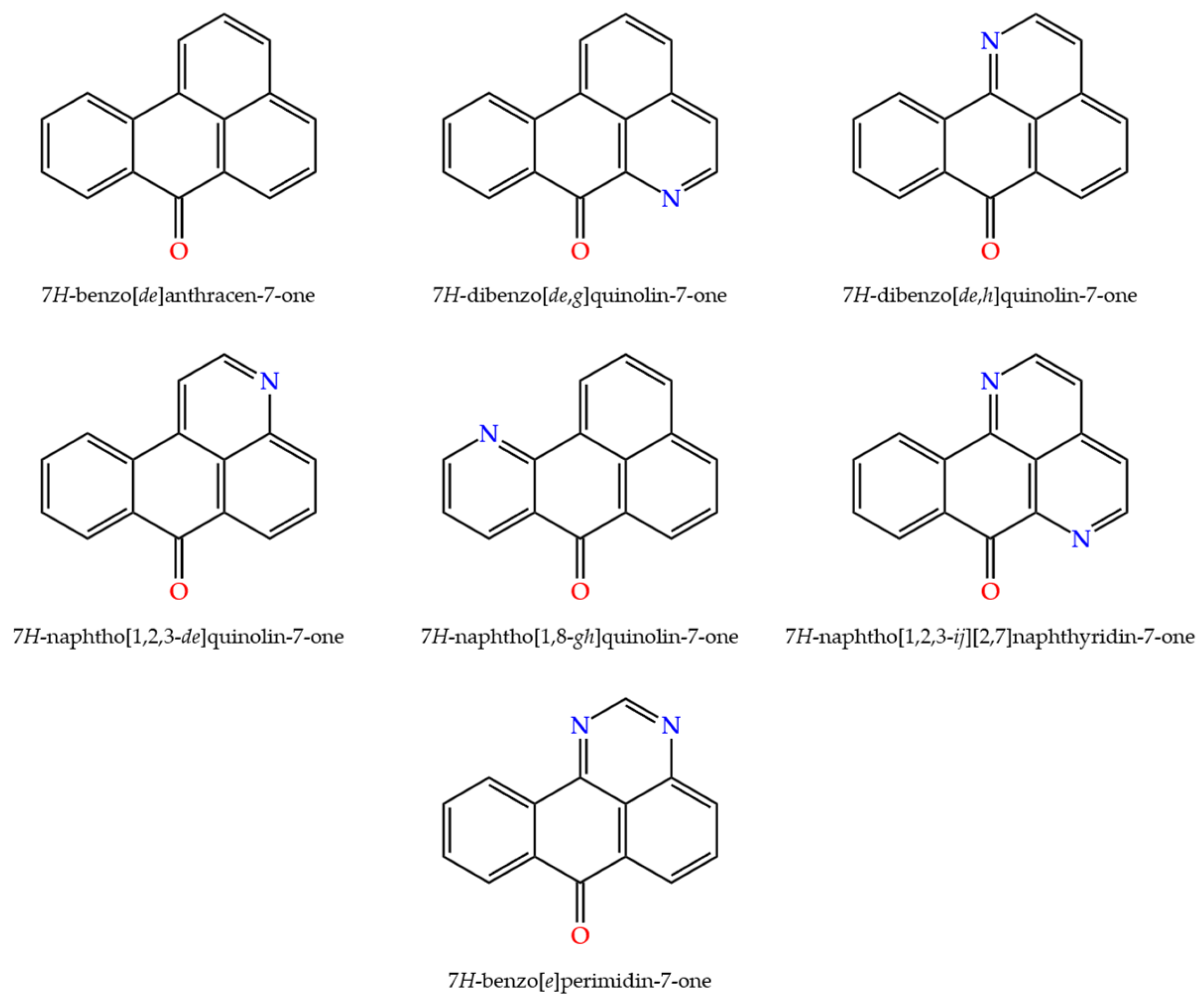
Benzanthrone derivatives featuring nitrogen atoms in the molecular core (see Figure 1), known as azabenzanthrones, have also been documented in the literature. Such compounds include the naturally occurring 6-azabenzanthrone (7H-dibenzo[de,g]quinolin-7-one), also known as oxoaporphine, and 1-azabenzanthrone (7H-dibenzo[de,h]quinolin-7-one), also known as oxoisoaporphine, which are alkaloids with anti-inflammatory, antiprotozoal, and anticancer activities [14,15,16,17,18,19,20,21,22,23,24,25,26,27,28,29]. The synthesis of substituted 3-azabenzanthrone derivatives and theoretical studies on their optical and nonlinear optical properties have also been reported [30,31]. More recently, the synthesis of 11-azabenzanthrone and its amino derivatives as photosensitizers for cancer therapy has been described [32].
Figure 1.
The key (di)(aza)benzanthrone derivatives.
It is possible to introduce several nitrogen atoms into the benzanthrone core. For example, sampangine or 1,6-diazabenzanthrone (7H-naphtho [1,2,3-ij][2,7]naphthyridin-7-one) derivatives are structurally unique natural alkaloids with versatile biological activities, ranging from heme inhibition and reactive oxygen species (ROS) generation to promising antifungal and anticancer effects [33,34,35]. Similarly, 1,3-diazabenzanthrone (7H-benzo[e]perimidin-7-one) derivatives were studied as DNA-intercalating agents with potential anticancer activity [36]. These molecules can be synthesized from 1-aminoanthraquinone by condensation with urea or dimethylacetamide, followed by cyclization. Although the therapeutic use of aza- and diazabenzanthrone derivatives is limited by their potential toxicity and metabolic factors, ongoing research into these derivatives is paving the way for novel and more effective compounds.
While the synthesis and bioactivity profiles of aza- and diazabenzanthrones have been reported, their luminescent properties have rarely been investigated. Beyond their biological activity, azabenzanthrones also exhibit intriguing electronic and optical characteristics that distinguish them from their parent benzanthrone analogs. The introduction of one or more nitrogen atoms into the aromatic framework modulates the electron distribution and intermolecular interactions, possibly resulting in an enhanced charge-transfer character and better visualization of biological specimens in confocal laser-scanning microscopy (CLSM). This microscopy method is widely used in scientific communities to study various biological processes. In consideration of the increase in human food-borne parasitic diseases, the diagnostic and detailed investigation of these causative agents remains an important aspect of modern life sciences.
Among the functionalized benzanthrone derivatives, the amidine group was found to exhibit favorable photophysical properties and robust performance as a CLSM dye [37]. To develop new luminophores with improved properties for practical use in CLSM, we synthesized a 7H-benzo[e]perimidin-7-one cyclic amidine derivative and investigated its photophysical properties.
2. Results and Discussion
2.1. Synthesis
Since the last century, 1-aminoanthraquinone has been of great practical importance, due to the possibility of its functionalization and the creation of new substances, including heterocyclic compounds [38]. Among the numerous heterocyclic derivatives, perimidines represent an interesting class of N-heterocycles that have seen significant development in recent years, due to their wide application in the life sciences, medicine, and industrial chemistry [39,40]. Various new approaches to the synthesis of benzoperimidines and their derivatives have been developed from 1-aminoanthraquinone [36,41] or 1-iodoanthraquinone [42].
In this work, we used a previously developed method based on the cyclization of an amidine obtained from 1-aminoanthraquinone with subsequent amination of the resulting benzoperimidine.
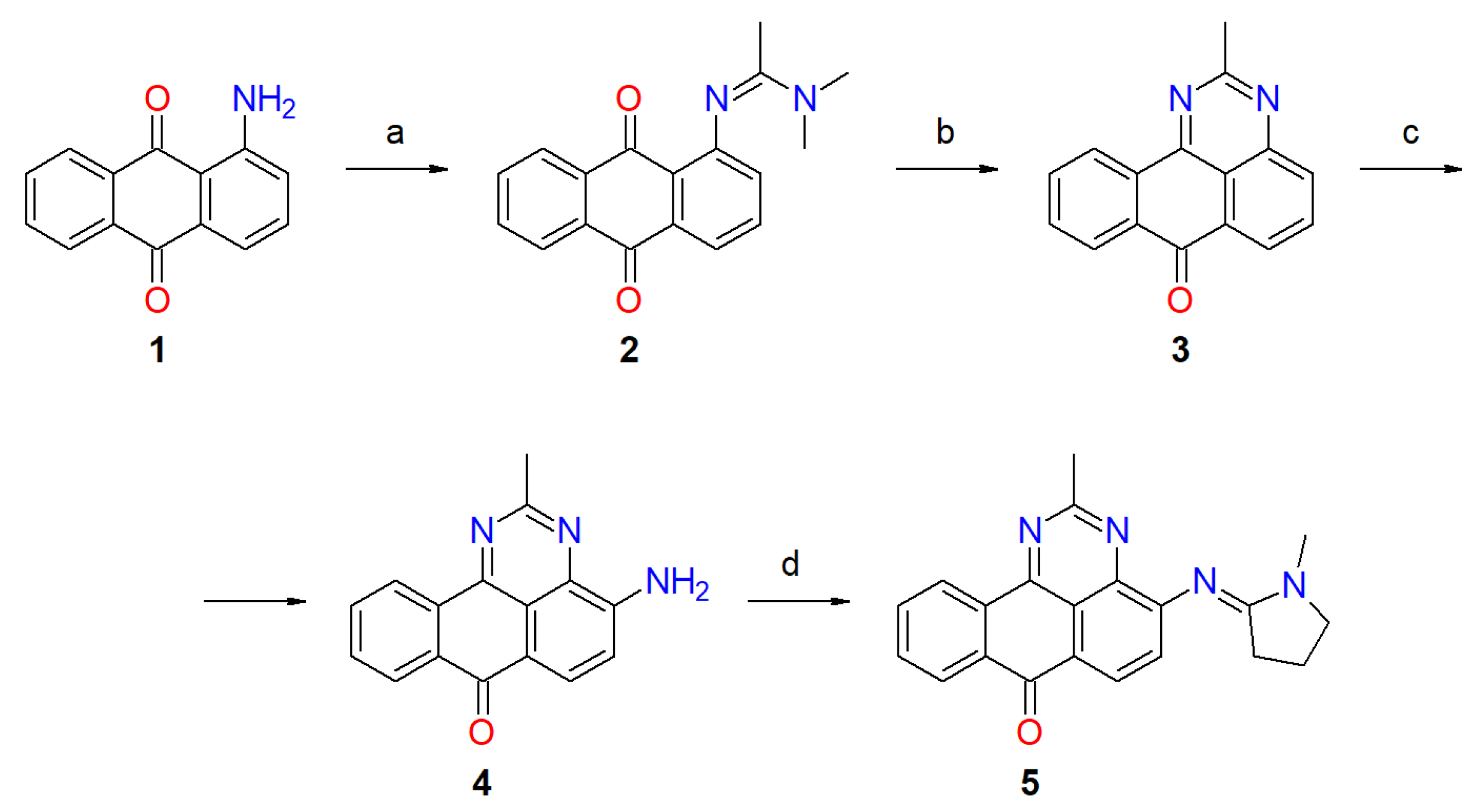
The synthesis of target compound 5 (Scheme 1) was guided by prior studies [41,43]. Commercially available 1-aminoanthraquinone (1) was first transformed into intermediate 2 through a Vilsmeier–Haack-type reaction with phosphorus oxychloride in the presence of N,N-dimethylacetamide. Subsequent condensation of 2 with ammonium chloride in methanol yielded intermediate 3, which was then directly aminated using hydroxylamine in diethylene glycol. The resulting amino derivative opens up broad and previously underexplored possibilities for modification via alkylation, acylation, condensation reactions, etc. Finally, applying the same reaction conditions used for the formation of 2, intermediate 4 was converted into the desired compound 5 via reaction with N-methylpyrrolidone. The chemical structures of the synthesized compounds were confirmed using NMR and IR spectroscopy methods, as well as mass spectrometry.
Scheme 1.
Synthesis of target benzoperimidine derivative 5. The reaction conditions and yields: (a): MeCN, DMAC, POCl3, 95%; (b): NH4OAc, EtOH, 90%; (c): NH2OH, diglycol, 73%; (d): N-methylpyrrolidone, POCl3, 63%.
For the amino group (NH2), the obtained infrared spectra showed two stretching vibration bands around 3300–3420 cm−1 for amine 4, two stretching vibration peaks of anthraquinone carbonyl groups (C=O) for derivatives 2–3 at around 1630–1670 cm−1, and one for benzoperimidine derivatives 4 and 5 carbonyl group at around 1660–1670 cm−1.
Confirmation of the obtained compounds’ structures was achieved using 1H-NMR spectroscopy. The characteristic signals of aromatic protons were identified. Hydrogens within the methyl groups of compound 5 appeared as two singlet signals at 3.20 and 3.02 ppm. The protons of the pyrrolidine ring showed two triplet signals at 3.55 and 2.53 ppm and a quintet at 2.06 ppm.
The mass of the synthesized compounds, determined from the results obtained from high-resolution mass spectrometry, was verified to match the calculated values.
2.2. X-Ray Crystallographic Study
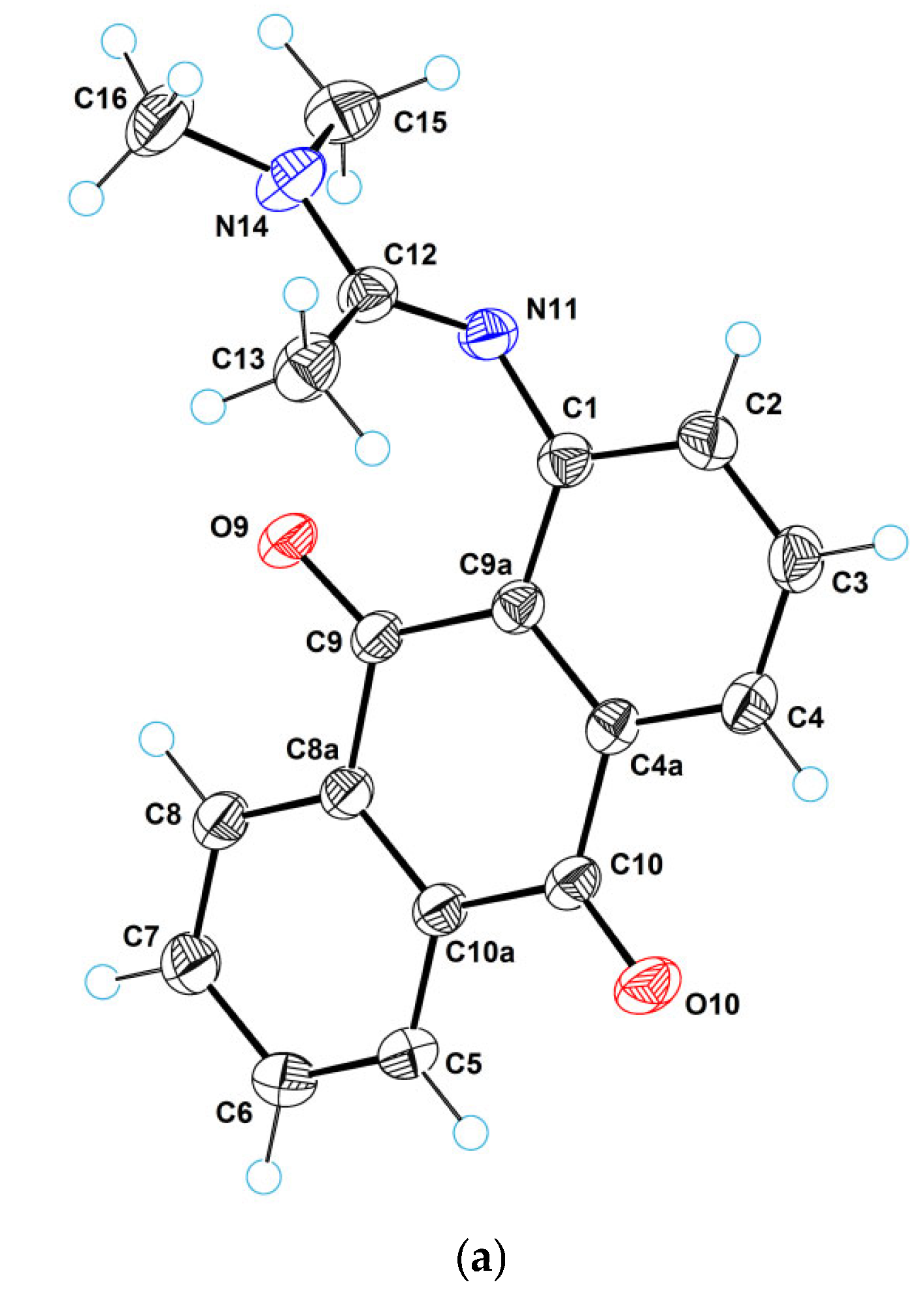
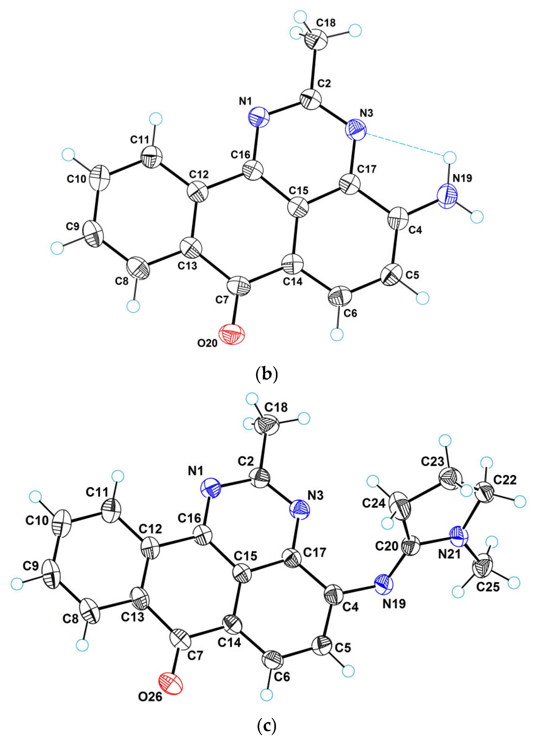
Figure 2 provides a perspective view of molecular structures 2, 4, and 5, respectively, with thermal ellipsoids and the atom numbering scheme. Molecule 2 is characterized by the usual geometry of the anthraquinone fragment. The planar dimethylacetimidamide group cannot be coplanar with the anthraquinone system, due to steric hindrance. That is why this group is turned and forms a dihedral angle of 81.9(2)° with the anthraquinone system. Such a high value of the dihedral angle does not promote conjugation; therefore, the length of the C1−N11 bond (1.384(1) Å) exceeds the standard value for Car−Nsp2 bonds [44].

Figure 2.
ORTEP diagrams for molecules 2 (a), 4 (b), and 5 (c).
In the crystal structure of compound 2, intermolecular bifurcated hydrogen bonds of CH···O···HC type are observed. These bonds are moderate, with the following parameters: C7···O10 = 3.181(1) Å (H7···O10 = 2.59(1)Å, C7−H7···O10 = 119(1)°); C8···O10 = 3.104(2) Å (H8···O10 = 2.43(2) Å, C8−H8···O10 = 125(2)°). By means of these bonds, molecular chains are formed in the crystal structure along the longest lattice parameter (along the parameter c). These chains are shown in Figure 3.
Figure 3.
A molecular chain in the crystal structure 2 viewed along the monoclinic axis.
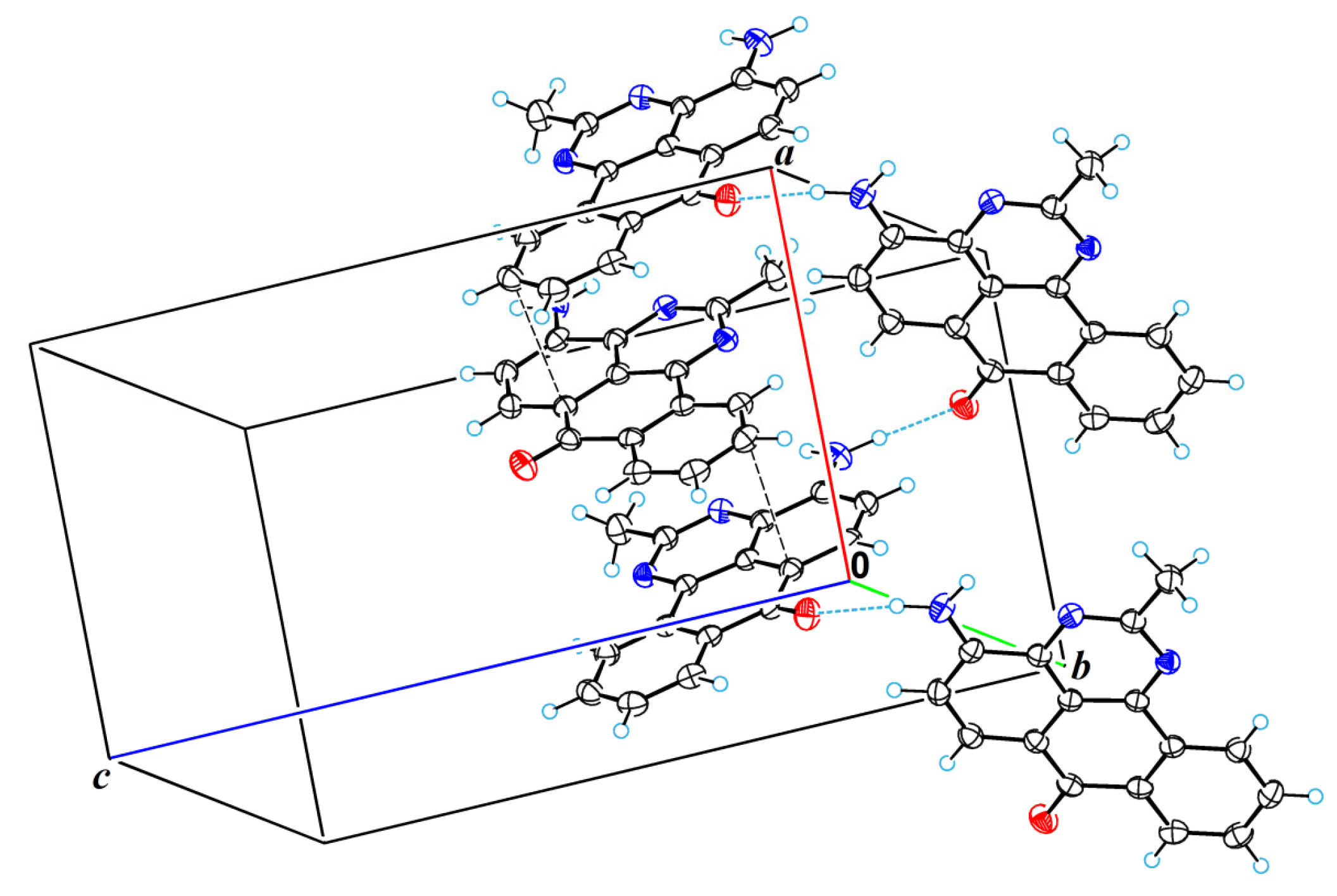
The polycyclic aromatic system in derivative 4 is planar and resembles the polycyclic system in benzanthrone. The atoms C18, N19, and O20 lie in the plane of the polycyclic system. The amino group is characterized by trigonal planar geometry and participates in the formation of hydrogen bonds. One of these bonds (the bond N19H···N3) is intramolecular with the length of 2.741(2) Å (H19b···N3 = 2.36(2) Å, N19−H···N3 = 104(3)°). The second hydrogen bond is intermolecular NH···O type bond with the length of 2.918(2) Å (H19a···O20 = 1.98(2) Å, N19−H19a···O20 = 164(2)°). Due to this intermolecular bond, molecular chains are formed in the crystal structure along the smallest lattice parameter (along parameter a). Intermolecular π-π stacking interactions with the smallest atom-atomic contact of 3.311(3) Å (C10···C14 contact) are also observed in the crystal structure. Figure 4 shows a fragment of the packing of 4 molecules in the crystal lattice.
Figure 4.
A fragment of molecular packing in the crystal structure 4 showing the intermolecular H-bonds and the shortest atom–atomic contacts.
2.3. Photophysical Properties
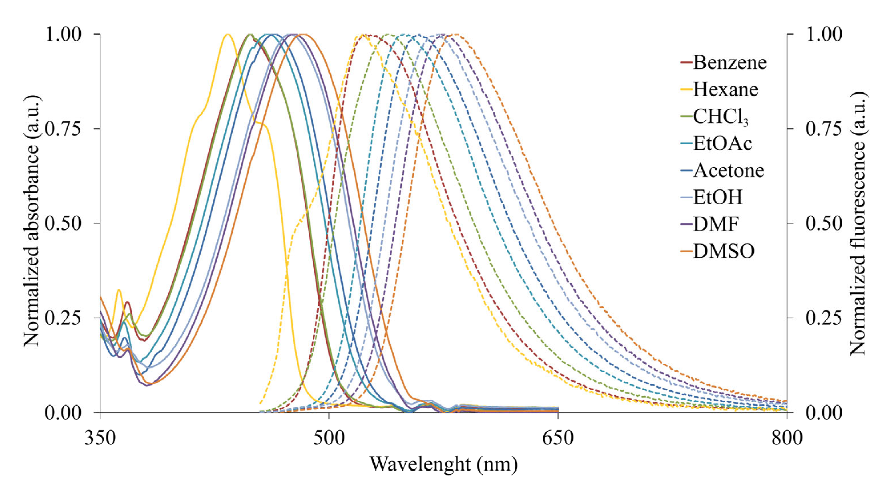
To investigate the photophysical characteristics of the synthesized compounds, UV–Vis absorption and fluorescence emission spectra in solvents of varying polarity (benzene, chloroform, ethyl acetate, acetone, N,N-dimethylformamide, dimethyl sulfoxide, and ethanol) were acquired. Table 1 provides a summary of the data, including the absorption maxima, molar attenuation coefficients, and emission maxima. For the studied compounds 4 and 5, the absorption and fluorescence spectra are shown in Figure 5 and Figure 6.
Table 1.
Absorption maxima (extinction coefficient) and fluorescence maxima (quantum yield) of the obtained compounds 2–5 in organic solvents (concentration 10–5 M).
Figure 5.
The UV–Vis absorption and fluorescence emission (dotted lines) spectra of compound 4 in various organic solvents.
Figure 6.
The UV–Vis absorption and fluorescence emission (dotted lines) spectra of compound 5 in various organic solvents.
The intermediates amidine 2 and perimidine 3 exhibit absorption in the region of 400–417 nm and 447–480 nm, respectively, and are practically non-luminescent. The introduction of an amino group into the benzoperimidine system results in the appearance of fluorescence in product 4 in the range of 520–585 nm. Furthermore, the synthesized cyclic amidine 5 also exhibits an emission that is bathochromically shifted by 60–70 nm compared to the initial amine 4. The largest Stokes shift of 5691 cm−1 is observed for derivative 5 in ethanol, and for amine 4, it is 3598 cm−1. The synthesized amidine 5 exhibits more pronounced emission and solvatochromic effects, which can be explained by more efficient intramolecular charge transfer from the amidine residue to the carbonyl group, compared to the transfer in the molecule of the starting amine 4. Thus, the substitution of hydrogen atoms contributes to the formation of more emissive and polarity-sensitive derivatives of 4-aminoperimidine.
2.4. Antimicrobial Activity
In recent years, particular attention has been paid to the use of anthrone derivatives in some biological studies, which has led to the need to evaluate their biological activity. Thus, the microbiological activity of some ammonium salts and metal complexes of anthraquinone and benzanthrone has been studied with the aim of developing substances for the production of colored antibacterial dressings and other antibacterial textile products [45,46,47,48]. The antimicrobial activity of target compounds is often studied in vitro using the agar diffusion method with various bacterial and yeast cultures [49].
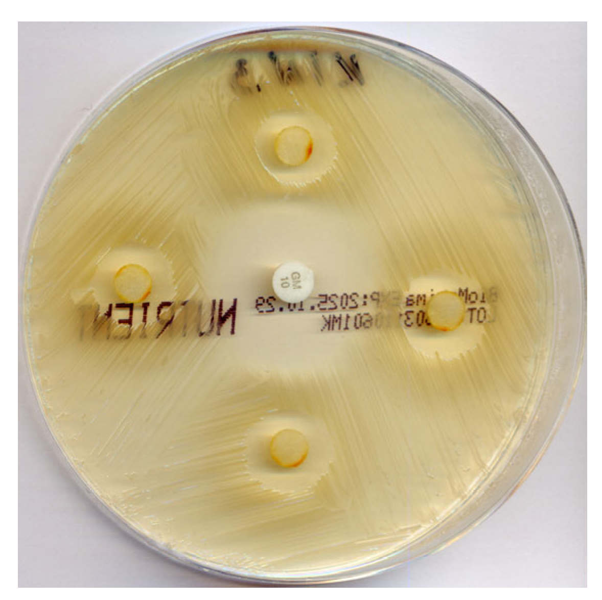
In this study, the antimicrobial activity of the obtained compounds was assessed using the Gram-positive bacteria Bacillus subtilis subsp. Spizizenii. For the growth of Bacillus subtilis subsp. Spizizenii with gentamicin (Gm, 10 µg), the inhibition zone diameters were 18–26 mm. As for the growth of Bacillus subtilis subsp. spizizenii with compounds 1, 2, 3, and 4 (10 µg), the inhibition zones were not detected. In turn, inhibition zone diameters with compound 5 (10 µg) ranged from 12 to 14 mm. Antimicrobial tests indicated the good antimicrobial activity of the synthesized compound 5, where the inhibition zones of the bacteria Bacillus subtilis subsp. Spizizenii were approximately half as small as those of gentamicin (see Table 2 and Figure 7).
Table 2.
Zones of inhibition of the growth of the test culture with the investigated compounds 1–4.
Figure 7.
Zones of inhibition of the growth of Bacillus subtilis subsp. Spizizenii with 10 µg of compound 5.
Thus, antimicrobial tests showed that 1-aminoanthraquinone does not exhibit antimicrobial activity, in contrast to its substituted amide, the activity of which against B. subtilis, as previously established [45], is only slightly inferior to that of the standard (gentamicin). It is obvious that an increase in the conjugated aromatic system leads to an increase in antimicrobial activity. Therefore, the antimicrobial activity of substituted diazabenzanthrone 5 is similar to the activity observed for benzanthrone derivatives [46].
2.5. Imaging of Opisthorchis felineus
According to the most recent estimates from the Global Burden of Disease Study, the total number of food-borne trematode infections, including opisthorchiasis, has reached approximately 44.5 million cases [50]. Among members of the family Opisthorchiidae, the most medically important species for humans are Opisthorchis felineus, O. viverrini, and Clonorchis sinensis [51]. Recent reviews suggest that about 1.6 million people are infected with O. felineus, 9–10 million with O. viverrini, and about 15 million with C. sinensis [52].
Diagnosis of opisthorchiasis in humans traditionally relies on the microscopic detection of parasite eggs in feces; however, its sensitivity decreases in light or chronic infections. Modern serological tests (ELISA) and antigen-detection assays demonstrate markedly higher diagnostic accuracy, with reported sensitivities of 94–100% and specificities of up to 99% [53,54,55]. Moreover, new urine antigen rapid diagnostic tests (OV-RDT) have shown comparable performance and can facilitate field-level diagnosis in endemic areas [55]. The implementation of such sensitive and affordable tools is critical for the early detection of asymptomatic and chronic infections.
Chronic opisthorchiasis is associated with biliary tract pathologies, including cholangitis, fibrosis, and cholangiocarcinoma (CCA) [56,57]. Experimental studies further demonstrate that O. felineus can induce biliary intraepithelial neoplasia, a precancerous lesion of the bile ducts, and produce oxysterol-like metabolites capable of causing DNA damage in host tissues [58].
O. felineus is regarded as a potential carcinogenic agent comparable to O. viverrini and C. sinensis [59]. Thus, given the high prevalence of opisthorchiasis and its association with cholangiocarcinoma, O. felinius was selected as the model organism for the development of a sensitive diagnostic tool using CLS microscopy.
The developed fluorophore 5 produced high-quality and high-resolution images, emphasizing the key internal structures of O. felineus (see Figure 8). Dye 5 effectively stained the internal organs, including the digestive and reproductive systems, and allowed clear visualization of the muscular structures at magnifications up to ×600.
Figure 8.
Adult Opisthorchis felineus stained with the examined dye 5, showing the internal structure: OS: oral sucker, P: pharynx, E: esophagus, IC: intestinal caeca, VG: vitellaria glands, O: ovary, UwE: uterus filled with eggs, T: testis. CLSM was performed using 488 nm and 638 nm lasers.
The results revealed elongated, flattened, and leaf-like trematodes with tapered anterior ends, which were typical of O. felineus. The specimen exhibited two small circular suckers—an oral sucker positioned almost terminally at the anterior end and a ventral sucker (acetabulum) lying slightly behind the anterior third part, which is also described as a key diagnostic feature separating Opisthorchis from Clonorchis.
Within the digestive system, the following were visualized: a short prepharynx, a globular pharynx behind the oral sucker, a short esophagus, and bifurcated thin straight intestinal caeca posteriorly nearly to the tail end, exhibiting the highest fluorescence intensity with strong red emission under 638 nm excitation. Within these regions, the fluorophore’s high tissue penetration and strong affinity for peptide- and protein-rich regions were shown.
The reproductive system was well obtained. The ovary was visualized as small, oval, and located in the midline just anterior to the testes. Two large, spherical, and symmetrical testes were located posteriorly. The uterus was coiled, filled with numerous operculated eggs occupying the central region anterior to the testes, in general situated between the ventral sucker and the testes. The vitellaria were lateral, extending from the midbody to the posterior end, providing yolk for the eggs. The eggs were small ovals, operculated with a prominent opercular shoulder at one end and a small terminal knob at the opposite end.
The body tegument was smooth and translucent, sometimes with faint granulation visible under higher magnification.
However, spikes in the muscle system were not clearly observed. Overall, the fluorophore enabled detailed visualization of the preserved specimens, yielding superior resolution and tissue contrast, particularly for muscular and reproductive structures.
3. Materials and Methods
3.1. Materials and Measurements
All of the reagents and solvents were obtained commercially and used without any additional purification. The assessment of the progress of reactions and the purity of the synthesized compounds was performed using TLC on MERCK (Boston, MA, USA) Silica gel 60 F254 plates in hexane/acetone (1:2) as an eluent and visualized under UV light. Melting points were determined using a METTLER TOLEDO™ (Greifensee, Switzerland) Melting Point System MP70 apparatus.
The IR spectrum was recorded using a Thermo Scientific Nicolet iS50 Spectrometer (Waltham, MA, USA, ATR accessory; no. of scans: 64; resolution: 4; data spacing: 0.482 cm−1). 1H- and 13C- NMR spectra were recorded using a Bruker Avance 500 MHz (Bruker Corporation, Billerica, MA, USA) in CDCl3 at ambient temperature, with solvent peaks as the internal reference. The chemical shift (δ) values are reported in ppm. High-resolution accurate mass measurements were performed employing an Orbitrap Exploris 120 (Thermo Fischer Scientific) operating at Full Scan mode at 120,000 resolution.
The fluorescence emission spectra were recorded using an FLSP920 (Edinburgh Instruments Ltd., Livingston, UK) spectrofluorometer in the visible range 450–800 nm, and the absorption spectra were obtained using the UV–visible spectrophotometer SPECORD® 80 (Analytik Jena AG, Jega, Germany). The spectral properties of the investigated compound were measured at an ambient temperature in quartz cuvettes of 10 mm in hexane, benzene, chloroform, ethyl acetate (EtOAc), acetone, ethanol (EtOH), dimethyl sulfoxide (DMSO), and dimethylformamide (DMF) with a concentration of 10−5 M. The quantum yields were measured relative to rhodamine 101 as the standard.
3.2. Synthesis and Characterization
2-Methyl-4-amino-7H-benzo[e]perimidin-7-one (4) was obtained by condensation of 1-aminoanthraquinone (1) with N,N-dimethylacetamide and subsequent cyclization of the resulting (N’-(9,10-dioxo-9,10-dihydroanthracen-1-yl)-N,N-dimethylethanimideamide (2) to 2-methyl-7H-benzo[e]perimidin-7-one (3) and its amination, according to the procedure described by Yan-Ping Li and coworkers [41].
2-Methyl-4-((1-methylpyrrolidin-2-ylidene)amino)-7H-benzo[e]perimidin-7-one (5) was synthesized as follows. POCl3 (1.86 mL, 20 mM) was added dropwise to a stirring solution of N-methylpyrrolidone (2.41 mL, 25 mM) in 10 mL of acetonitrile in a 50 mL round-bottom flask and left to stir for an hour. 2-methyl-4-amino-7H-benzo[e]perimidin-7-one (2.61 g, 10 mM) and 10 mL of acetonitrile were then added to the solution in one portion. The mixture was then heated at 50 °C for 2 h, cooled, poured onto 100 mL of water, and basified with 10% NaOH. The precipitate formed was filtered off, washed with water, and dried in air. Recrystallization from xylenes afforded a red solid in 63% yield, with a melting point of 179 °C and Rf = 0.57 (TLC, silica gel, DCM/acetone 1:1). 1H NMR (500 MHz, Chloroform-d) δ 8.96 (d, J = 7.7 Hz, 1H), 8.49 (d, J = 8.1 Hz, 1H), 8.46 (d, J = 7.7 Hz, 1H), 7.81 (t, J = 7.1 Hz, 1H), 7.75 (t, J = 7.4 Hz, 1H), 7.36 (d, J = 8.1 Hz, 1H), 3.55 (t, J = 6.9 Hz, 2H), 3.20 (s, 3H), 3.02 (s, 3H), 2.53 (t, J = 7.8 Hz, 2H), 2.06 (p, J = 7.4 Hz, 2H). 13C NMR (APT, 126 MHz, Chloroform-d) δ 181.43 (C=O), 163.86 (C), 156.89 (C), 156.35 (C), 144.30 (C), 135.01 (C), 134.41 (C), 133.15 (CH), 131.96 (CH), 130.41 (CH), 127.54 (CH), 125.42 (CH), 124.65 (CH), 121.60 (C), 119.12 (C), 51.71 (CH2), 31.88 (CH3), 29.09 (CH2), 27.18 (CH3), 19.96 (CH2). GC-MS: calculated for [C21H18N4O]: 342.2, found: 342.1.
3.3. Antimicrobial Activity Test
The antimicrobial activity of the compounds was tested in vitro via the agar diffusion method using the Gram-positive bacterium Bacillus subtilis subsp. Spizizenii [49].
Nutrient agar plates were seeded with cell suspensions of the test Bacillus Subtilis subsp. Spizizenii cultures. Commercial disks with gentamicin (Gm, 10 µg) and the studied compounds 2–5 (10 µg) were used as a standard antibacterial and antifungal agent, respectively. The plates were incubated at an appropriate temperature (+30 °C) and monitored for growth for 24–48 h. The growth of microorganisms was inhibited by diffusion of the test solutions, and the antimicrobial activity was indicated by the presence of clear inhibition zones around the wells.
3.4. Confocal Laser Scanning Microscopy Imaging
We visualized the internal structure of 30 Opisthorchis felineus specimens fixed in 70% ethanol more than 5 years ago and subsequently stained with the synthesized fluorescent dye 5.
A simple and rapid staining protocol was developed based on previous experience with other luminophores applied to biological specimens [37,60,61,62,63].
The staining procedure consisted of 4 main steps: staining, rinsing, clearing, and mounting. In the first step, each specimen was transferred from a microtube to a watch glass, and all residual fixative was removed. Subsequently, 2 mL of compound 5 fluorescent dye dissolved in ethanol was added for 5 min. In the second step, the staining solution was removed, and the specimen was rinsed 3 times with 2 mL of 70% ethanol. In the third step, after rising, the ethanol was removed, and 2 mL of the clearing agent (a 1:1 mixture of 70% ethanol and 100% xylene) was added. The clearing agent was applied for 30 s to increase the tissue transparency and improve the structural visibility. In the last step, the specimen was transferred to a microscopy slide, the clearing agent was removed, and the sample was mounted in Canada balsam. A coverslip was carefully placed over the mounted specimen.
Fluorescence imaging was performed using a confocal laser scanning microscope (Eclipse Ti-E; Nikon, Tokyo, Japan) equipped with a digital sight DS-U3 camera and an A1R MP high-speed multiphoton system (Nikon, Japan). Image acquisition and analysis were conducted using NIS-Elements AR software (version 3.2, 64-bit; Nikon, Japan). Excitation was provided by 488 nm and 638 nm lasers with FITC (500–550 nm) and Cy5 (662–737 nm) emission filters, respectively. Fluorescence signals were detected using the internal spectral detector, with the detection range set from 20 nm above the excitation wavelength to the red edge of the visible spectrum. No passive cut-off filters were applied in the optical path. Z-stack images were collected at 2.0 µm intervals, and morphological measurements were performed using the same software.
3.5. Single Crystal X-Ray Diffraction Analysis
For compounds 2, 4, and 5, diffraction data were collected at low temperature on a Rigaku (Tokyo, Japan) XtaLAB Synergy Dualflex HyPix diffractometer using copper monochromated Cu-Kα radiation (λ = 1.54184 Å). The crystal structures were solved using direct methods [64] and refined using full-matrix least squares [65]. All non-hydrogen atoms were refined in an anisotropic approximation. For further details, see the crystallographic data for 2, 4, and 5 deposited at the Cambridge Crystallographic Data Center as Supplementary Publications Numbers CCDC 2470312 (for 2), CCDC 2470313 (for 4), and CCDC 2470314 (for 5). These data are provided free of charge by the joint Cambridge Crystallographic Data Center and Fachinformationszentrum Karlsruhe Access Structures service.
4. Conclusions
In conclusion, the newly synthesized benzoperimidine derivative exhibits notable fluorescence properties with solvatochromic behavior, making it suitable for a wide variety of applications. The spectroscopic analysis confirmed the chemical structures, and the compound’s emission properties were thoroughly explored in various organic solvents. The X-ray crystallographic study of the studied compound contributes valuable information about the compound’s molecular arrangement and intermolecular interactions.
An assessment of antimicrobial activity on the soil bacterium Bacillus subtilis subsp. Spizizenii revealed a significant effect of the obtained dye on the growth of these bacteria. Overall, the synthesized compounds demonstrate potential for versatile applications, particularly in the investigation of parasitic organisms. The developed dye demonstrates strong fluorescence, high photostability, and specific affinity toward muscular and reproductive tissues, enabling clear imaging of multiple organ systems. The optimized staining and clearing protocol developed in this study ensures efficient sample preparation for CLSM and can serve as a basis for further bioimaging research using anthrone-based dyes.
Supplementary Materials
The following supporting information can be downloaded at https://www.mdpi.com/article/10.3390/molecules30224472/s1. 1H, 13C NMR spectra; MS (ESI) data.
Author Contributions
Conceptualization, E.K. and M.K.; methodology, A.M., L.M. and I.U.; validation, S.K. and I.R.; formal analysis, E.K. and S.B.; investigation, V.P., A.B. and I.R.; data curation, A.P. and A.M.; writing—original draft preparation, A.M. and E.K.; writing—review and editing, M.K.; visualization, A.P., L.M. and A.B.; supervision, E.K. and M.K.; project administration, E.K.; V.K. and L.L. provided samples and fluke species identification. All authors have read and agreed to the published version of the manuscript.
Funding
This work is supported by the Fundamental and applied research projects of the Latvian Council of Science, project No. lzp-2022/1-0436 “Novel fluorescent anthrone-derived functional materials for bioimaging applications”.
Institutional Review Board Statement
Not applicable.
Informed Consent Statement
Not applicable.
Data Availability Statement
Data are contained within the article or Supplementary Materials.
Conflicts of Interest
The authors declare no conflicts of interest.
References
- Grabchev, I.; Bojinov, V.; Moneva, I. The synthesis and application of fluorescent dyes based on 3-amino benzanthrone. Dyes Pigm. 2001, 48, 143–150. [Google Scholar] [CrossRef]
- Cao, L.; Xu, L.; Zhang, D.; Zhou, Y.; Zheng, Y.; Fu, Q.; Jiang, X.-F.; Lu, F. D-A dyad and D-A-D triad incorporating triphenylamine, benzanthrone and perylene diimide: Synthesis, electrochemical, linear and nonlinear optical properties. Chem. Phys. Lett. 2017, 682, 133–139. [Google Scholar] [CrossRef]
- Staneva, D.; Vasileva-Tonkova, E.; Grabchev, I. pH sensor potential and antimicrobial activity of a new PPA dendrimer modified with benzanthrone fluorophores in solution and on viscose fabric. J. Photochem. Photobiol. A Chem. 2019, 375, 24–29. [Google Scholar] [CrossRef]
- Staneva, D.; Betcheva, R. Synthesis and functional properties of new optical pH sensor based on benzo[de]anthracen-7-one immobilized on the viscose. Dyes Pigm. 2006, 74, 148–153. [Google Scholar] [CrossRef]
- Kirilova, E.M.; Puckins, A.I.; Romanovska, E.; Fleisher, M.; Belyakov, S.V. Novel amidine derivatives of benzanthrone: Effect of bromine atom on the spectral parameters. Acta Part A Mol. Biomol. Spectrosc. 2018, 202, 41–49. [Google Scholar] [CrossRef]
- Maļeckis, A.; Griškjāns, E.; Cvetinska, M.; Savicka, M.; Belyakov, S.; Kirilova, E. Synthesis, characterization, spectroscopic studies and evaluation of toxicological effect on growth of wheat sprouts (Triticum aestivum) of new benzanthrone α-aryl-α-aminophosphonates. J. Mol. Struct. 2022, 1277, 134838. [Google Scholar] [CrossRef]
- Maļeckis, A.; Cvetinska, M.; Kirjušina, M.; Mežaraupe, L.; Kecko, S.; Gavarāne, I.; Kiyan, V.; Lider, L.; Pavlova, V.; Savicka, M.; et al. A Comparative Study of New Fluorescent Anthraquinone and Benzanthrone α-Aminophosphonates: Synthesis, Spectroscopy, Toxicology, X-ray Crystallography, and Microscopy of Opisthorchis felineus. Molecules 2024, 29, 1143. [Google Scholar] [CrossRef] [PubMed]
- Grabchev, I.; Moneva, I. Synthesis and properties of benzanthrone derivatives as luminophore dyes for liquid crystals. Dyes Pigm. 1998, 37, 155–164. [Google Scholar] [CrossRef]
- Grabtchev, I.K.; Bojinov, V.B.; Moneva, I.T. Functional properties of azomethine substituted benzanthrone dyes for use in nematic liquid crystals. J. Mol. Struct. 1998, 471, 19–25. [Google Scholar] [CrossRef]
- Maļeckis, A.; Cvetinska, M.; Griškjāns, E.; Kirilova, E. Exploring dual solvatochromic traits in novel fluorescent benzanthrone ethynyl derivatives. J. Solution Chem. 2024, 53, 1074–1088. [Google Scholar] [CrossRef]
- Grabchev, I.; Moneva, I.; Wolarz, E.; Bauman, D. Fluorescent 3-oxy benzanthrone dyes in liquid crystalline media. Dyes Pigm. 2003, 58, 1–6. [Google Scholar] [CrossRef]
- Maļeckis, A.; Cvetinska, M.; Griškjāns, E.; Dmitrijevs, K.; Traskovskis, K.; Belyakov, S.; Kirilova, E. Benzanthrone sulfides: Synthesis, solvatochromism characterization and analysis of experimental photophysical parameters and theoretical calculations. Dyes Pigm. 2023, 219, 111599. [Google Scholar] [CrossRef]
- Maļeckis, A.; Romanovska-Dzalbe, E. Recent progress of benzanthrone chemistry: A condensed review. Chem. Pap. 2025, 79, 3463–3473. [Google Scholar] [CrossRef]
- Rodríguez-Arce, E.; Cancino, P.; Arias-Calderón, M.; Silva-Matus, P.; Saldías, M. Oxoisoaporphines and Aporphines: Versatile Molecules with Anticancer Effects. Molecules 2019, 25, 108. [Google Scholar] [CrossRef]
- Sun, J.; Zhan, X.; Wang, W.; Yang, X.; Liu, Y.; Yang, H.; Deng, J.; Yang, H. Natural aporphine alkaloids: A comprehensive review of phytochemistry, pharmacokinetics, anticancer activities, and clinical application. J. Adv. Res. 2023, 63, 231–253. [Google Scholar] [CrossRef]
- Liao, L.-S.; Tan, L.-J.; Chen, Y.; Yang, Q.-Y.; Choudhary, M.I.; Pan, Y.-M.; Tang, H.-T.; Su, G.-F.; Liang, H.; Chen, Z.-F. One-pot synthesis of oxoaporphines as potent antitumor agents and investigation of their mechanisms of actions. Eur. J. Med. Chem. 2022, 231, 114141. [Google Scholar] [CrossRef] [PubMed]
- Tang, H.; Wang, X.-D.; Wei, Y.-B.; Huang, S.-L.; Huang, Z.-S.; Tan, J.-H.; An, L.-K.; Wu, J.-Y.; Chan, A.S.-C.; Gu, L.-Q. Oxoisoaporphine alkaloid derivatives: Synthesis, DNA binding affinity and cytotoxicity. Eur. J. Med. Chem. 2007, 43, 973–980. [Google Scholar] [CrossRef]
- Guo, R.; Wang, C.-L.; Cao, X.-J.; Yao, X.-J.; Qiao, X.; Meng, Y.-T.; Zhang, T.; Zhang, Q. Rare oxoisoaporphine alkaloids from Menispermum dauricum with potential anti-inflammatory activity. Phytochemistry 2024, 225, 114170. [Google Scholar] [CrossRef]
- Lu, Y.; Wang, F.-Y.; Levine, M.S.; Shi, H.-R.; Wang, Y.; Xiong, X.; Yang, L.-M.; Shi, Y.-Q.; Zou, T.; Sessler, J.L.; et al. Oxoisoaporphine alkaloid Iridium(III) derivative: An immunogenic cell death inducer that engages the Autophagy-Dependent regulator Cathepsin D. J. Am. Chem. Soc. 2025, 147, 15216–15228. [Google Scholar] [CrossRef]
- Zhong, H.; Zhao, M.; Wu, C.; Zhang, J.; Chen, L.; Sun, J. Development of oxoisoaporphine derivatives with topoisomerase I inhibition and reversal of multidrug resistance in breast cancer MCF-7/ADR cells. Eur. J. Med. Chem. 2022, 235, 114300. [Google Scholar] [CrossRef]
- Melzer, B.; Bracher, F. A divergent approach to benzylisoquinoline-type and oxoaporphine alkaloids via regioselective direct ring metalation of alkoxy isoquinolines. Org. Biomol. Chem. 2015, 13, 7664–7672. [Google Scholar] [CrossRef]
- Qin, J.-L.; Meng, T.; Chen, Z.-F.; Xie, X.-L.; Qin, Q.-P.; He, X.-J.; Huang, K.-B.; Liang, H. Facile total synthesis of lysicamine and the anticancer activities of the RuII, RhIII, MnII and ZnII complexes of lysicamine. Oncotarget 2017, 8, 59359–59375. [Google Scholar] [CrossRef] [PubMed]
- Hufford, C.D.; Sharma, A.S.; Oguntimein, B.O. Antibacterial and antifungal activity of liriodenine and related oxoaporphine alkaloids. J. Pharm. Sci. 1980, 69, 1180–1183. [Google Scholar] [CrossRef] [PubMed]
- Huang, X.; Hao, N.; Wang, Q.; Li, R.; Zhang, G.; Chen, G.; Liu, S.; Che, Z. Non-food bioactive forest product liriodenine: Sources, chemistry, and bioactivities. Ind. Crops Prod. 2022, 187, 115447. [Google Scholar] [CrossRef]
- Zhang, J.; Chen, L.; Sun, J. Oxoisoaporphine alkaloids: Prospective Anti-Alzheimer’s disease, anticancer, and antidepressant agents. ChemMedChem 2018, 13, 1262–1274. [Google Scholar] [CrossRef]
- Qin, Q.-P.; Qin, J.-L.; Meng, T.; Yang, G.-A.; Wei, Z.-Z.; Liu, Y.-C.; Liang, H.; Chen, Z.-F. Preparation of 6/8/11-Amino/Chloro-Oxoisoaporphine and group-10 metal complexes and evaluation of their in vitro and in vivo antitumor activity. Sci. Rep. 2016, 6, 37644. [Google Scholar] [CrossRef]
- Qin, J.-L.; Shen, W.-Y.; Chen, Z.-F.; Zhao, L.-F.; Qin, Q.-P.; Yu, Y.-C.; Liang, H. Oxoaporphine Metal Complexes (CoII, NiII, ZnII) with High Antitumor Activity by Inducing Mitochondria-Mediated Apoptosis and S-phase Arrest in HepG2. Sci. Rep. 2017, 7, 46056. [Google Scholar] [CrossRef]
- Melzer, B.C.; Bracher, F. A novel approach to oxoisoaporphine alkaloids via regioselective metalation of alkoxy isoquinolines. Beilstein J. Org. Chem. 2017, 13, 1564–1571. [Google Scholar] [CrossRef]
- Wei, Y.-B.; Li, Y.-X.; Song, H.; Feng, X.-J. Design, synthesis and anticancer activity of oxoaporphine alkaloid derivatives. J. Enzyme Inhib. Med. Chem. 2014, 29, 722–727. [Google Scholar] [CrossRef]
- Tatke, D.R.; Seshadrif, S. Nucleophilic substitution reactions of azabenzanthrone derivatives. Dyes Pigm. 1986, 7, 153–158. [Google Scholar] [CrossRef]
- Lokhande, P.K.M.; Patil, D.S.; Kadam, M.M.; Sekar, N. Theoretical Investigation of Optical and Nonlinear optical (NLO) properties of 3-Azabenzanthrone analogues: DFT and TD-DFT Approach. ChemistrySelect 2019, 4, 10033–10045. [Google Scholar] [CrossRef]
- Zang, Q.; Yu, J.; Yu, W.; Qian, J.; Hu, R.; Tang, B.Z. Red-emissive azabenzanthrone derivatives for photodynamic therapy irradiated with ultralow light power density and two-photon imaging. Chem. Sci. 2018, 9, 5165–5171. [Google Scholar] [CrossRef]
- Rao, J.U.M.; Giri, G.S.; Hanumaiah, T.; Rao, K.V.J. Sampangine, a New Alkaloid from Cananga odorata. J. Nat. Prod. 1986, 49, 346–347. [Google Scholar] [CrossRef]
- Plodek, A.; König, M.; Bracher, F. Synthesis of the Azaoxoaporphine Alkaloid Sampangine and Ascididemin-Type Pyridoacridines through TMPMgCl·LiCl-Mediated Ring Closure. Eur. J. Org. Chem. 2015, 2015, 1302–1308. [Google Scholar] [CrossRef]
- Mahdi, F.; Morgan, J.B.; Liu, W.; Agarwal, A.K.; Jekabsons, M.B.; Liu, Y.; Zhou, Y.-D.; Nagle, D.G. Sampangine (a Copyrine Alkaloid) Exerts Biological Activities through Cellular Redox Cycling of Its Quinone and Semiquinone Intermediates. J. Nat. Prod. 2015, 78, 3018–3023. [Google Scholar] [CrossRef] [PubMed]
- Bu, X.; Deady, L.W.; Finlay, G.J.; Baguley, B.C.; Denny, W.A. Synthesis and Cytotoxic Activity of 7-Oxo-7H-dibenz[f,ij]isoquinoline and 7-Oxo-7H-benzo[e]perimidine Derivatives. J. Med. Chem. 2001, 44, 2004–2014. [Google Scholar] [CrossRef]
- Rubenina, I.; Gavarane, I.; Kirilova, E.; Mezaraupe, L.; Kirjusina, M. Comparison of the Benzanthrone Luminophores: They Are Not Equal for Rapid Examination of Parafasciolopsis fasciolaemorpha (Trematoda: Digenea). Biomolecules 2021, 11, 598. [Google Scholar] [CrossRef] [PubMed]
- Zollinger, H. Color Chemistry. Synthesis, Properties and Applications of Organic Dyes and Pigments. Angew. Chem. Int. Ed. 2004, 43, 5291–5292. [Google Scholar] [CrossRef]
- Sahiba, N.; Agarwal, S. Recent Advances in the Synthesis of Perimidines and their Applications. Top. Curr. Chem. 2020, 378, 44. [Google Scholar] [CrossRef]
- Pozharskii, A.F.; Gulevskaya, A.V.; Claramunt, R.M.; Alkorta, I.; Elguero, J. Perimidines: A unique π-amphoteric heteroaromatic system. Russ. Chem. Rev. 2020, 89, 1204–1260. [Google Scholar] [CrossRef]
- Li, Y.P.; Weng, X.; Ning, F.X.; Bin Ou, J.; Hou, J.Q.; Bin Luo, H.; Li, D.; Huang, Z.S.; Huang, S.L.; Gu, L.Q. 3D-QSAR studies of azaoxoisoaporphine, oxoaporphine, and oxoisoaporphine derivatives as anti-AChE and anti-AD agents by the CoMFA method. J. Mol. Graph. Model. 2013, 41, 61–67. [Google Scholar] [CrossRef]
- Baranov, D.S.; Fadeev, D.S. Synthesis of 2-octyloxy-7H-benzo[e]perimidin-7-one and 3-substituted 3H-benzo[e]perimidine-2,7-diones. Mendeleev Commun. 2016, 26, 174–176. [Google Scholar] [CrossRef]
- Ning, F.X.; Weng, X.; Huang, S.L.; Gu, L.J.; Huang, Z.S.; Gu, L.Q. A facile and efficient method for hydroxylation of azabenzanthrone compounds. Chin. Chem. Lett. 2010, 22, 41–44. [Google Scholar] [CrossRef]
- Allen, F.H.; Kennard, O.; Watson, D.G.; Brammer, L.; Orpen, A.G.; Taylor, R. Tables of bond lengths determined by X-ray and neutron diffraction. Part 1. Bond lengths in organic compounds. J. Chem. Soc. Perkin Trans 1987, 2, S1–S19. [Google Scholar]
- Yordanova, S.; Vasileva-Tonkova, E.; Staneva, D.; Stoyanov, S.; Grabchev, I. Synthesis and characterization of new water soluble 9,10-anthraquinone and evaluation of its antimicrobial activity. J. Mol. Struct. 2018, 1168, 22–27. [Google Scholar] [CrossRef]
- Staneva, D.; Vasileva-Tonkova, E.; Kukeva, R.; Stoyanova, R.; Grabchev, I. Synthesis, spectral characteristics and microbiological activity of benzanthrone derivatives and their Cu(II) complexes. J. Mol. Struct. 2019, 1197, 576–582. [Google Scholar] [CrossRef]
- Staneva, D.; Vasileva-Tonkova, E.; Grabchev, I. A New Bioactive Complex between Zn(II) and a Fluorescent Symmetrical Benzanthrone Tripod for an Antibacterial Textile. Materials 2019, 12, 3473. [Google Scholar] [CrossRef] [PubMed]
- Tsanova, A.; Stoyanova, V.; Jordanova, A.; Grabchev, I. Study of the Mechanism of the Antimicrobial Activity of Novel Water Soluble Ammonium Quaternary Benzanthrone on Model Membranes. J. Membr. Biol. 2020, 253, 247–256. [Google Scholar] [CrossRef]
- Balouiri, M.; Sadiki, M.; Ibnsouda, S.K. Methods for in vitro evaluating antimicrobial activity: A review. J. Pharm. Anal. 2016, 6, 71–79. [Google Scholar] [CrossRef]
- Liu, L.; Lu, L.D.; Yang, G.J.; Qian, M.B.; Yang, K.; Tan, F.; Zhou, X.N. Global, regional and national disease burden of food-borne trematodiases: Projections to 2030 based on the Global Burden of Disease Study 2021. Infect. Dis. Poverty 2024, 13, 95. [Google Scholar] [CrossRef] [PubMed]
- Pakharukova, M.Y.; Mordvinov, V.A. The liver fluke Opisthorchis felineus: Biology, epidemiology and carcinogenic potential. Trans. R. Soc. Trop. Med. Hyg. 2016, 110, 28–36. [Google Scholar] [CrossRef]
- Hu, Y.; Zhan, R.J.; Lu, S.L.; Zhang, Y.Y.; Zhou, M.Y.; Huang, H.; Wang, D.D.; Zhang, T.; Huang, Z.X.; Zhou, Y.F.; et al. Global distribution of zoonotic digenetic trematodes: A scoping review. Infect. Dis. Poverty 2024, 13, 46. [Google Scholar] [CrossRef] [PubMed]
- Sripa, J.; Chaiwong, T. Multi-epitope protein production and its application in the diagnosis of opisthorchiasis. Parasites Vectors 2024, 17, 206. [Google Scholar] [CrossRef] [PubMed]
- Bulashev, A.K.; Borovikov, S.N.; Serikova, S.S.; Suranshiev, Z.A.; Kiyan, V.S.; Eskendirova, S.Z. Development of an ELISA using anti-idiotypic antibody for diagnosis of opisthorchiasis. Folia Parasitol. 2016, 63, 025. [Google Scholar] [CrossRef]
- Taron, W.; Jamnongkan, W.; Kamjanlard, C.; Phetcharaburanin, J.; Wangwiwatsin, A.; Klanrit, P.; Namwat, N.; Techasen, A.; Titapun, A.; Boonmars, T.; et al. Development of ready-to-use microtiter plates for enhanced ELISA detection of Opisthorchis viverrini antigens in urine samples. Spectrochim. Acta A Mol. Biomol. Spectrosc. 2025, 343, 126589. [Google Scholar] [CrossRef]
- Sripa, B.; Kaewkes, S.; Sithithaworn, P.; Mairiang, E.; Laha, T.; Smout, M.; Pairojkul, C.; Bhudhisawasdi, V.; Tesana, S.; Thinkamrop, B.; et al. Liver fluke induces cholangiocarcinoma. PLoS Med. 2007, 4, e201. [Google Scholar] [CrossRef]
- Machicado, C.; Marcos, L.A. Carcinogenesis associated with parasites other than Schistosoma, Opisthorchis and Clonorchis: A systematic review. Int. J. Cancer 2016, 138, 2915–2921. [Google Scholar] [CrossRef]
- Gouveia, M.J.; Pakharukova, M.Y.; Laha, T.; Sripa, B.; Maksimova, G.A.; Rinaldi, G.; Brindley, P.J.; Mordvinov, V.A.; Amaro, T.; Santos, L.L.; et al. Infection with Opisthorchis felineus induces intraepithelial neoplasia of the biliary tract in a rodent model. Carcinogenesis 2017, 38, 929–937. [Google Scholar] [CrossRef] [PubMed]
- Fürst, T.; Keiser, J.; Utzinger, J. Global burden of human food-borne trematodiasis: A systematic review and meta-analysis. Lancet Infect. Dis. 2012, 12, 210–221. [Google Scholar] [CrossRef]
- Kokina, I.; Mickeviča, I.; Jahundoviča, I.; Ogurcovs, A.; Krasovska, M.; Jermaļonoka, M.; Mihailova, I.; Tamanis, E.; Gerbreders, V. Plant Explants Grown on Medium Supplemented with Fe3O4 Nanoparticles Have a Significant Increase in Embryogenesis. J. Nanomater. 2017, 3, 4587147. [Google Scholar] [CrossRef]
- Kirilova, E.M.; Kecko, S.; Mežaraupe, L.; Gavarāne, I.; Pučkins, A.; Mickeviča, I.; Rubeniņa, I.; Osipovs, S.; Bulanovs, A.; Pupiņš, M.; et al. Novel luminescent dyes for confocal laser scanning microscopy used in parasite Trematoda diagnostics. Acta Biochim. Pol. 2018, 65, 449–454. [Google Scholar] [CrossRef] [PubMed]
- Gavarane, I.; Kirilova, E.; Rubeniņa, I.; Mežaraupe, L.; Osipovs, S.; Deksne, G.; Pučkins, A.; Kokina, I.; Bulanovs, A.; Kirjušina, M. A Simple and Rapid Staining Technique for Sex Determination of Trichinella Larvae Parasites by Confocal Laser Scanning Microscopy. Microsc. Microanal. 2019, 25, 1491–1497. [Google Scholar] [CrossRef] [PubMed]
- Kirilova, E.; Mickeviča, I.; Mežaraupe, L.; Pučkins, A.; Rubeniņa, I.; Osipovs, S.; Kokina, I.; Bulanovs, A.; Kirjušina, M.; Gavarāne, I. Novel dye for detection of callus embryo by confocal laser scanning fluorescence microscopy. Luminescence 2019, 34, 353–359. [Google Scholar] [CrossRef]
- Sheldrick, G.M. SHELXT-Integrated Space-Group and Crystal-Structure Determination. Acta Crystallogr. 2015, 71, 3–8. [Google Scholar] [CrossRef]
- Bourhis, L.J.; Dolomanov, O.V.; Gildea, R.J.; Howard, J.A.K.; Puschmann, H. The anatomy of a comprehensive constrained, restrained refinement program for the modern computing environment—Olex2 dissected. Acta Crystallogr. 2015, 71, 59–75. [Google Scholar]
Disclaimer/Publisher’s Note: The statements, opinions and data contained in all publications are solely those of the individual author(s) and contributor(s) and not of MDPI and/or the editor(s). MDPI and/or the editor(s) disclaim responsibility for any injury to people or property resulting from any ideas, methods, instructions or products referred to in the content. |
© 2025 by the authors. Licensee MDPI, Basel, Switzerland. This article is an open access article distributed under the terms and conditions of the Creative Commons Attribution (CC BY) license (https://creativecommons.org/licenses/by/4.0/).