Abstract
The hitherto unknown thiol-disulfide redox potential (E0′) of the β93Cys residue in the HbS (β6Glu→Val) variant of human hemoglobin was calculated by MALDI-ToF mass spectrometry, which analyzes blood from a heterozygous carrier. To calculate the (E0′) value, a redox equilibrium model was adopted, and the previously calculated value for wild-type β-Hb chain (E0′ −121 mV) was used. An E0′ value of −130.5 ± 1.7 mV for the β93Cys residue of HbS was obtained, thus a more reducing value than E0′ in the wild-type isoform. Glutathionylation from this residue in the HbS tetramer lowers the extent of protein aggregation in fibrils and the clinical consequences, such as painful capillary occlusion and hemolysis. This finding confirmed the peculiar property of HbS as a more reactive scavenger of glutathione sulphinic acid (E0′ = −264 mV), which forms in the cytoplasm of red blood cells and reacts with structural and regulatory proteins, including hemoglobin. The ability to assess the erythrocyte oxidative status in sickle cell carriers can be developed into an additional functional test to rationally assess the effect of drug treatment and antioxidant dietary interventions on improving disease control.
1. Introduction
The sickle cell trait of red blood cells (RBCs) is the first molecular disease for which the genesis has been determined, starting with a DNA single-point mutation (CTC > CAC) in the β-globin gene and determining a single acid-to-neutral amino acid change, Glu→Val, at position 6, close to the N-terminal of a 150-amino-acid-long protein [,,].
The resulting HbS-variant hemoglobin tetramer carries two β6Glu→Val-variant chains and has different and unfavorable properties. HbS polymerization under deoxygenated conditions leads to the formation of fibrous precipitates within red blood cells, resulting in altered deformability and impaired oxygen delivery to tissues. This polymerization process, rather than a direct defect in oxygen binding, underlies the pathophysiology of sickle cell disease, including reduced resistance to physical effort and infections, in children. The β6Glu→Val α2β2 tetramers bind together through non-covalent, hydrophobic forces of the modified conformations [,] and generate fibrils that modify the natural discoidal, bi-concave shape of the healthy RBCs and their capacity to deform reversibly to enter the narrowest capillaries of the bone, joints, and internal organs. Irreversibly sickled RBCs cause, as the most critical clinical feature, capillary obstruction, bone pain, and painful swelling of joints due to thrombotic occlusion of micro-vessels, hemolysis, and stroke [,]. Sickle cell disease (SCD) affects about 7.7 million people worldwide, and a very recent editorial in Lancet Hematology highlights this as a neglected health priority for which more research and funding is needed [].
Systemic and cellular oxidative stress occurs as the physiological commitment of the organism to variations in the use of oxygen and produces reactive species through a host of different chemical and biological mechanisms. The biochemical antioxidant response of the organism at systemic and intracellular levels is to deactivate reactive species, especially by means of soluble molecular antioxidants and enzyme-catalyzed reactions, before such species can cause chemical and functional damage to bio-structures, such as cellular membranes, structural and functional proteins, enzymes, and DNA. Oxidative stress is either a physiological coping and signaling mechanism, in transient increased oxygen consumption, or a pathological event, when the organism is chronically exposed to low oxygenation and cannot express sufficient coping activity [,,].
The cysteine-containing non-ribosomal tripeptide glutathione (GSH) is a key component of the physiological antioxidant and toxicant response intracellular panel. Most of its antioxidant activity is carried through the reversible formal interconversion of the thiol(ate) functional group of cysteine to the disulfide form. It is worth noting that recently, a non-covalent binding of GSH to hemoglobin has been highlighted [,].
The process of reversible hemoglobin glutathionylation is a coping response of RBCs to oxidative stress from endogenous processes that generate hydrogen peroxide [] and from other substances, such as hydroperoxides [], peroxynitrite [], and nitrite [,]. It uses the two most abundant buffering species, hemoglobin, at 5 mM typical concentration, and glutathione, at 0.5 to 3.5 mM, as genetically controlled levels []. Glutathionyl–hemoglobin (Hb-SSG) is continuously generated and reduced [] as the key component of the intracellular antioxidant network [,]. Further details are in the Supplementary Materials.
Within the complex network of metabolic reactions in RBCs [,,,], the overall reversible disulfide–thiolate redox reactions constitute a prominent antioxidant buffer that protects more sensitive biological components and structures from the degrading effect of reactive electrophiles and oxygen-derived radical species.
Dynamic conformational studies show that glutathionylation of the reactive-thiol 93Cys residue is even enhanced in the β6Glu→Val Hb variant, compared to that of the wild-type protein []. In addition, glutathionylation weakens the intermolecular interaction necessary for association with fibrils [], thus lowering hemoglobin polymerization and improving RBC blood cell rheology and the symptoms of the disease.
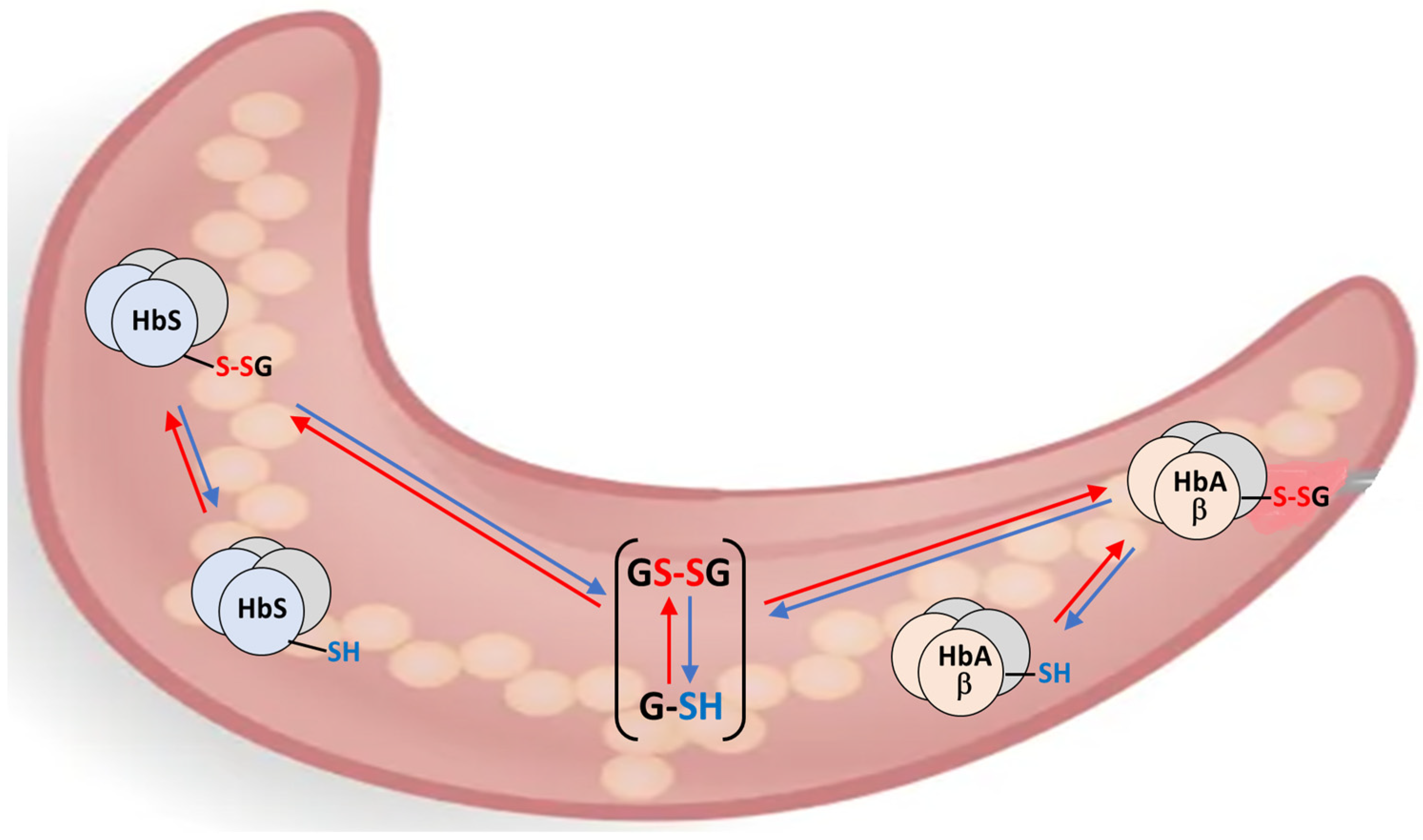
The response of the thiolome of RBCs to oxidative stress involves the reversible distribution of the glutathione pool among its soluble thiol (GSH), disulfide (GSSG), and hemoglobin-bound forms (Hb-SSG), as illustrated in the scheme of Figure 1 for the interplay of the glutathionylated forms of Hemoglobin A and S (HbA-S-SG, HbS-S-SG) in sickled RBCs.
Figure 1.
Interplay of three intra-erythrocyte processes of reversible glutathionylation in the sickle RBCs of a heterozygous HbS-variant carrier.
Therefore, the availability of a biochemical parameter able to assess the physiological response of RBCs to conditions that enhance oxidative stress would simplify understanding this complicated network of processes. The values of E0′ have been measured for the main components of the soluble thiolome utilizing solution and spectroscopic techniques [] and by extrapolation of gas-phase behavior to the solution state []. In addition, re-analysis [] of measured concentrations of GSH, GSSG, and Hb-SSG in an oxidant challenge ex vivo, in vitro experiment [] allowed calculation of the value of E0′ for the 93Cys thiol group of wild-type Hb.
The availability of blood from a carrier of the sickle cell β6Glu→Val variant of human β-Hb prompted an analysis of the composition of hemoglobin isoforms and main modifications obtained by MALDI-ToF mass spectrometry to apply a previously developed calculation for obtaining a value for the hitherto unknown thiol-disulfide redox potential (E0′) of the 93Cys residue in the modified protein. Availability of this value, in turn, assists in understanding the potentiality of the “redox potential” approach to study RBC physiopathology in conditions of chronic and transient oxidative stress.
2. Results
2.1. MALDI Mass Spectra of the Patient’s Hemolysates
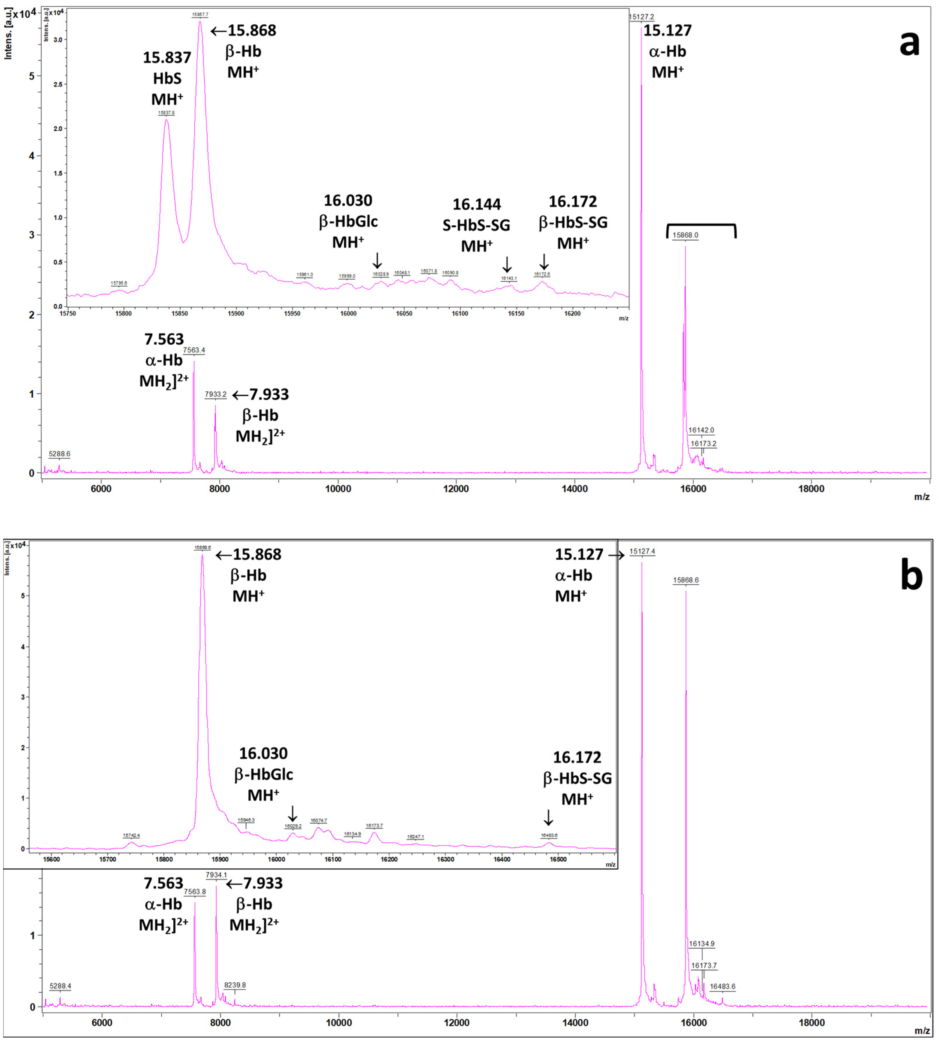
As part of a different research protocol that included assessing HbSSG in the blood of patients [], one subject used the anomalous MALDI mass spectrum reported in Figure 2a. Apart from minor differences in the relative proportions of the minor signals (vide infra), samples from the same patient (n = 2), taken at admission to and discharge from the hospital, showed very similar spectral profiles. As a reference, the spectrum from a wild-type hemoglobin subject is reported as Figure 2b.
Figure 2.
MALDI mass spectra of the hemolysate from (a) the profile of the subject carrying the β6Glu→Val variant of human Hb; (b) the wild-type hemoglobin subject (main analytical conditions are described in the Materials and Methods section). The expansions highlight the minor forms of the β-chain of the wild type (glycated and glutathonylated, in both panels) and, in panel (a), the unmodified and glutathionylated forms of the β6Glu→Val-variant chain.
The proteins in the samples were as summarized in Table A1. Briefly, the patient’s RBCs contain both the α-(m/z 15,127) and β-(m/z 15,868) wild-type hemoglobin chains and, unexpectedly at the time of analysis, an additional signal (m/z 15,837) that corresponds to the β6Glu→Val variant of the β-chain []. The intensity ratio of the two forms of hemoglobin is approximately 61% (β-Hb):39% β6Glu→Val), calculated as the average of four replicated sample depositions for each of the two obtained blood samples, as reported in Table A2.
We should raise awareness that relative MALDI intensities of different proteins may not accurately reflect their actual concentration ratios, due to possible differences in their overall ionization and desorption efficiency during the analysis. This uncontrolled factor may influence the calculation of the actual intra-erythrocyte concentrations of the two hemoglobin isoforms, with a likely minor influence also on the ratio of the glutathionylated to free-cysteine form of each. Nevertheless, due to the current lack of suitable standards, the response factors of the two isoforms are taken as identical for the purpose of the ensuing calculations.
The measured 3:2 proportion of the β-Hb and β6Glu→Val Hb chains points to a heterozygous carrier of the sickle cell trait. In addition, the spectrum does not show substantial levels of the micro-heterogeneous fetal γ forms centered at m/z 15,995 (isoform 137-G) and 16,009 (isoform 137-A), respectively [,]. These proteins would be present in large amounts in the RBCs of sickle cell patients treated with hydroxyurea, a drug that stimulates, in the still immature RBCs, the biosynthesis of the fetal chains [], as we evidenced previously by analyzing an unrelated sample of a homozygous-variant carrier subject undergoing the pharmacological treatment with hydroxyurea.
In addition, as is apparent from the inserts of Figure 2a,b, further minor peaks are present, corresponding to the known minor forms of hemoglobin: the glycated α- and β-forms (m/z 15,289 and 16,030, respectively) and the glutathionylated β-chain (m/z 16,172). One further signal, at m/z 16,142, corresponds to the glutathionylated form of β6Glu→Val chain []. Table A3 reports the mean % proportions of the isoforms in the samples.
2.2. Calculation of the Value of E0 for the 93Cys of HbS
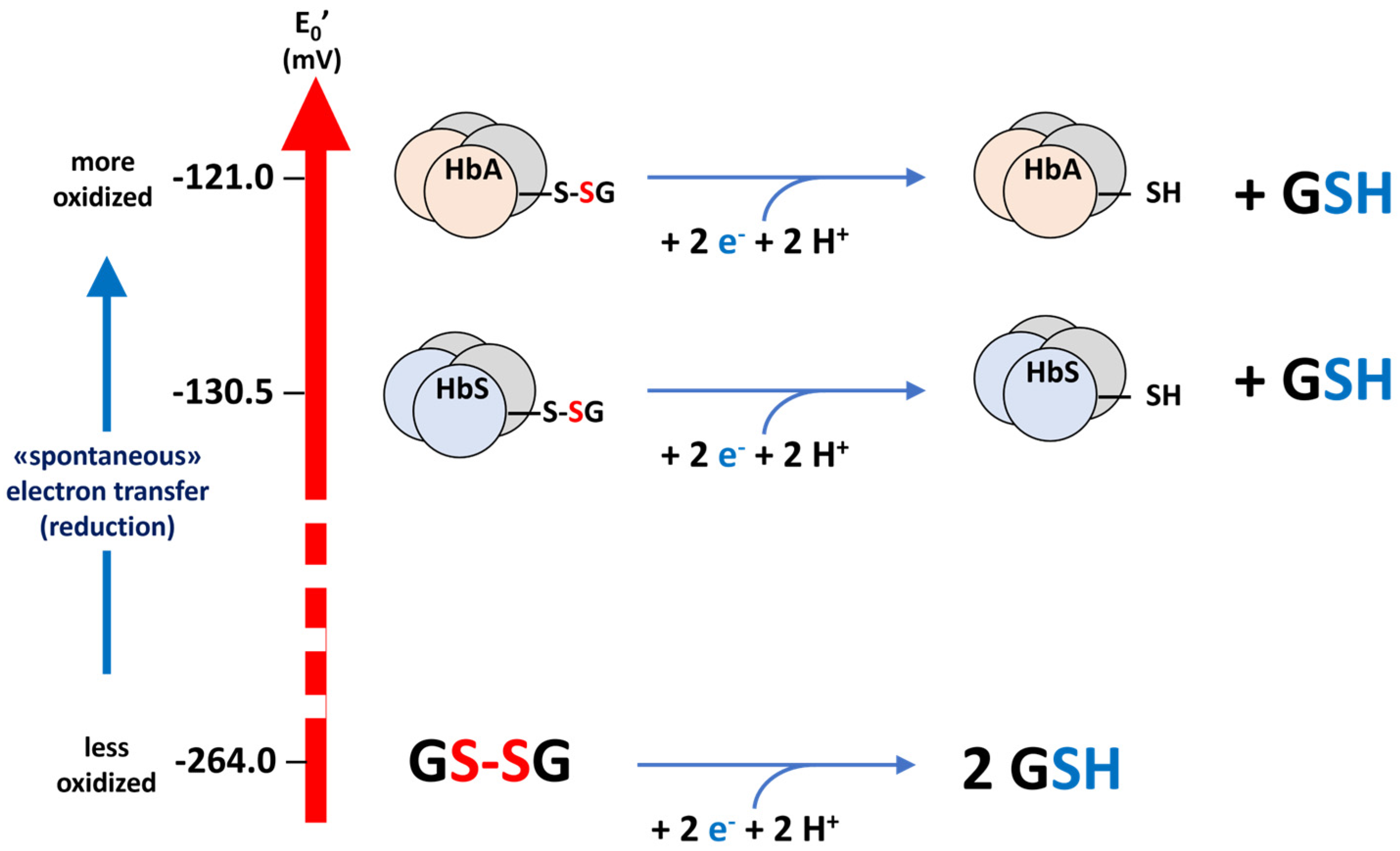
A value for the redox potential of the thiol group in the side chain of 93Cys in β6Glu→Val in HbS could be calculated from the MALDI-ToF mass spectra of the two samples, using the equilibrium method that was developed to calculate the corresponding value for the wild-type protein []. The complete calculation is reported in Appendix B. The values of hematocrit and total Hb concentration were used to calculate the total erythrocyte Hb concentration (line 3 of Table A4). In this case, the two forms of hemoglobin compete for the glutathione pool, and their concentrations were calculated from the total Hb content, while the relative proportions of the different forms were measured by mass spectrometry. Then, the calculated concentrations of the different Hb forms are used in the Nernst equation to calculate the value of E0′(β6Glu→Val-Hb), using the already available E0′(β-Hb) [] as reference. The calculated value of E0′(β6Glu→Val-Hb) of HbS is −130.5 ± 1.7 mV, as the mean of the eight determinations performed on the two samples. This value corresponds to a more negative potential, and therefore to a more nucleophilic (acid) 93Cys thiolate residue, which is thus more reactive towards thiolate exchange with GSSG (more oxidizable) and less reducible by GSH than the homologous one in the wild-type β chain of HbA. The three processes are shown in the scheme of Figure 3.
Figure 3.
E0′ potentials of the redox pairs of glutathione (GSSG/GSH) and of the 93Cys residue in HbS and HbA.
2.3. Application: The Oxidative Stress Level of the Patient Compared to That of In Vitro Oxidatively Challenged Wild-Type Healthy RBCs
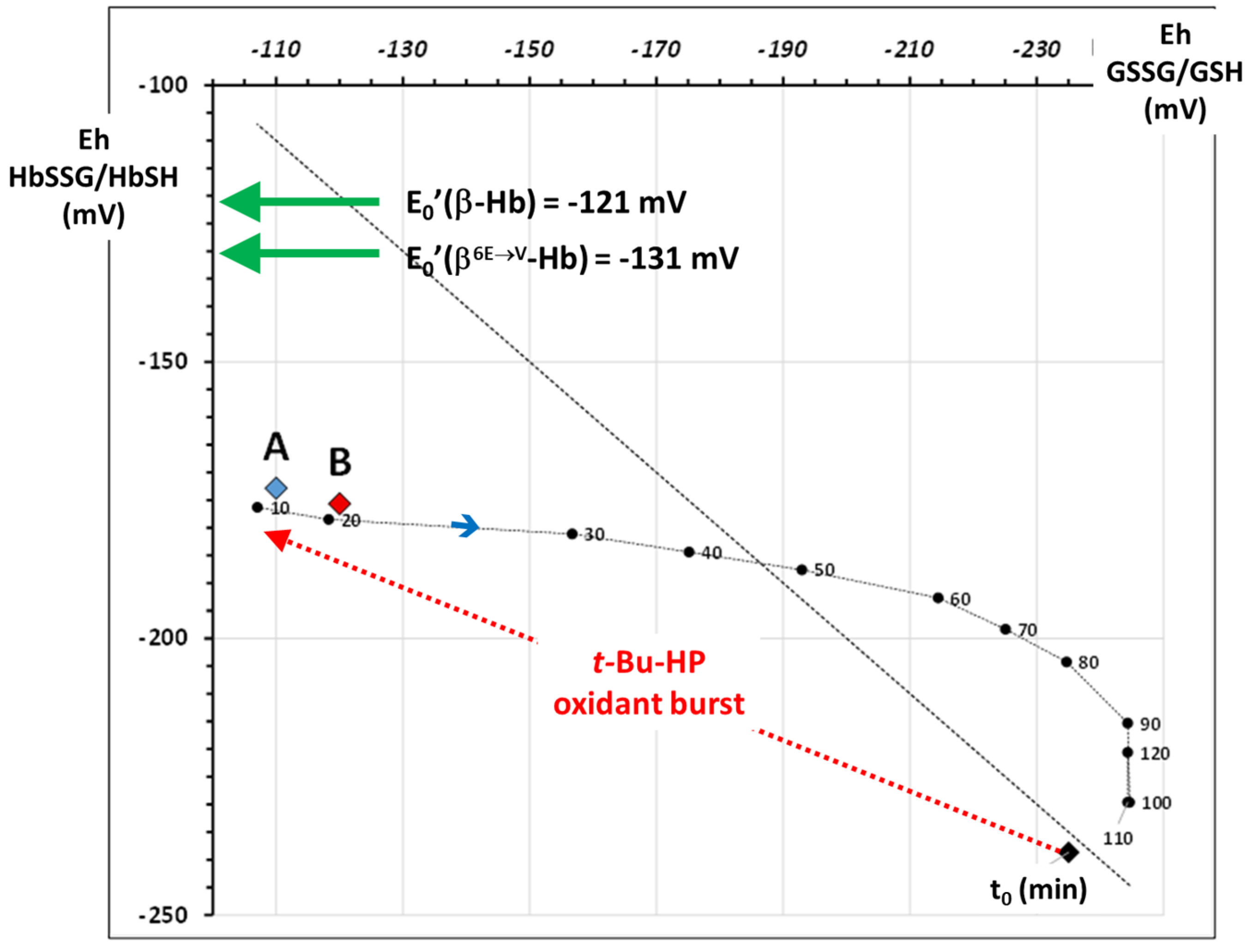
According to the estimated E0′ values for the two hemoglobin variants, in the two blood samples of the patient, the calculated concentration-dependent values of the Eh′ potential, considered at equilibrium, were −172.8 ± 0.1 mV at hospital admission and −175.7 ± 0.2 mV at discharge. A preliminary investigation of the meaning of this observation is developed below.
To frame the condition of chronic oxidative stress of the patient’s sickle RBCs, it is necessary to compare the values of Eh′ in the samples with a reference condition. One such suitable reference is that of complete reversible recovery after acute oxidative stress in a wild-type subject.
This is indeed possible by referring again to the results of [,]. Briefly, we used [] the original data of the previously published experiment [] to calculate the Eh‘ values of the two redox pairs, GSSG/GSH and HbSSG/HbSH, during the oxidant challenge and recovery phase, obtaining the time course of the experiment showed the displacement of the Eh potentials of both species from the “reduced” situation (equilibrium position close to the bisecting line) towards an oxidatively stressed one immediately after the addition of the oxidant and a gradual recovery of the conditions to the initial ones (Figure 4).
Figure 4.
Plot of the Eh values of the glutathione (X-axis) and hemoglobin (Y-axis) redox pairs calculated from the oxidant challenge experiment carried out on wild-type, healthy RBCs [,]. T0 indicates the Eh before the oxidant challenge; black numbers indicate the Eh at progressive times during the recovery phase (from 10 min to 110 min). The two green arrows indicate the E0′ of the two hemoglobins studied in this report. The red and blue A and B diamonds show the values of Eh for HbSSG calculated for the two examined blood patients’ samples at admission and discharge, respectively.
The same graph shows the Eh′ values of the two samples A and B taken from the patient at admission (A, Eh = −172.8 ± 0.1 mV; red diamond) and at discharge (B, Eh = −175.7 ± 0.2 mV; blue diamond). These values describe levels of chronic oxidative stress comparable to those of the in vitro RBCs in the first 10–20 min after the acute oxidative stress and before starting the recovery phase. In addition, the difference of −2.8 ± 0.2 mV between the two samples may point to a slight decrease in the level of oxidative stress during the hospital stay. The Eh of the patient at discharge is not only statistically lower than the one at admission but is also comparable to the value calculated after the 20 min recovery in the “in vitro” experiment on oxidized RBC [].
3. Discussion
We applied a previously developed method to assign a value to the hitherto unknown redox potential of the thiol group of the 93Cys residue in the β6Glu→Val variant of the hemoglobin β-chain. The obtained value, more reducing (more negative) than that of the wild-type species, can explain the empirically observed higher level of glutathionylation in the RBCs of variant carriers [].
It is of interest that the reactivity study of HbA and HbS towards the reference electrophilic reagent iodoacetic acid shows higher and faster alkylation of 93Cys in the wild-type than in the S-mutant form, both in the oxygenated and in the deoxy state []. This observation is a caveat towards adopting for proteins the straightforward correlation between a higher acidity and a more reducing potential that has been observed for small-molecule thiols [].
3.1. The “Thermodynamic”—“Kinetic” Electrochemical Model of Thiol Oxidative Stress in RBCs
The use of redox potential calculated with the Nernst equation can improve the interpretation of cellular responses to oxidative stress [,,].
From the simple side of data presentation, the redox potential Eh merges into a single calculated parameter of the two measured variables represented by the concentrations of the oxidized and reduced forms of a disulfide–thiol pair. This data transformation lowers the number of variables and simplifies data plotting.
In addition, from the side of the physical meaning of the measurements, the Nernstian potential of a disulfide–thiol pair depends not only on the concentration ratio but also on the total pool size of the species. This makes a difference from other redox systems, such as the NAD(P)H/NAD+ and FAD/FADH2, the Eh potential values of which only depend on the oxidized/reduced concentration ratio.
Furthermore, the relative values of the potentials calculated for different redox pairs, such as that of GSSG/GSH and that of Hb-SSG/Hb-SH, indicate the direction of the formal electron flux, as would the electromotive force of a reversible pile, where the lower-potential half-element “spontaneously donates” reducing equivalents to the higher-potential one. The use of these metrics requires the assessment of a fundamental constant for each species, the “normal” potential E0′, the value of which refers to standardized conditions of species concentration and specific conditions of temperature and pH for biologically relevant species.
The measurement of the E0′ of hemoglobin 93Cys, as well as the method that has been applied for the calculation, are thus embedded in a contemporary debate on the theoretically legitimate application of physico-chemical thermodynamic equilibrium models, such as the Nernstian electrochemical potential, to describe the biochemical phenomena inside cells and their substructures []. This highly conserved redox-active amino acid residue [] plays a fundamental role in the preservation of the structural and functional integrity of hemoglobin in the oxidizing environment of the RBC [,].
This simplified approach considers the biochemical phenomena that occur inside physically segregated compartments, such as living cells, as being driven by thermodynamic-kinetic movers that operate under the laws of chemical equilibria in solution. This approach is particularly appealing in RBC biochemistry, due to the occurrence, in a deliberately [,,,,] “simplified” model of RBC physiology, of the biochemical processes as they would in a conceivably “free solution state” of the RBC cytoplasm. This bio-organic reductionist paradigm is, in part, justified by the fact that the mature, circulating human RBCs do not have a nucleus, protein biosynthesis, or mitochondrial electron transport.
This trust in the paradigm had been pioneered by Schafer & Buettner [] and by Jones and collaborators [,,], who introduced the calculation of Nernstian potentials as a physically significant metrics for describing the relative levels of the free-thiol and disulfide forms of the soluble thiols in tissue, in the whole cells and in subcellular organelles in different stages of the response to physiological and stress-challenged conditions [].
As an example of the application of redox potential metrics, one experiment measured the circadian changes in concentrations of plasma cystine/cysteine and glutathione disulfide/glutathione redox pairs in healthy volunteers [] and calculated the respective Eh values from the measured concentrations. The time courses showed prominent and complex circadian variations, with Eh of the glutathione pair ranging from −118 to −123 mV, with maxima around 13:00 and 00:30 and minima reached around 08:00, 17:30, and 22:30, while that of the cysteine pair ranged from −73 to −79 mV, with the minimum reached at around 20:30 and the maxima at around 21:00 and 05:00. The minima for glutathione were shifted concerning those of cysteine that was introduced with the diet and was made available after food digestion.
A further example of the efficacy of the electrochemical equilibrium approach to highlight the in vitro resilience of RBCs to radical oxidant challenge has been reported in Section 2.3 above [,]. This experiment shows that the single oxidant burst did not irreversibly damage the redox homeostasis machinery of the healthy RBCs, which reacted to the oxidation of the intracellular glutathione and hemoglobin pools in a quantitatively reversible mode. The Eh metrics can thus be employed as a theoretically legitimate general frame to compare the effect of different conditions of oxidative stress, including the reversible physiological recovery phase, to an in vitro reference one. This is an appealing possibility in the dynamic study of oxidative stress phenomena occurring in vivo in patients and in subjects experiencing extreme environments.
However, in quantitative models of oxidative stress production and recovery, we should also consider the pivotal role of the enzymes that synthesize glutathione and recycle the disulfide-bound forms, as highlighted by Flohé [,]. His objection is, however, compatible with the results of the described oxidant challenge experiment, which measured resilience to a single oxidative exposure. It is reasonable to expect that repeated oxidative challenges without a suitable recovery phase, or chronic exposure to oxidizing conditions, will lead to a progressive inactivation of the redox enzymes [,]. A decrease in enzyme activity will, in turn, delay the recovery phase of the thiolome, causing high levels of the disulfide forms. This could be the case for several cohorts of chronically impaired subjects, such as heavy smokers [], refractory overweight subjects [], workers exposed to workplace pollutants [], and patients with chronic respiratory impairment [].
3.2. The S-Variant of Hemoglobin and Oxidative Stress in RBCs in Malaria Infection
The examined blood sample belongs to a subject whose heterozygous HbA+/HbS+ status is often associated with provenance from geographical regions with a high prevalence of malaria []. The evolutionary conservation of the heterozygous trait is due to the enhanced resistance of the carrier subjects to the effects of infection with P. falciparum, the causal agent of malaria [,]. According to abundant research, the peculiar physico-chemical properties of HbS make the cytoplasmic environment of the RBCs in the “sickled” shape less adapted to the growth of blood parasites, such as the developing forms of P. falciparum. Protein carbonylation is approximately fivefold higher in P. falciparum cultivated in heterozygous S-trait carriers than in wild-type RBCs []. The malaria parasite needs a specific redox environment to reside and multiply inside the RBCs [] and challenges the pro-oxidant response of host RBCs with a unique form of “improved”, dimeric glutathione, trypanothione, that has better antioxidant properties than the ordinary monomeric tripeptide [].
For this reason, while homozygous carriers of the sickle cell trait have poor chances of surviving in the reproductive age, heterozygous carriers, which express both the HbA and HbS, obtain an evolutionary advantage in geographical areas where malaria is endemic [].
The redox balance of RBCs in subjects with SCD has been studied through several different indicators, which include the measurement of the total, reduced (thiol), and soluble disulfide form of glutathione []. The approx. 35% lower level of total soluble glutathione measured in SCD patients was explained as increased binding to proteins, although without explicit measurement of this compartment. An approximate calculation based on the reported information shows that this level may correspond to a total glutathionylated hemoglobin pool in the 7% range, a level comparable to the 5% measured as the sum of the glutathionylated forms in our examined patient (Table A3).
Glutathionylation is a protective process that modifies the conformation of the tetramer of Hb-S and hampers its polymerization, thus mitigating the “sickling” deformation of RBC shape and the cascade of pathological phenomena caused to the affected subject by this functional impairment [,,,].
Several biomarkers of oxidative stress, including the levels of reduced glutathione and enzyme activities, were measured in plasma and in RBC hemolysates of SCD patients treated or not with hydroxyurea, compared to controls, both in vivo and in vitro [,]. In vivo, hydroxyurea is a putative precursor of nitric oxide, the binding of which to circulating thiols triggers the formation of thiol disulfides. Treatment of RBCs with hydroxyurea triggers a dose-dependent increase in antioxidant defenses (increased activity of Superoxide Dismutase and Glutathione Peroxidase(s), decreased Glutathione Reductase(s) and free glutathione). Concomitantly, there is a switch of the glucose metabolism towards the production of reducing power through the activity increase in Hexokinase and Glucose-6-phosphate Dehydrogenase [].
In the treatment of SCD, the failure of antioxidant treatments, such as administration of strongly reducing N-acetylcysteine (NAC) (E0 = −268 mV []) [] to prevent occlusive crises, can be rationalized as the consequence of an excessive imbalance of the redox equilibrium towards reduction, with decrease in the protective glutathionylated form of HbS and enhanced formation of intra-erythrocyte hemoglobin fibrils. Concerning the use of antioxidant therapy for SCD treatment, de Silva et al. [] call for the use of NAC and Melatonin in combination. A recent study [] measured oxidative stress in the RBCs of wild-type Hb young obese subjects undergoing a weight-loss protocol that included supplementation with melatonin. Contrary to expectations, the melatonin-treated arm showed enhanced rather than lowered levels of oxidative stress, including HbSSG. Understanding how and if this observation can be useful for SCD management calls for additional studies.
In conclusion, the availability of the E0′ redox potentials of the β-93Cys thiolate group of hemoglobin variants could be a useful tool that may add to the limited panel of oxidative stress biomarkers of RBC. The use of the Eh redox potential in addition to the simple % of HbSSG may more efficiently assess the effect of pharmacological and nutraceutical interventions [,,].
4. Materials and Methods
4.1. Patient History
The 41-year-old adult male subject to whom the blood samples belong was born in Cameroun. He had been admitted recently to the hospital for treatment of pneumonia and had been enrolled in a wide, approved research protocol aimed at studying biomarkers of hypoxia. Among the research biomarkers of the study, the measurement of glutathionyl–hemoglobin was included as an indicator of oxidative stress.
4.2. Blood Samples
Two blood samples were obtained, one at admission to and one at discharge from the hospital, from the healed patient. Pre-analytical blood treatment (coding, fractionation, and storage) was performed at the times of admission and discharge, and routine blood analyses were executed by the clinical laboratory. The measurement of glutathionyl–hemoglobin was performed after the end of all patients’ enrolment and discharge. Therefore, the condition of being an otherwise healthy carrier of the HbS hemoglobin variant became known to the investigators only during the post-acquisition review of the mass spectra, when the measurement of glutathionyl–hemoglobin was performed in the study samples batch.
4.3. Measurement of Glutathionyl–Hemoglobin
The measurement of glutathionyl–hemoglobin in the patient’s blood sample was performed during a routine research measurement that employed the published method []. Briefly, a micro-sample of 10 mM titrated hemolysate was measured by Matrix-Assisted Laser Desorption mass spectrometry in a Time-of-Flight instrument (MALDI-ToF) in a Bruker Autoflex II instrument (Bruker, Inc., Bremen, Germany).
Sinapinic acid (SA; 10 mM dissolved in 0.1% trifluoroacetic acid-acetonitrile) was used as MALDI matrix, in a 1:10,000 molar proportion to the hemolysate. An automated procedure was used for laser firing, and the sample was measured in quadruplicate.
4.4. Calculation of the Molecular Masses of Hemoglobin Proteins and Variants
The amino acid sequences of the wild-type and variant hemoglobin chains were obtained from the UniProt database (https://www.uniprot.org/uniprotkb/P68871/entry#structure, accessed on 4 November 2025); their elemental compositions and those of the post-translational modifications (glycation and glutathionylation) were calculated with a custom spreadsheet. The corresponding molecular masses and isotope pattern profiles were calculated from the elemental composition with the Envipat calculator (https://www.envipat.eawag.ch/index.php, accessed on 4 November 2025) [], at a value of mass resolution (M/ΔM) of 1000, to yield the results of Table A1. All results were downloaded to Excel spreadsheets for further elaboration.
4.5. Data Analysis
An original, custom spreadsheet was employed to analyze and integrate the profile mass spectra, extracted as raw, externally calibrated .txt files from the instrument analysis files. The area integration routine was modified in the spreadsheet to include the signals belonging to the unexpected protein species. The obtained m/z values (MH+ species) are reported in Table A1, as the mean and interval of the four separate determinations.
A specific worksheet was prepared to perform the calculations for the estimation of E0 from the raw data.
4.6. Calculation of the E0 Electrochemical Potential of the HbS Glutathionyl–Hemoglobin
The calculation of the E0′ electrochemical potential of the unexpected protein species was carried out by adopting the “equilibrium model”, according to the published method [] employed to calculate the E0′ of the wild-type glutathionyl–hemoglobin in an ex vivo, in vitro oxidant challenge experiment performed on human RBCs [].
Briefly, it is assumed that, in the measured sample, the relative concentrations of “reduced” (thiol-free) and “oxidized” (mixed-disulfide) forms of the glutathionyl–hemoglobins correspond to those derived from the Nernst equation for the electrochemical cell pair of Equation (1)
for which the redox equilibrium condition (Eeq) is calculated from Equation (2)
β-Hb-SSG/β-Hb-SH//β6Glu→Val Hb-SSG/β6Glu→Val Hb-SH
Eeq = E0(β-Hb) + RT/nF ln [β-Hb-SSG]/[β-Hb-SH]2 =
=E0β6Glu→Val Hb) + RT/nF ln [β6Glu→Val Hb-SSG]/[β6Glu→Val Hb-SH]
The value for E0′(β-Hb) is −121 mV [].
The calculation of actual intra-erythrocyte concentrations of the proteins in Equation (2) is performed by referring to their relative proportions (Table A2) to the total hemoglobin concentration, which is calculated from the hematocrit and total Hb concentration according to Equation (3).
HbRBC (mM) = 103*(10*HbWB (g/dL)/64.454)/Ht%
5. Conclusions
This is the first attempt to assign a value to the redox potential of the thiol group in the β-93Cys residue of the point-mutated β6Glu→Val isoform of human hemoglobin, responsible for the sickle cell trait. The obtained value confirms that the thiol group is more reducing and therefore more susceptible to oxidation, nucleophilic attack, and thiol–disulfide exchange in the mutation-carrying than in the wild-type chain (E0′ of −131 mV vs. −121 mV). This feature explains why HbS is more prone to glutathionylation.
The assessment of the redox status of RBCs by the stable biomarker HbSSG% and calculation of HbSSG Eh could improve the understanding of oxidative stress and help the rational treatment with diets and nutraceutical supplements.
Supplementary Materials
The following supporting information can be downloaded at: https://www.mdpi.com/article/10.3390/molecules30224342/s1. Figure S1. Main processes of oxidative stress in RBCs through the generation of hydrogen peroxide and organic hydroperoxides, and coping mechanisms by glutathione and hemoglobin; Figure S2. Main processes of restoration of the reduced thiol pool in RBCs. Refs. [,,] are cited in Supplementary Materials.
Author Contributions
Conceptualization, F.M.R.; methodology, F.M.R.; software, F.M.R.; validation, F.M.R., R.P. and M.S.; formal analysis, M.S. and S.O.; investigation, F.M.R.; resources, D.C. and R.P.; data curation, F.M.R. and M.B.; writing—original draft preparation, F.M.R.; writing—review and editing, F.M.R., R.P., M.B. and M.S.; visualization, F.M.R., A.S., C.M. and M.D.C.; supervision, R.P. and M.M.; project administration, R.P. and M.M.; funding acquisition, D.C. and R.P. All authors have read and agreed to the published version of the manuscript.
Funding
This research was funded by Italian MUR/PRIN 2022-LS2-D.M. 970 30.06.2023 “JANUS—The double face of hypoxia in health and disease”. A.S.’s work was also supported by this Grant.
Institutional Review Board Statement
The study was conducted in accordance with the Declaration of Helsinki and approved by the Comitato Etico Territoriale Lombardia 1 on 16 May 2025, N° CET 176-2025.
Informed Consent Statement
Informed consent was obtained from the subject involved in the study.
Data Availability Statement
The original contributions presented in this study are included in the article/Supplementary Materials. Further inquiries can be directed to the corresponding author.
Acknowledgments
Mass spectrometry measurement was accomplished with the instrument managed by the COSPECT instrument platform of Università degli Studi di Milano. We acknowledge Drs. Enrico Caneva, Marco Pappini, and Milda Stuknyte for their long-standing, friendly assistance during the measurements.
Conflicts of Interest
The authors declare no conflicts of interest.
Abbreviations
The following abbreviations are used in this manuscript:
| βHb | wild-type (6Glu) β-chain of human hemoglobin |
| β6Glu→Val Hb | 6Glu > Val point-mutated β-chain of human hemoglobin |
| E0′ | Standard electrochemical reduction potential (at “near-physiological” pH 7) |
| Eh | Concentration-dependent electrochemical reduction potential |
| GSH | Glutathione, “reduced” thiol form |
| GSSG | Glutathione, “oxidized” disulfide form |
| Prot-SSG | Glutathionylated form of generic protein |
| HbA | Main wild-type form of adult human hemoglobin α2β2 |
| HbS | Main mutated form of sickle cell hemoglobin α2(β6E→V)2 |
| HbA-SH | Main wild-type form of adult human hemoglobin α2β2 thiol form |
| HbS-SH | Main mutated form of sickle cell hemoglobin α2(β6E→V)2 thiol form |
| HbA-SSG | Glutathionyl–hemoglobin A, glutathionylated at β-93Cys |
| HbS-SSG | Glutathionyl–hemoglobin S, glutathionylated at β-93Cys |
| HbA1c | Glycated hemoglobin A |
| β-HbGlc | Glycated hemoglobin A (β-chain) |
| β6Glu→Val HbGlc | Glycated hemoglobin S |
| RBC | Red blood cell(s) |
| SCD | Sickle Cell Disease |
Appendix A. Characterization of the Main Proteins in the RBC Samples by MALDI-ToF Mass Spectrometry
The main proteins in the RBC samples were identified by matching the m/z values measured in the internally calibrated mass spectra to those calculated from the respective amino acid sequences and putatively occurring post-translational modifications (PTM), as summarized in Table A1. Signal intensities were obtained with the spreadsheet described in Ref. [].
Table A1.
Summary of the hemoglobin species identified in the mass spectra of the two blood samples from the HbS carrying patient.
Table A1.
Summary of the hemoglobin species identified in the mass spectra of the two blood samples from the HbS carrying patient.
| MH+ | Samp_1 | Samp_2 | ||||||
|---|---|---|---|---|---|---|---|---|
| Type | Protein | PTM 1 | Molecular Formula | m/z calc | m/z | Δ_m/z 2 | m/z | Δ_m/z 2 |
| wt | α-Hb | - | C685 H1071 N187 O194 S3 | 15,127.5 | 15,127.4 | 0.4 | 15,127.0 | 0.9 |
| wt | α-Hb | Glc | C691 H1081 N187 O199 S3 | 15,289.7 | 15,289.1 | 0.5 | 15,288.5 | 1.6 |
| wt | β-Hb | - | C724 H1119 N195 O201 S3 | 15,868.4 | 15,868.2 | 0.5 | 15,867.9 | 0.7 |
| wt | β-Hb | Glc | C730 H1129 N195 O206 S3 | 16,030.5 | 16,044.1 | 3.8 | 16,043.8 | 1.8 |
| wt | β-Hb | SSG | C734 H1134 N198 O207 S4 | 16,173.6 | 16,172.9 | 0.6 | 16,173.0 | 6.0 |
| β6Glu→Val | β-Hb | - | C724 H1121 N195 O199 S3 | 15,838.4 | 15,838.4 | 0.5 | 15,838.2 | 0.8 |
| β6Glu→Val | β-Hb | Glc | C730 H1131 N195 O204 S3 | 16,000.5 | 15,999.5 | 1.4 | 15,999.1 | 6.4 |
| β6Glu→Val | β-Hb | SSG | C734 H1136 N198 O205 S4 | 16,143.7 | 16,143.4 | 2.6 | 16,141.0 | 3.0 |
1 Post Translational Modification. 2 Max-min interval of centroid m/z, to be compared to the width of the isotope envelope at 1000 resolution, of approx. 14 Da.
Table A2.
Relative proportion (%) of β-Hb and β6Glu→Val Hb in the two blood samples from the HbS carrying patient 1.
Table A2.
Relative proportion (%) of β-Hb and β6Glu→Val Hb in the two blood samples from the HbS carrying patient 1.
| Repl | βHb 1 | β6Glu→Val βHb 1 | βHb 1 | β6Glu→Val βHb 1 |
|---|---|---|---|---|
| Samp1 | Samp2 | |||
| Repl_1 | 61.1 | 38.9 | 61.2 | 38.8 |
| Repl_2 | 60.3 | 39.7 | 60.6 | 39.4 |
| Repl_3 | 60.2 | 39.8 | 61.6 | 38.4 |
| Repl_4 | 60.4 | 39.6 | 61.1 | 38.9 |
| M | 60.5 | 39.5 | 61.2 | 38.8 |
| DS | 0.3 | 0.3 | 0.3 | 0.3 |
| CV% | 0.7 | 1.1 | 0.6 | 1.0 |
1 Integration from custom spreadsheet, only the species at m/z 15,868.4 (β-Hb) and 15,838.4 (β6Glu→Val Hb), respectively.
Table A3.
Proportions (%) of the major and post-translationally modified forms of βHb and β6Glu→Val Hb in the two blood samples from the HbS—carrying patient 1.
Table A3.
Proportions (%) of the major and post-translationally modified forms of βHb and β6Glu→Val Hb in the two blood samples from the HbS—carrying patient 1.
| Samp | A | Samp | B | |||||||||
|---|---|---|---|---|---|---|---|---|---|---|---|---|
| Replicates | Replicates | |||||||||||
| Species | 1 | 2 | 3 | 4 | M | SD | 1 | 2 | 3 | 4 | M | SD |
| β-HbSH | 55.8 | 55.6 | 54.9 | 55.5 | 55.5 | 0.4 | 56.6 | 56.1 | 56.4 | 56.4 | 56.4 | 0.2 |
| β-HbGlc | 3.7 | 3.2 | 3.6 | 3.3 | 3.4 | 0.2 | 3.3 | 3.3 | 4.2 | 3.6 | 3.6 | 0.4 |
| β-HbSSG | 2.7 | 2.7 | 2.7 | 2.7 | 2.7 | 0.0 | 2.2 | 2.2 | 2.2 | 2.2 | 2.2 | 0.0 |
| β6Glu→Val-HbSH | 35.5 | 36.6 | 36.3 | 36.4 | 36.2 | 0.5 | 35.8 | 36.4 | 35.2 | 35.9 | 35.8 | 0.5 |
| β6Glu→Val-HbGlc | <0.5 | <0.5 | <0.5 | <0.5 | <0.5 | <0.5 | <0.5 | <0.5 | ||||
| β6Glu→Val-HbSSG | 2.3 | 1.9 | 2.5 | 2.1 | 2.2 | 0.3 | 2.1 | 2.1 | 2.0 | 2.0 | 2.0 | 0.0 |
| ∑ all HbSSG | 4.9 | 0.2 | 4.2 | 0.0 | ||||||||
1 Integration from custom spreadsheet, sum of all considered species, α-Hb excluded.
Appendix B. Calculation of the E0 of S-Hb with the Equilibrium Method
The calculation with the equilibrium method is reported in Table A4 below and follows the method described in ref. [], separately for the two blood samples A (at admission) and B (at healed discharge).
Table A4.
Development of the calculation to estimate the value of E0′ for the thiol group of the Hb-S variant of hemoglobin from the composition of the protein forms in the two blood samples (A and B) from the β6E→V Hb-S—carrying patient.
Table A4.
Development of the calculation to estimate the value of E0′ for the thiol group of the Hb-S variant of hemoglobin from the composition of the protein forms in the two blood samples (A and B) from the β6E→V Hb-S—carrying patient.
| Samp | A | B | |||||||||||
| Hb (g/dL)a | 11.9 | 13.9 | |||||||||||
| Ht %a | 35.6% | 42.6% | |||||||||||
| [Hb] mM | 5.02 | 4.90 | |||||||||||
| Species | 1 | 2 | 3 | 4 | M | SD | 1 | 2 | 3 | 4 | M | SD | |
| β-HbA-SH | 55.8% | 55.6% | 54.9% | 55.5% | 55.5% | 0.4% | 56.6% | 56.1% | 56.4% | 56.4% | 56.4% | 0.2% | |
| β-HbGlc | 3.7% | 3.2% | 3.6% | 3.3% | 3.4% | 0.2% | 3.3% | 3.3% | 4.2% | 3.6% | 3.6% | 0.4% | |
| β-HbSSG | 2.7% | 2.7% | 2.7% | 2.7% | 2.7% | 0.0% | 2.2% | 2.2% | 2.2% | 2.2% | 2.2% | 0.0% | |
| β6Glu→Val HbA-SH | 35.5% | 36.6% | 36.3% | 36.4% | 36.2% | 0.5% | 35.8% | 36.4% | 35.2% | 35.9% | 35.8% | 0.5% | |
| β6Glu→Val HbGlc | <0.5% | <0.5% | <0.5% | <0.5% | <0.5% | <0.5% | <0.5% | <0.5% | |||||
| β6Glu→Val HbS-SSG | 2.3% | 1.9% | 2.5% | 2.1% | 2.2% | 0.3% | 2.1% | 2.1% | 2.0% | 2.0% | 2.0% | 0.05% | |
| β-HbSH | mM | 2.80 | 2.79 | 2.76 | 2.78 | 2.77 | 2.75 | 2.76 | 2.76 | ||||
| β-HbGlc | mM | 0.18 | 0.16 | 0.18 | 0.17 | 0.16 | 0.16 | 0.21 | 0.17 | ||||
| β-HbSSG | mM | 0.14 | 0.14 | 0.13 | 0.14 | 0.11 | 0.11 | 0.11 | 0.11 | ||||
| β6Glu→Val-HbSH | mM | 1.78 | 1.84 | 1.82 | 1.83 | 1.75 | 1.78 | 1.72 | 1.76 | ||||
| β6Glu→Val-HbGlc | mM | ||||||||||||
| β6Glu→Val HbSSG | mM | 0.12 | 0.10 | 0.13 | 0.11 | 0.10 | 0.10 | 0.10 | 0.10 | ||||
| Nernst calculation | |||||||||||||
| RT/nF (mV) | 29.5 | 29.5 | 29.5 | 29.5 | 29.5 | 29.5 | 29.5 | 29.5 | |||||
| E0_β-Hb (mV) | mV | −121 | −121 | −121 | −121 | −121 | −121 | −121 | −121 | ||||
| β _[OX]/[RED]^2 | 0.0174 | 0.0174 | 0.0177 | 0.0175 | 0.0139 | 0.0143 | 0.0142 | 0.0138 | |||||
| β6Glu→Val [OX]/[RED]^2 | 0.0372 | 0.0284 | 0.0378 | 0.0316 | 0.0333 | 0.0319 | 0.0331 | 0.0316 | |||||
| β_logRat | −1.758 | −1.760 | −1.752 | −1.758 | −1.857 | −1.846 | −1.848 | −1.860 | |||||
| β6Glu→Val _logRat | −1.430 | −1.547 | −1.422 | −1.501 | −1.477 | −1.496 | −1.480 | −1.501 | |||||
| A_Eh (mV) | −173 | −173 | −173 | −173 | −172.8 | ±0.1 | −176 | −175 | −176 | −176 | −175.7 | ±0.2 | |
| β6Glu→Val _Eh-E0 (mV) | −42 | −46 | −42 | −44 | −43.5 | ±1.8 | −44 | −44 | −44 | −44 | −43.9 | ±0.3 | |
| E0′β6Glu→Val-Hb (mV) | mV | −130.7 | −127.3 | −130.7 | −128.6 | −129.3 | ±1.7 | −132.2 | −131.3 | −131.9 | −131.6 | −131.7 | ±0.4 |
a Data from the clinical laboratory.
The estimated mean E0′ value resulting from the calculation for the disulfide–thiol redox process of the β6Glu→Val-93Cys thiol group is thus −130.5 ± 1.7 mV.
References
- Pauling, L.; Itano, H.A.; Singer, S.J.; Wells, I.C. Sickle Cell Anemia, a Molecular Disease. Science 1949, 110, 543–548. [Google Scholar] [CrossRef]
- Ingram, V.M. A specific chemical difference between the globins of normal human and sickle-cell anaemia haemoglobin. Nature 1956, 178, 792–794. [Google Scholar] [CrossRef] [PubMed]
- Ingram, V.M. Gene mutations in human haemoglobin: The chemical difference between normal and sickle cell haemoglobin. Nature 1957, 180, 326–328. [Google Scholar] [CrossRef] [PubMed]
- Garel, M.C.; Domenget, C.; Caburi-Martin, J.; Prehu, C.; Galacteros, F.; Beuzard, Y. Covalent binding of glutathione to hemoglobin. I. Inhibition of hemoglobin S polymerization. J. Biol. Chem. 1986, 261, 14704–14709. [Google Scholar] [CrossRef] [PubMed]
- Das, R.; Mitra, A.; Mitra, G.; Maity, D.; Bhat, V.; Pal, D.; Ross, C.; Kurpad, A.V.; Mandal, A.K. Molecular insights of inhibition in sickle hemoglobin polymerization upon glutathionylation: Hydrogen/deuterium exchange mass spectrometry and molecular dynamics simulation-based approach. Biochem. J. 2018, 475, 2153–2166. [Google Scholar] [CrossRef] [PubMed]
- Mandal, A.K.; Mitra, A.; Das, R. Sickle Cell Hemoglobin. Subcell. Biochem. 2020, 94, 297–322. [Google Scholar] [CrossRef] [PubMed]
- Agarwal, A.M.; Rets, A.V. Advances in Hemoglobinopathies and Thalassemia Evaluation. Clin. Lab. Med. 2024, 44, 441–453. [Google Scholar] [CrossRef] [PubMed]
- The Lancet Haematology. Sickle cell disease: A neglected health priority. Lancet Haematol. 2025, 12, e769. [Google Scholar] [CrossRef] [PubMed]
- Möller, M.N.; Orrico, F.; Villar, S.F.; López, A.C.; Silva, N.; Donzé, M.; Thomson, L.; Denicola, A. Oxidants and Antioxidants in the Redox Biochemistry of Human Red Blood Cells. ACS Omega 2022, 8, 147–168. [Google Scholar] [CrossRef] [PubMed] [PubMed Central]
- Daraghmeh, D.N.; Karaman, R. The Redox Process in Red Blood Cells: Balancing Oxidants and Antioxidants. Antioxidants 2024, 14, 36. [Google Scholar] [CrossRef] [PubMed] [PubMed Central]
- Spinelli, S.; Marino, A.; Remigante, A.; Morabito, R. Redox Homeostasis in Red Blood Cells: From Molecular Mechanisms to Antioxidant Strategies. Curr. Issues Mol. Biol. 2025, 47, 655. [Google Scholar] [CrossRef] [PubMed] [PubMed Central]
- Fenk, S.; Melnikova, E.V.; Anashkina, A.A.; Poluektov, Y.M.; Zaripov, P.I.; Mitkevich, V.A.; Tkachev, Y.V.; Kaestner, L.; Minetti, G.; Mairbäurl, H.; et al. Hemoglobin is an oxygen-dependent glutathione buffer adapting the intracellular reduced glutathione levels to oxygen availability. Redox Biol. 2022, 58, 102535. [Google Scholar] [CrossRef] [PubMed]
- Kuleshova, I.D.; Zaripov, P.I.; Poluektov, Y.M.; Anashkina, A.A.; Kaluzhny, D.N.; Parshina, E.Y.; Maksimov, G.V.; Mitkevich, V.A.; Makarov, A.A.; Petrushanko, I.Y. Changes in Hemoglobin Properties in Complex with Glutathione and after Glutathionylation. Int. J. Mol. Sci. 2023, 24, 13557. [Google Scholar] [CrossRef] [PubMed]
- Regazzoni, L.; Panusa, A.; Yeum, K.-J.; Carini, M.; Aldini, G. Hemoglobin Glutathionylation Can Occur Through Cysteine Sulfenic Acid Intermediate: Electrospray Ionization LTQ-Orbitrap Hybrid Mass Spectrometry Studies. J. Chromatogr. B 2009, 877, 3456–3461. [Google Scholar] [CrossRef] [PubMed]
- Colombo, G.; Dalle-Donne, I.; Giustarini, D.; Gagliano, N.; Portinaro, N.; Colombo, R.; Rossi, R.; Milzani, A.D.G. Cellular Redox Potential and Hemoglobin S-Glutathionylation in Human and Rat Erythrocytes: A Comparative Study. Blood Cells Mol. Dis. 2010, 44, 133–139. [Google Scholar] [CrossRef]
- Minetti, M.; Pietraforte, D.; Carbone, V.; Salzano, A.M.; Scorza, G.; Marino, G. Scavenging of Peroxynitrite by Oxyhemoglobin and Identification of Modified Globin Residues. Biochemistry 2000, 39, 6689–6697. [Google Scholar] [CrossRef]
- Böhmer, A.; Pich, A.; Schmidt, M.; Haghiki, A.; Tsikas, D. Evidence by chromatography and mass spectrometry that inorganic nitrite induces S-glutathionylation of hemoglobin in human red blood cells. J. Chromatogr. B 2016, 1019, 72–82. [Google Scholar] [CrossRef]
- Kosmachevskaya, O.V.; Nasybullina, E.I.; Pokidova, O.V.; Sanina, N.A.; Topunov, A.F. Effects of Nitrosyl Iron Complexes with Thiol, Phosphate, and Thiosulfate Ligands on Hemoglobin. Int. J. Mol. Sci. 2024, 25, 7194. [Google Scholar] [CrossRef] [PubMed]
- Van ‘t Erve, T.J.; Wagner, B.A.; Ryckman, K.K.; Raife, T.J.; Buettner, G.R. The concentration of glutathione in human erythrocytes is a heritable trait. Free Radic. Biol. Med. 2013, 65, 742–749. [Google Scholar]
- Mieyal, J.J.; Starke, D.W.; Gravina, S.A.; Dothey, C.; Chung, J.S. Thioltransferase in Human Red Blood Cells: Purification and Properties. Biochemistry 1991, 30, 6088–6097. [Google Scholar] [CrossRef]
- Scirè, A.; Casari, G.; Romaldi, B.; de Bari, L.; Antognelli, C.; Armeni, T. Glutathionyl Hemoglobin and Its Emerging Role as a Clinical Biomarker of Chronic Oxidative Stress. Antioxidants 2023, 12, 1976. [Google Scholar] [CrossRef]
- Deponte, M. Glutathione catalysis and the reaction mechanisms of glutathione-dependent enzymes. Biochim. Biophys. Acta 2013, 1830, 3217–3266. [Google Scholar] [CrossRef]
- Key, A.; Haiman, Z.; Palsson, B.O.; D’Alessandro, A. Modeling Red Blood Cell Metabolism in the Omics Era. Metabolites 2023, 13, 1145. [Google Scholar] [CrossRef] [PubMed] [PubMed Central]
- Spinelli, S.; Marino, A.; Morabito, R.; Remigante, A. Interplay Between Metabolic Pathways and Increased Oxidative Stress in Human Red Blood Cells. Cells 2024, 13, 2026. [Google Scholar] [CrossRef] [PubMed] [PubMed Central]
- D’Alessandro, A. Red blood cell metabolism: A window on systems health towards clinical metabolomics. Curr. Opin. Hematol. 2025, 32, 111–119. [Google Scholar] [CrossRef]
- Haiman, Z.B.; Key, A.; D’Alessandro, A.; Palsson, B.O. RBC-GEM: A genome-scale metabolic model for systems biology of the human red blood cell. PLoS Comput. Biol. 2025, 21, e1012109. [Google Scholar] [CrossRef] [PubMed]
- Thom, G.G.; Kallanagowdar, C.; Somjee, S.S.; Velez, M.C.; Yu, L.C.; Hempe, J.M. Characterization of S-glutathionyl hemoglobin in homozygous sickle cell disease. J. Pediatr. Hematol. Oncol. 2009, 31, 895–900. [Google Scholar] [CrossRef] [PubMed]
- Muralidharan, M.; Mitra, A.; Maity, D.; Pal, D.; Mandal, A.K. Structural analysis of glutathionyl hemoglobin using native mass spectrometry. J. Struct. Biol. 2019, 208, 107386. [Google Scholar] [CrossRef] [PubMed]
- Rabenstein, D.L.; Millis, K.K. Nuclear magnetic resonance study of the thioltransferase-catalyzed glutathione/glutathione disulfide interchange reaction. Biochim. Biophys. Acta 1995, 1249, 29–36. [Google Scholar] [CrossRef] [PubMed]
- Rubino, F.M.; Pitton, M.; Caneva, E.; Pappini, M.; Colombi, A. Thiol-disulfide redox equilibria of glutathione metaboloma compounds investigated by tandem mass spectrometry. Rapid Commun. Mass. Spectrom. 2008, 22, 3935–3948. [Google Scholar] [CrossRef] [PubMed]
- Rubino, F.M. The Redox Potential of the β-93-Cysteine Thiol Group in Human Hemoglobin Estimated from In Vitro Oxidant Challenge Experiments. Molecules 2021, 26, 2528. [Google Scholar] [CrossRef] [PubMed]
- Duca, L.; Ottolenghi, S.; Coppola, S.; Rinaldo, R.; Dei Cas, M.; Rubino, F.M.; Paroni, R.; Samaja, M.; Chiumello, D.A.; Motta, I. Differential Redox State and Iron Regulation in Chronic Obstructive Pulmonary Disease, Acute Respiratory Distress Syndrome and Coronavirus Disease 2019. Antioxidants 2021, 10, 1460. [Google Scholar] [CrossRef] [PubMed] [PubMed Central]
- Houston, C.T.; Reilly, J.P. Rapid Analysis of Hemoglobin from Whole Human Blood by Matrix-assisted Laser Desorption/Ionization Time-of-flight Mass Spectrometry. Rapid Commun. Mass Spectrom. 1997, 11, 1435–1439. [Google Scholar] [CrossRef]
- Ehrmann, D.C.; Rose, K.; Calcutt, M.W.; Beller, A.B.; Hill, S.; Rogers, T.J.; Steele, S.D.; Hachey, D.L.; Aschner, J.L. Glutathionylated γG and γA subunits of hemoglobin F: A novel post-translational modification found in extremely premature infants by LC-MS and nanoLC-MS/MS. J. Mass Spectrom. 2014, 49, 178–183. [Google Scholar] [CrossRef] [PubMed]
- Zhang, Q.; Wang, G.; Sun, D.; Lin, W.; Yan, T.; Wu, Y.; Wu, M.; Chen, J.; Zou, S.; Xie, W.; et al. MALDI-TOF-MS for Rapid Screening and Typing of β-Globin Variant and β-Thalassemia through Direct Measurements of Intact Globin Chains. Clin. Chem. 2022, 68, 1541–1551. [Google Scholar] [CrossRef] [PubMed]
- Lanzkron, S.; Strouse, J.J.; Wilson, R.; Beach, M.C.; Haywood, C.; Park, H.; Witkop, C.; Bass, E.B.; Segal, J.B. Systematic review: Hydroxyurea for the treatment of adults with sickle cell disease. Ann. Intern. Med. 2008, 148, 939–955. [Google Scholar] [CrossRef] [PubMed]
- Mitra, A.; Muralidharan, M.; Srivastava, D.; Das, R.; Bhat, V.; Mandal, A.K. Assessment of Cysteine Reactivity of Human Hemoglobin at Its Residue Level: A Mass Spectrometry-Based Approach. Hemoglobin 2017, 41, 300–305. [Google Scholar] [CrossRef]
- Bulaj, G.; Kortemme, T.; Goldenberg, D.P. Ionization-reactivity relationships for cysteine thiols in polypeptides. Biochemistry 1998, 37, 8965–8972. [Google Scholar] [CrossRef] [PubMed]
- Martinovich, G.G.; Cherenkevich, S.N.; Sauer, H. Intracellular redox state: Towards quantitative description. Eur. Biophys. J. 2005, 34, 937–942. [Google Scholar] [CrossRef]
- Kemp, M.; Go, Y.-M.; Jones, D.P. Nonequilibrium Thermodynamics of Thiol/Disulfide Redox Systems: A Perspective on Redox Systems Biology. Free Radic. Biol. Med. 2008, 44, 921–937. [Google Scholar] [CrossRef]
- Khazim, K.; Giustarini, D.; Rossi, R.; Verkaik, D.; Cornell, J.E.; Cunningham, S.E.; Mohammad, M.; Trochta, K.; Lorenzo, C.; Folli, F.; et al. Glutathione redox potential is low and glutathionylated and cysteinylated hemoglobin levels are elevated in maintenance hemodialysis patients. Transl. Res. 2013, 162, 16–25. [Google Scholar] [CrossRef] [PubMed] [PubMed Central]
- Alayash, A.I. βCysteine 93 in human hemoglobin: A gateway to oxidative stability in health and disease. Lab. Investig. 2021, 101, 4–11. [Google Scholar] [CrossRef]
- Anashkina, A.A.; Simonenko, S.Y.; Orlov, Y.L.; Petrushanko, I.Y. Glutathione Non-Covalent Binding Sites on Hemoglobin and Major Glutathionylation Target betaCys93 Are Conservative among Both Hypoxia-Sensitive and Hypoxia-Tolerant Mammal Species. Int. J. Mol. Sci. 2024, 25, 53. [Google Scholar] [CrossRef] [PubMed]
- Meng, F.; Kassa, T.; Strader, M.B.; Soman, J.; Olson, J.S.; Alayash, A.I. Substitutions in the β subunits of sickle-cell hemoglobin improve oxidative stability and increase the delay time of sickle-cell fiber formation. J. Biol. Chem. 2019, 294, 4145–4159. [Google Scholar] [CrossRef]
- Sae-Lee, W.; McCafferty, C.L.; Verbeke, E.J.; Havugimana, P.C.; Papoulas, O.; McWhite, C.D.; Houser, J.R.; Vanuytsel, K.; Murphy, G.J.; Drew, K.; et al. The protein organization of a red blood cell. Cell Rep. 2022, 40, 111103. [Google Scholar] [CrossRef] [PubMed]
- Schafer, F.Q.; Buettner, G.R. Redox Environment of the Cell as Viewed Through the Redox State of the Glutathione Disulfide/Glutathione Couple. Free Radic. Biol. Med. 2001, 30, 1191–1212. [Google Scholar]
- Jones, D.P. Redox Potential of [GSH]/[GSSG] Couple: Assay and Biological Significance. Methods Enzym. 2002, 348, 93–112. [Google Scholar]
- Jones, D.P.; Sies, H. The Redox Code. Antioxid. Redox Signal 2015, 23, 734–746. [Google Scholar] [CrossRef] [PubMed]
- Blanco, R.A.; Ziegler, T.R.; Carlson, B.A.; Cheng, P.Y.; Park, Y.; Cotsonis, G.A.; Accardi, C.J.; Jones, D.P. Diurnal variation in glutathione and cysteine redox states in human plasma. Am. J. Clin. Nutr. 2007, 86, 1016–1023. [Google Scholar] [CrossRef] [PubMed]
- Berndt, C.; Lillig, C.H.; Flohé, L. Redox regulation by glutathione needs enzymes. Front. Pharmacol. 2014, 5, 168. [Google Scholar] [CrossRef] [PubMed] [PubMed Central]
- Flohé, L. The Fairytale of the GSSG/GSH Redox Potential. Biochim. Biophys. Acta BBA Gen. Subj. 2013, 1830, 3139–3142. [Google Scholar] [CrossRef]
- Niwa, T. Protein glutathionylation and oxidative stress. J. Chromatogr. B Anal. Technol. Biomed. Life Sci. 2007, 855, 59–65. [Google Scholar] [CrossRef] [PubMed]
- Tamma, G.; Valenti, G. Evaluating the Oxidative Stress in Renal Diseases: What Is the Role for S-Glutathionylation? Antioxid. Redox Signal. 2016, 25, 147–164. [Google Scholar] [CrossRef] [PubMed]
- Rubino, F.M.; Della Noce, C.; Vigna, L.; De Giuseppe, R.; Novembrino, C.; De Liso, F.; Maiavacca, R.; Patrini, L.; Riboldi, L.; Bamonti, F. Measurement of Glutathionylated Haemoglobin by MAL-DI-ToF Mass Spectrometry as a Biomarker of Oxidative Stress in Heavy Smokers and in Occupational Obese Subjects. Int. J. Anal. Mass Spectrom. Chromatogr. 2013, 1, 22–30. [Google Scholar] [CrossRef]
- Primavera, A.; Fustinoni, S.; Biroccio, A.; Ballerini, S.; Urbani, A.; Bernardini, S.; Federici, G.; Capucci, E.; Manno, M.; Bello, M.L. Glutathione Transferases and Glutathionylated Hemoglobin in Workers Exposed to Low Doses of 1,3-Butadiene. Cancer Epidemiol. Biomark. Prev. 2008, 17, 3004–3012. [Google Scholar] [CrossRef] [PubMed]
- Laval, G.; Peyrégne, S.; Zidane, N.; Harmant, C.; Renaud, F.; Patin, E.; Prugnolle, F.; Quintana-Murci, L. Recent Adaptive Acquisition by African Rainforest Hunter-Gatherers of the Late Pleistocene Sickle-Cell Mutation Suggests Past Differences in Malaria Exposure. Am. J. Hum. Genet. 2019, 104, 553–561. [Google Scholar] [CrossRef] [PubMed]
- Carter, R.; Mendis, K.N. Evolutionary and historical aspects of the burden of malaria. Clin. Microbiol. Rev. 2002, 15, 564–594, Erratum in Clin. Microbiol. Rev. 2003, 16, 173. [Google Scholar] [CrossRef] [PubMed]
- Esoh, K.; Wonkam, A. Evolutionary history of sickle-cell mutation: Implications for global genetic medicine. Hum. Mol. Genet. 2021, 30, R119–R128. [Google Scholar] [CrossRef] [PubMed]
- Díaz-Castillo, A.; Contreras-Puentes, N.; Alvear-Sedán, C.; Moneriz-Pretell, C.; Rodríguez-Cavallo, E.; Mendez-Cuadro, D. Sickle Cell Trait Induces Oxidative Damage on Plasmodium falciparum Proteome at Erythrocyte Stages. Int. J. Mol. Sci. 2019, 20, 5769. [Google Scholar] [CrossRef] [PubMed] [PubMed Central]
- Flohé, L.; Hecht, H.J.; Steinert, P. Glutathione and trypanothione in parasitic hydroperoxide metabolism. Free Radic. Biol. Med. 1999, 27, 966–984. [Google Scholar] [CrossRef] [PubMed]
- Gizi, A.; Papassotiriou, I.; Apostolakou, F.; Lazaropoulou, C.; Papastamataki, M.; Kanavaki, I.; Kalotychou, V.; Goussetis, E.; Kattamis, A.; Rombos, I.; et al. Assessment of oxidative stress in patients with sickle cell disease: The glutathione system and the oxidant-antioxidant status. Blood Cells Mol. Dis. 2011, 46, 220–225. [Google Scholar] [CrossRef] [PubMed]
- Mitra, G.; Muralidharan, M.; Narayanan, S.; Pinto, J.; Srinivasan, K.; Mandal, A.K. Glutathionylation induced structural changes in oxy human hemoglobin analyzed by backbone amide hydrogen/deuterium exchange and MALDI-mass spectrometry. Bioconjug. Chem. 2012, 23, 2344–2353. [Google Scholar] [CrossRef] [PubMed]
- Renó, C.O.; Barbosa, A.R.; de Carvalho, S.S.; Pinheiro, M.B.; Rios, D.R.; Cortes, V.F.; Barbosa, L.A.; Santos, H.L. Oxidative stress assessment in sickle cell anemia patients treated with hydroxyurea. Ann. Hematol. 2020, 99, 937–945. [Google Scholar] [CrossRef] [PubMed]
- Renó, C.O.; Maia, G.A.S.; Nogueira, L.S.; de Barros Pinheiro, M.; Rios, D.R.A.; Cortes, V.F.; de Oliveira Barbosa, L.A.; de Lima Santos, H. Biochemical Evaluation of the Effects of Hydroxyurea in Vitro on Red Blood Cells. Antioxidants 2021, 10, 1599. [Google Scholar] [CrossRef]
- Bolarinwa, A.B.; Oduwole, O.; Okebe, J.; Ogbenna, A.A.; Otokiti, O.E.; Olatinwo, A.T. Antioxidant supplementation for sickle cell disease. Cochrane Database Syst. Rev. 2024, 2024, CD013590. [Google Scholar] [CrossRef] [PubMed]
- da Silva, D.G.; Ricci, O., Jr.; de Almeida, E.A.; Bonini-Domingos, C.R. Potential utility of melatonin as an antioxidant therapy in the management of sickle cell anemia. J. Pineal Res. 2015, 58, 178–188. [Google Scholar] [CrossRef] [PubMed]
- Rigamonti, A.E.; Rubino, F.M.; Caroli, D.; Bondesan, A.; Mai, S.; Cella, S.G.; Centofanti, L.; Paroni, R.; Sartorio, A. Effects of Melatonin on Exercise-Induced Oxidative Stress in Adults with Obesity Undergoing a Multidisciplinary Body Weight Reduction Program. J. Clin. Med. 2024, 13, 5216. [Google Scholar] [CrossRef] [PubMed] [PubMed Central]
- Ohemeng, A.; Boadu, I. The role of nutrition in the pathophysiology and management of sickle cell disease among children: A review of literature. Crit. Rev. Food Sci. Nutr. 2018, 58, 2299–2305. [Google Scholar] [CrossRef]
- Kalpatthi, R.; Novelli, E.M. Measuring success: Utility of biomarkers in sickle cell disease clinical trials and care. Hematol. Am. Soc. Hematol. Educ. Program. 2018, 2018, 482–492. [Google Scholar] [CrossRef] [PubMed] [PubMed Central]
- Orsi, B.C.; Gorski, D.; Krul, N.E.; Wiens, A.; Brito, M.; Tonin, F.S.; Pontarolo, R. The effects of nutritional supplementation for children and adolescents with sickle cell disease: A systematic review and meta-analyses. Clin. Nutr. 2025, 47, 157–168. [Google Scholar] [CrossRef]
- Rubino, F.M.; Ottolenghi, S.; Brizzolari, A.; Maioli, C.; Samaja, M.; Paroni, R. Enhanced-Precision Measurement of Glutathionyl Hemoglobin by MALDI-ToF MS. Molecules 2023, 28, 497. [Google Scholar] [CrossRef] [PubMed]
- Loos, M.; Gerber, C.; Corona, F.; Hollender, J.; Singer, H. Accelerated Isotope Fine Structure Calculation Using Pruned Transition Trees. Anal. Chem. 2015, 87, 5738–5744. [Google Scholar] [CrossRef] [PubMed]
- Gravina, S.A.; Mieyal, J.J. Thioltransferase is a specific glutathionyl mixed disulfide oxidoreductase. Biochemistry 1993, 32, 3368–3376. [Google Scholar] [CrossRef]
- Mieyal, J.J.; Gallogly, M.M.; Qanungo, S.; Sabens, E.A.; Shelton, M.D. Molecular mechanisms and clinical implications of reversible protein S-glutathionylation. Antioxid. Redox Signal. 2008, 10, 1941–1988. [Google Scholar] [CrossRef]
- Lang, L.; Reinert, P.; Diaz, C.; Deponte, M. The dithiol mechanism of class I glutaredoxins promotes specificity for glutathione as a reducing agent. Redox Biol. 2024, 78, 103410. [Google Scholar] [CrossRef] [PubMed]
Disclaimer/Publisher’s Note: The statements, opinions and data contained in all publications are solely those of the individual author(s) and contributor(s) and not of MDPI and/or the editor(s). MDPI and/or the editor(s) disclaim responsibility for any injury to people or property resulting from any ideas, methods, instructions or products referred to in the content. |
© 2025 by the authors. Licensee MDPI, Basel, Switzerland. This article is an open access article distributed under the terms and conditions of the Creative Commons Attribution (CC BY) license (https://creativecommons.org/licenses/by/4.0/).