

In Silico Design, Synthesis, and Evaluation of Novel Enantiopure Isoxazolidines as Promising Dual Inhibitors of α-Amylase and α-Glucosidase

 , , ,
, , ,  ,
,
Abstract
1. Introduction
2. Results
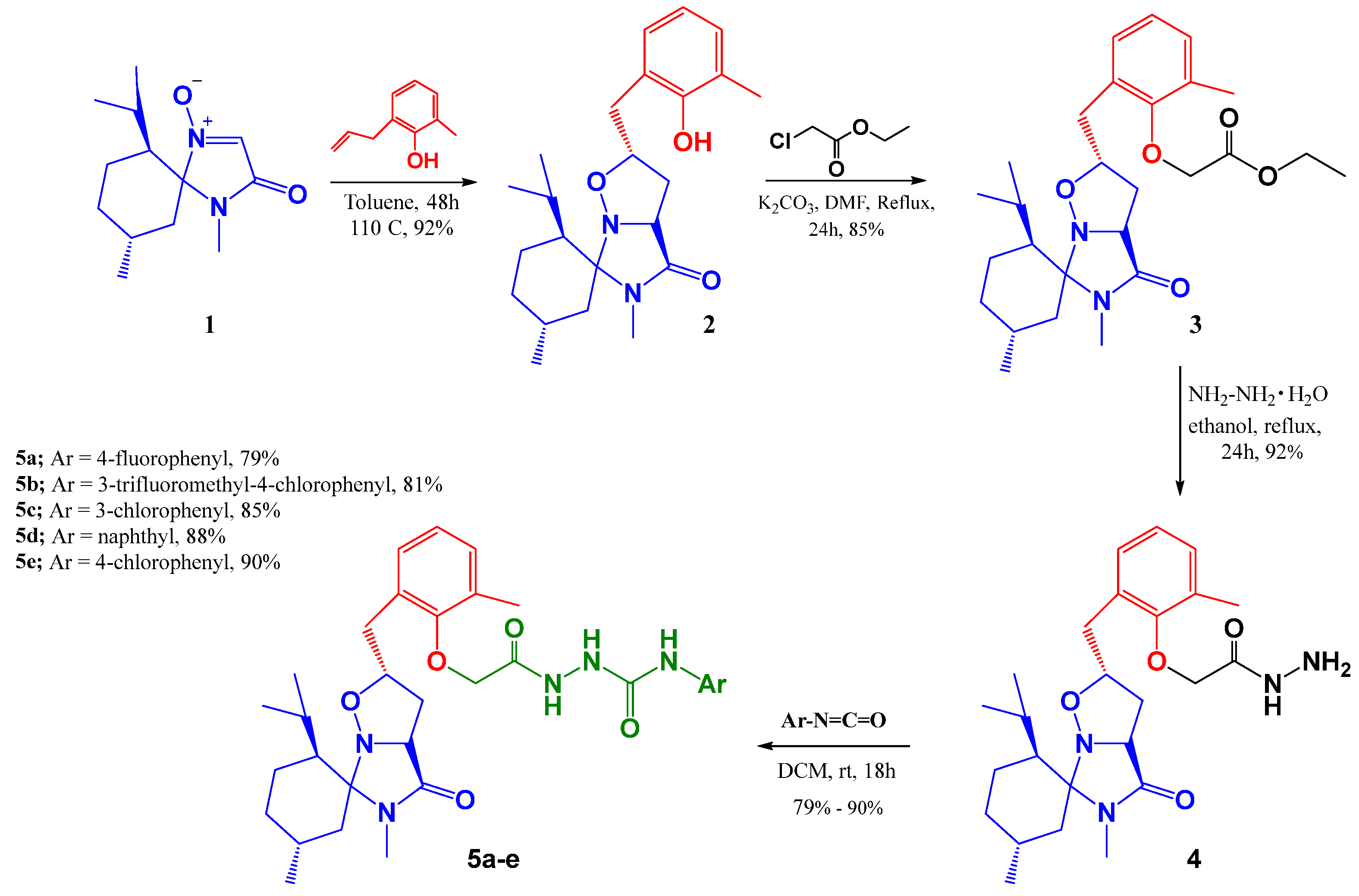
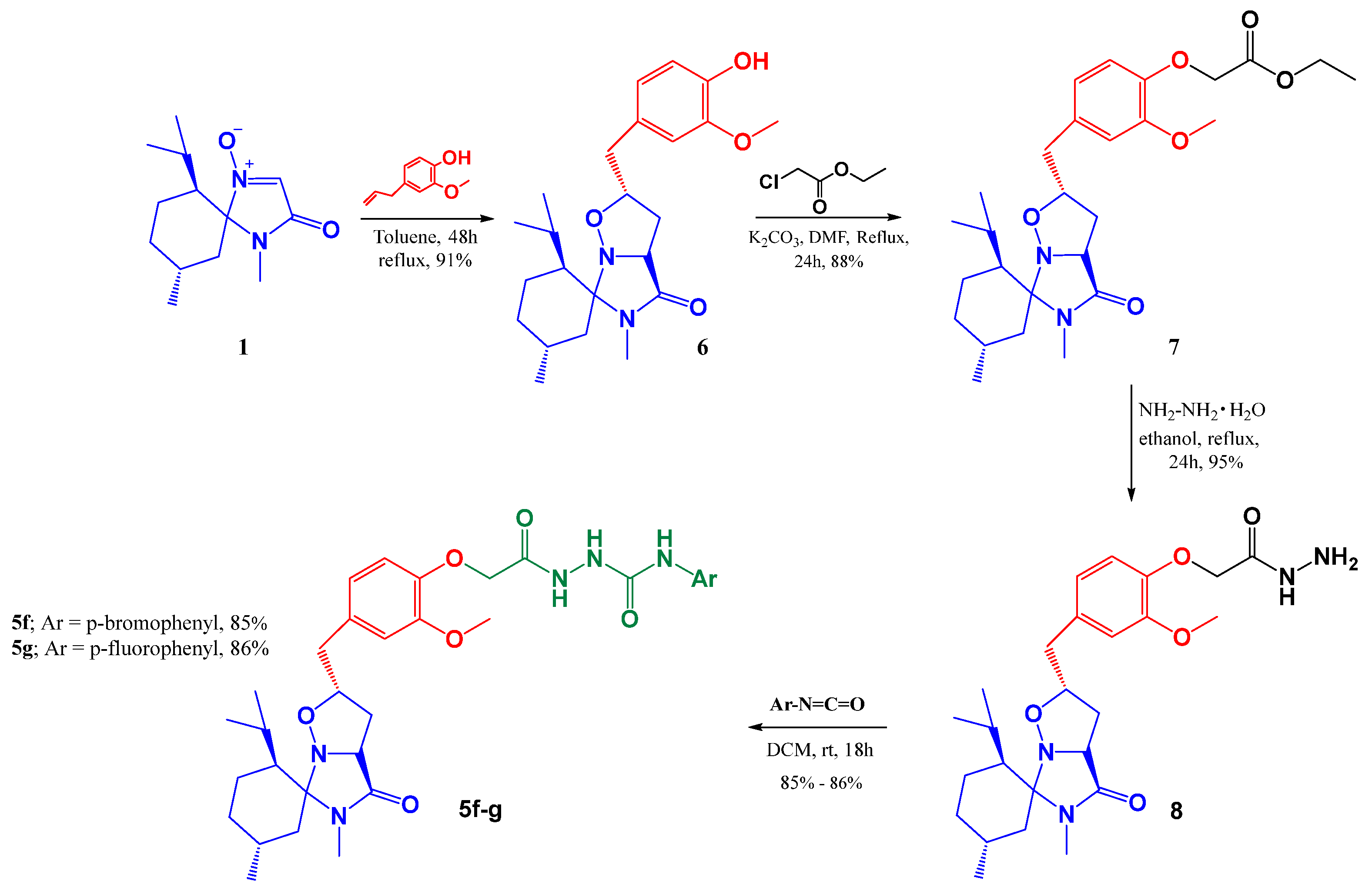
2.1. Chemistry
2.2. In Vitro α-Amylase and α-Glucosidase Inhibitory Activities
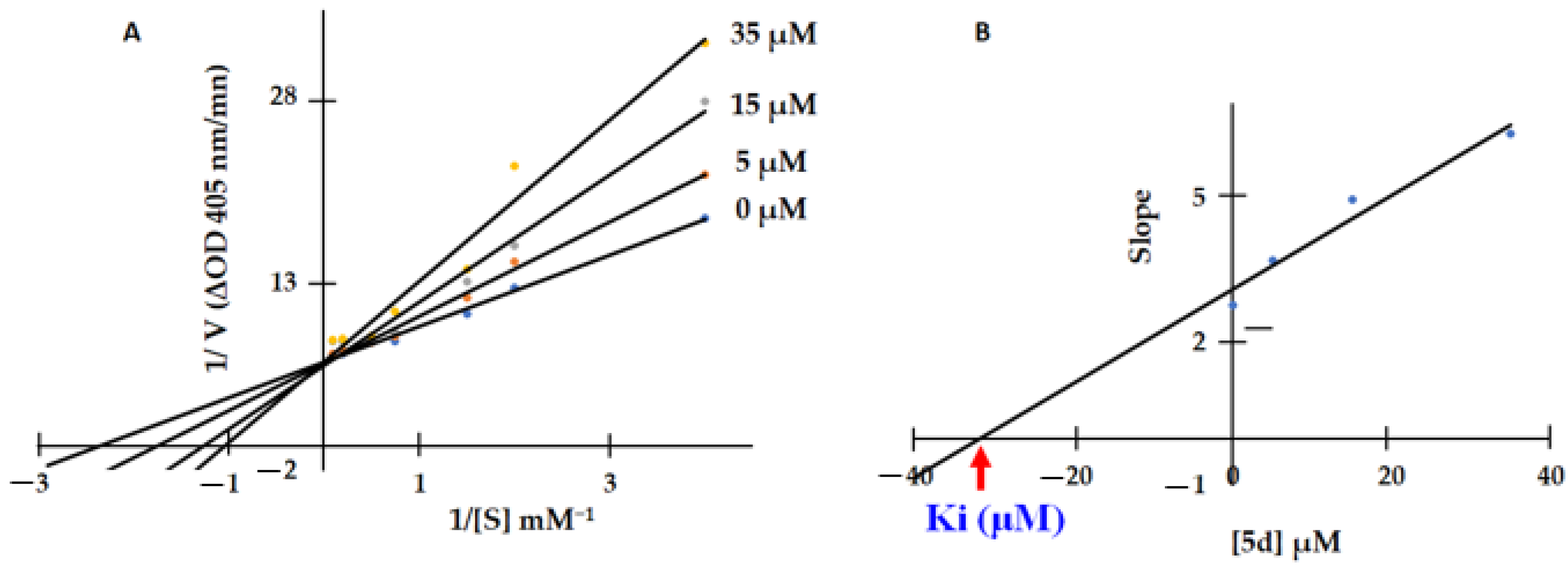
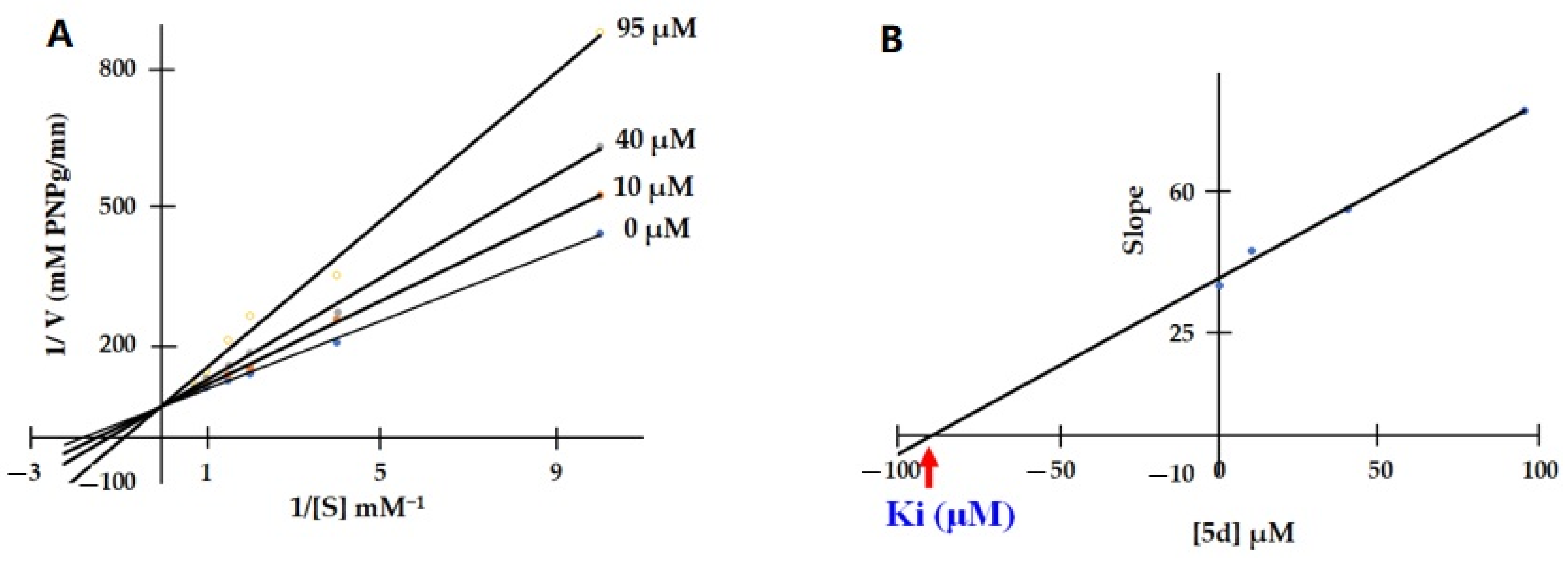
2.3. Enzyme Kinetic Studies
2.3.1. Mode of α-Amylase Inhibition
2.3.2. Mode of α-Glucosidase Inhibition
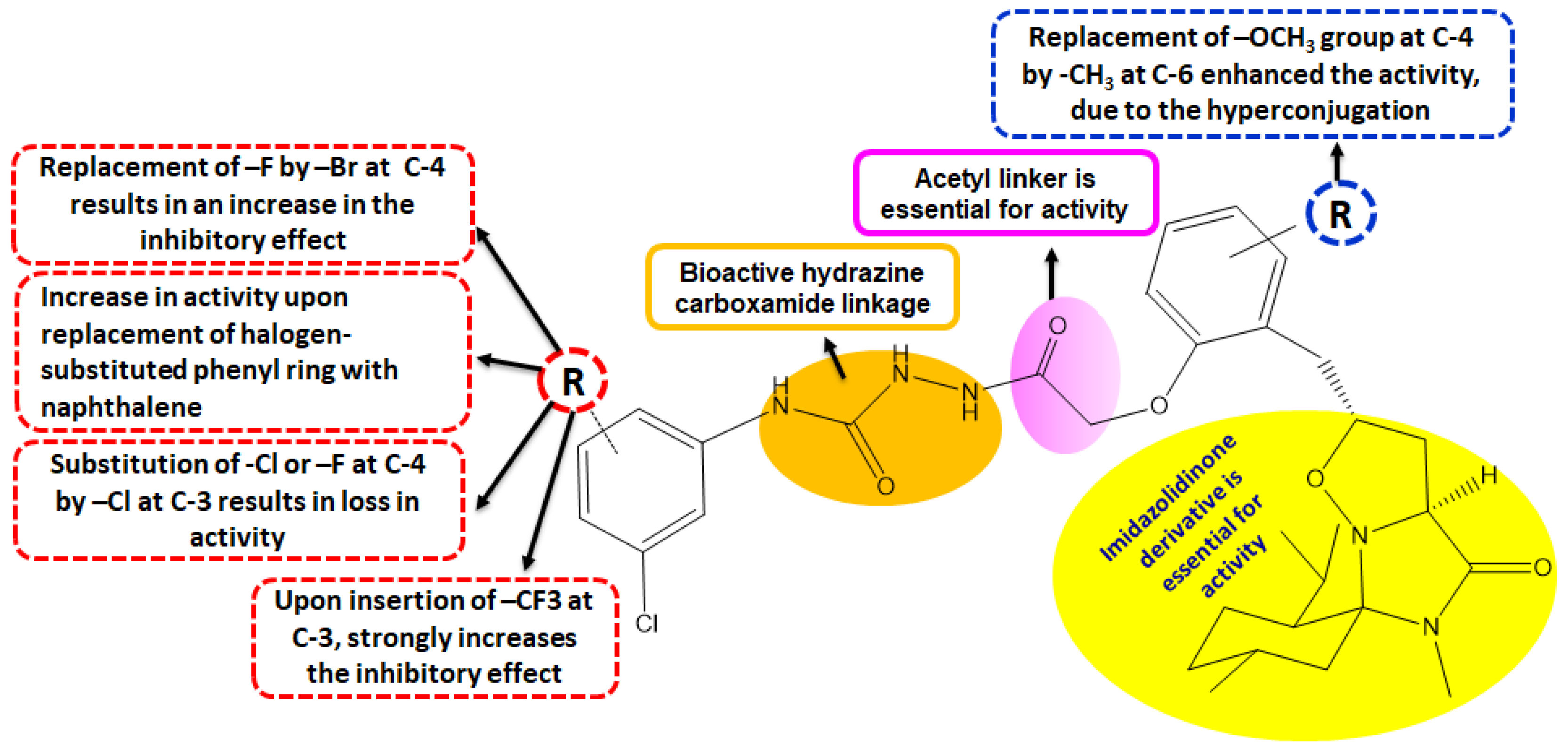
2.4. Structure–Activity Relationship (SAR) Investigation
2.5. ADMET Analysis
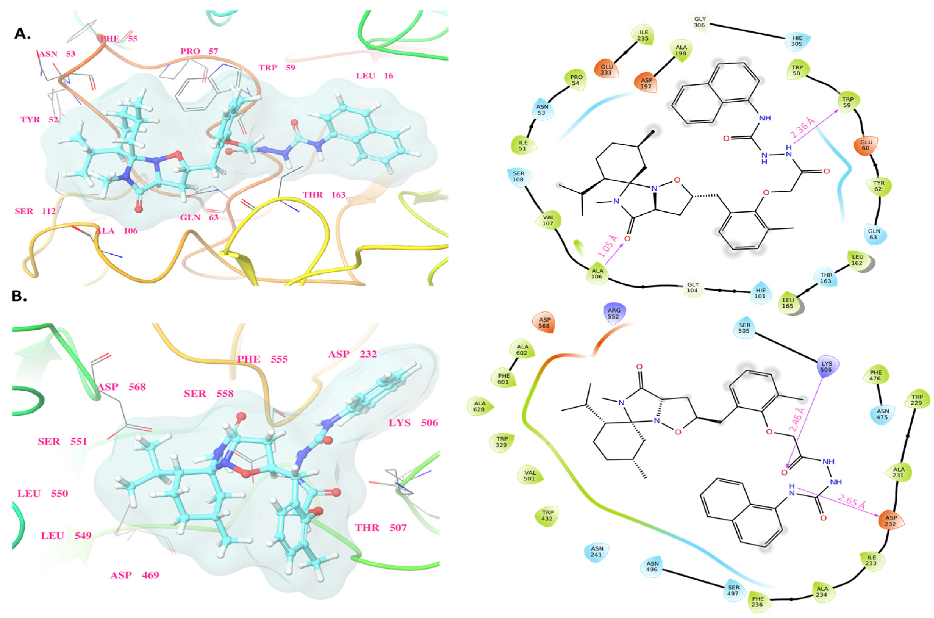
2.6. Molecular Docking Study
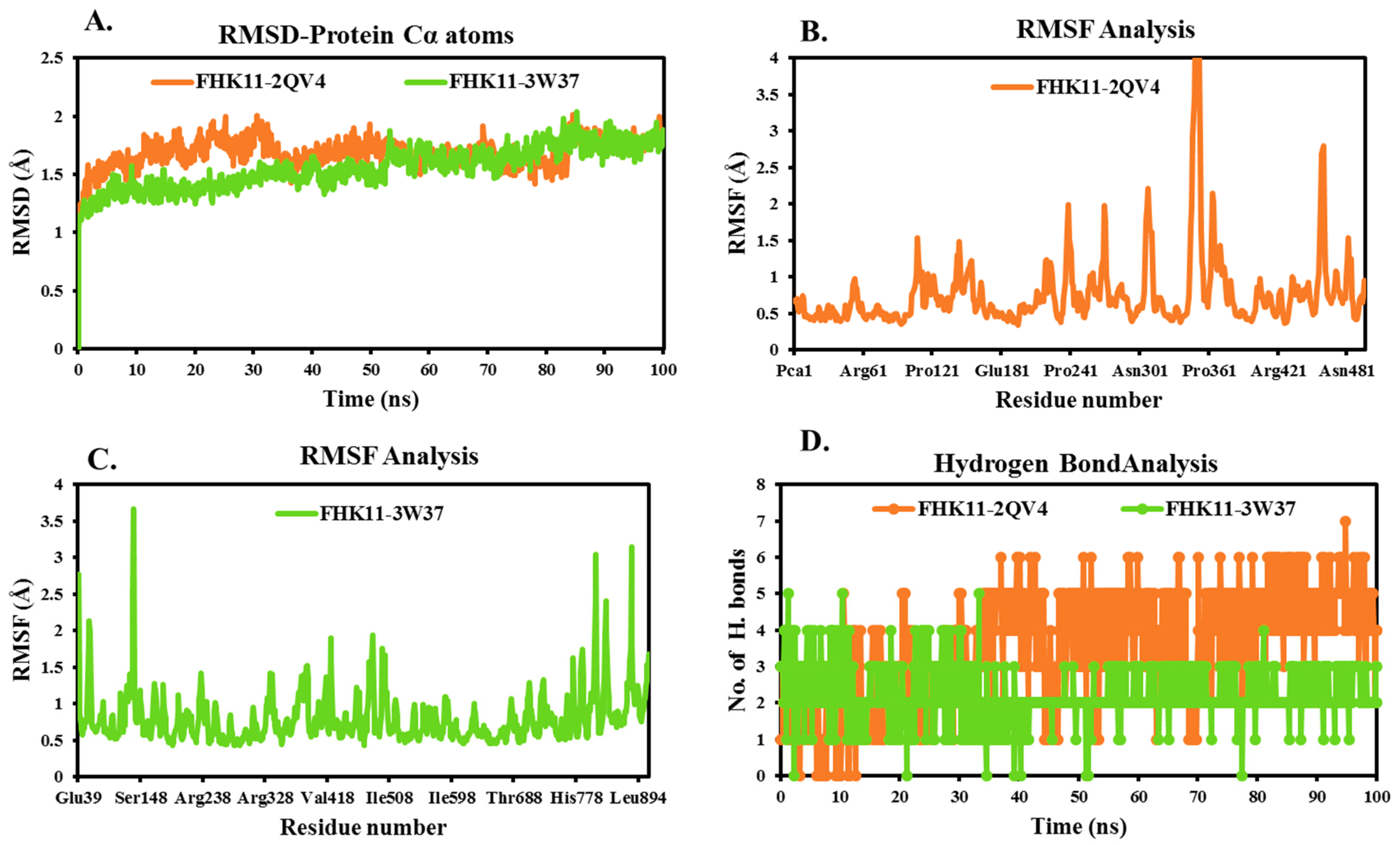
2.7. MD Simulation
3. Materials and Methods
3.1. Chemistry
3.1.1. General Methods
3.1.2. General Procedure (A) for the Preparation of Cycloadduct 2 and 6
3.1.3. General Procedure B for the Preparation of Ester 3 and 7
3.1.4. General Procedure C for the Preparation of Hydrazide 4 and 8
3.1.5. General Procedure D for the Preparation of Compounds 5a–g
3.2. α-Amylase and α-Glucosidase Inhibition Assays
3.3. Kinetic Studies
3.4. Molecular Docking Study
3.5. Molecular Dynamic (MD) Simulation
3.6. ADMET Property Predictions
3.7. Statistical Analysis
4. Conclusions
Supplementary Materials
Author Contributions
Funding
Institutional Review Board Statement
Informed Consent Statement
Data Availability Statement
Conflicts of Interest
References
- Hinault, C.; Caroli-Bosc, P.; Bost, F.; Chevalier, N. Critical Overview on Endocrine Disruptors in Diabetes Mellitus. Int. J. Mol. Sci. 2023, 24, 4537. [Google Scholar] [CrossRef] [PubMed]
- Popoviciu, M.S.; Kaka, N.; Sethi, Y.; Patel, N.; Chopra, H.; Cavalu, S. Type 1 Diabetes Mellitus and Autoimmune Diseases: A Critical Review of the Association and the Application of Personalized Medicine. J. Pers. Med. 2023, 13, 422. [Google Scholar] [CrossRef] [PubMed]
- Lee, J.; Yun, J.-S.; Ko, S.-H. Advanced Glycation End Products and Their Effect on Vascular Complications in Type 2 Diabetes Mellitus. Nutrients 2022, 14, 3086. [Google Scholar] [CrossRef] [PubMed]
- Li, Y.; Liu, Y.; Liu, S.; Gao, M.; Wang, W.; Chen, K.; Huang, L.; Liu, Y. Diabetic vascular diseases: Molecular mechanisms and therapeutic strategies. Signal Transduct. Target. Ther. 2023, 8, 152. [Google Scholar] [CrossRef]
- Ramos, H.; Hernández, C.; Simó, R.; Simó-Servat, O. Inflammation: The Link between Neural and Vascular Impairment in the Diabetic Retina and Therapeutic Implications. Int. J. Mol. Sci. 2023, 24, 8796. [Google Scholar] [CrossRef] [PubMed]
- Rajlic, S.; Treede, H.; Münzel, T.; Daiber, A.; Duerr, G.D. Early Detection Is the Best Prevention—Characterization of Oxidative Stress in Diabetes Mellitus and Its Consequences on the Cardiovascular System. Cells 2023, 12, 583. [Google Scholar] [CrossRef] [PubMed]
- Leyane, T.S.; Jere, S.W.; Houreld, N.N. Oxidative Stress in Ageing and Chronic Degenerative Pathologies: Molecular Mechanisms Involved in Counteracting Oxidative Stress and Chronic Inflammation. Int. J. Mol. Sci. 2022, 23, 7273. [Google Scholar] [CrossRef]
- Available online: https://idf.org/about-diabetes/diabetes-facts-figures/ (accessed on 10 June 2023).
- Bocanegra, A.; Macho-González, A.; Garcimartín, A.; Benedí, J.; Sánchez-Muniz, F.J. Whole Alga, Algal Extracts, and Compounds as Ingredients of Functional Foods: Composition and Action Mechanism Relationships in the Prevention and Treatment of Type-2 Diabetes Mellitus. Int. J. Mol. Sci. 2021, 22, 3816. [Google Scholar] [CrossRef]
- Papoutsis, K.; Zhang, J.; Bowyer, M.C.; Brunton, N.; Gibney, E.R.; Lyng, J. Fruit, vegetables, and mushrooms for the preparation of extracts with α-amylase and α-glucosidase inhibition properties: A review. Food Chem. 2021, 338, 128119. [Google Scholar] [CrossRef]
- Ghannay, S.; Snoussi, M.; Messaoudi, S.; Kadri, A.; Aouadi, K. Novel enantiopure isoxazolidine and C-alkyl imine oxide derivatives as potential hypoglycemic agents: Design, synthesis, dual inhibitors of α-amylase and α-glucosidase, ADMET and molecular docking study. Bioorg. Chem. 2020, 104, 104270. [Google Scholar] [CrossRef]
- Gong, L.; Feng, D.; Wang, T.; Ren, Y.; Liu, Y.; Wang, J. Inhibitors of α-amylase and α-glucosidase: Potential linkage for whole cereal foods on prevention of hyperglycemia. Food Sci. Nutr. 2020, 8, 6320–6337. [Google Scholar] [CrossRef] [PubMed]
- Kadri, A.; Aouadi, K. In vitro antimicrobial and α-glucosidase inhibitory potential of enantiopure cycloalkylglycine derivatives: Insights into their in silico pharmacokinetic, druglikeness, and medicinal chemistry properties. J. Appl. Pharm. Sci. 2020, 10, 107–115. [Google Scholar]
- Haguet, Q.; Le Joubioux, F.; Chavanelle, V.; Groult, H.; Schoonjans, N.; Langhi, C.; Michaux, A.; Otero, Y.F.; Boisseau, N.; Peltier, S.L.; et al. Inhibitory Potential of α-Amylase, α-Glucosidase, and Pancreatic Lipase by a Formulation of Five Plant Extracts: TOTUM-63. Int. J. Mol. Sci. 2023, 24, 3652. [Google Scholar] [CrossRef]
- Aouadi, K.; Hajlaoui, H.; Arraouadi, S.; Ghannay, S.; Snoussi, M.; Kadri, A. Phytochemical Profiling, Antimicrobial and α-Glucosidase Inhibitory Potential of Phenolic-Enriched Extracts of the Aerial Parts from Echium humile Desf.: In Vitro Combined with In Silico Approach. Plants 2022, 11, 1131. [Google Scholar] [CrossRef] [PubMed]
- Galicia-Garcia, U.; Benito-Vicente, A.; Jebari, S.; Larrea-Sebal, A.; Siddiqi, H.; Uribe, K.B.; Ostolaza, H.; Martín, C. Pathophysiology of Type 2 Diabetes Mellitus. Int. J. Mol. Sci. 2020, 21, 6275. [Google Scholar] [CrossRef] [PubMed]
- Salib, A.; Cayabyab, F.; Yoshihara, E. Stem Cell-Derived Islets for Type 2 Diabetes. Int. J. Mol. Sci. 2022, 23, 5099. [Google Scholar] [CrossRef] [PubMed]
- Moelands, S.V.; Lucassen, P.L.; Akkermans, R.P.; De Grauw, W.J.; Van de Laar, F.A. Alpha-glucosidase inhibitors for prevention or delay of type 2 diabetes mellitus and its associated complications in people at increased risk of developing type 2 diabetes mellitus. Cochrane Database Syst. Rev. 2018, 12, CD005061. [Google Scholar] [CrossRef]
- Hossain, U.; Das, A.K.; Ghosh, S.; Sil, P.C. An overview on the role of bioactive α-glucosidase inhibitors in ameliorating diabetic complications. Food Chem Toxicol. 2020, 145, 111738. [Google Scholar] [CrossRef]
- Ghabi, A.; Brahmi, J.; Alminderej, F.; Messaoudi, S.; Vidald, S.; Kadri, A.; Aouadi, K. Multifunctional isoxazolidine derivatives as α-amylase and α-glucosidase inhibitors. Bioorg. Chem. 2020, 98, 103713. [Google Scholar] [CrossRef]
- Wei, H.; Qiao, C.; Liu, G.; Yang, Z.; Li, C.C. Stereoselective Total Syntheses of (−)-Flueggine A and (+)-Virosaine B. Angew. Chem. Int. Ed. 2013, 52, 620–624. [Google Scholar] [CrossRef]
- Zhao, B.-X.; Wang, Y.; Zhang, D.-M.; Jiang, R.-W.; Wang, G.-C.; Shi, J.-M.; Huang, X.-J.; Chen, W.-M.; Che, C.-T.; Ye, W.-C. Flueggines A and B, Two New Dimeric Indolizidine Alkaloids from Flueggea virosa. Org. Lett. 2011, 13, 3888–3891. [Google Scholar] [CrossRef]
- Shao, H.; Fang, K.; Wang, Y.P.; Zhang, X.M.; Ding, T.M.; Zhang, S.Y.; Chen, Z.M.; Tu, Y.Q. Total Synthesis of Fawcettimine-Type Alkaloid, Lycojaponicumin A. Org Lett. 2020, 15, 3775–3779. [Google Scholar] [CrossRef]
- Alminderej, F.; Ghannay, S.; Elsamani, M.O.; Alhawday, F.; Albadri, A.E.A.E.; Elbehairi, S.E.I.; Alfaifi, M.Y.; Kadri, A.; Aouadi, K. In Vitro and In Silico Evaluation of Antiproliferative Activity of New Isoxazolidine Derivatives Targeting EGFR: Design, Synthesis, Cell Cycle Analysis, and Apoptotic Inducers. Pharmaceuticals 2023, 16, 1025. [Google Scholar] [CrossRef] [PubMed]
- Al-Adhreai, A.; ALSaeedy, M.; Alrabie, A.; Al-horaibi, S.A.; Al-Qadsy, I.; Alezzy, A.A.; AL-Odayni, A.B.; Saeed, W.S.; Farooqui, M. Enhanced synthesis of novel multisubstituted isoxazolidines as potential antimicrobial and antioxidant agents using zinc (II) catalyst, and in silico studies. J. Mol. Struct. 2023, 1292, 136146. [Google Scholar] [CrossRef]
- Kumar, K.R.R.; Mallesha, H.; Rangappa, K.S. Synthesis and Characterization of 5-Substituted Novel Isoxazolidines Derived from 1,3-Dipolar Cycloaddition of Nitrones with Olefins: Studies of Antibacterial and Antifungal Activities Cycloaddition of Nitrones with Olefins. Synth. Commun. 2003, 33, 1545–1555. [Google Scholar] [CrossRef]
- Al-Adhreai, A.; ALSaeedy, M.; Alrabie, A.; Al-Qadsy, I.; Dawbaa, S.; Alaizeri, Z.M.; Alhadlaq, H.A.; Al-Kubati, A.; Ahamed, M.; Farooqui, M. Design and synthesis of novel enantiopure Bis(5-Isoxazolidine) derivatives: Insights into their antioxidant and antimicrobial potential via in silico drug-likeness, pharmacokinetic, medicinal chemistry properties, and molecular docking studies. Heliyon 2022, 8, e09746. [Google Scholar] [CrossRef] [PubMed]
- Jones, R.C.F.; Martin, J.N. The Chemistry of Heterocyclic Compounds, Vol. 59: Synthetic Applications of 1,3-Dipolar Cycloaddition Chemistry Toward Heterocycles and Natural Products; Padwa, A., Pearson, W.H., Eds.; Wiley: Hoboken, NJ, USA, 2002; pp. 1–81. [Google Scholar]
- Zhang, G.-L.; Rücker, G.; Breitmaier, E.; Nieger, M.; Mayer, R.; Steinbeck, C. Alkaloids from Dactylicapnos torulosa. Phytochemistry 1995, 40, 299–305. [Google Scholar] [CrossRef]
- Serna, A.V.; Kürti, L.; Siitonen, J.H. Synthesis of (±)-Setigerumine I: Biosynthetic Origins of the Elusive Racemic Papaveraceae Isoxazolidine Alkaloids. Angew. Chem. 2021, 60, 27236–27240. [Google Scholar] [CrossRef]
- Aouadi, K.; Jeanneau, E.; Msadek, M.; Praly, J.-P. New Synthetic Routes toward Enantiopure (2S,3R,4R)-4-Hydroxyisoleucine by 1,3-Dipolar Cycloaddition of a Chiral Nitrone to C4 Alkenes. Synthesis 2007, 21, 3399–3405. [Google Scholar]
- Othman, I.M.M.M.; Mahross, H.; Gad-Elkareem, M.A.M.; Rudrapal, M.; Gogoi, N.; Chetia, D.; Aouadi, K.; Snoussi, M.; Kadri, A. Toward a treatment of antibacterial and antifungal infections: Design, synthesis and in vitro activity of novel arylhydrazothiazolylsulfonamides analogues and their insight of DFT, docking and molecular dynamic simulations. J. Mol. Struct. 2021, 1243, 130862. [Google Scholar] [CrossRef]
- Ghannay, S.; Bakari, S.; Msaddek, M.; Vidal, S.; Kadri, A.; Aouadi, K. Design, synthesis, molecular properties and in vitro antioxidant and antibacterial potential of novel enantiopure isoxazolidine derivatives. Arab. J. Chem. 2020, 13, 2121–2131. [Google Scholar] [CrossRef]
- Radwan, H.A.; Ahmad, I.; Othman, I.M.M.; Gad-Elkareem, M.A.M.; Patel, H.; Aouadi, K.; Snoussi, M.; Kadri, A. Design, synthesis, in vitro anticancer and antimicrobial evaluation, SAR analysis, molecular docking and dynamic simulation of new pyrazoles, triazoles and pyridazines based isoxazole. J. Mol. Struct. 2022, 1264, 133312. [Google Scholar] [CrossRef]
- Westermann, B.; Walter, A.; Flörke, U.; Altenbach, H.-J. Chiral auxiliary based approach toward the synthesis of C-glycosylated amino acids. Org. Lett. 2001, 3, 1375. [Google Scholar] [CrossRef] [PubMed]
- Vogt, A.; Altenbach, H.-J.; Kirschbaum, M.; Hahn, M.G.; Matthäus, M.S.P.; Hermann, A.R. EP 976721, 2000. Chem. Abstr. 2000, 132, 108296j. [Google Scholar]
- Shah, P.; Westwell, A.D. The role of fluorine in medicinal chemistry. J. Enzym. Inhib. Med. Chem. 2007, 22, 527–540. [Google Scholar] [CrossRef] [PubMed]
- Othman, I.M.M.; Gad-Elkareem, M.A.M.; Anouar, E.H.; Snoussi, M.; Aouadi, K.; Kadri, A. Novel fused pyridine derivatives containing pyrimidine moiety as prospective tyrosyl-tRNA synthetase inhibitors: Design, synthesis, pharmacokinetics and molecular docking studies. J. Mol. Struct. 2020, 1219, 128651. [Google Scholar] [CrossRef]
- Badraoui, R.; Rebai, T.; Elkahoui, S.; Alreshidi, M.; Veettil, V.N.; Noumi, E.; Al-Motair, K.A.; Aouadi, K.; Kadri, A.; De Feo, V.; et al. Allium subhirsutum L. as a Potential Source of Antioxidant and Anticancer Bioactive Molecules: HR-LCMS Phytochemical Profiling, In Vitro and In Vivo Pharmacological Study. Antioxidants 2020, 9, 1003. [Google Scholar] [CrossRef]
- Othman, I.M.M.; Gad-Elkareem, M.A.M.; Anouar, E.H.; Aouadi, K.; Kadri, A.; Snoussi, M. Design, synthesis ADMET and molecular docking of new imidazo [4,5-b]pyridine-5-thione derivatives as potential tyrosyl-tRNA synthetase inhibitors. Bioorg. Chem. 2020, 102, 104105. [Google Scholar] [CrossRef]
- Shinde, A.D.; Nandurkar, Y.M.; Bhalekar, S.; Walunj, Y.S.; Ugale, S.; Ahmad, I.; Patel, H.; Chavan, A.P.; Mhaske, P.C. Investigation of new 1,2,3-triazolyl-quinolinyl-propan-2-ol derivatives as potential antimicrobial agents: In vitro and in silico approach. J. Biomol. Struct. Dyn. 2023, 1–17. [Google Scholar] [CrossRef]
- Elsaman, T.; Ahmad, I.; Eltayib, E.M.; Suliman Mohamed, M.; Yusuf, O.; Saeed, M.; Patel, H.; Mohamed, M.A. Flavonostilbenes natural hybrids from Rhamnoneuron balansae as potential antitumors targeting ALDH1A1: Molecular docking, ADMET, MM-GBSA calculations and molecular dynamics studies. J. Biomol. Struct. Dyn. 2023, 1–18. [Google Scholar] [CrossRef]
- Williams, L.K.; Li, C.; Withers, S.G.; Brayer, G.D. Order and Disorder: Differential structural impacts of myricetin and ethyl caffeate on human amylase, an antidiabetic target. J. Med. Chem. 2012, 55, 10177–10186. [Google Scholar] [CrossRef] [PubMed]
- Rydberg, E.H.; Li, C.; Maurus, R.; Overall, C.M.; Brayer, G.D.; Withers, S.G. Mechanistic analyses of catalysis in human pancreatic α-amylase: Detailed kinetic and structural studies of mutants of three conserved carboxylic acids. Biochemistry 2002, 41, 4492–4502. [Google Scholar] [CrossRef]
- Anigboro, A.A.; Avwioroko, O.J.; Ohwokevwo, O.A.; Pessu, B.; Tonukari, N.J. Phytochemical profile, antioxidant, α-amylase inhibition, binding interaction and docking studies of Justicia carnea bioactive compounds with αamylase. Biophys. Chem. 2021, 269, 106529. [Google Scholar] [CrossRef] [PubMed]
- Şahin, İ.; Çeşme, M.; Yüce, N.; Tümer, F. Discovery of new 1, 4-disubstituted 1, 2, 3-triazoles: In silico ADME profiling, molecular docking and biological evaluation studies. J. Biomol. Struct. Dyn. 2022, 20, 1–14. [Google Scholar] [CrossRef] [PubMed]
- Lekmine, S.; Benslama, O.; Kadi, K.; Martín-García, A.I.; Yilmaz, M.A.; Akkal, S.; Boumegoura, A.; Alhomida, A.S.; Ola, M.S.; Ali, A. LC/MS-MS Analysis of Phenolic Compounds in Hyoscyamus albus L. Extract: In Vitro Antidiabetic Activity, In Silico Molecular Docking, and In Vivo Investigation against STZ-Induced Diabetic Mice. Pharmaceuticals 2023, 16, 1015. [Google Scholar] [CrossRef] [PubMed]
- Arshad, A.; Ahemad, S.; Saleem, H.; Saleem, M.; Zengin, G.; Abdallah, H.H.; Tousif, M.I.; Ahemad, N.; Fawzi Mahomoodally, M. RP-UHPLC-MS Chemical Profiling, Biological and In Silico Docking Studies to Unravel the Therapeutic Potential of Heliotropium crispum Desf. as a Novel Source of Neuroprotective Bioactive Compounds. Biomolecules 2021, 11, 53. [Google Scholar] [CrossRef] [PubMed]
- Ogunyemi, O.M.; Gyebi, G.A.; Saheed, A.; Paul, J.; Nwaneri-Chidozie, V.; Olorundare, O.; Adebayo, J.; Koketsu, M.; Aljarba, N.; Alqahtani, S.; et al. Inhibition Mechanism of Alpha-Amylase, a Diabetes Target, by a Steroidal Pregnane and Pregnane Glycosides Derived from Gongronema Latifolium Benth. Front. Mol. Biosci. 2022, 9, 866719. [Google Scholar] [CrossRef]
- Alqahtani, A.S.; Hidayathulla, S.; Rehman, M.T.; ElGamal, A.A.; Al-Massarani, S.; Razmovski-Naumovski, V.; Alqahtani, M.S.; El Dib, R.A.; AlAjmi, M.F. Alpha-Amylase and Alpha-Glucosidase Enzyme Inhibition and Antioxidant Potential of 3-Oxolupenal and Katononic Acid Isolated from Nuxia oppositifolia. Biomolecules 2020, 10, 61. [Google Scholar] [CrossRef]
- Dalli, M.; Daoudi, N.E.; Abrigach, F.; Azizi, S.; Bnouham, M.; Kim, B.; Gseyra, N. In vitro α-amylase and hemoglobin glycation inhibitory potential of Nigella sativa essential oil, and molecular docking studies of its principal components. Front. Pharmacol. 2022, 13, 1–13. [Google Scholar] [CrossRef]
- Tagami, T.; Yamashita, K.; Okuyama, M.; Mori, H.; Yao, M.; Kimura, A. Molecular basis for the recognition of long-chain substrates by plant & α-glucosidase. J. Biol. Chem. 2013, 288, 19296–19303. [Google Scholar]
- Aulifa, D.L.; Adnyana, I.K.; Sukrasno, S.; Levita, J. Inhibitory activity of xanthoangelol isolated from Ashitaba (Angelica keiskei Koidzumi) towards α-glucosidase and dipeptidyl peptidase-IV: In silico and in vitro studies. Heliyon 2022, 8, E09501. [Google Scholar] [CrossRef] [PubMed]
- Liu, S.-K.; Hao, H.; Bian, Y.; Ge, Y.-X.; Lu, S.; Xie, H.-X.; Wang, K.-M.; Tao, H.; Yuan, C.; Zhang, J.; et al. Discovery of New α-Glucosidase Inhibitors: Structure-Based Virtual Screening and Biological Evaluation. Front. Chem. 2021, 9, 639279. [Google Scholar] [CrossRef] [PubMed]
- Reddy, V.N.V.L.S.; Akhila, M.; Subrahmanyam, C.V.S.; Trimurtulu, G.; Raghavendra, N.M. Isolation in vitro antidiabetic, antioxidant activity and molecular docking studies of pentacyclic triterpenoids from syzygium alternifolium(wt.) walp bark. IOSR J. Pharm. Biol. Sci. 2015, 10, 148–154. [Google Scholar]
- Ahmed Atto Al-Shuaeeb, R.; Abd El-Mageed, H.; Ahmed, S.; Mohamed, H.S.; Hamza, Z.S.; Rafi, M.O.; Ahmad, I.; Patel, H. In silico investigation and potential therapeutic approaches of isoquinoline alkaloids for neurodegenerative diseases: Computer-aided drug design perspective. J. Biomol. Struct. Dyn. 2023, 41, 1–13. [Google Scholar] [CrossRef] [PubMed]
- Sepehri, N.; Azizian, H.; Ghadimi, R.; Abedinifar, F.; Mojtabavi, S.; Faramarzi, M.A.; Moghadamnia, A.A.; Zabihi, E.; Mohebbi, G.; Larijani, B.; et al. New 4,5-diphenylimidazole-acetamide-1,2,3-triazole hybrids as potent α-glucosidase inhibitors: Synthesis, in vitro and in silico enzymatic and toxicity evaluations. Monatsh. Chem. 2021, 152, 679–693. [Google Scholar] [CrossRef]
- Saleem, F.; Kanwal; Mohammed Khan, K.; Chigurupati, S.; Andriani, Y.; Solangi, M.; Hameed, S.; Abdel Hafez, A.A.M.; Begum, F.; Arif Lodhi, M.; et al. Dicyanoanilines as potential and dual inhibitors of α-amylase and α-glucosidase enzymes: Synthesis, characterization, in vitro, in silico, and kinetics studies. Arab. J. Chem. 2022, 15, 103651. [Google Scholar] [CrossRef]
- Jagatap, V.R.; Ahmad, I.; Sriram, D.; Kumari, J.; Adu, D.K.; Ike, B.W.; Ghai, M.; Ansari, S.A.; Ansari, I.A.; Wetchoua, P.O.M.; et al. Isoflavonoid and Furanochromone Natural Products as Potential DNA Gyrase Inhibitors: Computational, Spectral, and Antimycobacterial Studies. ACS Omega 2023, 8, 16228–16240. [Google Scholar] [CrossRef]
- Sayed, H.M.; Ramadan, M.A.; Salem, H.H.; Ahmad, I.; Patel, H.; Fayed, M.A. Phytochemical Investigation, In Silico/In Vivo Analgesic, and Anti-inflammatory Assessment of the Egyptian Cassia occidentalis L. Steroids 2023, 196, 109245. [Google Scholar] [CrossRef]
- Khan, M.; Khan, S.; Alshammary, F.L.; Zaidi, S.; Singh, V.; Ahmad, I.; Patel, H.; Gupta, V.K.; Haque, S. In silico analysis to identify potential antitubercular molecules in Morus alba through virtual screening and molecular dynamics simulations. J. Biomol. Struct. Dyn. 2023, 1–8. [Google Scholar] [CrossRef]
- Abdullahi, M.; Uzairu, A.; Gideon Shallangwa, A.; Andrew Mamza, P.; Tukur Ibrahim, M.; Ahmad, I.; Patel, H. Structurebased drug design, molecular dynamics simulation, ADMET, and quantum chemical studies of some thiazolinones targeting influenza neuraminidase. J. Biomol. Struct. Dyn. 2023, 41, 13829–13843. [Google Scholar] [CrossRef]
- Noumi, E.; Ahmad, I.; Adnan, M.; Merghni, A.; Patel, H.; Haddaji, N.; Bouali, N.; Alabbosh, K.F.; Ghannay, S.; Aouadi, K.; et al. GC/MS Profiling, Antibacterial, Anti-Quorum Sensing, and Antibiofilm Properties of Anethum graveolens L. Essential Oil: Molecular Docking Study and In-Silico ADME Profiling. Plants 2023, 12, 1997. [Google Scholar] [CrossRef] [PubMed]
- Zala, A.R.; Kumar, D.; Razakhan, U.; Rajani, D.P.; Ahmad, I.; Patel, H.; Kumari, P. Molecular modeling and biological investigation of novel s-triazine linked benzothiazole and coumarin hybrids as antimicrobial and antimycobacterial agents. J. Biomol. Struct. Dyn. 2023, 1–12. [Google Scholar] [CrossRef] [PubMed]
- Owen, A.E.; Chima, C.M.; Ahmad, I.; Emori, W.; Agwamba, E.C.; Cheng, C.R.; Benjamin, I.; Patel, H.; Ahuekwe, E.F.; Ojong, M.A.; et al. Antibacterial Potential of Trihydroxycyclohexa-2,4-Diene-1-Carboxylic Acid: Insight from DFT, Molecular Docking, and Molecular Dynamic Simulation. Polycycl. Aromat. Compd. 2023, 1–24. [Google Scholar] [CrossRef]


| Entry | Structure | Coupling Constant | Literature [20] |
|---|---|---|---|
| 5a |  | J3,4 (cis) = 8.4 Hz J3,4 (trans)~0 Hz J4,5 (trans) = 8.8 Hz J4,5 (cis) = 3.0 Hz | J3,4 (cis) ≥ 6.6 Hz J3,4 (trans)~0 Hz J4,5 (trans) > 8 Hz J4,5 (cis) < 6 Hz |
| 5b | 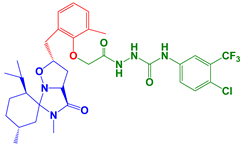 | J3,4 (cis) = 8.4 Hz J3,4 (trans)~0 Hz J4,5 (trans) = 9.2 Hz | |
| 5c | 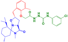 | J3,4 (cis) = 8.0 Hz J3,4 (trans)~0 Hz J4,5 (trans) = 8.8 Hz J4,5 (cis) = 3.0 Hz | |
| 5d | 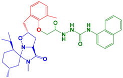 | J3,4 (cis) = 8.4 Hz J3,4 (trans)~0 Hz J4,5 (trans) = 8.8 Hz J4,5 (cis) = 3.2 Hz | |
| 5e | 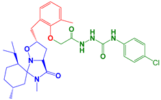 | J3,4 (cis) = 8.4 Hz J3,4 (trans)~0 Hz J4,5 (trans) = 8.8 Hz J4,5 (cis) = 4.0 Hz | |
| 5f | 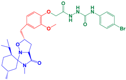 | J3,4 (cis) = 8.4 Hz J3,4 (trans)~0 Hz J4,5 (cis) = 3.2 Hz | |
| 5g | 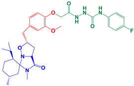 | J3,4 (cis) = 8.4 Hz J3,4 (trans)~0 Hz |
| Entry | α-Amylase IC50 (μM) | α-Glucosidase IC50 (μM) |
|---|---|---|
| 5a | 120.3 ± 0.306 c | 152.3 ± 0.367 c |
| 5b | 67.4 ± 0.202 a | 118.9 ± 0.325 ab |
| 5c | 92.28 ± 0.276 b | 120.9 ± 0.333 b |
| 5d | 53.03 ± 0.106 a | 94.33 ± 0.282 a |
| 5e | 232.8 ± 0.517 e | 258.7 ± 0.521 e |
| 5f | 183.8 ± 0.498 d | 204.6 ± 0.41 d |
| 5g | 134.8 ± 0.411 c | 170.8 ± 0.358 c |
| Acarbose | 296.6 ± 0.825 | 780.4 ± 0.346 |
| Entry | 5d | Reference |
|---|---|---|
| Absorption | ||
| Water solubility | −4.275 | - |
| Caco2 permeability | 0.857 | >0.9 |
| Intestinal absorption (human) | 86.437 | <30% is poorly |
| Skin Permeability (log Kp) | −2.736 | >−2.5 is low |
| Distribution | ||
| VDss (human) | −0.048 | Low is <−0.15, High is >0.45 |
| Fraction unbound (human) | 0.024 | - |
| BBB permeability | −1.395 | Poorly is <−1, High is >0.3 |
| CNS permeability | −2.131 | Penetrate is >−2, Unable is <−3 |
| Metabolism | ||
| CYP2D6 substrate | Yes | No |
| CYP3A4 substrate | Yes | - |
| CYP1A2 inhibitior | No | No |
| CYP2C19 inhibitior | Yes | No |
| CYP2C9 inhibitior | Yes | No |
| CYP2D6 inhibitior | No | No |
| CYP3A4 inhibitior | Yes | No |
| Excretion | ||
| Total clearance | −0.065 | - |
| Renal OCT2 substrate | No | - |
| Toxicity | ||
| AMES toxicity | No | No |
| Max. tolerated dose (human) | 0.216 | Low is ≤0.477, High is >0.477 |
| hERG I inhibitor | No | No |
| Oral Rat Acute Toxicity (LD50) | 3.006 | - |
| Oral Rat Chronic Toxicity (LOAEL) | 0.632 | - |
| Skin sensitisation | No | No |
| Compounds | α-Amylase Enzyme (PDB Code: 2QV4) | α-Glucosidase Enzyme (PDB Code: 3W37) |
|---|---|---|
| 5a | −5.355 | −5.329 |
| 5b | −5.607 | −5.343 |
| 5c | −5.245 | −5.55 |
| 5d | −5.623 | −5.373 |
| 5e | −3.315 | −3.039 |
| 5f | −4.451 | −3.276 |
| 5g | −5.367 | −4.415 |
Disclaimer/Publisher’s Note: The statements, opinions and data contained in all publications are solely those of the individual author(s) and contributor(s) and not of MDPI and/or the editor(s). MDPI and/or the editor(s) disclaim responsibility for any injury to people or property resulting from any ideas, methods, instructions or products referred to in the content. |
© 2024 by the authors. Licensee MDPI, Basel, Switzerland. This article is an open access article distributed under the terms and conditions of the Creative Commons Attribution (CC BY) license (https://creativecommons.org/licenses/by/4.0/).
Share and Cite
Alhawday, F.; Alminderej, F.; Ghannay, S.; Hammami, B.; Albadri, A.E.A.E.; Kadri, A.; Aouadi, K. In Silico Design, Synthesis, and Evaluation of Novel Enantiopure Isoxazolidines as Promising Dual Inhibitors of α-Amylase and α-Glucosidase. Molecules 2024, 29, 305. https://doi.org/10.3390/molecules29020305
Alhawday F, Alminderej F, Ghannay S, Hammami B, Albadri AEAE, Kadri A, Aouadi K. In Silico Design, Synthesis, and Evaluation of Novel Enantiopure Isoxazolidines as Promising Dual Inhibitors of α-Amylase and α-Glucosidase. Molecules. 2024; 29(2):305. https://doi.org/10.3390/molecules29020305
Chicago/Turabian StyleAlhawday, Fahad, Fahad Alminderej, Siwar Ghannay, Bechir Hammami, Abuzar E. A. E. Albadri, Adel Kadri, and Kaiss Aouadi. 2024. "In Silico Design, Synthesis, and Evaluation of Novel Enantiopure Isoxazolidines as Promising Dual Inhibitors of α-Amylase and α-Glucosidase" Molecules 29, no. 2: 305. https://doi.org/10.3390/molecules29020305
APA StyleAlhawday, F., Alminderej, F., Ghannay, S., Hammami, B., Albadri, A. E. A. E., Kadri, A., & Aouadi, K. (2024). In Silico Design, Synthesis, and Evaluation of Novel Enantiopure Isoxazolidines as Promising Dual Inhibitors of α-Amylase and α-Glucosidase. Molecules, 29(2), 305. https://doi.org/10.3390/molecules29020305