

Comprehensive Biological Potential, Phytochemical Profiling Using GC-MS and LC-ESI-MS, and In-Silico Assessment of Strobilanthes glutinosus Nees: An Important Medicinal Plant

 , ,
, ,
Abstract
1. Introduction
2. Results
2.1. Phytochemical Composition
2.2. Antioxidant Assays
2.3. In Vitro Enzyme Inhibition Activity
2.4. In Silico Analysis
3. Discussion
4. Materials and Methods
4.1. Plant Collection and Extraction
4.2. Phytochemical Analysis
4.2.1. GC-MS Analysis
4.2.2. LC-ESI-MS Analysis
4.3. Antioxidant Activity
4.3.1. Radical Scavenging Activity
4.3.2. Reducing Power Assays
4.3.3. Total Antioxidant Activity
4.3.4. Metal Chelating Activity
4.4. Enzyme Inhibitory Activities
4.5. In Silico Analysis
4.6. Statistical Analysis
5. Conclusions
Supplementary Materials
Author Contributions
Funding
Institutional Review Board Statement
Informed Consent Statement
Data Availability Statement
Conflicts of Interest
References
- Zahra, W.; Rai, S.N.; Birla, H.; Singh, S.S.; Rathore, A.S.; Dilnashin, H.; Keswani, C.; Singh, S.P. Economic Importance of Medicinal Plants in Asian Countries. In Bioeconomy for Sustainable Development; Springer: Singapore, 2020; pp. 359–377. [Google Scholar] [CrossRef]
- Jamshidi-Kia, F.; Lorigooini, Z.; Amini-Khoei, H. Medicinal plants: Past history and future perspective. J. Herbmed Pharmacol. 2018, 7, 1–7. [Google Scholar] [CrossRef]
- Silverman, R.B.; Holladay, M.W. The Organic Chemistry of Drug Design and Drug Action; Academic Press: Cambridge, MA, USA, 2014. [Google Scholar] [CrossRef]
- Williams, J.T.; Ahmad, Z. Priorities for Medicinal Plants Research and Development in Pakistan; Medicinal and Aromatic Plants Program in Asia (MAP PA): New Delhi, India, 1999. [Google Scholar]
- Newman, D.J.; Cragg, G.M.; Snader, K.M. The influence of natural products upon drug discovery. Nat. Prod. Rep. 2000, 17, 215–234. [Google Scholar] [CrossRef] [PubMed]
- Adomako-Bonsu, A.G.; Chan, S.L.F.; Pratten, M.; Fry, J.R. Antioxidant activity of rosmarinic acid and its principal metabolites in chemical and cellular systems: Importance of physico-chemical characteristics. Toxicol. Vitr. 2017, 40, 248–255. [Google Scholar] [CrossRef] [PubMed]
- Behravan, E.; Razavi, B.M.; Hosseinzadeh, H. Review of Plants and Their Constituents in the Therapy of Cerebral Ischemia. Phytother. Res. 2014, 28, 1265–1274. [Google Scholar] [CrossRef]
- Moylan, E.C.; Bennett, J.R.; Carine, M.A.; Olmstead, R.G.; Scotland, R.W. Phylogenetic relationships among Strobilanthes sl (Acanthaceae): Evidence from ITS nrDNA, trnL-F cpDNA, and morphology. Am. J. Bot. 2004, 91, 724–735. [Google Scholar] [CrossRef]
- Terao, H. Taxonomic Study of the Genus Strobilanthes Blume (Acanthaceae): Generic Delimitation and Infrageneric Classification. Ph.D. Thesis, Kyoto University, Kyoto, Japan, 1983. [Google Scholar]
- Ajaib, M.; Ishtiaq, M.; Shafi, F.; Bhatti, K.H.; Zahid, M.T. Antimicrobial and antioxidant analysis of Strobilanthes glutinous: An unexplored medicinal plant. Biosci. Res. 2020, 17, 1521–1534. [Google Scholar]
- Nguyen, H.P.; Schug, K.A. The advantages of ESI-MS detection in conjunction with HILIC mode separations: Fundamentals and applications. J. Sep. Sci. 2008, 31, 1465–1480. [Google Scholar] [CrossRef]
- Lin, D.; Xiao, M.; Zhao, J.; Li, Z.; Xing, B.; Li, X.; Kong, M.; Li, L.; Zhang, Q.; Liu, Y.; et al. An Overview of Plant Phenolic Compounds and Their Importance in Human Nutrition and Management of Type 2 Diabetes. Molecules 2016, 21, 1374. [Google Scholar] [CrossRef]
- Siddique, H.R.; Saleem, M. Beneficial health effects of lupeol triterpene: A review of preclinical studies. Life Sci. 2011, 88, 285–293. [Google Scholar] [CrossRef]
- Yu, F.R.; Lian, X.Z.; Guo, H.Y.; McGuire, P.M.; Li, R.D.; Wang, R.; Yu, F.H. Isolation and characterization of methyl esters and derivatives from Euphorbia kansui (Euphorbiaceae) and their inhibitory effects on the human SGC-7901 cells. J. Pharm. Pharm. Sci. 2005, 8, 528–535. [Google Scholar]
- Banjarnahor, S.D.S.; Artanti, N. Antioxidant properties of flavonoids. Med. J. Indones. 2014, 23, 239–244. [Google Scholar] [CrossRef]
- Foti, M.C. Antioxidant properties of phenols. J. Pharm. Pharmacol. 2007, 59, 1673–1685. [Google Scholar] [CrossRef]
- Khurshid, U.; Ahmad, S.; Saleem, H.; Nawaz, H.A.; Zengin, G.; Locatelli, M.; Mahomoodally, M.F.; Abidin, S.A.Z.; Tousif, M.I.; Ahemad, N. Phytochemical composition and in vitro pharmacological investigations of Neurada procumbens L. (Neuradaceae): A multidirectional approach for industrial products. Ind. Crop. Prod. 2019, 142, 111861. [Google Scholar] [CrossRef]
- Pervaiz, I.; Saleem, H.; Sarfraz, M.; Tousif, M.I.; Khurshid, U.; Ahmad, S.; Zengin, G.; Sinan, K.I.; Locatelli, M.; Mahomoodally, F.M.; et al. Multidirectional insights into the phytochemical, biological, and multivariate analysis of the famine food plant (Calligonum polygonoides L.).: A novel source of bioactive phytocompounds. Food Res. Int. 2020, 137, 109606. [Google Scholar] [CrossRef]
- Adegbola, P.I.; Adetutu, A.; Olaniyi, T.D. Antioxidant activity of Amaranthus species from the Amaranthaceae family—A review. S. Afr. J. Bot. 2020, 133, 111–117. [Google Scholar] [CrossRef]
- Jiang, M.-Z. In vitro and in vivo studies of antioxidant activities of flavonoids from Adiantum capillus-veneris L. Afr. J. Pharm. Pharmacol. 2011, 5, 2079–2085. [Google Scholar] [CrossRef]
- Nimse, S.B.; Pal, D. Free radicals, natural antioxidants, and their reaction mechanisms. RSC Adv. 2015, 5, 27986–28006. [Google Scholar] [CrossRef]
- Khan, S.; Nazir, M.; Raiz, N.; Saleem, M.; Zengin, G.; Fazal, G.; Mukhtar, M.; Tousif, M.I.; Tareen, R.B.; Abdallah, H.H.; et al. Phytochemical profiling, in vitro biological properties and in silico studies on Caragana ambigua stocks (Fabaceae): A comprehensive approach. Ind. Crop. Prod. 2019, 131, 117–124. [Google Scholar] [CrossRef]
- Khokhar, S.; Apenten, R.O. Iron binding characteristics of phenolic compounds: Some tentative structure–activity relations. Food Chem. 2003, 81, 133–140. [Google Scholar] [CrossRef]
- Yanishlieva, N.V.; Marinova, E.M.; Gordon, M.H.; Raneva, V.G. Antioxidant activity and mechanism of action of thymol and carvacrol in two lipid systems. Food Chem. 1999, 64, 59–66. [Google Scholar] [CrossRef]
- Tchimene, M.K.; Nwaehujor, C.O.; Ezenwali, M.; Okoli, C.C.; Iwu, M.M. Free radical scavenging activity of lupeol isolated from the methanol leaf extract of Crateva adansonii Oliv. (Capparidaceae). Int. J. Pharmacogn. Phytochem. Res. 2016, 8, 419–426. [Google Scholar]
- Owen, R.; Mier, W.; Giacosa, A.; Hull, W.; Spiegelhalder, B.; Bartsch, H. Phenolic compounds and squalene in olive oils: The concentration and antioxidant potential of total phenols, simple phenols, secoiridoids, lignansand squalene. Food Chem. Toxicol. 2000, 38, 647–659. [Google Scholar] [CrossRef]
- Campbell, R.K. Type 2 diabetes: Where we are today: An overview of disease burden, current treatments, and treatment strategies. J. Am. Pharm. Assoc. 2009, 49, S3–S9. [Google Scholar] [CrossRef]
- Etxeberria, U.; de la Garza, A.L.; Campión, J.; Martínez, J.A.; I Milagro, F. Antidiabetic effects of natural plant extracts via inhibition of carbohydrate hydrolysis enzymes with emphasis on pancreatic alpha amylase. Expert Opin. Ther. Targets 2012, 16, 269–297. [Google Scholar] [CrossRef]
- Joseph, S.; Kumar, L.; Bai, V.N. Evaluation of anti-diabetic activity of Strobilanthes cuspidata in alloxan induced diabetic rats and the effect of bioactive compounds on inhibition of [alpha]-amylase enzyme. J. Pharmacogn. Phytochem. 2016, 5, 169. [Google Scholar]
- Muddathir, A.; Yamauchi, K.; Batubara, I.; Mohieldin, E.; Mitsunaga, T. Anti-tyrosinase, total phenolic content and antioxidant activity of selected Sudanese medicinal plants. S. Afr. J. Bot. 2017, 109, 9–15. [Google Scholar] [CrossRef]
- Jdey, A.; Falleh, H.; Ben Jannet, S.; Hammi, K.M.; Dauvergne, X.; Ksouri, R.; Magné, C. Phytochemical investigation and antioxidant, antibacterial and anti-tyrosinase performances of six medicinal halophytes. S. Afr. J. Bot. 2017, 112, 508–514. [Google Scholar] [CrossRef]
- Mocan, A.; Zengin, G.; Uysal, A.; Gunes, E.; Mollica, A.; Degirmenci, N.S.; Alpsoy, L.; Aktumsek, A. Biological and chemical insights of Morina persica L.: A source of bioactive compounds with multifunctional properties. J. Funct. Foods 2016, 25, 94–109. [Google Scholar] [CrossRef]
- Hayat, M.M.; Uzair, M. Biological potential and GC-MS analysis of phytochemicals of Farsetia hamiltonii (Royle). Biomed. Res. 2019, 30, 609–616. [Google Scholar] [CrossRef]
- Qamar, M.; Akhtar, S.; Ismail, T.; Sestili, P.; Tawab, A.; Ahmed, N. Anticancer and anti-inflammatory perspectives of Pakistan’s indigenous berry Grewia asiatica Linn (Phalsa). J. Berry Res. 2020, 10, 115–131. [Google Scholar] [CrossRef]
- Re, R.; Pellegrini, N.; Proteggente, A.; Pannala, A.; Yang, M.; Rice-Evans, C. Antioxidant activity applying an improved ABTS radical cation decolorization assay. Free Radic. Biol. Med. 1999, 26, 1231–1237. [Google Scholar] [CrossRef]
- Zengin, G.; Nithiyanantham, S.; Locatelli, M.; Ceylan, R.; Uysal, S.; Aktumsek, A.; Selvi, P.K.; Maskovic, P. Screening of in vitro antioxidant and enzyme inhibitory activities of different extracts from two uninvestigated wild plants: Centranthus longiflorus subsp. longiflorus and Cerinthe minor subsp. auriculata. Eur. J. Integr. Med. 2016, 8, 286–292. [Google Scholar] [CrossRef]
- Berk, S.; Tepe, B.; Arslan, S.; Sarikurkcu, C. Screening of the antioxidant, antimicrobial and DNA damage protection potentials of the aqueous extract of Asplenium ceterach DC. Afr. J. Biotechnol. 2011, 10, 8902–8908. [Google Scholar]
- Grochowski, D.M.; Uysal, S.; Aktumsek, A.; Granica, S.; Zengin, G.; Ceylan, R.; Locatelli, M.; Tomczyk, M. In vitro enzyme inhibitory properties, antioxidant activities, and phytochemical profile of Potentilla thuringiaca. Phytochem. Lett. 2017, 20, 365–372. [Google Scholar] [CrossRef]
- Kitamura, M.; Okuyama, M.; Tanzawa, F.; Mori, H.; Kitago, Y.; Watanabe, N.; Kimura, A.; Tanaka, I.; Yao, M. Structural and Functional Analysis of a Glycoside Hydrolase Family 97 Enzyme from Bacteroides thetaiotaomicron. J. Biol. Chem. 2008, 283, 36328–36337. [Google Scholar] [CrossRef]
- Ho, Y.-L. Evaluation of antinociceptive, anti-inflammatory and antipyretic effects of Strobilanthes cusia leaf extract in male mice and rats. Am. J. Chin. Med. 2003, 31, 61–69. [Google Scholar] [CrossRef]
- Mitscher, L.A.; Baker, W. Tuberculosis: A search for novel therapy starting with natural products. Med. Res. Rev. 1998, 18, 363–374. [Google Scholar] [CrossRef]
- Tsai, Y.-C. Antiviral action of tryptanthrin isolated from Strobilanthes cusia leaf against human coronavirus NL63. Biomolecules 2020, 10, 366. [Google Scholar] [CrossRef]
- Agarwal, R.; Rangari, V. Antiinflammatory and antiarthritic activities of lupeol and 19 alpha-H lupeol isolated from Strobilanthus callosus and Strobilanthus ixiocephala roots. Indian J. Pharmacol. 2003, 35, 384–387. [Google Scholar]
- Kumar, S. Acute and chronic inflammation studies of Strobilanthes callosus leaves extract on rat model. Inflammopharmacology 2013, 21, 233–239. [Google Scholar] [CrossRef]
- Singh, B.; Sahu, P.; Sharma, M. Anti-inflammatory and antimicrobial activities of triterpenoids from Strobilanthes callosus Nees. Phytomedicine 2002, 9, 2355–2359. [Google Scholar] [CrossRef]
- Abou Muamar, A.F.H. Isolation, Identification and Evaluation of Antibacterial Activity of the Semi-purified Compound from Strobilanthes crispus (L. Bremek). Master’s Thesis, Universiti Putra Malaysia, Serdang, Malaysia, 1999. [Google Scholar]
- Bakar, M.F.A. Effects of Strobilanthes crispus Crude and Tea Extracts in Streptozotocin-Induced Hyperglycemic Rats. Master’s Thesis, Universiti Putra Malaysia, Serdang, Malaysia, 2005. [Google Scholar]
- Fadzelly, A.; Asmah, R.; Fauziah, O. Effects of Strobilanthes crispus tea aqueous extracts on glucose and lipid profile in normal and streptozotocin-induced hyperglycemic rats. Plant Foods Hum. Nutr. 2006, 61, 6–11. [Google Scholar] [CrossRef]
- Ghasemzadeh, A.; Jaafar, H.Z.; Rahmat, A. Phytochemical constituents and biological activities of different extracts of Strobilanthes crispus (L.) Bremek leaves grown in different locations of Malaysia. BMC Complement. Altern. Med. 2015, 15, 1–10. [Google Scholar] [CrossRef]
- Balasubramaniam, G.; Sekar, M.; Varadarajan, M.; Badami, S. Antioxidant and hepatoprotective activities of Strobilanthes kunthianus against carbon tetrachloride-induced hepatotoxicity in rats. Pharmacogn. J. 2020, 12, 1143–1151. [Google Scholar] [CrossRef]


| Sr. | RT | % Area | Name of Compound | Mol. Weight | Mol. Formula | Chem. Class |
|---|---|---|---|---|---|---|
| 1 | 3.07 | 0.69 | Ethylbenzene | 106 | C8H10 | Alkylbenzene |
| 2 | 3.14 | 4.14 | Benzene, 1,3-dimethyl- | 160 | C8H4Cl6 | Alkylbenzene |
| 3 | 3.37 | 1.75 | p-Xylene | 106.16 | C8H10 | Hydrocarbon |
| 4 | 10.41 | 0.27 | Phenol, 2,5-bis(1,1-dimethylether | 206.32 | C14H22O | Phenol |
| 5 | 14.12 | 0.29 | 2-Pentadecanone, 6,10,14-trimet, … | 268.5 | C18H36O | Sesquiterpenoids |
| 6 | 14.33 | 0.90 | 9-Octadecene | 252.5 | C18H36 | Hydrocarbon |
| 7 | 15.02 | 0.27 | 2-Cyclopenten-1-one, 2-pentyl- | 152.3 | C10H16O | Cyclic ketones |
| 8 | 15.07 | 3.57 | Hexadecanoic acid, methyl ester | 270.5 | C17H34O2 | Fatty acid |
| 9 | 17.27 | 0.94 | 9,12-Octadecadienoic acid, meth, … | 294.5 | C19H34O2 | Fatty acid |
| 10 | 17.90 | 2.40 | (R)-(-)-14-Methyl-8-hexadecyn-1-ol | 252.4 | C17H32O | Hydrocarbon |
| 11 | 18.18 | 0.41 | 2-Piperidinone, N-[4-bromo-n-bu, … | 234.1 | C9H16BrNO | Delta-lactams |
| 12 | 27.56 | 0.34 | Pyridine-3-carboxamide, oxime, … | 137.4 | C6H7 N3O | Oxime |
| 13 | 27.97 | 0.44 | 2-Ethylacridine | 207.2 | C15H13N | Acridine |
| 14 | 28.18 | 0.47 | Cyclotrisiloxane, hexamethyl- | 222.4 | C6H18O3Si3 | Organosilicon |
| 15 | 28.47 | 1.33 | Eicosane | 282.5 | C20H42 | Alkane |
| 16 | 28.64 | 0.74 | Cholesta-6,22,24-triene, 4,4-di, … | 394.7 | C29H46 | Sterol |
| 17 | 29.37 | 1.18 | 1,3,5-Trisilacyclohexane, 1,1-d, … | 339.0 | C3H6Cl6Si3 | Hetrocyclic |
| 18 | 29.97 | 5.53 | Cholest-5-en-3-ol (3.beta.)-, c, … | 386.7 | C27H46O | Cholesterol |
| 19 | 30.82 | 2.42 | Ergosta-4,6,22-trien-3.beta.-ol | 396.6 | C28H44O | Sterol |
| 20 | 31.02 | 0.85 | Phenylacetic acid, 2-(1-adamant, … | 298.4 | C2oH26O2 | Ethyl ester |
| 21 | 31.12 | 0.74 | Benz[b]-1,4-oxazepine-4(5H)-thi, … | 207.2 | C11H13NOS | Alkyl benzene |
| 22 | 32.35 | 0.43 | 2,4-Cyclohexadien-1-one, 3,5-bi, … | 184.1 | C12H8O2 | Cyclohexadien |
| 23 | 33.00 | 0.77 | 1H-Indole, 1-methyl-2-phenyl- | 207.2 | C15H13N | Phenyl indole |
| 24 | 33.24 | 0.76 | 1-Bromoeicosane | 361.4 | C20H41Br | Alkane |
| 25 | 33.84 | 2.29 | Campesterol | 400.7 | C28H48O | Sterol |
| 26 | 35.92 | 5.74 | Stigmasterol, 22,23-dihydro- | 412.7 | C29H48O | Sterol |
| 27 | 36.65 | 0.46 | beta.-Amyrin | 426.7 | C30H50O | Triterpenoid |
| 28 | 37.28 | 2.52 | Lup-20(29)-en-3-one | 424.7 | C30H48O | Triterpenoid |
| 29 | 37.89 | 4.58 | Lupeol | 426.7 | C30H50O | Triterpenoid |
| Sr. | RT | % Area | Name of Compound | Mol. Weight | Mol. Formula | Chem. Class |
|---|---|---|---|---|---|---|
| 1 | 3.06 | 0.13 | Ethylbenzene | 106.1 | C8H10 | Ar. hydrocarbon |
| 2 | 3.13 | 1.31 | p-Xylene | 106.1 | C8H10 | Ar. hydrocarbon |
| 3 | 3.37 | 0.64 | o-Xylene | 106.1 | C8H10 | Ar. hydrocarbon |
| 4 | 6.63 | 0.03 | m-Mentha-4,8-diene, (1S,3S)-(+)- | 136.2 | C10H16 | Ar. hydrocarbon |
| 5 | 7.40 | 0.01 | 1H-Inden-1-one, 2,3-dihydro-3,4, … | 174.2 | C12H14O | Indanones |
| 6 | 7.60 | 0.01 | Decane, 3,8-dimethyl- | 170.3 | C12H26 | Ali. hydrocarbon |
| 7 | 8.94 | 0.02 | 1-Tetradecene | 196.3 | C14H28 | Ali. hydrocarbon |
| 8 | 9.77 | 0.02 | Nonadecane | 268.5 | C19H40 | Ali. hydrocarbon |
| 9 | 10.19 | 0.05 | Pentacosane | 352.7 | C20H52 | Ali. hydrocarbon |
| 10 | 10.42 | 0.09 | Phenol, 2,5-bis(1,1-dimethyleth, … | 206.3 | C14H22O | Phenol |
| 11 | 10.71 | 0.01 | Octacosane | 394.8 | C28H58 | Ali. hydrocarbon |
| 12 | 11.30 | 0.03 | 1-Hexadecene | 224.4 | C16H32 | Ali. hydrocarbon |
| 13 | 11.38 | 0.06 | Hexadecane | 226.4 | C16H34 | Ali. hydrocarbon |
| 14 | 11.93 | 0.05 | 2-Undecene, 5-methyl- | 168.32 | C12H24 | Ali. hydrocarbon |
| 15 | 12.08 | 0.02 | Hexadecane, 2-methyl- | 240.5 | C17H36 | Ali. hydrocarbon |
| 16 | 12.18 | 0.03 | Pentadecane | 212.4 | C15H32 | Ali. hydrocarbon |
| 17 | 12.49 | 0.16 | Heptadecane | 240.5 | C17H36 | Ali. hydrocarbon |
| 18 | 12.56 | 0.08 | Pentadecane, 2,6,10,14-tetramet, … | 268.5 | C19H40 | Ali. hydrocarbon |
| 19 | 12.63 | 0.04 | Hentriacontane | 436.8 | C31H64 | Ali. hydrocarbon |
| 20 | 12.97 | 0.05 | Tetratetracontane | 619.2 | C44H90 | Ali. hydrocarbon |
| 21 | 13.18 | 0.04 | Heptadecane, 2-methyl- | 215.4 | C18H38 | Ali. hydrocarbon |
| 22 | 13.27 | 0.03 | Heptadecane, 3-methyl- | 254.9 | C18H38 | Ali. hydrocarbon |
| 23 | 13.51 | 0.03 | 1-Octadecene | 252.6 | C18H36 | Ali. hydrocarbon |
| 24 | 13.70 | 0.14 | Hexadecane, 2,6,10,14- phytane) | 282.5 | C20H42 | Diterpene |
| 25 | 14.12 | 0.02 | 7-Oxabicyclo [4.1.0]heptane, 1,5, … | 194.2 | C12H18O2 | Ali. hydrocarbon |
| 26 | 14.16 | 0.03 | Tetradecane, 5-methyl- | 212.4 | C15H32 | Ali. hydrocarbon |
| 27 | 14.24 | 0.02 | Pentadecane | 212.4 | C15H32 | Ali. hydrocarbon |
| 28 | 14.54 | 0.05 | Tetrapentacontane, 1,54-dibromo- | 917.2 | C54H108Br2 | Ali. hydrocarbon |
| 29 | 14.66 | 0.05 | Nonadecane, 9-methyl- | 282.5 | C20H42 | Ali. hydrocarbon |
| 30 | 14.84 | 0.02 | Cyclotetradecane, 1,7,11-trimet, … | 280.5 | C20H40 | Diterpene |
| 31 | 15.07 | 0.13 | Pentadecanoic acid, 14-methyl-, … | 256.4 | C16H32O2 | Fatty acid |
| 32 | 15.12 | 0.02 | 7,9-Di-tert-butyl-1-oxaspiro(4, … | 276.4 | C17H24O3 | Flavanoids |
| 33 | 15.36 | 0.02 | Octadecane, 1-chloro- | 288.9 | C18H37Cl | Alkyl chloride |
| 34 | 15.58 | 0.02 | Cyclopentadecane | 210.4 | C15H30 | Alkane |
| 35 | 15.90 | 0.03 | 1-Nonadecene | 266.5 | C19H38 | Ali. hydrocarbon |
| 36 | 16.48 | 0.30 | Heneicosane | 296.6 | C21H44 | Ali. hydrocarbon |
| 37 | 16.73 | 0.04 | Octadecane | 254.5 | C18H38 | Ali. hydrocarbon |
| 38 | 16.91 | 0.08 | Nonadecane | 268.5 | C19H40 | Ali. hydrocarbon |
| 39 | 16.98 | 0.11 | Cycloeicosane | 280.5 | C20H40 | Alkane |
| 40 | 17.46 | 0.14 | 1-Docosene | 308.6 | C22H44 | Ali. hydrocarbon |
| 41 | 17.55 | 0.12 | 2-Eicosanol, (.+/−.)- | 298.5 | C20H42O | Phenol |
| 42 | 17.69 | 0.16 | tert-Hexadecanethiol | 258.2 | C16H34S | Thiol |
| 43 | 18.39 | 0.17 | Tridecane, 6-cyclohexyl- | 266.5 | C19H38 | Ar. hydrocarbon |
| 44 | 18.50 | 0.21 | Hexadecanoic acid, butyl ester | 312.5 | C20H40O2 | Fatty acid ester |
| 45 | 19.19 | 0.72 | Nonahexacontanoic acid | 999.8 | C69H138O2 | Fatty acid |
| 46 | 19.31 | 0.19 | Nonadecane, 1-chloro- | 303 | C19H39Cl | Alkane |
| 47 | 19.97 | 0.24 | Docosane | 310.6 | C22H46 | Alkane |
| 48 | 20.22 | 0.33 | Tricosane | 324.6 | C23H48 | Alkane |
| 49 | 20.28 | 0.23 | Cyclotetradecane, 1,7,11-trimet, … | 280.5 | C20H40 | Alkane |
| 50 | 20.35 | 0.32 | Nonadecane, 1-chloro- | 302.9 | C19H39Cl | Alkane |
| 51 | 20.43 | 0.40 | 1-Chloroeicosane | 317.0 | C20H41Cl | Alkyl halide |
| 52 | 20.61 | 0.81 | Docosane | 310.6 | C22H46 | Alkane |
| 53 | 20.69 | 0.24 | Octadecane | 254.5 | C18H38 | Alkane |
| 54 | 21.41 | 0.21 | Hexadecane, 1-iodo- | 352.34 | C16H33I | Alkyl halide |
| 55 | 21.64 | 1.01 | 1-Chloroeicosane | 317.0 | C20H41Cl | Alkyl halide |
| 56 | 21.80 | 0.38 | 1-Tricosene | 322.6 | C23H46 | Alkene |
| 57 | 21.91 | 0.74 | 1-Nonadecene | 266.5 | C19H38 | Alkene |
| 58 | 22.03 | 1.06 | Hexadecane, 1-iodo- | 352.34 | C16H33I | Alkyl Halide |
| 59 | 22.66 | 1.01 | 1-Hexacosene | 364.7 | C 26H52 | Alkene |
| 60 | 22.95 | 0.99 | Pentacosane | 352.7 | C25H52 | Alkane |
| 61 | 23.07 | 1.24 | Hexacosane | 366.71 | C26H54 | Alkane |
| 62 | 23.45 | 1.46 | Nonadecane, 9-methyl- | 282.5 | C20H42 | Alkane |
| 63 | 23.54 | 0.45 | Hexadecane, 2-methyl- | 240.5 | C17H36 | Alkane |
| 64 | 23.71 | 1.05 | Di-n-octyl phthalate | 390.6 | C24H38O4 | Benzoic acid esters |
| 65 | 24.05 | 0.32 | Nonahexacontanoic acid | 999.8 | C69H138O2 | Fatty acid |
| 66 | 24.12 | 0.46 | Ethanol, 2-(octadecyloxy)- | 314.5 | C20H42O2 | Phenol |
| 67 | 24.19 | 0.26 | 1-Chloroeicosane | 317.0 | C20H41Cl | Alkyl halide |
| 68 | 24.26 | 1.01 | Octadecane | 254.4 | C18H38 | Alkane |
| 69 | 24.73 | 0.77 | 1-Decanol, 2-hexyl- | 242.44 | C16H34O | Alchohol |
| 70 | 24.85 | 1.41 | Nonadecane, 9-methyl- | 282.5 | C20H42 | Alkane |
| 71 | 24.91 | 0.79 | Octadecane, 1-iodo- | 380.4 | C18H37I | Alkyl halide |
| 72 | 26.12 | 0.74 | Tricosane | 324.6 | C23H48 | Alkane |
| 73 | 26.24 | 2.61 | Heptacosane, 1-chloro- | 415.2 | C27H55Cl | Alkyl halide |
| 74 | 27.21 | 1.36 | Heptacosane | 380.7 | C27H56 | Alkane |
| 75 | 27.58 | 2.56 | Octacosane | 394.7 | C28H58 | Alkane |
| 76 | 28.89 | 3.64 | Eicosane | 282.5 | C20H42 | Ali. hydrocarbon |
| 77 | 31.40 | 2.01 | Heneicosane, 3-methyl- | 310.6 | C22H46 | Alkane |
| 78 | 33.24 | 0.45 | 1-Bromoeicosane | 361.4 | C20H41Br | Alkyl Halide |
| 79 | 34.60 | 0.09 | Z-14-Nonacosane | 406.8 | C29H58 | Alkanes |
| 80 | 35.49 | 0.08 | Methoxyacetic acid, heptadecyl, … | 314.5 | C19H38O3 | Ester |
| 81 | 36.25 | 0.11 | Tetratriacontane, 17-hexadecyl- | 703.3 | C50H102 | Alkanes |
| Sr. | RT (min) | % Area | Tentative Identification | Mol. Formula | Mol. Mass | Adduct | Chemical Class |
|---|---|---|---|---|---|---|---|
| 1 | 10.95 | 0.49 | Aesculetin | C9H6O4 | 177 | [−H] | Coumarin |
| 2 | 11.70 | 0.22 | Echinospine | C10H9NO | 159 | [−H] | Other |
| 3 | 11.91 | 1.57 | Syringic acid | C9H10O5 | 199 | [−H] | Phenol |
| 4 | 12.25 | 0.36 | Daidzein | C15H10O4 | 253 | [−H] | Flavonoid |
| 5 | 12.46 | 5.84 | Hispidulin | C16H12O6 | 255 | [−H] | Flavonoid |
| 6 | 12.70 | 0.47 | Emmotin A | C16H22O4 | 277 | [−H] | Terpenoid |
| 7 | 2.21 | 0.55 | p-coumaryl malic acid | C13 H12 O7 | 279 | [−H] | Phenol |
| 8 | 13.06 | 2.74 | Oleic acid | C18H34O2 | 281 | [−H] | Fatty acid |
| 9 | 2.37 | 0.33 | Catechin | C15H14O6 | 289.5 | [−H] | Phenol |
| 10 | 13.32 | 1.15 | Gingerol | C17C26O4 | 293.50 | [−H] | Phenol |
| 11 | 2.63 | 0.25 | 8-Prenylnaringenin | C20H20O5 | 295.00 | [HCOO] | Flavonoid |
| 12 | 13.40 | 0.68 | Caffeoyl tartaric acid | C13H12 O9 | 311.00 | [−H] | Phenolic acid |
| 13 | 13.61 | 0.94 | p-coumaric acid hexoside | C15H18O8 | 325.00 | [−H] | Phenolic acid |
| 14 | 14.09 | 1.28 | Sesamolinol | C20H20O7 | 371.00 | [−H] | Lignan |
| 15 | 14.29 | 1.16 | Oleuropein aglycone | C19H20O8 | 377 | [−H] | Phenol |
| 16 | 14.49 | 0.93 | Campesterol | C28H48O | 400 | [−H] | Terpenoid |
| 17 | 14.71 | 1.58 | Beta-amyrin | C30H50O | 425 | [−H] | Terpenoid |
| 18 | 15.01 | 0.33 | (−)-Epicatechin 3-O-gallate | C22H18O10 | 441.50 | [−H] | Flavonoid |
| 19 | 15.99 | 1.88 | Myricetin 3-O-arabinoside | C20H18O12 | 449.50 | [−H] | Flavonoid |
| 20 | 16.16 | 1.46 | 3-Hydroxyphloretin 2′-O-glucoside | C21H24O11 | 451.15 | [−H] | Glucoside |
| 21 | 16.34 | 4.32 | Lariciresinol-sesquilignan | C30H36O10 | 555.00 | [−H] | Lignan |
| 22 | 16.76 | 1.76 | Pratensein 7-O-β-d-glucoside 6″O-malonate | C25H23O13 | 549.50 | [−H] | Flavonoid |
| 23 | 17.05 | 0.49 | Luteolin 7- rutinoside | C27H30O15 | 593 | [−H] | Flavonoid |
| Sr. | RT (min) | % Area | Tentative Identification | Mol. Formula | Mol. Mass | Adduct | Chemical Class |
|---|---|---|---|---|---|---|---|
| 1 | 1.50 | 1.36 | Betaine | C5H11NO2 | 118.00 | [+H] | Amino acid |
| 2 | 2.28 | 0.94 | Gentesic acid | C7H6O4 | 156.00 | [+H] | Phenolic acid |
| 3 | 2.71 | 1.44 | Azelaic acid | C9H16O4 | 189.00 | [+H] | Dicarboxylic acid |
| 4 | 3.06 | 2.57 | Angustifoline | C14H22N2O | 235.00 | [+H] | Alkaloid |
| 5 | 0.57 | 0.66 | Apigenin | C15H10O5 | 271.00 | [+H] | Flavonoid |
| 6 | 0.95 | 0.35 | Linoleinic acid | C18H30O2 | 279.17 | [+H] | Fatty acid |
| 7 | 1.24 | 0.62 | Linoleic acid | C18H32O2 | 281.50 | [+H] | Fatty acid |
| 8 | 3.43 | 0.68 | Eriodictyol | C15H12O6 | 289.00 | [+H] | Flavonoid |
| 9 | 3.60 | 0.44 | Catechin | C15H14O6 | 291.00 | [+H] | Phenol |
| 10 | 3.98 | 0.27 | Gallic acid hexoside | C13H16O10 | 331.50 | [+H] | Phenolic glycoside |
| 11 | 4.28 | 0.38 | 7dehydro cholesterin | C27H44O | 385 | [+H] | Terpenoid |
| 12 | 5.90 | 1.68 | α-tocopherol | C29H50O2 | 429.30 | [+H] | Terpenoid |
| 13 | 6.21 | 1.74 | 5-OH liquiritin | C21H22O10 | 435.30 | [+H] | Flavonoid |
| 14 | 6.99 | 2.89 | Ligstroside | C25H32O12 | 523.30 | [+H] | Phenolic glycoside |
| 15 | 6.42 | 2.33 | Scutellarin | C21H18O12 | 463.30 | [+H] | Flavonoid |
| 16 | 7.33 | 1.46 | Agnuside | C22H26O11 | 467.50 | [+H] | Other |
| 17 | 7.57 | 5.18 | di-O-acetyldarutoside | C30H48O10 | 567.50 | [+H] | Phenol |
| 18 | 7.87 | 4.78 | Rutin | C27H30O16 | 611.20 | [+H] | Flavonoid |
| 19 | 8.32 | 4.56 | Quercetin-6,4′-dimethoxy-3-fructo-rhamnoside | C21H20O11 | 655.50 | [+H] | Flavonoid |
| 20 | 8.84 | 7.23 | Quercetin rhamnoside-feruloyl-hexoside | C31H28O15 | 743.55 | [+H] | Flavonoid |
| 21 | 9.97 | 5.99 | Quercetin 3-O-rhamnosyl-glucoside 7-O-rhamnoside | C27H30O16 | 875.50 | [+H] | Flavonoid |
| Extract/Fractions | Radical Scavenging Assays | Reducing Power Assays | Total Antioxidant Capacity | Ferrous Ion Chelation | ||
|---|---|---|---|---|---|---|
| DPPH (mg TE/g Extract) | ABTS (mg TE/g Extract) | CUPRAC (mg TE/g Extract) | FRAP (mg TE/g Extract) | Phosphomolybdenum (mg TE/g Extract) | Metal Chelation (mg EDTAE/g Extract) | |
| Methanol | 56.217 ± 0.66 a | 63.469 ± 0.045 a | 245.116 ± 4.240 a | 87.126 ± 0.083 a | 96.015 ± 0.476 b | 17.038 ± 0.0769 b |
| n-butanol | 47.920 ± 0.166 c | 47.669 ± 0.078 c | 162.629 ± 6.372 b | 75.097 ± 0.054 b | 6.544 ± 0.748 d | 7.692 ± 0.0769 d |
| Chloroform | 50.130 ± 0.108 b | 53.574 ± 5.183 b | 84.693 ± 2.780 c | 65.007 ± 0.361 c | 60.698 ± 0.079 c | 12.384 ± 8.423 c |
| n-hexane | 9.804 ± 1.234 d | 12.761 ± 0.045 d | 59.878 ± 4.865 d | 34.971 ± 1.820 d | 119.587 ± 0.555 a | 25.346 ± 0.192 a |
| Extract/Fractions | α-Amylase (mmol ACAE/g Extract) | α-Glucosidase (mmol ACAE/g Extract) | Tyrosinase (mg KAE/g Extract) |
|---|---|---|---|
| Methanol | 166.758 ± 1.72 b | 294.195 ± 3.036 b | 9.86 ± 1.4 a |
| n-butanol | 107.007 ± 1.104 c | 118.64 ± 1.224 c | 8.52 ± 1.82 b |
| Chloroform | 501.407 ± 2.982 a | 605.854 ± 6.252 a | 6.91 ± 1.35 c |
| n-hexane | 85.859 ± 0.510 d | 93.572 ± 0.965 d | NA |
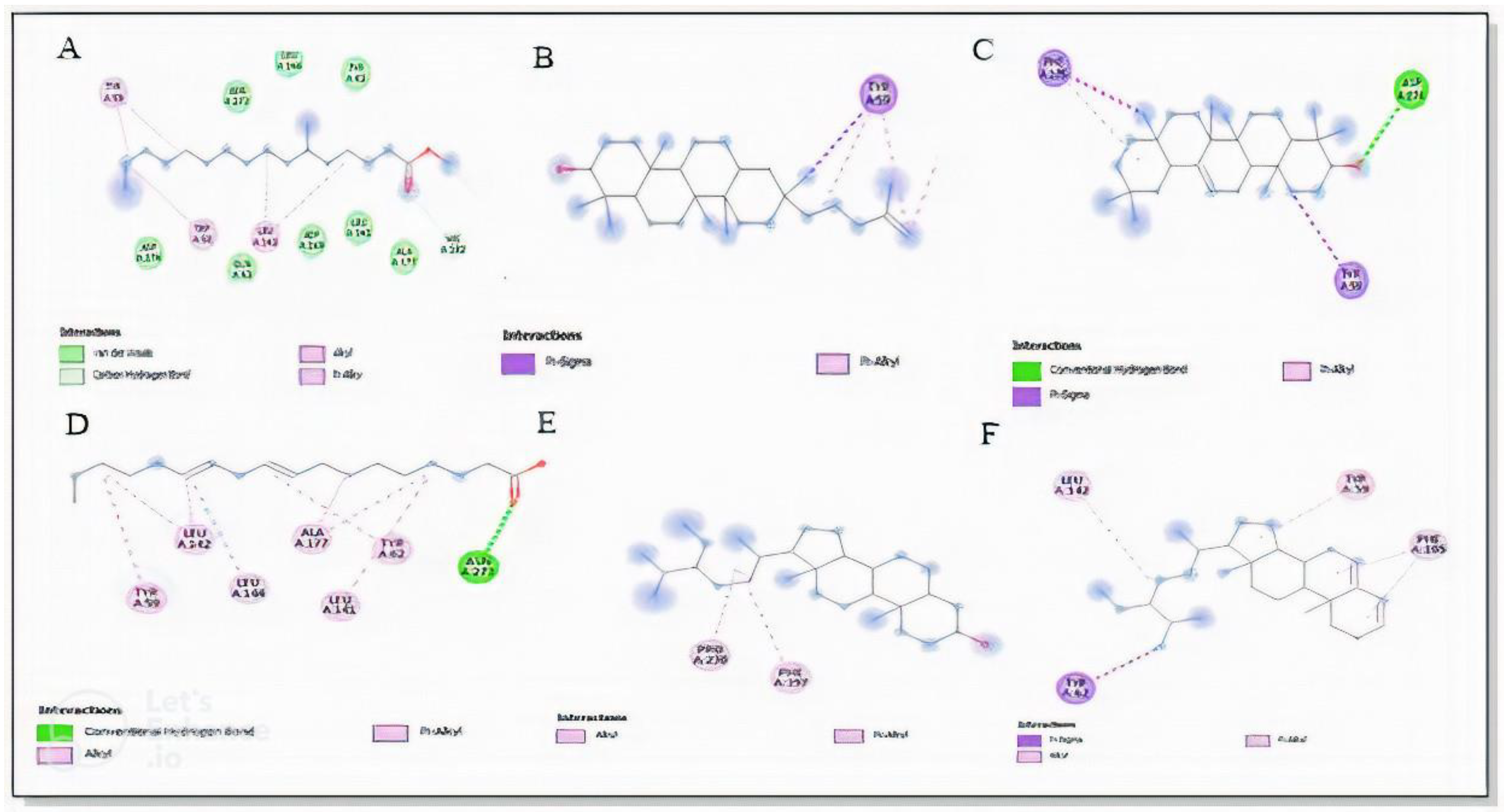
| Enzyme | Ligands | Binding Energy (kcal/mol) | Electrostatic/Hydrophobic Interaction |
|---|---|---|---|
| α-glucosidase | Acarbose | −6.6 | Hydrogen bond (Thr448, Asn443, Ala514, Asp441, Glu432, Arg437, His348) C-H bond (Gln438) |
| Lupeol | −6.9 | Alkyl interaction (Lys398, Trp394, Val380, Trp354) Hydrogen bond (Ala229, Asn301) Van-dar walls (Pro230, Arg340, Phe357, Ala378, Gly402, Glu377, Val335, Leu227, Met302, Glu396, Asp379, Glu231) | |
| Sitosterol | −7.5 | Alkyl interaction (Leu45, Ala444) C-H bond (Leu433) Van-dar walls (Ala434, Arg450, Met407, Thr445) | |
| 9,12-octadecadienoic acid | −4.1 | Alkyl interaction (Leu446) Van-dar walls (Asp411, Thr410, Leu373, Leu45, Asp440, Ser44, Gln438, Pro408, Glu432, Leu431) | |
| β-Amyrin | −8.4 | Alkyl interaction (Pro230) | |
| Hexadacanoic acid | −3.9 | Hydrogen bond (Aal380) C-H bond (Lyc398, Gly998) Alkyl interaction (Val335) | |
| Stigmasterol | −7.5 | Alkyl interaction (Val335, Ala343, Met302, Val334, Phe397, Phe297) | |
| α-amylase | Lupeol | −7.6 | Pi-sigma (Tyr59) Pi-alkyl (Trp60) |
| Sitosterol | −5.1 | Pi-alkyl (Pro230, Phe397) | |
| 9,12-octadecadienoic acid | −4.9 | Hydrogen bond (Asn273) Alkyl interaction (Tyr59, Leu142, Met302, Ala177, Leu141) | |
| β-Amyrin | −8.4 | Hydrogen bond (Asp274) Pi-sigma (Phe105) Pi-alkyl (Tyr59) | |
| Hexadacanoic acid | −4.6 | Van-dar walls (Asp274, Tyr62, Ala177, Asp176, Gln63, Asp269, Leu144, Asn273) Pi-alkyl (Tyr59, Trp58, Leu142) C-H bond (His102, Gln208) | |
| Stigmasterol | −9.1 | Pi-sigma (Tyr59) Pi-alkyl (Tyr59, Leu142, Phe105) |
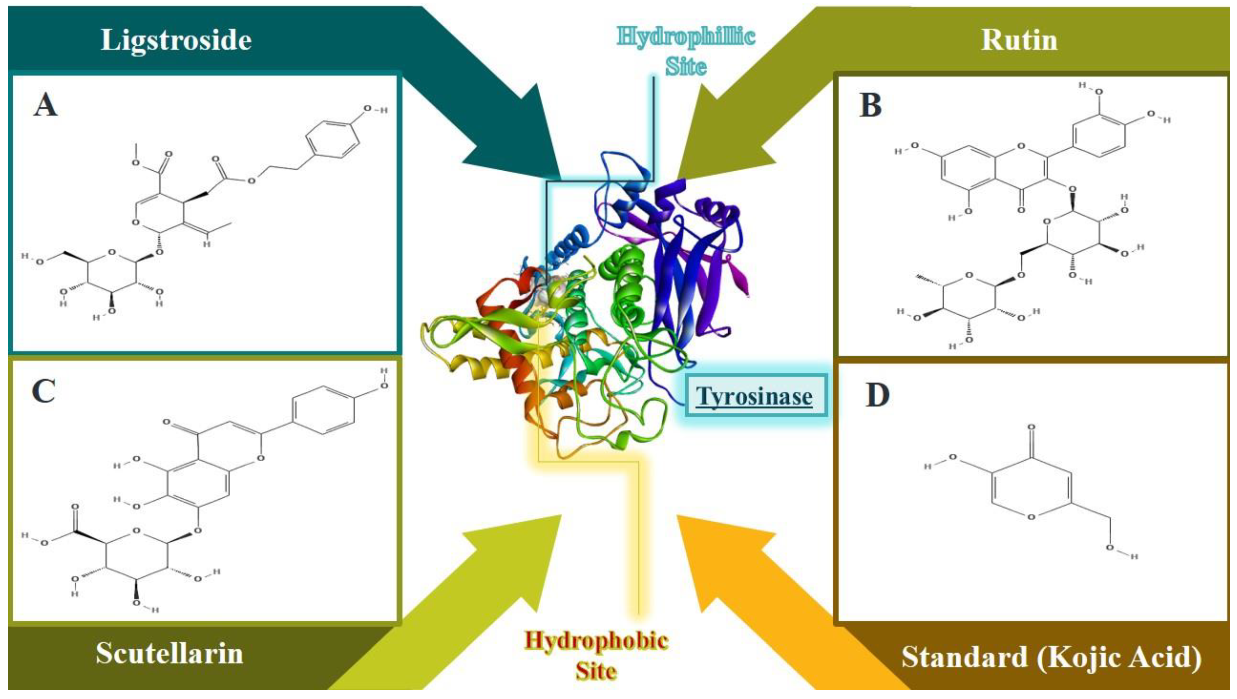
| Enzyme | Ligand | Binding Affinity (Kcal/mol) | Amino Acids Interactions |
|---|---|---|---|
| Tyrosinase | Ligstroside | −8.0 | Unfavorable Accaptor: (TYRA352) Pi Sigma: (VALA366) Conventional Hydrogen Bond: (ASNA15, GLNA294, GLYA360, GLYA361) Carbon Hydrogen: (SERA351, PHEA355) |
| Rutin | −8.9 | Amide-Pi Stacked: (PHEA355) Pi-Alkyl: (PROA298, LYSA359) Conventional Hydrogen Bond: (GLNA294, THRA345, VALA358, GLYA360, GLYA361) Van der Waals: (VALA13, GLYA299, VALA300, THRA343, ASPA344, ALAA346, SERA351, TYRA352, PROA363, VALA366) | |
| Scutellarin | −8.6 | Pi-Pi Stacked: (PHEA355) Pi-Alkyl: (ALAA295, PROA298) Conventional Hydrogen Bond: (GLNA294) Carbon Hydrogen: (THRA343) Van der Waals: (SERA291, TYRA297, GLYA299, VALA300, TRPA301, THRA345, SERA351, TYRA352, PROA363, VALA366) | |
| Kojic acid (Standard) | −5.3 | Pi-Pi Stacked: (PHEA355) |
Publisher’s Note: MDPI stays neutral with regard to jurisdictional claims in published maps and institutional affiliations. |
© 2022 by the authors. Licensee MDPI, Basel, Switzerland. This article is an open access article distributed under the terms and conditions of the Creative Commons Attribution (CC BY) license (https://creativecommons.org/licenses/by/4.0/).
Share and Cite
Aziz, M.; Ahmad, S.; Khurshid, U.; Pervaiz, I.; Lodhi, A.H.; Jan, N.; Khurshid, S.; Arshad, M.A.; Ibrahim, M.M.; Mersal, G.A.M.; et al. Comprehensive Biological Potential, Phytochemical Profiling Using GC-MS and LC-ESI-MS, and In-Silico Assessment of Strobilanthes glutinosus Nees: An Important Medicinal Plant. Molecules 2022, 27, 6885. https://doi.org/10.3390/molecules27206885
Aziz M, Ahmad S, Khurshid U, Pervaiz I, Lodhi AH, Jan N, Khurshid S, Arshad MA, Ibrahim MM, Mersal GAM, et al. Comprehensive Biological Potential, Phytochemical Profiling Using GC-MS and LC-ESI-MS, and In-Silico Assessment of Strobilanthes glutinosus Nees: An Important Medicinal Plant. Molecules. 2022; 27(20):6885. https://doi.org/10.3390/molecules27206885
Chicago/Turabian StyleAziz, Marya, Saeed Ahmad, Umair Khurshid, Irfan Pervaiz, Arslan Hussain Lodhi, Nasrullah Jan, Sameera Khurshid, Muhammad Adeel Arshad, Mohamed M. Ibrahim, Gaber A. M. Mersal, and et al. 2022. "Comprehensive Biological Potential, Phytochemical Profiling Using GC-MS and LC-ESI-MS, and In-Silico Assessment of Strobilanthes glutinosus Nees: An Important Medicinal Plant" Molecules 27, no. 20: 6885. https://doi.org/10.3390/molecules27206885
APA StyleAziz, M., Ahmad, S., Khurshid, U., Pervaiz, I., Lodhi, A. H., Jan, N., Khurshid, S., Arshad, M. A., Ibrahim, M. M., Mersal, G. A. M., Alenazi, F. S., Awadh Saleh Alamri, A., Butt, J., Saleem, H., & El-Bahy, Z. M. (2022). Comprehensive Biological Potential, Phytochemical Profiling Using GC-MS and LC-ESI-MS, and In-Silico Assessment of Strobilanthes glutinosus Nees: An Important Medicinal Plant. Molecules, 27(20), 6885. https://doi.org/10.3390/molecules27206885