
Imaging Constructs: The Rise of Iron Oxide Nanoparticles


 , , ,
, , , 

Abstract
1. Introduction
2. Techniques for Synthesis of Iron Oxide Core for Imaging Purposes
2.1. Co-Precipitation
2.2. Thermal Decomposition
2.3. Hydrothermal
2.4. Sol-Gel
2.5. Microemulsion
2.6. Other Used Methods
3. Surface Shell Engineering of Magnetic Materials for Imaging Purposes
3.1. Biopolymeric Shells
3.1.1. Chitosan and Its Derivatives
3.1.2. Dextran and Derivatives
3.1.3. Heparin
3.1.4. Albumin
3.1.5. Gelatin
3.1.6. Alginate
3.2. Synthetic Biocompatible Polymers
3.2.1. Silica Shells
3.2.2. PEG
4. Imaging Pre-Clinical and Clinical Studies of Core-Shell Iron Oxide Agents
5. Authors Contributions
6. Conclusions and Perspectives
Author Contributions
Funding
Conflicts of Interest
Abbreviations
| AEAPTMS | [N-(2-aminoethyl)-3-aminopropyl] trimethoxysilane |
| Ala | Alanine |
| AOT | Sodium 2-ethylhexyl sulfosuccinate |
| APTMS | (3-aminopropyl) trimethoxysilane |
| APTS | (3-aminopropyl) triethoxysilane |
| Asp | Aspartic acid |
| BSA | Bovine serum albumin |
| CD20 | b-lymphocyte antigen |
| CD41 | Integrin alpha-2b |
| CD61 | Integrin beta-3 |
| CT | Computed tomography |
| CTAB | Cetyltrimethyl ammonium bromide |
| Cy5.5 | Cyanine-5.5 |
| DBS | Sodium dodecylbenzene sulphonate |
| DLS | Dynamic light scattering |
| DPPE | 1,2-Bis(diphenylphosphino)ethane |
| DSPE | 1,2-Distearoyl-sn-glycero-3-phosphorylethanolamine |
| DSS | Dioctyl sodium dodecyl sulphate |
| EGF | Epidermal growth factor |
| FDA | Food and Drug Administration |
| Fe3O4 | Magnetite |
| γ-Fe2O3 | Maghemite |
| α-Fe2O3 | Hematite |
| FITC | Fluorescein isothiocyanate |
| FT-IR | Fourier-transform infrared spectroscopy |
| Glu | Glutamic acid |
| HA | Hyaluronic acid |
| HER2 | Human epidermal growth factor receptor 2 |
| hMSCs | Human mesenchymal stem cells |
| HPC | Hexadecylpyridinium chloride |
| HSA | Human serum albumin |
| IONPS | Iron oxide nanoparticles |
| Leu | Leucine |
| Lys | Lysine |
| MDA2 | Malondialdehyde 2 |
| MICAD | Molecular imaging and contrast agent database |
| MPI | Magnetic particle imaging |
| MRI | Magnetic resonance imaging |
| MTT | 3-(4,5-dimethylthiazol-2-yl)-2,5-diphenyl-2H-tetrazolium bromide |
| MTD | Maximum tolerated dose |
| NIH | National Institutes of Health |
| NIRF | Near-infrared fluorescence |
| NMR | Nuclear magnetic resonance |
| PAA | Poly(acrylic acid) |
| PAMAM | Poly (amidoamine) |
| PEG | Polyethylene glycol |
| PEI | Polyethylenimine |
| PEO | Polyethyleneoxide |
| Phe | Phenylalanine |
| PGA | Poly glutamic acid |
| PLA | Poly lactic acid |
| PLGA | Poly (lactic-co glycolic acid) |
| PS | Polystyrene |
| PVA | Polyvinyl alcohol |
| PVP | Polyvinylpyrrolidone |
| rHSA | Recombinant human serum albumin; |
| RITC | Rhodamine B isothiocyanate |
| SDS | Sodium dodecylsulfonate |
| SPECT | Single-photon emission computed tomography |
| SPIO | Superparamagnetic iron oxide |
| SPIONs | Superparamagnetic iron oxide nanoparticles |
| t1/2 | Half-life |
| TEM | Transmission electron microscopy |
| T | Tesla |
| Tyr | Tyrosine |
| US | Ultrasound |
| USPIO | Ultrasmall superparamagnetic iron oxide |
| USPIONs | Ultrasmall superparamagnetic iron oxide nanoparticles |
| Val | Valine |
| VCAM-1 | Vascular cell adhesion molecule-1 |
| VSM | Vibrating sample magnetometer |
| VT680 | Vivotag 680 |
| XRD | X-ray diffraction analysis |
References
- Hong, H.; Zhang, Y.; Sun, J.; Cai, W. Molecular Imaging and Therapy of Cancer with Radiolabeled Nanoparticles. Nano Today 2009, 4, 399–413. [Google Scholar] [CrossRef] [PubMed]
- Fass, L. Imaging and Cancer: A Review. Mol. Oncol. 2008, 2, 115–152. [Google Scholar] [CrossRef] [PubMed]
- Barreto, J.A.; O’Malley, W.; Kubeil, M.; Graham, B.; Stephan, H.; Spiccia, L. Nanomaterials: Applications in Cancer Imaging and Therapy. Adv. Mater. 2011, 23, H18–H40. [Google Scholar] [CrossRef]
- Janib, S.M.; Moses, A.S.; MacKay, J.A. Imaging and Drug Delivery Using Theranostic Nanoparticles. Adv. Drug Deliv. Rev. 2010, 62, 1052–1063. [Google Scholar] [CrossRef]
- Ellis, C.M.; Pellico, J.; Davis, J.J. Magnetic Nanoparticles Supporting Bio-Responsive T1/T2 Magnetic Resonance Imaging. Materials 2019, 12, 4096. [Google Scholar] [CrossRef]
- National Institute of Biomedical Imaging and Bioengineering (NIBIB). Available online: https://www.nibib.nih.gov/Science-Education/Science-Topics/Nuclear-Medicine (accessed on 11 April 2021).
- Assadi, M.; Afrasiabi, K.; Nabipour, I.; Seyedabadi, M. Nanotechnology and Nuclear Medicine; Research and Preclinical Applications. Hell. J. Nucl. Med. 2011, 14, 149–159. [Google Scholar] [PubMed]
- Morales-Avila, E.; Ferro-Flores, G.; Ocampo-García, B.E.; de María Ramírez, F. Radiolabeled Nanoparticles for Molecular Imaging. In Molecular Imaging; Schaller, B., Ed.; IntechOpen: London, UK, 2012; ISBN 978-953-51-0359-2. [Google Scholar]
- Sun, G.; Xu, J.; Hagooly, A.; Rossin, R.; Li, Z.; Moore, D.A.; Hawker, C.J.; Welch, M.J.; Wooley, K.L. Strategies for Optimized Radiolabeling of Nanoparticles for in Vivo PET Imaging. Adv. Mater. 2007, 19, 3157–3162. [Google Scholar] [CrossRef]
- Gomes, C.M.; Abrunhosa, A.J.; Ramos, P.; Pauwels, E.K.J. Molecular Imaging with SPECT as a Tool for Drug Development. Adv. Drug Deliv. Rev. 2011, 63, 547–554. [Google Scholar] [CrossRef] [PubMed]
- Brigger, I.; Dubernet, C.; Couvreur, P. Nanoparticles in Cancer Therapy and Diagnosis. Adv. Drug Deliv. Rev. 2002, 54, 631–651. [Google Scholar] [CrossRef]
- Phillips, M.A.; Gran, M.L.; Peppas, N.A. Targeted Nanodelivery of Drugs and Diagnostics. Nano Today 2010, 5, 143–159. [Google Scholar] [CrossRef] [PubMed]
- Jeong, H.-J.; Lee, B.C.; Ahn, B.-C.; Kang, K.W. Development of Drugs and Technology for Radiation Theragnosis. Nucl. Eng. Technol. 2016, 48, 597–607. [Google Scholar] [CrossRef][Green Version]
- Huang, H.-C.; Barua, S.; Sharma, G.; Dey, S.K.; Rege, K. Inorganic Nanoparticles for Cancer Imaging and Therapy. J. Control. Release 2011, 155, 344–357. [Google Scholar] [CrossRef]
- Molecular Imaging and Contrast Agent Database (MICAD); National Center for Biotechnology Information (US): Bethesda, MD, USA, 2004–2013. Available online: https://www.ncbi.nlm.nih.gov/books/NBK5330/ (accessed on 18 March 2021).
- Ali, A.; Zafar, H.; Zia, M.; ul Haq, I.; Phull, A.R.; Ali, J.S.; Hussain, A. Synthesis, Characterization, Applications, and Challenges of Iron Oxide Nanoparticles. Nanotechnol. Sci. Appl. 2016, 9, 49–67. [Google Scholar] [CrossRef]
- Laurent, S.; Forge, D.; Port, M.; Roch, A.; Robic, C.; Vander Elst, L.; Muller, R.N. Magnetic Iron Oxide Nanoparticles: Synthesis, Stabilization, Vectorization, Physicochemical Characterizations, and Biological Applications. Chem. Rev. 2008, 108, 2064–2110. [Google Scholar] [CrossRef] [PubMed]
- Laurent, S.; Bridot, J.-L.; Elst, L.V.; Muller, R.N. Magnetic Iron Oxide Nanoparticles for Biomedical Applications. Future Med. Chem. 2010, 2, 427–449. [Google Scholar] [CrossRef] [PubMed]
- Ramimoghadam, D.; Bagheri, S.; Hamid, S.B.A. Progress in Electrochemical Synthesis of Magnetic Iron Oxide Nanoparticles. J. Magn. Magn. Mater. 2014, 368, 207–229. [Google Scholar] [CrossRef]
- Avasthi, A.; Caro, C.; Pozo-Torres, E.; Leal, M.P.; García-Martín, M.L. Magnetic Nanoparticles as MRI Contrast Agents. Top. Curr. Chem. 2020, 378, 40. [Google Scholar] [CrossRef] [PubMed]
- Caspani, S.; Magalhães, R.; Araújo, J.P.; Sousa, C.T. Magnetic Nanomaterials as Contrast Agents for MRI. Materials 2020, 13, 2586. [Google Scholar] [CrossRef] [PubMed]
- Chouhan, R.S.; Horvat, M.; Ahmed, J.; Alhokbany, N.; Alshehri, S.M.; Gandhi, S. Magnetic Nanoparticles—A Multifunctional Potential Agent for Diagnosis and Therapy. Cancers 2021, 13, 2213. [Google Scholar] [CrossRef]
- Niculescu, A.-G.; Chircov, C.; Grumezescu, A.M. Magnetite Nanoparticles: Synthesis Methods—A Comparative Review. Methods 2021, in press. [Google Scholar] [CrossRef]
- Fatima, H.; Kim, K.-S. Iron-Based Magnetic Nanoparticles for Magnetic Resonance Imaging. Adv. Powder Technol. 2018, 29, 2678–2685. [Google Scholar] [CrossRef]
- Khalafalla, S.; Reimers, G. Preparation of Dilution-Stable Aqueous Magnetic Fluids. IEEE Trans. Magn. 1980, 16, 178–183. [Google Scholar] [CrossRef]
- Massart, R. Preparation of Aqueous Magnetic Liquids in Alkaline and Acidic Media. IEEE Trans. Magn. 1981, 17, 1247–1248. [Google Scholar] [CrossRef]
- Dadfar, S.M.; Roemhild, K.; Drude, N.I.; von Stillfried, S.; Knüchel, R.; Kiessling, F.; Lammers, T. Iron Oxide Nanoparticles: Diagnostic, Therapeutic and Theranostic Applications. Adv. Drug Deliv. Rev. 2019, 138, 302–325. [Google Scholar] [CrossRef]
- Adewunmi, A.A.; Kamal, M.S.; Solling, T.I. Application of Magnetic Nanoparticles in Demulsification: A Review on Synthesis, Performance, Recyclability, and Challenges. J. Pet. Sci. Eng. 2021, 196, 107680. [Google Scholar] [CrossRef]
- Xia, T.; Wang, J.; Wu, C.; Meng, F.; Shi, Z.; Lian, J.; Feng, J.; Meng, J. Novel Complex-Co-precipitation Route to Form High Quality Triethanolamine-Coated Fe3O4 Nanocrystals: Their High Saturation Magnetizations and Excellent Water Treatment Properties. CrystEngComm 2012, 14, 5741–5744. [Google Scholar] [CrossRef]
- Majidi, S.; Zeinali Sehrig, F.; Farkhani, S.M.; Soleymani Goloujeh, M.; Akbarzadeh, A. Current Methods for Synthesis of Magnetic Nanoparticles. Artif. Cells Nanomed. Biotechnol. 2016, 44, 722–734. [Google Scholar] [CrossRef]
- Lu, A.-H.; Salabas, E.L.; Schüth, F. Magnetic Nanoparticles: Synthesis, Protection, Functionalization, and Application. Angew. Chem. Int. Ed. 2007, 46, 1222–1244. [Google Scholar] [CrossRef]
- Wu, W.; He, Q.; Jiang, C. Magnetic Iron Oxide Nanoparticles: Synthesis and Surface Functionalization Strategies. Nanoscale Res. Lett. 2008, 3, 397–415. [Google Scholar] [CrossRef]
- Xu, C.; Lee, J.; Teja, A.S. Continuous Hydrothermal Synthesis of Lithium Iron Phosphate Particles in Subcritical and Supercritical Water. J. Supercrit. Fluids 2008, 44, 92–97. [Google Scholar] [CrossRef]
- Al-Rawi, N.N.; Anwer, B.A.; Al-Rawi, N.H.; Uthman, A.T.; Ahmed, I.S. Magnetism in Drug Delivery: The Marvels of Iron Oxides and Substituted Ferrites Nanoparticles. Saudi Pharm. J. 2020, 28, 876–887. [Google Scholar] [CrossRef] [PubMed]
- Soenen, S.J.H.; De Cuyper, M. Assessing Cytotoxicity of (Iron Oxide-Based) Nanoparticles: An Overview of Different Methods Exemplified with Cationic Magnetoliposomes. Contrast Media Mol. Imaging 2009, 4, 207–219. [Google Scholar] [CrossRef] [PubMed]
- Caruntu, D.; Caruntu, G.; Chen, Y.; O’Connor, C.J.; Goloverda, G.; Kolesnichenko, V.L. Synthesis of Variable-Sized Nanocrystals of Fe3O4 with High Surface Reactivity. Chem. Mater. 2004, 16, 5527–5534. [Google Scholar] [CrossRef]
- Iacovita, C.; Fizeșan, I.; Pop, A.; Scorus, L.; Dudric, R.; Stiufiuc, G.; Vedeanu, N.; Tetean, R.; Loghin, F.; Stiufiuc, R.; et al. In Vitro Intracellular Hyperthermia of Iron Oxide Magnetic Nanoparticles, Synthesized at High Temperature by a Polyol Process. Pharmaceutics 2020, 12, 424. [Google Scholar] [CrossRef] [PubMed]
- Cheng, C.; Xu, F.; Gu, H. Facile Synthesis and Morphology Evolution of Magnetic Iron Oxide Nanoparticles in Different Polyol Processes. New J. Chem. 2011, 35, 1072. [Google Scholar] [CrossRef]
- Hachani, R.; Lowdell, M.; Birchall, M.; Hervault, A.; Mertz, D.; Begin-Colin, S.; Thanh, N.T.K. Polyol Synthesis, Functionalisation, and Biocompatibility Studies of Superparamagnetic Iron Oxide Nanoparticles as Potential MRI Contrast Agents. Nanoscale 2016, 8, 3278–3287. [Google Scholar] [CrossRef]
- Alphandéry, E. Applications of Magnetosomes Synthesized by Magnetotactic Bacteria in Medicine. Front. Bioeng. Biotechnol. 2014, 2, 5. [Google Scholar] [CrossRef] [PubMed]
- Vargas, G.; Cypriano, J.; Correa, T.; Leão, P.; Bazylinski, D.A.; Abreu, F. Applications of Magnetotactic Bacteria, Magnetosomes and Magnetosome Crystals in Biotechnology and Nanotechnology: Mini-Review. Molecules 2018, 23, 2438. [Google Scholar] [CrossRef] [PubMed]
- Zhao, D. Bacteriogenic Magnetic Nanoparticles as Magnetic Resonance Imaging Contrast Agents. Transl. Cancer Res. 2017, 6, S512–S514. [Google Scholar] [CrossRef]
- González-Carreño, T.; Morales, M.P.; Gracia, M.; Serna, C.J. Preparation of Uniform γ-Fe2O3 Particles with Nanometer Size by Spray Pyrolysis. Mater. Lett. 1993, 18, 151–155. [Google Scholar] [CrossRef]
- Teoh, W.Y.; Amal, R.; Mädler, L. Flame Spray Pyrolysis: An Enabling Technology for Nanoparticles Design and Fabrication. Nanoscale 2010, 2, 1324–1347. [Google Scholar] [CrossRef]
- Sequeira, C.A.C. Electrochemical Synthesis of Iron Oxide Nanoparticles for Biomedical Application. Org. Med. Chem. Int. J. 2018, 5. [Google Scholar] [CrossRef]
- Fu, X.; Cai, J.; Zhang, X.; Li, W.-D.; Ge, H.; Hu, Y. Top-down Fabrication of Shape-Controlled, Monodisperse Nanoparticles for Biomedical Applications. Adv. Drug Deliv. Rev. 2018, 132, 169–187. [Google Scholar] [CrossRef] [PubMed]
- Valencia, P.M.; Farokhzad, O.C.; Karnik, R.; Langer, R. Microfluidic Technologies for Accelerating the Clinical Translation of Nanoparticles. Nat. Nanotechnol. 2012, 7, 623–629. [Google Scholar] [CrossRef]
- Shrimal, P.; Jadeja, G.; Patel, S. A Review on Novel Methodologies for Drug Nanoparticle Preparation: Microfluidic Approach. Chem. Eng. Res. Des. 2020, 153, 728–756. [Google Scholar] [CrossRef]
- Suryawanshi, P.L.; Sonawane, S.H.; Bhanvase, B.A.; Ashokkumar, M.; Pimplapure, M.S.; Gogate, P.R. Synthesis of Iron Oxide Nanoparticles in a Continuous Flow Spiral Microreactor and Corning® Advanced FlowTM Reactor. Green Process. Synth. 2018, 7, 1–11. [Google Scholar] [CrossRef]
- Dodi, G.; Hritcu, D.; Lisa, G.; Popa, M.I. Core–Shell Magnetic Chitosan Particles Functionalized by Grafting: Synthesis and Characterization. Chem. Eng. J. 2012, 203, 130–141. [Google Scholar] [CrossRef]
- Jain, T.K.; Reddy, M.K.; Morales, M.A.; Leslie-Pelecky, D.L.; Labhasetwar, V. Biodistribution, Clearance, and Biocompatibility of Iron Oxide Magnetic Nanoparticles in Rats. Mol. Pharm. 2008, 5, 316–327. [Google Scholar] [CrossRef] [PubMed]
- Cojocaru, F.D.; Balan, V.; Popa, I.M.; Munteanu, A.; Anghelache, A.; Verestiuc, L. Magnetic Composite Scaffolds for Potential Applications in Radiochemotherapy of Malignant Bone Tumors. Medicina 2019, 55, 153. [Google Scholar] [CrossRef] [PubMed]
- Muthiah, M.; Park, I.-K.; Cho, C.-S. Surface Modification of Iron Oxide Nanoparticles by Biocompatible Polymers for Tissue Imaging and Targeting. Biotechnol. Adv. 2013, 31, 1224–1236. [Google Scholar] [CrossRef] [PubMed]
- Zhu, N.; Ji, H.; Yu, P.; Niu, J.; Farooq, M.U.; Akram, M.W.; Udego, I.O.; Li, H.; Niu, X. Surface Modification of Magnetic Iron Oxide Nanoparticles. Nanomaterials 2018, 8, 810. [Google Scholar] [CrossRef]
- Ansari, S.; Ficiarà, E.; Ruffinatti, F.; Stura, I.; Argenziano, M.; Abollino, O.; Cavalli, R.; Guiot, C.; D’Agata, F. Magnetic Iron Oxide Nanoparticles: Synthesis, Characterization and Functionalization for Biomedical Applications in the Central Nervous System. Materials 2019, 12, 465. [Google Scholar] [CrossRef] [PubMed]
- Palanisamy, S.; Wang, Y.-M. Superparamagnetic Iron Oxide Nanoparticulate System: Synthesis, Targeting, Drug Delivery and Therapy in Cancer. Dalton Trans. 2019, 48, 9490–9515. [Google Scholar] [CrossRef]
- Narayanaswamy, R.; Kanagesan, S.; Pandurangan, A.; Padmanabhan, P. Basics to different imaging techniques, different nanobiomaterials for image enhancement. In Nanobiomaterials in Medical Imaging; Elsevier: Amsterdam, The Netherlands, 2016; pp. 101–129. ISBN 978-0-323-41736-5. [Google Scholar]
- Nelson, N.; Port, J.; Pandey, M. Use of Superparamagnetic Iron Oxide Nanoparticles (SPIONs) via Multiple Imaging Modalities and Modifications to Reduce Cytotoxicity: An Educational Review. J. Nanotheranostics 2020, 1, 105–135. [Google Scholar] [CrossRef]
- Gholipour, N.; Akhlaghi, M.; Mokhtari Kheirabadi, A.; Geramifar, P.; Beiki, D. Development of Ga-68 Labeled, Biotinylated Thiosemicarbazone Dextran-Coated Iron Oxide Nanoparticles as Multimodal PET/MRI Probe. Int. J. Biol. Macromol. 2020, 148, 932–941. [Google Scholar] [CrossRef] [PubMed]
- Unterweger, H.; Dézsi, L.; Matuszak, J.; Janko, C.; Poettler, M.; Jordan, J.; Bäuerle, T.; Szebeni, J.; Fey, T.; Boccaccini, A.; et al. Dextran-Coated Superparamagnetic Iron Oxide Nanoparticles for Magnetic Resonance Imaging: Evaluation of Size-Dependent Imaging Properties, Storage Stability and Safety. Int. J. Nanomed. 2018, 13, 1899–1915. [Google Scholar] [CrossRef] [PubMed]
- Balan, V.; Dodi, G.; Tudorachi, N.; Ponta, O.; Simon, V.; Butnaru, M.; Verestiuc, L. Doxorubicin-Loaded Magnetic Nanocapsules Based on N-Palmitoyl Chitosan and Magnetite: Synthesis and Characterization. Chem. Eng. J. 2015, 279, 188–197. [Google Scholar] [CrossRef]
- Zheng, S.; Han, J.; Jin, Z.; Kim, C.-S.; Park, S.; Kim, K.-P.; Park, J.-O.; Choi, E. Dual Tumor-Targeted Multifunctional Magnetic Hyaluronic Acid Micelles for Enhanced MR Imaging and Combined Photothermal-Chemotherapy. Colloids Surf. B Biointerfaces 2018, 164, 424–435. [Google Scholar] [CrossRef] [PubMed]
- Tudorachi, N.; Chiriac, A.P.; Nita, L.E.; Mustata, F.; Diaconu, A.; Balan, V.; Rusu, A.; Lisa, G. Studies on the Nanocomposites Based on Carboxymethyl Starch-g-Lactic Acid-Co-Glycolic Acid Copolymer and Magnetite. J. Therm. Anal. Calorim. 2018, 131, 1867–1880. [Google Scholar] [CrossRef]
- Vismara, E.; Bongio, C.; Coletti, A.; Edelman, R.; Serafini, A.; Mauri, M.; Simonutti, R.; Bertini, S.; Urso, E.; Assaraf, Y.G.; et al. Albumin and Hyaluronic Acid-Coated Superparamagnetic Iron Oxide Nanoparticles Loaded with Paclitaxel for Biomedical Applications. Molecules 2017, 22, 1030. [Google Scholar] [CrossRef]
- Castelló, J.; Gallardo, M.; Busquets, M.A.; Estelrich, J. Chitosan (or Alginate)-Coated Iron Oxide Nanoparticles: A Comparative Study. Colloids Surf. A Physicochem. Eng. Asp. 2015, 468, 151–158. [Google Scholar] [CrossRef]
- Tomitaka, A.; Jo, J.; Aoki, I.; Tabata, Y. Preparation of Biodegradable Iron Oxide Nanoparticles with Gelatin for Magnetic Resonance Imaging. Inflamm. Regen. 2014, 34, 45–55. [Google Scholar] [CrossRef][Green Version]
- Farokhi, M.; Mottaghitalab, F.; Saeb, M.R.; Thomas, S. Functionalized Theranostic Nanocarriers with Bio-Inspired Polydopamine for Tumor Imaging and Chemo-Photothermal Therapy. J. Control. Release 2019, 309, 203–219. [Google Scholar] [CrossRef] [PubMed]
- Wu, M.; Zhang, D.; Zeng, Y.; Wu, L.; Liu, X.; Liu, J. Nanocluster of Superparamagnetic Iron Oxide Nanoparticles Coated with Poly (Dopamine) for Magnetic Field-Targeting, Highly Sensitive MRI and Photothermal Cancer Therapy. Nanotechnology 2015, 26, 115102. [Google Scholar] [CrossRef] [PubMed]
- Ge, Y.; Zhang, Y.; He, S.; Nie, F.; Teng, G.; Gu, N. Fluorescence Modified Chitosan-Coated Magnetic Nanoparticles for High-Efficient Cellular Imaging. Nanoscale Res. Lett. 2009, 4, 287–295. [Google Scholar] [CrossRef] [PubMed]
- Unsoy, G.; Yalcin, S.; Khodadust, R.; Gunduz, G.; Gunduz, U. Synthesis Optimization and Characterization of Chitosan-Coated Iron Oxide Nanoparticles Produced for Biomedical Applications. J. Nanopart. Res. 2012, 14, 964. [Google Scholar] [CrossRef]
- Rhee, I.; Hong, S.; Chang, Y. Chitosan-Coated Ferrite (Fe3O4) Nanoparticles as a T2 Contrast Agent for Magnetic Resonance Imaging. J. Korean Phys. Soc. 2010, 56, 868–873. [Google Scholar] [CrossRef]
- Khmara, I.; Strbak, O.; Zavisova, V.; Koneracka, M.; Kubovcikova, M.; Antal, I.; Kavecansky, V.; Lucanska, D.; Dobrota, D.; Kopcansky, P. Chitosan-Stabilized Iron Oxide Nanoparticles for Magnetic Resonance Imaging. J. Magn. Magn. Mater. 2019, 474, 319–325. [Google Scholar] [CrossRef]
- Hajiramezanali, M.; Atyabi, F.; Mosayebnia, M.; Akhlaghi, M.; Geramifar, P.; Jalilian, A.R.; Mazidi, S.M.; Yousefnia, H.; Shahhosseini, S.; Beiki, D. 68Ga-Radiolabeled Bombesin-Conjugated to Trimethyl Chitosan-Coated Superparamagnetic Nanoparticles for Molecular Imaging: Preparation, Characterization and Biological Evaluation. Int. J. Nanomed. 2019, 14, 2591–2605. [Google Scholar] [CrossRef]
- Su, H.; Han, X.; He, L.; Deng, L.; Yu, K.; Jiang, H.; Wu, C.; Jia, Q.; Shan, S. Synthesis and Characterization of Magnetic Dextran Nanogel Doped with Iron Oxide Nanoparticles as Magnetic Resonance Imaging Probe. Int. J. Biol. Macromol. 2019, 128, 768–774. [Google Scholar] [CrossRef]
- Hsieh, W.-J.; Liang, C.-J.; Chieh, J.-J.; Wang, S.-H.; Lai, I.-R.; Chen, J.-H.; Chang, F.-H.; Tseng, W.-K.; Yang, S.-Y.; Wu, C.-C.; et al. In Vivo Tumor Targeting and Imaging with Anti-Vascular Endothelial Growth Factor Antibody-Conjugated Dextran-Coated Iron Oxide Nanoparticles. Int. J. Nanomed. 2012, 7, 2833–2842. [Google Scholar] [CrossRef][Green Version]
- Mishra, S.K.; Kumar, B.S.H.; Khushu, S.; Tripathi, R.P.; Gangenahalli, G. Increased Transverse Relaxivity in Ultrasmall Superparamagnetic Iron Oxide Nanoparticles Used as MRI Contrast Agent for Biomedical Imaging. Contrast Media Mol. Imaging 2016, 11, 350–361. [Google Scholar] [CrossRef]
- Jafari, A.; Salouti, M.; Shayesteh, S.F.; Heidari, Z.; Rajabi, A.B.; Boustani, K.; Nahardani, A. Synthesis and Characterization of Bombesin-Superparamagnetic Iron Oxide Nanoparticles as a Targeted Contrast Agent for Imaging of Breast Cancer Using MRI. Nanotechnology 2015, 26, 075101. [Google Scholar] [CrossRef]
- Torres Martin de Rosales, R.; Tavaré, R.; Glaria, A.; Varma, G.; Protti, A.; Blower, P.J. 99mTc-Bisphosphonate-Iron Oxide Nanoparticle Conjugates for Dual-Modality Biomedical Imaging. Bioconjug. Chem. 2011, 22, 455–465. [Google Scholar] [CrossRef] [PubMed]
- Liu, G.; Hong, R.Y.; Guo, L.; Li, Y.G.; Li, H.Z. Preparation, Characterization and MRI Application of Carboxymethyl Dextran Coated Magnetic Nanoparticles. Appl. Surf. Sci. 2011, 257, 6711–6717. [Google Scholar] [CrossRef]
- Tao, C.; Zheng, Q.; An, L.; He, M.; Lin, J.; Tian, Q.; Yang, S. T1-Weight Magnetic Resonance Imaging Performances of Iron Oxide Nanoparticles Modified with a Natural Protein Macromolecule and an Artificial Macromolecule. Nanomaterials 2019, 9, 170. [Google Scholar] [CrossRef] [PubMed]
- Li, H.; Yan, K.; Shang, Y.; Shrestha, L.; Liao, R.; Liu, F.; Li, P.; Xu, H.; Xu, Z.; Chu, P.K. Folate-Bovine Serum Albumin Functionalized Polymeric Micelles Loaded with Superparamagnetic Iron Oxide Nanoparticles for Tumor Targeting and Magnetic Resonance Imaging. Acta Biomater. 2015, 15, 117–126. [Google Scholar] [CrossRef] [PubMed]
- Ma, H.; Qi, X.; Maitani, Y.; Nagai, T. Preparation and Characterization of Superparamagnetic Iron Oxide Nanoparticles Stabilized by Alginate. Int. J. Pharm. 2007, 333, 177–186. [Google Scholar] [CrossRef] [PubMed]
- Ma, H.L.; Xu, Y.F.; Qi, X.R.; Maitani, Y.; Nagai, T. Superparamagnetic Iron Oxide Nanoparticles Stabilized by Alginate: Pharmacokinetics, Tissue Distribution, and Applications in Detecting Liver Cancers. Int. J. Pharm. 2008, 354, 217–226. [Google Scholar] [CrossRef] [PubMed]
- Torres Martin de Rosales, R.; Tavaré, R.; Paul, R.L.; Jauregui-Osoro, M.; Protti, A.; Glaria, A.; Varma, G.; Szanda, I.; Blower, P.J. Synthesis of 64Cu(II)-Bis(Dithiocarbamatebisphosphonate) and Its Conjugation with Superparamagnetic Iron Oxide Nanoparticles: In Vivo Evaluation as Dual-Modality PET-MRI Agent. Angew. Chem. Int. Ed. 2011, 50, 5509–5513. [Google Scholar] [CrossRef] [PubMed]
- Zhang, B.; Li, Q.; Yin, P.; Rui, Y.; Qiu, Y.; Wang, Y.; Shi, D. Ultrasound-Triggered BSA/SPION Hybrid Nanoclusters for Liver-Specific Magnetic Resonance Imaging. ACS Appl. Mater. Interfaces 2012, 4, 6479–6486. [Google Scholar] [CrossRef]
- Sun, W.; Yang, J.; Zhu, J.; Zhou, Y.; Li, J.; Zhu, X.; Shen, M.; Zhang, G.; Shi, X. Immobilization of Iron Oxide Nanoparticles within Alginate Nanogels for Enhanced MR Imaging Applications. Biomater. Sci. 2016, 4, 1422–1430. [Google Scholar] [CrossRef] [PubMed]
- Widder, D.J.; Greif, W.L.; Widder, K.J.; Edelman, R.R.; Brady, T.J. Magnetite Albumin Microspheres: A New MR Contrast Material. AJR Am. J. Roentgenol. 1987, 148, 399–404. [Google Scholar] [CrossRef] [PubMed]
- Ye, M.; Zhou, J.; Zhong, Y.; Xu, J.; Hou, J.; Wang, X.; Wang, Z.; Guo, D. SR-A-Targeted Phase-Transition Nanoparticles for the Detection and Treatment of Atherosclerotic Vulnerable Plaques. ACS Appl. Mater. Interfaces 2019, 11, 9702–9715. [Google Scholar] [CrossRef] [PubMed]
- Choi, H.; Choi, S.R.; Zhou, R.; Kung, H.F.; Chen, I.-W. Iron Oxide Nanoparticles as Magnetic Resonance Contrast Agent for Tumor Imaging via Folate Receptor-Targeted Delivery1. Acad. Radiol. 2004, 11, 996–1004. [Google Scholar] [CrossRef]
- Xu, S.; Wang, J.; Wei, Y.; Zhao, H.; Tao, T.; Wang, H.; Wang, Z.; Du, J.; Wang, H.; Qian, J.; et al. In Situ One-Pot Synthesis of Fe2O3@BSA Core-Shell Nanoparticles as Enhanced T1-Weighted Magnetic Resonance Imagine Contrast Agents. ACS Appl. Mater. Interfaces 2020, 12, 56701–56711. [Google Scholar] [CrossRef]
- Wang, X.; Tu, M.; Tian, B.; Yi, Y.; Wei, Z.; Wei, F. Synthesis of Tumor-Targeted Folate Conjugated Fluorescent Magnetic Albumin Nanoparticles for Enhanced Intracellular Dual-Modal Imaging into Human Brain Tumor Cells. Anal. Biochem. 2016, 512, 8–17. [Google Scholar] [CrossRef]
- Rosenberger, I.; Schmithals, C.; Vandooren, J.; Bianchessi, S.; Milani, P.; Locatelli, E.; Israel, L.L.; Hübner, F.; Matteoli, M.; Lellouche, J.-P.; et al. Physico-Chemical and Toxicological Characterization of Iron-Containing Albumin Nanoparticles as Platforms for Medical Imaging. J. Control. Release 2014, 194, 130–137. [Google Scholar] [CrossRef]
- Skaat, H.; Margel, S. Synthesis of Fluorescent-Maghemite Nanoparticles as Multimodal Imaging Agents for Amyloid-Beta Fibrils Detection and Removal by a Magnetic Field. Biochem. Biophys. Res. Commun. 2009, 386, 645–649. [Google Scholar] [CrossRef]
- Shen, F.; Poncet-Legrand, C.; Somers, S.; Slade, A.; Yip, C.; Duft, A.M.; Winnik, F.M.; Chang, P.L. Properties of a Novel Magnetized Alginate for Magnetic Resonance Imaging. Biotechnol. Bioeng. 2003, 83, 282–292. [Google Scholar] [CrossRef]
- Yuk, S.H.; Oh, K.S.; Cho, S.H.; Lee, B.S.; Kim, S.Y.; Kwak, B.-K.; Kim, K.; Kwon, I.C. Glycol Chitosan/Heparin Immobilized Iron Oxide Nanoparticles with a Tumor-Targeting Characteristic for Magnetic Resonance Imaging. Biomacromolecules 2011, 12, 2335–2343. [Google Scholar] [CrossRef]
- Moore, A.; Marecos, E.; Bogdanov, A.; Weissleder, R. Tumoral Distribution of Long-Circulating Dextran-Coated Iron Oxide Nanoparticles in a Rodent Model. Radiology 2000, 214, 568–574. [Google Scholar] [CrossRef] [PubMed]
- Mandeville, J.B.; Moore, J.; Chesler, D.A.; Garrido, L.; Weissleder, R.; Weisskoff, R.M. Dynamic Liver Imaging with Iron Oxide Agents: Effects of Size and Biodistribution on Contrast. Magn. Reson. Med. 1997, 37, 885–890. [Google Scholar] [CrossRef]
- Borny, R.; Lechleitner, T.; Schmiedinger, T.; Hermann, M.; Tessadri, R.; Redhammer, G.; Neumüller, J.; Kerjaschki, D.; Berzaczy, G.; Erman, G.; et al. Nucleophilic Cross-Linked, Dextran Coated Iron Oxide Nanoparticles as Basis for Molecular Imaging: Synthesis, Characterization, Visualization and Comparison with Previous Product. Contrast Media Mol. Imaging 2015, 10, 18–27. [Google Scholar] [CrossRef] [PubMed]
- Shin, T.-H.; Kim, P.K.; Kang, S.; Cheong, J.; Kim, S.; Lim, Y.; Shin, W.; Jung, J.-Y.; Lah, J.D.; Choi, B.W.; et al. High-Resolution T1 MRI via Renally Clearable Dextran Nanoparticles with an Iron Oxide Shell. Nat. Biomed. Eng. 2021, 5, 252–263. [Google Scholar] [CrossRef]
- Thorek, D.L.J.; Ulmert, D.; Diop, N.-F.M.; Lupu, M.E.; Doran, M.G.; Huang, R.; Abou, D.S.; Larson, S.M.; Grimm, J. Non-Invasive Mapping of Deep-Tissue Lymph Nodes in Live Animals Using a Multimodal PET/MRI Nanoparticle. Nat. Commun. 2014, 5, 3097. [Google Scholar] [CrossRef] [PubMed]
- Naha, P.C.; Zaki, A.A.; Hecht, E.; Chorny, M.; Chhour, P.; Blankemeyer, E.; Yates, D.M.; Witschey, W.R.T.; Litt, H.I.; Tsourkas, A.; et al. Dextran Coated Bismuth-Iron Oxide Nanohybrid Contrast Agents for Computed Tomography and Magnetic Resonance Imaging. J. Mater. Chem. B 2014, 2, 8239–8248. [Google Scholar] [CrossRef]
- Fu, C.; Zhou, H.; Wang, Y.; Liu, D.; Li, J.; Deng, H.; Qi, X.; Chen, T.; Zhang, L.-M.; Li, G. One-Pot Synthesis of Dextran-Coated Iron Oxide Nanoclusters for Real-Time Regional Lymph Node Mapping. Int. J. Nanomed. 2017, 12, 3365–3374. [Google Scholar] [CrossRef]
- Jarrett, B.R.; Frendo, M.; Vogan, J.; Louie, A.Y. Size-Controlled Synthesis of Dextran Sulfate Coated Iron Oxide Nanoparticles for Magnetic Resonance Imaging. Nanotechnology 2007, 18, 035603. [Google Scholar] [CrossRef] [PubMed]
- Tu, C.; Ng, T.S.C.; Sohi, H.K.; Palko, H.A.; House, A.; Jacobs, R.E.; Louie, A.Y. Receptor-Targeted Iron Oxide Nanoparticles for Molecular MR Imaging of Inflamed Atherosclerotic Plaques. Biomaterials 2011, 32, 7209–7216. [Google Scholar] [CrossRef]
- Jarrett, B.R.; Gustafsson, B.; Kukis, D.L.; Louie, A.Y. Synthesis of 64Cu-Labeled Magnetic Nanoparticles for Multimodal Imaging. Bioconjug. Chem. 2008, 19, 1496–1504. [Google Scholar] [CrossRef]
- Liu, S.; Jia, B.; Qiao, R.; Yang, Z.; Yu, Z.; Liu, Z.; Liu, K.; Shi, J.; Ouyang, H.; Wang, F.; et al. A Novel Type of Dual-Modality Molecular Probe for MR and Nuclear Imaging of Tumor: Preparation, Characterization and in Vivo Application. Mol. Pharm. 2009, 6, 1074–1082. [Google Scholar] [CrossRef]
- Lee, C.-M.; Jang, D.; Kim, J.; Cheong, S.-J.; Kim, E.-M.; Jeong, M.-H.; Kim, S.-H.; Kim, D.W.; Lim, S.T.; Sohn, M.-H.; et al. Oleyl-Chitosan Nanoparticles Based on a Dual Probe for Optical/MR Imaging in Vivo. Bioconjug. Chem. 2011, 22, 186–192. [Google Scholar] [CrossRef] [PubMed]
- Xie, M.; Wang, Z.; Lu, Q.; Nie, S.; Butch, C.J.; Wang, Y.; Dai, B. Ultracompact Iron Oxide Nanoparticles with a Monolayer Coating of Succinylated Heparin: A New Class of Renal-Clearable and Nontoxic T1 Agents for High-Field MRI. ACS Appl. Mater. Interfaces 2020, 12, 53994–54004. [Google Scholar] [CrossRef]
- Sherwood, J.; Rich, M.; Lovas, K.; Warram, J.; Bolding, M.S.; Bao, Y. T1-Enhanced MRI-Visible Nanoclusters for Imaging-Guided Drug Delivery. Nanoscale 2017, 9, 11785–11792. [Google Scholar] [CrossRef]
- Gervits, N.E.; Gippius, A.A.; Tkachev, A.V.; Demikhov, E.I.; Starchikov, S.S.; Lyubutin, I.S.; Vasiliev, A.L.; Chekhonin, V.P.; Abakumov, M.A.; Semkina, A.S.; et al. Magnetic Properties of Biofunctionalized Iron Oxide Nanoparticles as Magnetic Resonance Imaging Contrast Agents. Beilstein J. Nanotechnol. 2019, 10, 1964–1972. [Google Scholar] [CrossRef]
- Ostroverkhov, P.; Semkina, A.; Naumenko, V.; Plotnikova, E.; Yakubovskaya, R.; Vodopyanov, S.; Abakumov, A.; Majouga, A.; Grin, M.; Chekhonin, V.; et al. HSA-Coated Magnetic Nanoparticles for MRI-Guided Photodynamic Cancer Therapy. Pharmaceutics 2018, 10, 284. [Google Scholar] [CrossRef]
- Murata, Y.; Jo, J.; Tabata, Y. Preparation of Gelatin Nanospheres Incorporating Quantum Dots and Iron Oxide Nanoparticles for Multimodal Cell Imaging. J. Biomater. Sci. Polym. Ed. 2017, 28, 555–568. [Google Scholar] [CrossRef] [PubMed]
- Pellico, J.; Ruiz-Cabello, J.; Saiz-Alía, M.; del Rosario, G.; Caja, S.; Montoya, M.; Fernández de Manuel, L.; Morales, M.P.; Gutiérrez, L.; Galiana, B.; et al. Fast Synthesis and Bioconjugation of 68Ga Core-Doped Extremely Small Iron Oxide Nanoparticles for PET/MR Imaging. Contrast Media Mol. Imaging 2016, 11, 203–210. [Google Scholar] [CrossRef]
- Groult, H.; Poupard, N.; Herranz, F.; Conforto, E.; Bridiau, N.; Sannier, F.; Bordenave, S.; Piot, J.-M.; Ruiz-Cabello, J.; Fruitier-Arnaudin, I.; et al. Family of Bioactive Heparin-Coated Iron Oxide Nanoparticles with Positive Contrast in Magnetic Resonance Imaging for Specific Biomedical Applications. Biomacromolecules 2017, 18, 3156–3167. [Google Scholar] [CrossRef] [PubMed]
- Osborne, E.A.; Atkins, T.M.; Gilbert, D.A.; Kauzlarich, S.M.; Liu, K.; Louie, A.Y. Rapid Microwave-Assisted Synthesis of Dextran-Coated Iron Oxide Nanoparticles for Magnetic Resonance Imaging. Nanotechnology 2012, 23, 215602. [Google Scholar] [CrossRef]
- Wong, R.M.; Gilbert, D.A.; Liu, K.; Louie, A.Y. Rapid Size-Controlled Synthesis of Dextran-Coated, 64Cu-Doped Iron Oxide Nanoparticles. ACS Nano 2012, 6, 3461–3467. [Google Scholar] [CrossRef] [PubMed]
- Tzameret, A.; Ketter-Katz, H.; Edelshtain, V.; Sher, I.; Corem-Salkmon, E.; Levy, I.; Last, D.; Guez, D.; Mardor, Y.; Margel, S.; et al. In Vivo MRI Assessment of Bioactive Magnetic Iron Oxide/Human Serum Albumin Nanoparticle Delivery into the Posterior Segment of the Eye in a Rat Model of Retinal Degeneration. J. Nanobiotechnol. 2019, 17, 3. [Google Scholar] [CrossRef]
- Levy, I.; Sher, I.; Corem-Salkmon, E.; Ziv-Polat, O.; Meir, A.; Treves, A.J.; Nagler, A.; Kalter-Leibovici, O.; Margel, S.; Rotenstreich, Y. Bioactive Magnetic near Infra-Red Fluorescent Core-Shell Iron Oxide/Human Serum Albumin Nanoparticles for Controlled Release of Growth Factors for Augmentation of Human Mesenchymal Stem Cell Growth and Differentiation. J. Nanobiotechnol. 2015, 13, 34. [Google Scholar] [CrossRef] [PubMed]
- Tsai, Z.-T.; Wang, J.-F.; Kuo, H.-Y.; Shen, C.-R.; Wang, J.-J.; Yen, T.-C. In Situ Preparation of High Relaxivity Iron Oxide Nanoparticles by Coating with Chitosan: A Potential MRI Contrast Agent Useful for Cell Tracking. J. Magn. Magn. Mater. 2010, 322, 208–213. [Google Scholar] [CrossRef]
- Kania, G.; Sternak, M.; Jasztal, A.; Chlopicki, S.; Błażejczyk, A.; Nasulewicz-Goldeman, A.; Wietrzyk, J.; Jasiński, K.; Skórka, T.; Zapotoczny, S.; et al. Uptake and Bioreactivity of Charged Chitosan-Coated Superparamagnetic Nanoparticles as Promising Contrast Agents for Magnetic Resonance Imaging. Nanomedicine 2018, 14, 131–140. [Google Scholar] [CrossRef]
- Xiao, Y.; Lin, Z.T.; Chen, Y.; Wang, H.; Deng, Y.L.; Le, D.E.; Bin, J.; Li, M.; Liao, Y.; Liu, Y.; et al. High Molecular Weight Chitosan Derivative Polymeric Micelles Encapsulating Superparamagnetic Iron Oxide for Tumor-Targeted Magnetic Resonance Imaging. Int. J. Nanomed. 2015, 10, 1155–1172. [Google Scholar] [CrossRef]
- Lee, D.Y. Highly Effective T2 MR Contrast Agent Based on Heparinized Superparamagnetic Iron Oxide Nanoparticles. Macromol. Res. 2011, 19, 843–847. [Google Scholar] [CrossRef]
- Lee, J.; Jung, M.J.; Hwang, Y.H.; Lee, Y.J.; Lee, S.; Lee, D.Y.; Shin, H. Heparin-Coated Superparamagnetic Iron Oxide for in Vivo MR Imaging of Human MSCs. Biomaterials 2012, 33, 4861–4871. [Google Scholar] [CrossRef]
- Saraswathy, A.; Nazeer, S.S.; Nimi, N.; Arumugam, S.; Shenoy, S.J.; Jayasree, R.S. Synthesis and Characterization of Dextran Stabilized Superparamagnetic Iron Oxide Nanoparticles for In Vivo MR Imaging of Liver Fibrosis. Carbohydr. Polym. 2014, 101, 760–768. [Google Scholar] [CrossRef]
- Dai, F.; Du, M.; Liu, Y.; Liu, G.; Liu, Q.; Zhang, X. Folic Acid-Conjugated Glucose and Dextran Coated Iron Oxide Nanoparticles as MRI Contrast Agents for Diagnosis and Treatment Response of Rheumatoid Arthritis. J. Mater. Chem. B 2014, 2, 2240–2247. [Google Scholar] [CrossRef]
- Barrow, M.; Taylor, A.; Nieves, D.J.; Bogart, L.K.; Mandal, P.; Collins, C.M.; Moore, L.R.; Chalmers, J.J.; Lévy, R.; Williams, S.R.; et al. Tailoring the Surface Charge of Dextran-Based Polymer Coated SPIONs for Modulated Stem Cell Uptake and MRI Contrast. Biomater. Sci. 2015, 3, 608–616. [Google Scholar] [CrossRef] [PubMed]
- Chauhan, R.P.; Mathur, R.; Singh, G.; Kaul, A.; Bag, N.; Singh, S.; Kumar, H.; Patra, M.; Mishra, A.K. Evaluation of Folate Conjugated Superparamagnetic Iron Oxide Nanoparticles for Scintigraphic/Magnetic Resonance Imaging. J. Biomed. Nanotechnol. 2013, 9, 323–334. [Google Scholar] [CrossRef]
- Chen, T.-J.; Cheng, T.-H.; Chen, C.-Y.; Hsu, S.C.N.; Cheng, T.-L.; Liu, G.-C.; Wang, Y.-M. Targeted Herceptin-Dextran Iron Oxide Nanoparticles for Noninvasive Imaging of HER2/Neu Receptors Using MRI. J. Biol. Inorg. Chem. 2009, 14, 253–260. [Google Scholar] [CrossRef] [PubMed]
- Unterweger, H.; Janko, C.; Schwarz, M.; Dézsi, L.; Urbanics, R.; Matuszak, J.; Őrfi, E.; Fülöp, T.; Bäuerle, T.; Szebeni, J.; et al. Non-Immunogenic Dextran-Coated Superparamagnetic Iron Oxide Nanoparticles: A Biocompatible, Size-Tunable Contrast Agent for Magnetic Resonance Imaging. Int. J. Nanomed. 2017, 12, 5223–5238. [Google Scholar] [CrossRef]
- Ziemian, S.; Löwa, N.; Kosch, O.; Bajj, D.; Wiekhorst, F.; Schütz, G. Optimization of Iron Oxide Tracer Synthesis for Magnetic Particle Imaging. Nanomaterials 2018, 8, 180. [Google Scholar] [CrossRef]
- Lindemann, A.; Lüdtke-Buzug, K.; Fräderich, B.M.; Gräfe, K.; Pries, R.; Wollenberg, B. Biological Impact of Superparamagnetic Iron Oxide Nanoparticles for Magnetic Particle Imaging of Head and Neck Cancer Cells. Int. J. Nanomed. 2014, 9, 5025–5040. [Google Scholar] [CrossRef]
- You, D.G.; Saravanakumar, G.; Son, S.; Han, H.S.; Heo, R.; Kim, K.; Kwon, I.C.; Lee, J.Y.; Park, J.H. Dextran Sulfate-Coated Superparamagnetic Iron Oxide Nanoparticles as a Contrast Agent for Atherosclerosis Imaging. Carbohydr. Polym. 2014, 101, 1225–1233. [Google Scholar] [CrossRef] [PubMed]
- Lind, K.; Kresse, M.; Debus, N.P.; Müller, R.H. A Novel Formulation for Superparamagnetic Iron Oxide (SPIO) Particles Enhancing MR Lymphography: Comparison of Physicochemical Properties and the In Vivo Behaviour. J. Drug Target. 2002, 10, 221–230. [Google Scholar] [CrossRef]
- Abdollah, M.R.A.; Carter, T.J.; Jones, C.; Kalber, T.L.; Rajkumar, V.; Tolner, B.; Gruettner, C.; Zaw-Thin, M.; Baguña Torres, J.; Ellis, M.; et al. Fucoidan Prolongs the Circulation Time of Dextran-Coated Iron Oxide Nanoparticles. ACS Nano 2018, 12, 1156–1169. [Google Scholar] [CrossRef]
- Wan, X.; Song, Y.; Song, N.; Li, J.; Yang, L.; Li, Y.; Tan, H. The Preliminary Study of Immune Superparamagnetic Iron Oxide Nanoparticles for the Detection of Lung Cancer in Magnetic Resonance Imaging. Carbohydr. Res. 2016, 419, 33–40. [Google Scholar] [CrossRef] [PubMed]
- Zheng, S.; Huang, M.; Hong, R.; Deng, S.; Cheng, L.; Gao, B.; Badami, D. RGD-Conjugated Iron Oxide Magnetic Nanoparticles for Magnetic Resonance Imaging Contrast Enhancement and Hyperthermia. J. Biomater. Appl. 2014, 28, 1051–1059. [Google Scholar] [CrossRef]
- Misri, R.; Meier, D.; Yung, A.C.; Kozlowski, P.; Häfeli, U.O. Development and Evaluation of a Dual-Modality (MRI/SPECT) Molecular Imaging Bioprobe. Nanomed. Nanotechnol. Biol. Med. 2012, 8, 1007–1016. [Google Scholar] [CrossRef] [PubMed]
- Boros, E.; Bowen, A.M.; Josephson, L.; Vasdev, N.; Holland, J.P. Chelate-Free Metal Ion Binding and Heat-Induced Radiolabeling of Iron Oxide Nanoparticles. Chem. Sci. 2015, 6, 225–236. [Google Scholar] [CrossRef]
- Jang, E.S.; Lee, S.Y.; Cha, E.-J.; Sun, I.-C.; Kwon, I.C.; Kim, D.; Kim, Y.I.; Kim, K.; Ahn, C.-H. Fluorescent Dye Labeled Iron Oxide/Silica Core/Shell Nanoparticle as a Multimodal Imaging Probe. Pharm. Res. 2014, 31, 3371–3378. [Google Scholar] [CrossRef]
- Yuan, H.; Wilks, M.Q.; Normandin, M.D.; El Fakhri, G.; Kaittanis, C.; Josephson, L. Heat-Induced Radiolabeling and Fluorescence Labeling of Feraheme Nanoparticles for PET/SPECT Imaging and Flow Cytometry. Nat. Protoc. 2018, 13, 392–412. [Google Scholar] [CrossRef]
- Suzuki, M.; Bachelet-Violette, L.; Rouzet, F.; Beilvert, A.; Autret, G.; Maire, M.; Menager, C.; Louedec, L.; Choqueux, C.; Saboural, P.; et al. Ultrasmall Superparamagnetic Iron Oxide Nanoparticles Coated with Fucoidan for Molecular MRI of Intraluminal Thrombus. Nanomedicine 2015, 10, 73–87. [Google Scholar] [CrossRef] [PubMed]
- Erdal, E.; Demirbilek, M.; Yeh, Y.; Akbal, Ö.; Ruff, L.; Bozkurt, D.; Cabuk, A.; Senel, Y.; Gumuskaya, B.; Algın, O.; et al. A Comparative Study of Receptor-Targeted Magnetosome and HSA-Coated Iron Oxide Nanoparticles as MRI Contrast-Enhancing Agent in Animal Cancer Model. Appl. Biochem. Biotechnol. 2018, 185, 91–113. [Google Scholar] [CrossRef]
- Bar-Shir, A.; Avram, L.; Yariv-Shoushan, S.; Anaby, D.; Cohen, S.; Segev-Amzaleg, N.; Frenkel, D.; Sadan, O.; Offen, D.; Cohen, Y. Alginate-Coated Magnetic Nanoparticles for Noninvasive MRI of Extracellular Calcium. NMR Biomed. 2014, 27, 774–783. [Google Scholar] [CrossRef]
- Hobson, N.J.; Weng, X.; Siow, B.; Veiga, C.; Ashford, M.; Thanh, N.T.; Schätzlein, A.G.; Uchegbu, I.F. Clustering Superparamagnetic Iron Oxide Nanoparticles Produces Organ-Targeted High-Contrast Magnetic Resonance Images. Nanomedicine 2019, 14, 1135–1152. [Google Scholar] [CrossRef]
- Xie, M.; Liu, S.; Butch, C.J.; Liu, S.; Wang, Z.; Wang, J.; Zhang, X.; Nie, S.; Lu, Q.; Wang, Y. Succinylated Heparin Monolayer Coating Vastly Increases Superparamagnetic Iron Oxide Nanoparticle T2 Proton Relaxivity. Nanoscale 2019, 11, 12905–12914. [Google Scholar] [CrossRef]
- Su, H.; Liu, Y.; Wang, D.; Wu, C.; Xia, C.; Gong, Q.; Song, B.; Ai, H. Amphiphilic Starlike Dextran Wrapped Superparamagnetic Iron Oxide Nanoparticle Clusters as Effective Magnetic Resonance Imaging Probes. Biomaterials 2013, 34, 1193–1203. [Google Scholar] [CrossRef]
- Lee, P.-W.; Hsu, S.-H.; Wang, J.-J.; Tsai, J.-S.; Lin, K.-J.; Wey, S.-P.; Chen, F.-R.; Lai, C.-H.; Yen, T.-C.; Sung, H.-W. The Characteristics, Biodistribution, Magnetic Resonance Imaging and Biodegradability of Superparamagnetic Core–Shell Nanoparticles. Biomaterials 2010, 31, 1316–1324. [Google Scholar] [CrossRef] [PubMed]
- Lachowicz, D.; Szpak, A.; Malek-Zietek, K.E.; Kepczynski, M.; Muller, R.N.; Laurent, S.; Nowakowska, M.; Zapotoczny, S. Biocompatible and Fluorescent Superparamagnetic Iron Oxide Nanoparticles with Superior Magnetic Properties Coated with Charged Polysaccharide Derivatives. Colloids Surf. B Biointerfaces 2017, 150, 402–407. [Google Scholar] [CrossRef]
- Yan, L.; Luo, L.; Amirshaghaghi, A.; Miller, J.; Meng, C.; You, T.; Busch, T.M.; Tsourkas, A.; Cheng, Z. Dextran-Benzoporphyrin Derivative (BPD) Coated Superparamagnetic Iron Oxide Nanoparticle (SPION) Micelles for T2-Weighted Magnetic Resonance Imaging and Photodynamic Therapy. Bioconjug. Chem. 2019, 30, 2974–2981. [Google Scholar] [CrossRef] [PubMed]
- Kumar, K.; Nightingale, A.M.; Krishnadasan, S.H.; Kamaly, N.; Wylenzinska-Arridge, M.; Zeissler, K.; Branford, W.R.; Ware, E.; deMello, A.J.; deMello, J.C. Direct Synthesis of Dextran-Coated Superparamagnetic Iron Oxide Nanoparticles in a Capillary-Based Droplet Reactor. J. Mater. Chem. 2012, 22, 4704. [Google Scholar] [CrossRef]
- Li, H.; Yin, Y.; Xiang, Y.; Liu, H.; Guo, R. A Novel 3D Printing PCL/GelMA Scaffold Containing USPIO for MRI-Guided Bile Duct Repair. Biomed. Mater. 2020, 15, 045004. [Google Scholar] [CrossRef] [PubMed]
- Dash, M.; Chiellini, F.; Ottenbrite, R.M.; Chiellini, E. Chitosan—A Versatile Semi-Synthetic Polymer in Biomedical Applications. Prog. Polym. Sci. 2011, 36, 981–1014. [Google Scholar] [CrossRef]
- Rostami, E. Progresses in Targeted Drug Delivery Systems Using Chitosan Nanoparticles in Cancer Therapy: A Mini-Review. J. Drug Deliv. Sci. Technol. 2020, 58, 101813. [Google Scholar] [CrossRef]
- Agrawal, P.; Strijkers, G.J.; Nicolay, K. Chitosan-Based Systems for Molecular Imaging. Adv. Drug Deliv. Rev. 2010, 62, 42–58. [Google Scholar] [CrossRef]
- Anitha, A.; Sowmya, S.; Kumar, P.T.S.; Deepthi, S.; Chennazhi, K.P.; Ehrlich, H.; Tsurkan, M.; Jayakumar, R. Chitin and Chitosan in Selected Biomedical Applications. Prog. Polym. Sci. 2014, 39, 1644–1667. [Google Scholar] [CrossRef]
- Balan, V.; Malihin, S.; Verestiuc, L. Chitosan-Based Systems for Theranostic Applications. In Functional Chitosan; Jana, S., Jana, S., Eds.; Springer: Singapore, 2019; pp. 343–384. ISBN 978-981-150-262-0. [Google Scholar]
- Mohammed, L.; Gomaa, H.G.; Ragab, D.; Zhu, J. Magnetic Nanoparticles for Environmental and Biomedical Applications: A Review. Particuology 2017, 30, 1–14. [Google Scholar] [CrossRef]
- Hemalatha, T.; Prabu, P.; Gunadharini, D.N.; Gowthaman, M.K. Fabrication and Characterization of Dual Acting Oleyl Chitosan Functionalised Iron Oxide/Gold Hybrid Nanoparticles for MRI and CT Imaging. Int. J. Biol. Macromol. 2018, 112, 250–257. [Google Scholar] [CrossRef]
- Estelrich, J.; Sánchez-Martín, M.J.; Busquets, M.A. Nanoparticles in Magnetic Resonance Imaging: From Simple to Dual Contrast Agents. Int. J. Nanomed. 2015, 10, 1727–1741. [Google Scholar] [CrossRef]
- Sun, J.-Z.; Sun, Y.-C.; Sun, L. Synthesis of Surface Modified Fe3O4 Super Paramagnetic Nanoparticles for Ultrasound Examination and Magnetic Resonance Imaging for Cancer Treatment. J. Photochem. Photobiol. B Biol. 2019, 197, 111547. [Google Scholar] [CrossRef]
- Chung, I.J.; Jeon, S.I.; Cha, E.-J.; Byun, Y.; Kwon, I.C.; Kim, Y.I.; Kim, K.; Ahn, C.-H. Activatable NIRF/MRI Dual Imaging Probe Using Bio-Inspired Coating of Glycol Chitosan on Superparamagnetic Iron Oxide Nanoparticles. J. Ind. Eng. Chem. 2019, 76, 403–409. [Google Scholar] [CrossRef]
- Molday, R.S.; MacKenzie, D. Immunospecific Ferromagnetic Iron-Dextran Reagents for the Labeling and Magnetic Separation of Cells. J. Immunol. Methods 1982, 52, 353–367. [Google Scholar] [CrossRef]
- Magin, R.L.; Bacic, G.; Niesman, M.R.; Alameda, J.C.; Wright, S.M.; Swartz, H.M. Dextran Magnetite as a Liver Contrast Agent. Magn. Reson. Med. 1991, 20, 1–16. [Google Scholar] [CrossRef]
- Wabler, M.; Zhu, W.; Hedayati, M.; Attaluri, A.; Zhou, H.; Mihalic, J.; Geyh, A.; DeWeese, T.L.; Ivkov, R.; Artemov, D. Magnetic Resonance Imaging Contrast of Iron Oxide Nanoparticles Developed for Hyperthermia Is Dominated by Iron Content. Int. J. Hyperth. 2014, 30, 192–200. [Google Scholar] [CrossRef] [PubMed]
- Gleich, B.; Weizenecker, J. Tomographic Imaging Using the Nonlinear Response of Magnetic Particles. Nature 2005, 435, 1214–1217. [Google Scholar] [CrossRef] [PubMed]
- Kim, S.J.; Lewis, B.; Steiner, M.-S.; Bissa, U.V.; Dose, C.; Frank, J.A. Superparamagnetic Iron Oxide Nanoparticles for Direct Labeling of Stem Cells and In Vivo MRI Tracking. Contrast Media Mol. Imaging 2016, 11, 55–64. [Google Scholar] [CrossRef] [PubMed]
- Sonoda, A.; Nitta, N.; Tsuchiya, K.; Nitta-Seko, A.; Ohta, S.; Otani, H.; Murata, K. A Novel Blood-Pooling MR Contrast Agent: Carboxymethyl-Diethylaminoethyl Dextran Magnetite. Mol. Med. Rep. 2016, 14, 5195–5198. [Google Scholar] [CrossRef] [PubMed]
- Sharkey, J.; Starkey Lewis, P.J.; Barrow, M.; Alwahsh, S.M.; Noble, J.; Livingstone, E.; Lennen, R.J.; Jansen, M.A.; Carrion, J.G.; Liptrott, N.; et al. Functionalized Superparamagnetic Iron Oxide Nanoparticles Provide Highly Efficient Iron-Labeling in Macrophages for Magnetic Resonance-Based Detection In Vivo. Cytotherapy 2017, 19, 555–569. [Google Scholar] [CrossRef]
- Das Merces, A.A.D.; Ferreira, R.d.S.; Silva, K.J.S.; Salu, B.R.; Maciel, J.d.C.; Aguiar, J.A.O.; Tashima, A.K.; Oliva, M.L.V.; de Carvalho, L.B., Jr. Identification of Blood Plasma Proteins Using Heparin-Coated Magnetic Chitosan Particles. Carbohydr. Polym. 2020, 247, 116671. [Google Scholar] [CrossRef] [PubMed]
- Shin, T.-H.; Choi, Y.; Kim, S.; Cheon, J. Recent Advances in Magnetic Nanoparticle-Based Multi-Modal Imaging. Chem. Soc. Rev. 2015, 44, 4501–4516. [Google Scholar] [CrossRef]
- Severino, P.; da Silva, C.F.; Andrade, L.N.; de Lima Oliveira, D.; Campos, J.; Souto, E.B. Alginate Nanoparticles for Drug Delivery and Targeting. Curr. Pharm. Des. 2019, 25, 1312–1334. [Google Scholar] [CrossRef]
- Joglekar, M.; Trewyn, B.G. Polymer-Based Stimuli-Responsive Nanosystems for Biomedical Applications. Biotechnol. J. 2013, 8, 931–945. [Google Scholar] [CrossRef]
- Mojica Pisciotti, M.L.; Lima, E.; Vasquez Mansilla, M.; Tognoli, V.E.; Troiani, H.E.; Pasa, A.A.; Creczynski-Pasa, T.B.; Silva, A.H.; Gurman, P.; Colombo, L.; et al. In Vitro and in Vivo Experiments with Iron Oxide Nanoparticles Functionalized with DEXTRAN or Polyethylene Glycol for Medical Applications: Magnetic Targeting. J. Biomed. Mater. Res. B Appl. Biomater. 2014, 102, 860–868. [Google Scholar] [CrossRef]
- Gao, Z.; Hou, Y.; Zeng, J.; Chen, L.; Liu, C.; Yang, W.; Gao, M. Tumor Microenvironment-Triggered Aggregation of Antiphagocytosis 99mTc-Labeled Fe3O4 Nanoprobes for Enhanced Tumor Imaging In Vivo. Adv. Mater. 2017, 29, 1701095. [Google Scholar] [CrossRef]
- Wang, Q.; Xiao, A.; Liu, Y.; Zou, Q.; Zhou, Q.; Wang, H.; Yang, X.; Zheng, C.; Yang, Y.; Zhu, Y. One-Step Preparation of Nano-in-Micro Poly(Vinyl Alcohol) Embolic Microspheres and Used for Dual-Modal T1/T2-Weighted Magnetic Resonance Imaging. Nanomedicine 2018, 14, 2551–2561. [Google Scholar] [CrossRef]
- Llop, J.; Jiang, P.; Marradi, M.; Gómez-Vallejo, V.; Echeverría, M.; Yu, S.; Puigivila, M.; Baz, Z.; Szczupak, B.; Pérez-Campaña, C.; et al. Visualisation of Dual Radiolabelled Poly(Lactide-Co-Glycolide) Nanoparticle Degradation In Vivo Using Energy-Discriminant SPECT. J. Mater. Chem. B 2015, 3, 6293–6300. [Google Scholar] [CrossRef]
- Thong-On, B.; Rutnakornpituk, M. Controlled Magnetite Nanoclustering in the Presence of Glycidyl-Functionalized Thermo-Responsive Poly(N-Isopropylacrylamide). Eur. Polym. J. 2016, 85, 519–531. [Google Scholar] [CrossRef]
- Thapa, B.; Diaz-Diestra, D.; Beltran-Huarac, J.; Weiner, B.R.; Morell, G. Enhanced MRI T 2 Relaxivity in Contrast-Probed Anchor-Free PEGylated Iron Oxide Nanoparticles. Nanoscale Res. Lett. 2017, 12, 312. [Google Scholar] [CrossRef]
- Liu, D.; Wu, W.; Ling, J.; Wen, S.; Gu, N.; Zhang, X. Effective PEGylation of Iron Oxide Nanoparticles for High Performance In Vivo Cancer Imaging. Adv. Funct. Mater. 2011, 21, 1498–1504. [Google Scholar] [CrossRef]
- Zeng, J.; Jia, B.; Qiao, R.; Wang, C.; Jing, L.; Wang, F.; Gao, M. In Situ 111In-Doping for Achieving Biocompatible and Non-Leachable 111In-Labeled Fe3O4 Nanoparticles. Chem. Commun. 2014, 50, 2170–2172. [Google Scholar] [CrossRef]
- Feng, B.; Hong, R.Y.; Wang, L.S.; Guo, L.; Li, H.Z.; Ding, J.; Zheng, Y.; Wei, D.G. Synthesis of Fe3O4/APTES/PEG Diacid Functionalized Magnetic Nanoparticles for MR Imaging. Colloids Surf. A Physicochem. Eng. Asp. 2008, 328, 52–59. [Google Scholar] [CrossRef]
- Xiong, F.; Hu, K.; Yu, H.; Zhou, L.; Song, L.; Zhang, Y.; Shan, X.; Liu, J.; Gu, N. A Functional Iron Oxide Nanoparticles Modified with PLA-PEG-DG as Tumor-Targeted MRI Contrast Agent. Pharm. Res. 2017, 34, 1683–1692. [Google Scholar] [CrossRef] [PubMed]
- Yang, H.; Zhuang, Y.; Sun, Y.; Dai, A.; Shi, X.; Wu, D.; Li, F.; Hu, H.; Yang, S. Targeted Dual-Contrast T1- and T2-Weighted Magnetic Resonance Imaging of Tumors Using Multifunctional Gadolinium-Labeled Superparamagnetic Iron Oxide Nanoparticles. Biomaterials 2011, 32, 4584–4593. [Google Scholar] [CrossRef] [PubMed]
- Yazdani, F.; Fattahi, B.; Azizi, N. Synthesis of Functionalized Magnetite Nanoparticles to Use as Liver Targeting MRI Contrast Agent. J. Magn. Magn. Mater. 2016, 406, 207–211. [Google Scholar] [CrossRef]
- Ledda, M.; Fioretti, D.; Lolli, M.G.; Papi, M.; Di Gioia, C.; Carletti, R.; Ciasca, G.; Foglia, S.; Palmieri, V.; Marchese, R.; et al. Biocompatibility Assessment of Sub-5 Nm Silica-Coated Superparamagnetic Iron Oxide Nanoparticles in Human Stem Cells and in Mice for Potential Application in Nanomedicine. Nanoscale 2020, 12, 1759–1778. [Google Scholar] [CrossRef] [PubMed]
- Sharma, R.; Xu, Y.; Kim, S.W.; Schueller, M.J.; Alexoff, D.; Smith, S.D.; Wang, W.; Schlyer, D. Carbon-11 Radiolabeling of Iron-Oxide Nanoparticles for Dual-Modality PET/MR Imaging. Nanoscale 2013, 5, 7476. [Google Scholar] [CrossRef]
- Hurley, K.R.; Lin, Y.-S.; Zhang, J.; Egger, S.M.; Haynes, C.L. Effects of Mesoporous Silica Coating and Post-Synthetic Treatment on the Transverse Relaxivity of Iron Oxide Nanoparticles. Chem. Mater. 2013, 25, 1968–1978. [Google Scholar] [CrossRef]
- Tsiapa, I.; Efthimiadou, E.K.; Fragogeorgi, E.; Loudos, G.; Varvarigou, A.D.; Bouziotis, P.; Kordas, G.C.; Mihailidis, D.; Nikiforidis, G.C.; Xanthopoulos, S.; et al. (99m)Tc-Labeled Aminosilane-Coated Iron Oxide Nanoparticles for Molecular Imaging of Aνβ3-Mediated Tumor Expression and Feasibility for Hyperthermia Treatment. J. Colloid Interface Sci. 2014, 433, 163–175. [Google Scholar] [CrossRef]
- King, A.M.; Bray, C.; Hall, S.C.L.; Bear, J.C.; Bogart, L.K.; Perrier, S.; Davies, G.-L. Exploring Precision Polymers to Fine-Tune Magnetic Resonance Imaging Properties of Iron Oxide Nanoparticles. J. Colloid Interface Sci. 2020, 579, 401–411. [Google Scholar] [CrossRef] [PubMed]
- Lee, J.E.; Lee, N.; Kim, H.; Kim, J.; Choi, S.H.; Kim, J.H.; Kim, T.; Song, I.C.; Park, S.P.; Moon, W.K.; et al. Uniform Mesoporous Dye-Doped Silica Nanoparticles Decorated with Multiple Magnetite Nanocrystals for Simultaneous Enhanced Magnetic Resonance Imaging, Fluorescence Imaging, and Drug Delivery. J. Am. Chem. Soc. 2010, 132, 552–557. [Google Scholar] [CrossRef]
- Lee, D.Y.; Kang, S.; Lee, Y.; Kim, J.Y.; Yoo, D.; Jung, W.; Lee, S.; Jeong, Y.Y.; Lee, K.; Jon, S. PEGylated Bilirubin-Coated Iron Oxide Nanoparticles as a Biosensor for Magnetic Relaxation Switching-Based ROS Detection in Whole Blood. Theranostics 2020, 10, 1997–2007. [Google Scholar] [CrossRef]
- Yue-Jian, C.; Juan, T.; Fei, X.; Jia-Bi, Z.; Ning, G.; Yi-Hua, Z.; Ye, D.; Liang, G. Synthesis, Self-Assembly, and Characterization of PEG-Coated Iron Oxide Nanoparticles as Potential MRI Contrast Agent. Drug Dev. Ind. Pharm. 2010, 36, 1235–1244. [Google Scholar] [CrossRef]
- Cheng, D.; Li, X.; Zhang, C.; Tan, H.; Wang, C.; Pang, L.; Shi, H. Detection of Vulnerable Atherosclerosis Plaques with a Dual-Modal Single-Photon-Emission Computed Tomography/Magnetic Resonance Imaging Probe Targeting Apoptotic Macrophages. ACS Appl. Mater. Interfaces 2015, 7, 2847–2855. [Google Scholar] [CrossRef]
- Park, J.C.; Yu, M.K.; An, G.I.; Park, S.-I.; Oh, J.; Kim, H.J.; Kim, J.-H.; Wang, E.K.; Hong, I.-H.; Ha, Y.S.; et al. Facile Preparation of a Hybrid Nanoprobe for Triple-Modality Optical/PET/MR Imaging. Small 2010, 6, 2863–2868. [Google Scholar] [CrossRef]
- Madru, R.; Tran, T.A.; Axelsson, J.; Ingvar, C.; Bibic, A.; Ståhlberg, F.; Knutsson, L.; Strand, S.-E. (68)Ga-Labeled Superparamagnetic Iron Oxide Nanoparticles (SPIONs) for Multi-Modality PET/MR/Cherenkov Luminescence Imaging of Sentinel Lymph Nodes. Am. J. Nucl. Med. Mol. Imaging 2013, 4, 60–69. [Google Scholar]
- Yang, X.; Hong, H.; Grailer, J.J.; Rowland, I.J.; Javadi, A.; Hurley, S.A.; Xiao, Y.; Yang, Y.; Zhang, Y.; Nickles, R.J.; et al. CRGD-Functionalized, DOX-Conjugated, and 64Cu-Labeled Superparamagnetic Iron Oxide Nanoparticles for Targeted Anticancer Drug Delivery and PET/MR Imaging. Biomaterials 2011, 32, 4151–4160. [Google Scholar] [CrossRef]
- Chen, F.; Ellison, P.A.; Lewis, C.M.; Hong, H.; Zhang, Y.; Shi, S.; Hernandez, R.; Meyerand, M.E.; Barnhart, T.E.; Cai, W. Chelator-Free Synthesis of a Dual-Modality PET/MRI Agent. Angew. Chem. Int. Ed. 2013, 52, 13319–13323. [Google Scholar] [CrossRef] [PubMed]
- Tromsdorf, U.I.; Bruns, O.T.; Salmen, S.C.; Beisiegel, U.; Weller, H. A Highly Effective, Nontoxic T1 MR Contrast Agent Based on Ultrasmall PEGylated Iron Oxide Nanoparticles. Nano Lett. 2009, 9, 4434–4440. [Google Scholar] [CrossRef]
- Lu, C.; Dong, P.; Pi, L.; Wang, Z.; Yuan, H.; Liang, H.; Ma, D.; Chai, K.Y. Hydroxyl-PEG-Phosphonic Acid-Stabilized Superparamagnetic Manganese Oxide-Doped Iron Oxide Nanoparticles with Synergistic Effects for Dual-Mode MR Imaging. Langmuir 2019, 35, 9474–9482. [Google Scholar] [CrossRef]
- Kim, S.; Chae, M.K.; Yim, M.S.; Jeong, I.H.; Cho, J.; Lee, C.; Ryu, E.K. Hybrid PET/MR Imaging of Tumors Using an Oleanolic Acid-Conjugated Nanoparticle. Biomaterials 2013, 34, 8114–8121. [Google Scholar] [CrossRef]
- Iqbal, M.Z.; Ma, X.; Chen, T.; Zhang, L.; Ren, W.; Xiang, L.; Wu, A. Silica-Coated Super-Paramagnetic Iron Oxide Nanoparticles (SPIONPs): A New Type Contrast Agent of T1 Magnetic Resonance Imaging (MRI). J. Mater. Chem. B 2015, 3, 5172–5181. [Google Scholar] [CrossRef]
- Joshi, H.M.; De, M.; Richter, F.; He, J.; Prasad, P.V.; Dravid, V.P. Effect of Silica Shell Thickness of Fe3O4–SiOx Core–Shell Nanostructures on MRI Contrast. J. Nanopart. Res. 2013, 15, 1448. [Google Scholar] [CrossRef]
- Huang, Y.-K.; Su, C.-H.; Chen, J.-J.; Chang, C.-T.; Tsai, Y.-H.; Syu, S.-F.; Tseng, T.-T.; Yeh, C.-S. Fabrication of Silica-Coated Hollow Carbon Nanospheres Encapsulating Fe3O4 Cluster for Magnetical and MR Imaging Guided NIR Light Triggering Hyperthermia and Ultrasound Imaging. ACS Appl. Mater. Interfaces 2016, 8, 14470–14480. [Google Scholar] [CrossRef] [PubMed]
- Ye, F.; Laurent, S.; Fornara, A.; Astolfi, L.; Qin, J.; Roch, A.; Martini, A.; Toprak, M.S.; Muller, R.N.; Muhammed, M. Uniform Mesoporous Silica Coated Iron Oxide Nanoparticles as a Highly Efficient, Nontoxic MRI T(2) Contrast Agent with Tunable Proton Relaxivities. Contrast Media Mol. Imaging 2012, 7, 460–468. [Google Scholar] [CrossRef]
- Peng, Y.-K.; Lui, C.N.P.; Lin, T.-H.; Chang, C.; Chou, P.-T.; Yung, K.K.L.; Tsang, S.C.E. Multifunctional Silica-Coated Iron Oxide Nanoparticles: A Facile Four-in-One System for In Situ Study of Neural Stem Cell Harvesting. Faraday Discuss. 2014, 175, 13–26. [Google Scholar] [CrossRef]
- Thomas, G.; Boudon, J.; Maurizi, L.; Moreau, M.; Walker, P.; Severin, I.; Oudot, A.; Goze, C.; Poty, S.; Vrigneaud, J.-M.; et al. Innovative Magnetic Nanoparticles for PET/MRI Bimodal Imaging. ACS Omega 2019, 4, 2637–2648. [Google Scholar] [CrossRef]
- Ruiz-de-Angulo, A.; Zabaleta, A.; Gómez-Vallejo, V.; Llop, J.; Mareque-Rivas, J.C. Microdosed Lipid-Coated (67)Ga-Magnetite Enhances Antigen-Specific Immunity by Image Tracked Delivery of Antigen and CpG to Lymph Nodes. ACS Nano 2016, 10, 1602–1618. [Google Scholar] [CrossRef]
- Li, J.; Zheng, L.; Cai, H.; Sun, W.; Shen, M.; Zhang, G.; Shi, X. Polyethyleneimine-Mediated Synthesis of Folic Acid-Targeted Iron Oxide Nanoparticles for In Vivo Tumor MR Imaging. Biomaterials 2013, 34, 8382–8392. [Google Scholar] [CrossRef] [PubMed]
- Tan, H.; Xue, J.M.; Shuter, B.; Li, X.; Wang, J. Synthesis of PEOlated Fe3O4@SiO2 Nanoparticles via Bioinspired Silification for Magnetic Resonance Imaging. Adv. Funct. Mater. 2010, 20, 722–731. [Google Scholar] [CrossRef]
- Navarro-Palomares, E.; González-Saiz, P.; Renero-Lecuna, C.; Martín-Rodríguez, R.; Aguado, F.; González-Alonso, D.; Fernández Barquín, L.; González, J.; Bañobre-López, M.; Fanarraga, M.L.; et al. Dye-Doped Biodegradable Nanoparticle SiO2 Coating on Zinc- and Iron-Oxide Nanoparticles to Improve Biocompatibility and for in Vivo Imaging Studies. Nanoscale 2020, 12, 6164–6175. [Google Scholar] [CrossRef] [PubMed]
- Das, R.K.; Pramanik, A.; Majhi, M.; Mohapatra, S. Magnetic Mesoporous Silica Gated with Doped Carbon Dot for Site-Specific Drug Delivery, Fluorescence, and MR Imaging. Langmuir 2018, 34, 5253–5262. [Google Scholar] [CrossRef]
- Beg, M.S.; Mohapatra, J.; Pradhan, L.; Patkar, D.; Bahadur, D. Porous Fe3O4-SiO2 Core-Shell Nanorods as High-Performance MRI Contrast Agent and Drug Delivery Vehicle. J. Magn. Magn. Mater. 2017, 428, 340–347. [Google Scholar] [CrossRef]
- Yallapu, M.M.; Foy, S.P.; Jain, T.K.; Labhasetwar, V. PEG-Functionalized Magnetic Nanoparticles for Drug Delivery and Magnetic Resonance Imaging Applications. Pharm. Res. 2010, 27, 2283–2295. [Google Scholar] [CrossRef]
- Li, J.; Hu, Y.; Yang, J.; Sun, W.; Cai, H.; Wei, P.; Sun, Y.; Zhang, G.; Shi, X.; Shen, M. Facile Synthesis of Folic Acid-Functionalized Iron Oxide Nanoparticles with Ultrahigh Relaxivity for Targeted Tumor MR Imaging. J. Mater. Chem. B 2015, 3, 5720–5730. [Google Scholar] [CrossRef] [PubMed]
- Hu, F.; MacRenaris, K.W.; Waters, E.A.; Liang, T.; Schultz-Sikma, E.A.; Eckermann, A.L.; Meade, T.J. Ultrasmall, Water-Soluble Magnetite Nanoparticles with High Relaxivity for Magnetic Resonance Imaging. J. Phys. Chem. C Nanomater. Interfaces 2009, 113, 20855–20860. [Google Scholar] [CrossRef]
- Xiao, N.; Gu, W.; Wang, H.; Deng, Y.; Shi, X.; Ye, L. T1-T2 Dual-Modal MRI of Brain Gliomas Using PEGylated Gd-Doped Iron Oxide Nanoparticles. J. Colloid Interface Sci. 2014, 417, 159–165. [Google Scholar] [CrossRef] [PubMed]
- Malinge, J.; Géraudie, B.; Savel, P.; Nataf, V.; Prignon, A.; Provost, C.; Zhang, Y.; Ou, P.; Kerrou, K.; Talbot, J.-N.; et al. Liposomes for PET and MR Imaging and for Dual Targeting (Magnetic Field/Glucose Moiety): Synthesis, Properties, and In Vivo Studies. Mol. Pharm. 2017, 14, 406–414. [Google Scholar] [CrossRef] [PubMed]
- Pinho, S.L.C.; Pereira, G.A.; Voisin, P.; Kassem, J.; Bouchaud, V.; Etienne, L.; Peters, J.A.; Carlos, L.; Mornet, S.; Geraldes, C.F.G.C.; et al. Fine Tuning of the Relaxometry of γ-Fe2O3@SiO2 Nanoparticles by Tweaking the Silica Coating Thickness. ACS Nano 2010, 4, 5339–5349. [Google Scholar] [CrossRef]
- Ji, X.; Shao, R.; Elliott, A.M.; Stafford, R.J.; Esparza-Coss, E.; Bankson, J.A.; Liang, G.; Luo, Z.-P.; Park, K.; Markert, J.T.; et al. Bifunctional Gold Nanoshells with a Superparamagnetic Iron Oxide-Silica Core Suitable for Both MR Imaging and Photothermal Therapy. J. Phys. Chem. C Nanomater. Interfaces 2007, 111, 6245–6251. [Google Scholar] [CrossRef]
- Lee, H.-Y.; Li, Z.; Chen, K.; Hsu, A.R.; Xu, C.; Xie, J.; Sun, S.; Chen, X. PET/MRI Dual-Modality Tumor Imaging Using Arginine-Glycine-Aspartic (RGD)-Conjugated Radiolabeled Iron Oxide Nanoparticles. J. Nucl. Med. 2008, 49, 1371–1379. [Google Scholar] [CrossRef]
- Karahaliloglu, Z.; Kilicay, E.; Hazer, B. PLinaS-g-PEG Coated Magnetic Nanoparticles as a Contrast Agent for Hepatocellular Carcinoma Diagnosis. J. Biomater. Sci. Polym. Ed. 2020, 31, 1580–1603. [Google Scholar] [CrossRef]
- Gómez-Vallejo, V.; Puigivila, M.; Plaza-García, S.; Szczupak, B.; Piñol, R.; Murillo, J.L.; Sorribas, V.; Lou, G.; Veintemillas, S.; Ramos-Cabrer, P.; et al. PEG-Copolymer-Coated Iron Oxide Nanoparticles That Avoid the Reticuloendothelial System and Act as Kidney MRI Contrast Agents. Nanoscale 2018, 10, 14153–14164. [Google Scholar] [CrossRef]
- Yan, F.; Xu, H.; Anker, J.; Kopelman, R.; Ross, B.; Rehemtulla, A.; Reddy, R. Synthesis and Characterization of Silica-Embedded Iron Oxide Nanoparticles for Magnetic Resonance Imaging. J. Nanosci. Nanotechnol. 2004, 4, 72–76. [Google Scholar] [CrossRef] [PubMed]
- Burke, B.P.; Baghdadi, N.; Clemente, G.S.; Camus, N.; Guillou, A.; Kownacka, A.E.; Domarkas, J.; Halime, Z.; Tripier, R.; Archibald, S.J. Final Step Gallium-68 Radiolabelling of Silica-Coated Iron Oxide Nanorods as Potential PET/MR Multimodal Imaging Agents. Faraday Discuss. 2014, 175, 59–71. [Google Scholar] [CrossRef]
- Burke, B.P.; Baghdadi, N.; Kownacka, A.E.; Nigam, S.; Clemente, G.S.; Al-Yassiry, M.M.; Domarkas, J.; Lorch, M.; Pickles, M.; Gibbs, P.; et al. Chelator Free Gallium-68 Radiolabelling of Silica Coated Iron Oxide Nanorods via Surface Interactions. Nanoscale 2015, 7, 14889–14896. [Google Scholar] [CrossRef]
- Abushrida, A.; Elhuni, I.; Taresco, V.; Marciani, L.; Stolnik, S.; Garnett, M.C. A Simple and Efficient Method for Polymer Coating of Iron Oxide Nanoparticles. J. Drug Deliv. Sci. Technol. 2020, 55, 101460. [Google Scholar] [CrossRef]
- Lu, Y.; Xu, Y.-J.; Zhang, G.-B.; Ling, D.; Wang, M.-Q.; Zhou, Y.; Wu, Y.-D.; Wu, T.; Hackett, M.J.; Hyo Kim, B.; et al. Iron Oxide Nanoclusters for T1 Magnetic Resonance Imaging of Non-Human Primates. Nat. Biomed. Eng. 2017, 1, 637–643. [Google Scholar] [CrossRef]
- Xiao, S.; Yu, X.; Zhang, L.; Zhang, Y.; Fan, W.; Sun, T.; Zhou, C.; Liu, Y.; Liu, Y.; Gong, M.; et al. Synthesis Of PEG-Coated, Ultrasmall, Manganese-Doped Iron Oxide Nanoparticles with High Relaxivity For T1/T2 Dual-Contrast Magnetic Resonance Imaging. Int. J. Nanomed. 2019, 14, 8499–8507. [Google Scholar] [CrossRef]
- Liu, T.; Shi, S.; Liang, C.; Shen, S.; Cheng, L.; Wang, C.; Song, X.; Goel, S.; Barnhart, T.E.; Cai, W.; et al. Iron Oxide Decorated MoS2 Nanosheets with Double PEGylation for Chelator-Free Radiolabeling and Multimodal Imaging Guided Photothermal Therapy. ACS Nano 2015, 9, 950–960. [Google Scholar] [CrossRef]
- Calcagno, V.; Vecchione, R.; Quagliariello, V.; Marzola, P.; Busato, A.; Giustetto, P.; Profeta, M.; Gargiulo, S.; Cicco, C.D.; Yu, H.; et al. Oil Core-PEG Shell Nanocarriers for In Vivo MRI Imaging. Adv. Healthc. Mater. 2019, 8, 1801313. [Google Scholar] [CrossRef]
- Arami, H.; Krishnan, K.M. Highly Stable Amine Functionalized Iron Oxide Nanoparticles Designed for Magnetic Particle Imaging (MPI). IEEE Trans. Magn. 2013, 49, 3500–3503. [Google Scholar] [CrossRef]
- Wu, M.; Li, X.; Guo, Q.; Li, J.; Xu, G.; Li, G.; Wang, J.; Zhang, X. Magnetic Mesoporous Silica Nanoparticles-Aided Dual MR/NIRF Imaging to Identify Macrophage Enrichment in Atherosclerotic Plaques. Nanomedicine 2020, 32, 102330. [Google Scholar] [CrossRef] [PubMed]
- Lee, H.-Y.; Lee, S.-H.; Xu, C.; Xie, J.; Lee, J.-H.; Wu, B.; Koh, A.L.; Wang, X.; Sinclair, R.; Wang, S.X.; et al. Synthesis and Characterization of PVP-Coated Large Core Iron Oxide Nanoparticles as an MRI Contrast Agent. Nanotechnology 2008, 19, 165101. [Google Scholar] [CrossRef] [PubMed]
- Hurley, K.R.; Ring, H.L.; Etheridge, M.; Zhang, J.; Gao, Z.; Shao, Q.; Klein, N.D.; Szlag, V.M.; Chung, C.; Reineke, T.M.; et al. Predictable Heating and Positive MRI Contrast from a Mesoporous Silica-Coated Iron Oxide Nanoparticle. Mol. Pharm. 2016, 13, 2172–2183. [Google Scholar] [CrossRef]
- Lazaro-Carrillo, A.; Filice, M.; Guillén, M.J.; Amaro, R.; Viñambres, M.; Tabero, A.; Paredes, K.O.; Villanueva, A.; Calvo, P.; del Puerto Morales, M.; et al. Tailor-Made PEG Coated Iron Oxide Nanoparticles as Contrast Agents for Long Lasting Magnetic Resonance Molecular Imaging of Solid Cancers. Mater. Sci. Eng. C 2020, 107, 110262. [Google Scholar] [CrossRef] [PubMed]
- Mykhaylyk, O.; Sobisch, T.; Almstätter, I.; Sanchez-Antequera, Y.; Brandt, S.; Anton, M.; Döblinger, M.; Eberbeck, D.; Settles, M.; Braren, R.; et al. Silica-Iron Oxide Magnetic Nanoparticles Modified for Gene Delivery: A Search for Optimum and Quantitative Criteria. Pharm. Res. 2012, 29, 1344–1365. [Google Scholar] [CrossRef] [PubMed]
- Kunzmann, A.; Andersson, B.; Vogt, C.; Feliu, N.; Ye, F.; Gabrielsson, S.; Toprak, M.S.; Buerki-Thurnherr, T.; Laurent, S.; Vahter, M.; et al. Efficient Internalization of Silica-Coated Iron Oxide Nanoparticles of Different Sizes by Primary Human Macrophages and Dendritic Cells. Toxicol. Appl. Pharmacol. 2011, 253, 81–93. [Google Scholar] [CrossRef] [PubMed]
- Mathieu, P.; Coppel, Y.; Respaud, M.; Nguyen, Q.T.; Boutry, S.; Laurent, S.; Stanicki, D.; Henoumont, C.; Novio, F.; Lorenzo, J.; et al. Silica Coated Iron/Iron Oxide Nanoparticles as a Nano-Platform for T2 Weighted Magnetic Resonance Imaging. Molecules 2019, 24, 4629. [Google Scholar] [CrossRef] [PubMed]
- Yoo, M.-K.; Park, I.-K.; Lim, H.-T.; Lee, S.-J.; Jiang, H.-L.; Kim, Y.-K.; Choi, Y.-J.; Cho, M.-H.; Cho, C.-S. Folate-PEG-Superparamagnetic Iron Oxide Nanoparticles for Lung Cancer Imaging. Acta Biomater. 2012, 8, 3005–3013. [Google Scholar] [CrossRef] [PubMed]
- Naseroleslami, M.; Parivar, K.; Khoei, S.; Aboutaleb, N. Magnetic Resonance Imaging of Human-Derived Amniotic Membrane Stem Cells Using PEGylated Superparamagnetic Iron Oxide Nanoparticles. Cell J. 2016, 18, 332–339. [Google Scholar] [CrossRef]
- Liu, D.-F.; Qian, C.; An, Y.-L.; Chang, D.; Ju, S.-H.; Teng, G.-J. Magnetic Resonance Imaging of Post-Ischemic Blood–Brain Barrier Damage with PEGylated Iron Oxide Nanoparticles. Nanoscale 2014, 6, 15161–15167. [Google Scholar] [CrossRef]
- Yoo, E.; Cheng, H.A.; Nardacci, L.E.; Beaman, D.J.; Drinnan, C.T.; Lee, C.; Fishbein, K.W.; Spencer, R.G.; Fisher, O.Z.; Doiron, A.L. Activatable Interpolymer Complex-Superparamagnetic Iron Oxide Nanoparticles as Magnetic Resonance Contrast Agents Sensitive to Oxidative Stress. Colloids Surf. B Biointerfaces 2017, 158, 578–588. [Google Scholar] [CrossRef] [PubMed]
- Wang, H.; Kumar, R.; Nagesha, D.; Duclos, R.I.; Sridhar, S.; Gatley, S.J. Integrity of 111In-Radiolabeled Superparamagnetic Iron Oxide Nanoparticles in the Mouse. Nucl. Med. Biol. 2015, 42, 65–70. [Google Scholar] [CrossRef]
- Pöselt, E.; Kloust, H.; Tromsdorf, U.; Janschel, M.; Hahn, C.; Maßlo, C.; Weller, H. Relaxivity Optimization of a PEGylated Iron-Oxide-Based Negative Magnetic Resonance Contrast Agent for T2-Weighted Spin-Echo Imaging. ACS Nano 2012, 6, 1619–1624. [Google Scholar] [CrossRef]
- Alwi, R.; Telenkov, S.; Mandelis, A.; Leshuk, T.; Gu, F.; Oladepo, S.; Michaelian, K. Silica-Coated Super Paramagnetic Iron Oxide Nanoparticles (SPION) as Biocompatible Contrast Agent in Biomedical Photoacoustics. Biomed. Opt. Express 2012, 3, 2500–2509. [Google Scholar] [CrossRef]
- Xue, S.; Wang, Y.; Wang, M.; Zhang, L.; Du, X.; Gu, H.; Zhang, C. Iodinated Oil-Loaded, Fluorescent Mesoporous Silica-Coated Iron Oxide Nanoparticles for Magnetic Resonance Imaging/Computed Tomography/Fluorescence Trimodal Imaging. Int. J. Nanomed. 2014, 9, 2527–2538. [Google Scholar] [CrossRef]
- Tong, S.; Hou, S.; Zheng, Z.; Zhou, J.; Bao, G. Coating Optimization of Superparamagnetic Iron Oxide Nanoparticles for High T2 Relaxivity. Nano Lett. 2010, 10, 4607–4613. [Google Scholar] [CrossRef]
- Hsu, F.-T.; Wei, Z.-H.; Hsuan, Y.C.-Y.; Lin, W.; Su, Y.-C.; Liao, C.-H.; Hsieh, C.-L. MRI Tracking of Polyethylene Glycol-Coated Superparamagnetic Iron Oxide-Labelled Placenta-Derived Mesenchymal Stem Cells toward Glioblastoma Stem-like Cells in a Mouse Model. Artif. Cells Nanomed. Biotechnol. 2018, 46, S448–S459. [Google Scholar] [CrossRef] [PubMed]
- Khandhar, A.P.; Keselman, P.; Kemp, S.J.; Ferguson, R.M.; Goodwill, P.W.; Conolly, S.M.; Krishnan, K.M. Evaluation of PEG-Coated Iron Oxide Nanoparticles as Blood Pool Tracers for Preclinical Magnetic Particle Imaging. Nanoscale 2017, 9, 1299–1306. [Google Scholar] [CrossRef] [PubMed]
- Deng, L.-H.; Jiang, H.; Lu, F.-L.; Wang, H.-W.; Pu, Y.; Wu, C.-Q.; Tang, H.-J.; Xu, Y.; Chen, T.-W.; Zhu, J.; et al. Size and PEG Length-Controlled PEGylated Monocrystalline Superparamagnetic Iron Oxide Nanocomposite for MRI Contrast Agent. Int. J. Nanomed. 2021, 16, 201–211. [Google Scholar] [CrossRef]
- Park, Y.C.; Smith, J.B.; Pham, T.; Whitaker, R.D.; Sucato, C.A.; Hamilton, J.A.; Bartolak-Suki, E.; Wong, J.Y. Effect of PEG Molecular Weight on Stability, T2 Contrast, Cytotoxicity, and Cellular Uptake of Superparamagnetic Iron Oxide Nanoparticles (SPIONs). Colloids Surf. B Biointerfaces 2014, 119, 106–114. [Google Scholar] [CrossRef]
- Thomas, R.G.; Moon, M.J.; Lee, H.; Sasikala, A.R.K.; Kim, C.S.; Park, I.-K.; Jeong, Y.Y. Hyaluronic Acid Conjugated Superparamagnetic Iron Oxide Nanoparticle for Cancer Diagnosis and Hyperthermia Therapy. Carbohydr. Polym. 2015, 131, 439–446. [Google Scholar] [CrossRef]
- Alipour, A.; Soran-Erdem, Z.; Utkur, M.; Sharma, V.K.; Algin, O.; Saritas, E.U.; Demir, H.V. A New Class of Cubic SPIONs as a Dual-Mode T1 and T2 Contrast Agent for MRI. Magn. Reson. Imaging 2018, 49, 16–24. [Google Scholar] [CrossRef]
- Tang, Y.; Zhang, C.; Wang, J.; Lin, X.; Zhang, L.; Yang, Y.; Wang, Y.; Zhang, Z.; Bulte, J.W.M.; Yang, G.-Y. MRI/SPECT/Fluorescent Tri-Modal Probe for Evaluating the Homing and Therapeutic Efficacy of Transplanted Mesenchymal Stem Cells in a Rat Ischemic Stroke Model. Adv. Funct. Mater. 2015, 25, 1024–1034. [Google Scholar] [CrossRef] [PubMed]
- Patel, D.; Kell, A.; Simard, B.; Deng, J.; Xiang, B.; Lin, H.-Y.; Gruwel, M.; Tian, G. Cu2+-Labeled, SPION Loaded Porous Silica Nanoparticles for Cell Labeling and Multifunctional Imaging Probes. Biomaterials 2010, 31, 2866–2873. [Google Scholar] [CrossRef] [PubMed]
- Liong, M.; Lu, J.; Kovochich, M.; Xia, T.; Ruehm, S.G.; Nel, A.E.; Tamanoi, F.; Zink, J.I. Multifunctional Inorganic Nanoparticles for Imaging, Targeting, and Drug Delivery. ACS Nano 2008, 2, 889–896. [Google Scholar] [CrossRef]
- Huang, L.; Ao, L.; Wang, W.; Hu, D.; Sheng, Z.; Su, W. Multifunctional Magnetic Silica Nanotubes for MR Imaging and Targeted Drug Delivery. Chem. Commun. 2015, 51, 3923–3926. [Google Scholar] [CrossRef]
- Raschzok, N.; Langer, C.M.; Schmidt, C.; Lerche, K.H.; Billecke, N.; Nehls, K.; Schlüter, N.B.; Leder, A.; Rohn, S.; Mogl, M.T.; et al. Functionalizable Silica-Based Micron-Sized Iron Oxide Particles for Cellular Magnetic Resonance Imaging. Cell Transplant. 2013, 22, 1959–1970. [Google Scholar] [CrossRef]
- Dai, L.; Liu, Y.; Wang, Z.; Guo, F.; Shi, D.; Zhang, B. One-Pot Facile Synthesis of PEGylated Superparamagnetic Iron Oxide Nanoparticles for MRI Contrast Enhancement. Mater. Sci. Eng. C Mater. Biol. Appl. 2014, 41, 161–167. [Google Scholar] [CrossRef] [PubMed]
- Zhang, C.; Wängler, B.; Morgenstern, B.; Zentgraf, H.; Eisenhut, M.; Untenecker, H.; Krüger, R.; Huss, R.; Seliger, C.; Semmler, W.; et al. Silica- and Alkoxysilane-Coated Ultrasmall Superparamagnetic Iron Oxide Particles: A Promising Tool to Label Cells for Magnetic Resonance Imaging. Langmuir 2007, 23, 1427–1434. [Google Scholar] [CrossRef]
- Sandiford, L.; Phinikaridou, A.; Protti, A.; Meszaros, L.K.; Cui, X.; Yan, Y.; Frodsham, G.; Williamson, P.A.; Gaddum, N.; Botnar, R.M.; et al. Bisphosphonate-Anchored PEGylation and Radiolabeling of Superparamagnetic Iron Oxide: Long-Circulating Nanoparticles for In Vivo Multimodal (T1 MRI-SPECT) Imaging. ACS Nano 2013, 7, 500–512. [Google Scholar] [CrossRef] [PubMed]
- Belkahla, H.; Antunes, J.C.; Lalatonne, Y.; Sainte Catherine, O.; Illoul, C.; Journé, C.; Jandrot-Perrus, M.; Coradin, T.; Gigoux, V.; Guenin, E.; et al. USPIO-PEG Nanoparticles Functionalized with a Highly Specific Collagen-Binding Peptide: A Step towards MRI Diagnosis of Fibrosis. J. Mater. Chem. B 2020, 8, 5515–5528. [Google Scholar] [CrossRef] [PubMed]
- Yang, K.M.; Cho, H.-I.; Choi, H.J.; Piao, Y. Synthesis of Water Well-Dispersed PEGylated Iron Oxide Nanoparticles for MR/Optical Lymph Node Imaging. J. Mater. Chem. B 2014, 2, 3355–3364. [Google Scholar] [CrossRef]
- Campbell, J.L.; Arora, J.; Cowell, S.F.; Garg, A.; Eu, P.; Bhargava, S.K.; Bansal, V. Quasi-Cubic Magnetite/Silica Core-Shell Nanoparticles as Enhanced MRI Contrast Agents for Cancer Imaging. PLoS ONE 2011, 6, e21857. [Google Scholar] [CrossRef]
- Foglia, S.; Ledda, M.; Fioretti, D.; Iucci, G.; Papi, M.; Capellini, G.; Lolli, M.G.; Grimaldi, S.; Rinaldi, M.; Lisi, A. In Vitro Biocompatibility Study of Sub-5 Nm Silica-Coated Magnetic Iron Oxide Fluorescent Nanoparticles for Potential Biomedical Application. Sci. Rep. 2017, 7, 46513. [Google Scholar] [CrossRef]
- Jokerst, J.V.; Lobovkina, T.; Zare, R.N.; Gambhir, S.S. Nanoparticle PEGylation for Imaging and Therapy. Nanomedicine 2011, 6, 715–728. [Google Scholar] [CrossRef]
- Kozma, G.T.; Shimizu, T.; Ishida, T.; Szebeni, J. Anti-PEG Antibodies: Properties, Formation, Testing and Role in Adverse Immune Reactions to PEGylated Nano-Biopharmaceuticals. Adv. Drug Deliv. Rev. 2020, 154–155, 163–175. [Google Scholar] [CrossRef]
- Park, J.Y.; Daksha, P.; Lee, G.H.; Woo, S.; Chang, Y. Highly Water-Dispersible PEG Surface Modified Ultra Small Superparamagnetic Iron Oxide Nanoparticles Useful for Target-Specific Biomedical Applications. Nanotechnology 2008, 19, 365603. [Google Scholar] [CrossRef]
- Loudos, G.; Kagadis, G.C.; Psimadas, D. Current Status and Future Perspectives of in Vivo Small Animal Imaging Using Radiolabeled Nanoparticles. Eur. J. Radiol. 2011, 78, 287–295. [Google Scholar] [CrossRef] [PubMed]
- Fernández-Barahona, I.; Muñoz-Hernando, M.; Ruiz-Cabello, J.; Herranz, F.; Pellico, J. Iron Oxide Nanoparticles: An Alternative for Positive Contrast in Magnetic Resonance Imaging. Inorganics 2020, 8, 28. [Google Scholar] [CrossRef]
- Carvalho, A.; Domingues, I.; Gonçalves, M.C. Core–Shell Superparamagnetic Nanoparticles with Interesting Properties as Contrast Agents for MRI. Mater. Chem. Phys. 2015, 168, 42–49. [Google Scholar] [CrossRef]
- Song, Y.; Wang, R.; Rong, R.; Ding, J.; Liu, J.; Li, R.; Liu, Z.; Li, H.; Wang, X.; Zhang, J.; et al. Synthesis of Well-Dispersed Aqueous-Phase Magnetite Nanoparticles and Their Metabolism as an MRI Contrast Agent for the Reticuloendothelial System. Eur. J. Inorg. Chem. 2011, 2011, 3303–3313. [Google Scholar] [CrossRef]
- Hu, J.; Obayemi, J.; Malatesta, K.; Yurkow, E.; Adler, D.; Soboyejo, W. Luteinizing Hormone-Releasing Hormone (LHRH) Conjugated Magnetite Nanoparticles as MRI Contrast Agents for Breast Cancer Imaging. Appl. Sci. 2020, 10, 5175. [Google Scholar] [CrossRef]
- Periyathambi, P.; Sastry, T.P.; Anandasadagopan, S.K.; Manickavasagam, K. Macrophages Mediated Diagnosis of Rheumatoid Arthritis Using Fibrin Based Magnetic Nanoparticles as MRI Contrast Agents. Biochim. Biophys. Acta Gen. Subj. 2017, 1861, 2992–3001. [Google Scholar] [CrossRef]
- Wang, J.; Zhang, B.; Yang, G.; Su, L.; Wang, L.; Gao, F. Transferrin-Conjugated Superparamagnetic Iron Oxide Nanoparticles as In Vivo Magnetic Resonance Imaging Contrast Agents. J. Nanosci. Nanotechnol. 2020, 20, 2018–2024. [Google Scholar] [CrossRef]
- Tomanek, B.; Iqbal, U.; Blasiak, B.; Abulrob, A.; Albaghdadi, H.; Matyas, J.R.; Ponjevic, D.; Sutherland, G.R. Evaluation of Brain Tumor Vessels Specific Contrast Agents for Glioblastoma Imaging. Neuro-Oncol. 2012, 14, 53–63. [Google Scholar] [CrossRef] [PubMed][Green Version]
- National Institutes of Health (NIH); US National Library of Medicine (NLM). Available online: https://www.clinicaltrials.gov (accessed on 10 March 2021).
- Tassa, C.; Shaw, S.Y.; Weissleder, R. Dextran-Coated Iron Oxide Nanoparticles: A Versatile Platform for Targeted Molecular Imaging, Molecular Diagnostics, and Therapy. Acc. Chem. Res. 2011, 44, 842–852. [Google Scholar] [CrossRef] [PubMed]
- Medscape. Dosing & Uses. Available online: https://reference.medscape.com/drug/feraheme-ferumoxytol-999457 (accessed on 20 May 2021).
- Wang, Y.-X.J. Superparamagnetic Iron Oxide Based MRI Contrast Agents: Current Status of Clinical Application. Quant. Imaging Med. Surg. 2011, 1, 35–40. [Google Scholar] [CrossRef] [PubMed]
- Liu, T.; Bai, R.; Zhou, H.; Wang, R.; Liu, J.; Zhao, Y.; Chen, C. The Effect of Size and Surface Ligands of Iron Oxide Nanoparticles on Blood Compatibility. RSC Adv. 2020, 10, 7559–7569. [Google Scholar] [CrossRef]
- Gardikiotis, I.; Manole, A.; Azoicăi, D. Quality of life with mastectomy for breast cancer, in terms of patients’ responses of sf-36 questionnaire. Med. Surg. J. 2015, 119, 529–535. [Google Scholar]
- Dodi, G.; Hritcu, D.; Draganescu, D.; Popa, M.I. Iron Oxide Nanoparticles for Magnetically Assisted Patterned Coatings. J. Magn. Magn. Mater. 2015, 388, 49–58. [Google Scholar] [CrossRef]
- Dodi, G.; Uritu, C.M.; Gardikiotis, I.; Mihai, C.T.; Agrigoroaie, L.; Furcea, M.; Tamba, B.I.; Stefanescu, C. Radiolabelled Multifunctional Hybrid Magnetic Nanoparticle Probe for Dual-Modality (SPECT and MRI) Medical In Vivo Imaging. Annual Congress of the European Association of Nuclear Medicine October 12–16, 2019, Barcelona, Spain—Abstracts. Eur. J. Nucl. Med. Mol. Imaging 2019, 46 (Suppl. 1), S1–S952. [Google Scholar] [CrossRef]


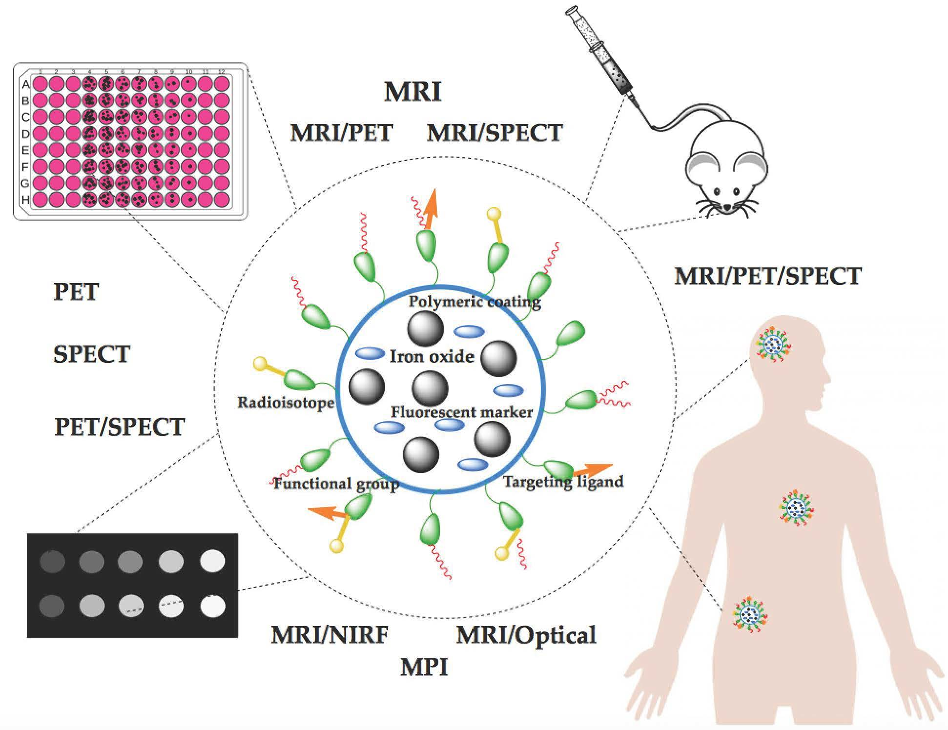
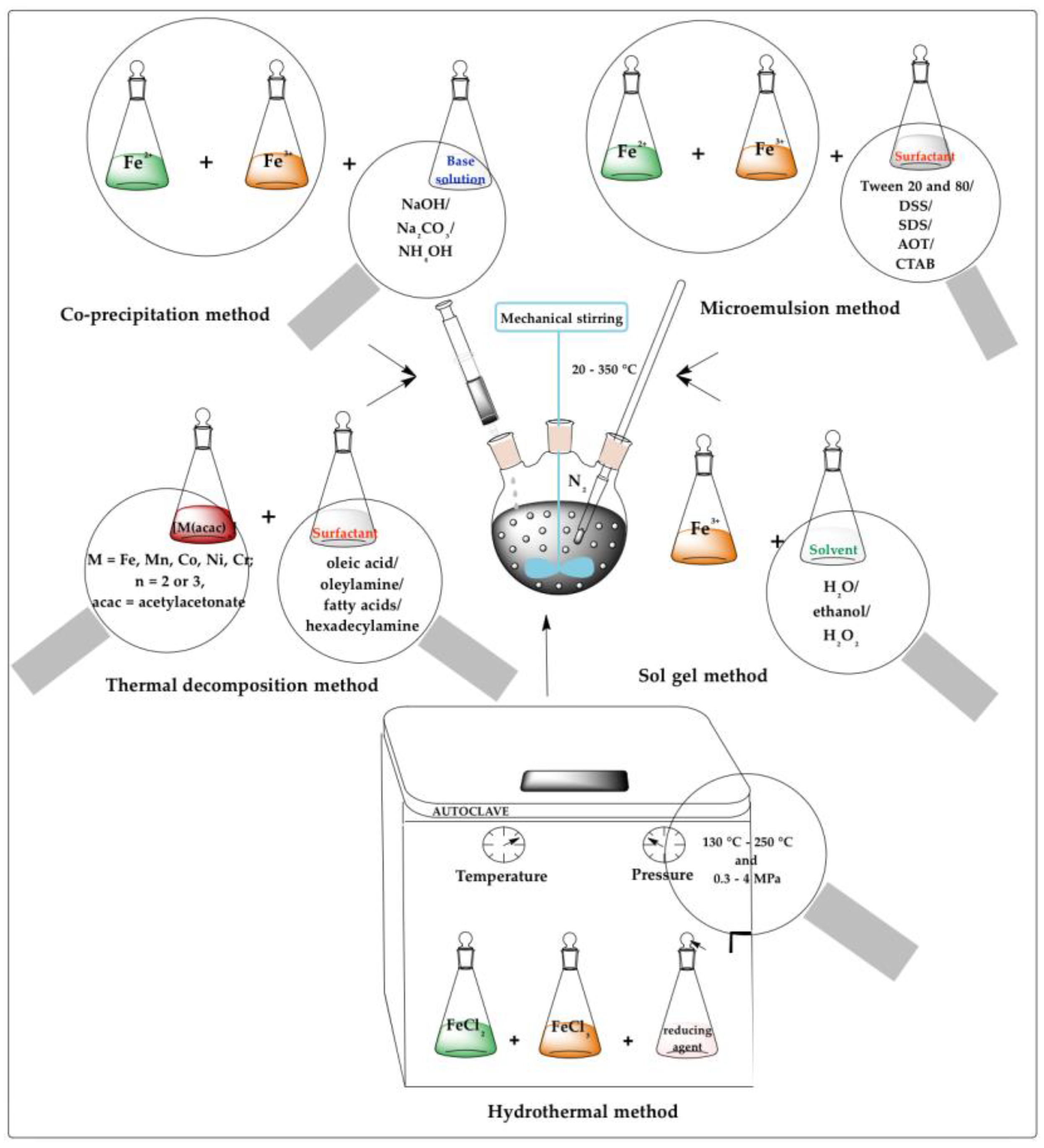
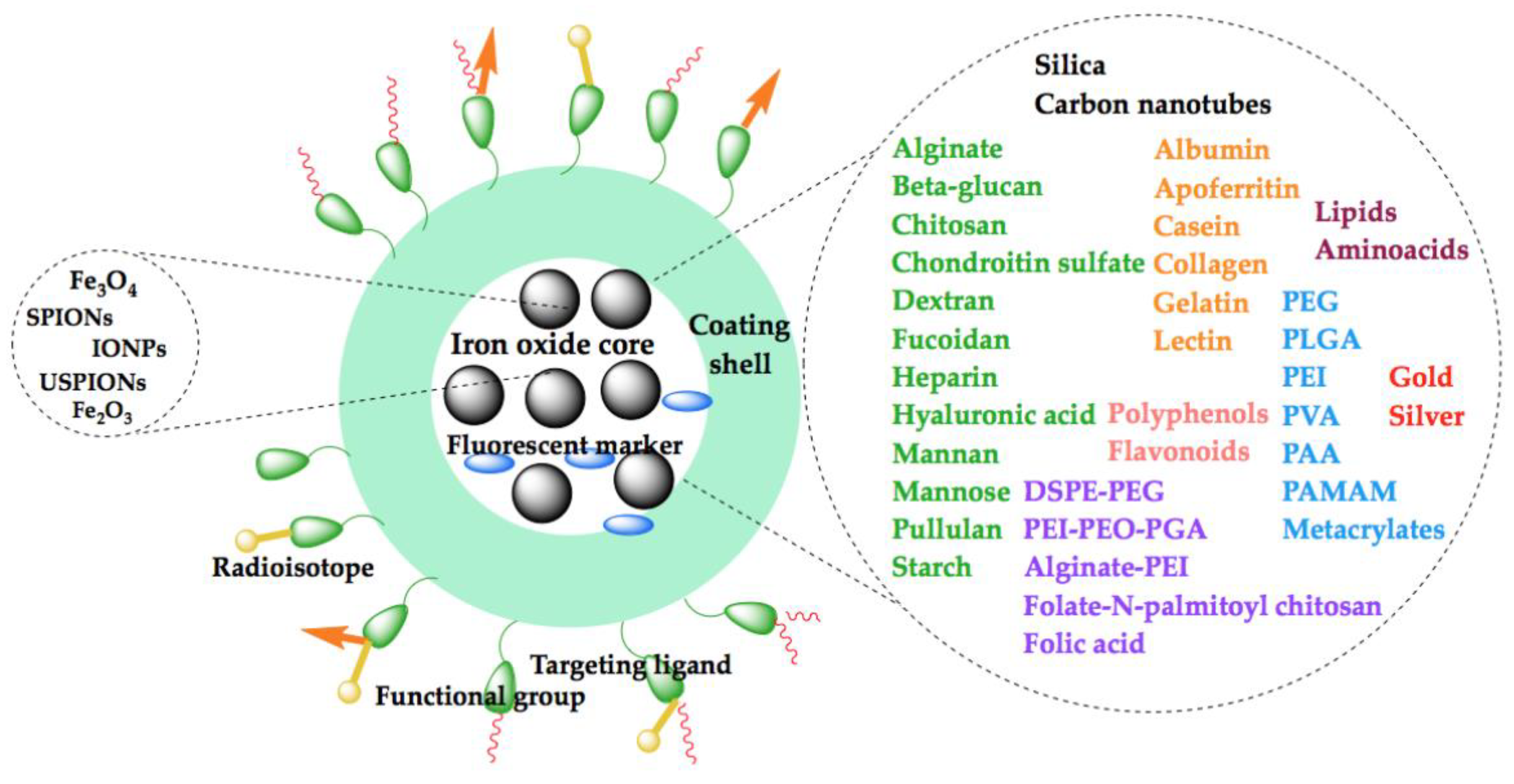
| Molecule | Target Region | Level of Research |
|---|---|---|
| Lactoferrin-conjugated PEG-coated Fe3O4 nanoparticles | Lactoferrin receptors | In vitro, rodents |
| Lactoferrin-conjugated SPIONs | ||
| Anti-ligand–induced binding sites antibody conjugated to microparticles of iron oxide | Platelet glycoprotein GPIIb/IIIa receptor (CD61/CD41) | |
| Anti-malondialdehyde-modified low-density lipoprotein MDA2 monoclonal antibody–labelled lipid-coated SPIONs | Malondialdehyde-modified low-density lipoprotein (antigen) | |
| Anti-malondialdehyde-modified low-density lipoprotein MDA2 monoclonal antibody–labelled lipid-coated USPIONs | ||
| Anti-vascular cell adhesion molecule antibody M/K-2.7 and anti-P-selectin antibody RB40.34 conjugated microparticles of iron oxide | VCAM-1 and P-selectin | |
| Anti-vascular cell adhesion molecule antibody M/K-2.7–conjugated microparticles of iron oxide | VCAM-1 | |
| USPIO-cyclo (Cys-Asn-Asn-Ser-Lys-Ser-His-Thr-Cys) | ||
| Avidin-coated baculoviral vectors-biotinylated USPIONs | - | |
| CLIO-(H-2Kd)-Lys-Tyr-Asp-Lys-Ala-Asp-Val-Phe-Leu | H-2Kd-restricted β cell-specific T cell receptor | |
| Complement receptor type 2-conjugated gold/SPIONs | Complement C3 fragments | |
| Cross-linked iron oxide-transactivator transcription | Adsorptive endocytosis, phagocytosis | |
| Cyclo(Arg-Gly-Asp-d-Try-Glu) conjugated to USPIONs | Integrin αvβ3 | |
| FluidMAG iron nanoparticle-labelled mesenchymal stem cells | Cell imaging | |
| Poly(N,N-dimethylacrylamide)-coated maghemite nanoparticles | ||
| Amine-modified silica-coated polyhedral SPIO–labelled rabbit bone marrow–derived mesenchymal stem cells | ||
| Glycol chitosan/heparin-immobilized gold-deposited iron oxide nanoparticles | Fibrinogen-derived product in tumour stroma | |
| Iron oxide–ferritin nanocages | Non-targeted | |
| Thiol-modified PEG-conjugated gold/SPIONs | ||
| Magnetic iron microbeads coupled with HEA-125 monoclonal antibody | Epithelial cell adhesion molecule | |
| H18/7 F(ab’)2 E-selectin monoclonal antibody conjugated to cross-linked iron oxide nanoparticles | E-selectin | |
| MES-1 F(ab’)2 E-selectin monoclonal antibody conjugated to USPIONs | ||
| Sialy Lewisx mimetic conjugated to PEGylated USPIONs | ||
| Sialy Lewisx mimetic conjugated to USPIONs | ||
| Monoclonal antibody against antigen A7 coupled to ferromagnetic lignosite particles | Antigen A7 | |
| Octreotide conjugated to PEGylated USPIONs | Somatostatin receptors | |
| Ovarian cancer antigen 183B2 monoclonal antibody conjugated to USPIONs | Ovarian cancer antigen 183B2 | |
| Saposin C-dioleylphosphatidylserine nanovesicles coupled with iron oxides | Phospholipids | |
| Trastuzumab-dextran iron oxide nanoparticles | HER2 or ErbB2/neu receptor | |
| Trastuzumab-manganese-doped iron oxide nanoparticles | EGF HER2 receptor | |
| ZHER2:342 Affibody-PEG-SPIONs | EGF | |
| Single-chain anti-epidermal growth factor receptor antibody fragment conjugated to magnetic iron oxide nanoparticles | ||
| USPIO-Leu-Ile-Lys-Lys-Pro-Phe | Phosphatidylserine | |
| USPIONs conjugated with Ile-Pro-Leu-Pro-Phe-Tyr-Asn | ß-amyloid (Aß42) peptide | |
| USPIO-anti-CD20 monoclonal antibody | CD20 antigen | |
| PEG–coated and folic acid–conjugated SPONs | Folate receptor | |
| N-Alkyl-polyethylenimine 2 kDa–stabilized SPIONs | Mesenchymal stem cell labelling | |
| SPION stabilized by alginate | RES | |
| Ferumoxsil (Siloxane-coated SPIO) | In vitro, rodents, humans | |
| Citrate-coated (184th variant) SPIONs | Phagocytosis | In vitro, rodents non-primate non-rodent mammals, humans |
| Ferumoxides (Dextran-coated SPIO) | RES | |
| Iron oxide nanoparticles-poly-l-lysine complex | - | |
| Ferumoxtran (USPIONs) | RES | In vitro, rodents, non-primate non-rodent mammals, non-human primates, humans |
| Molecule | Source of Signal | Target Region | Level of Research |
|---|---|---|---|
| 99mTc-Diethylenetriamine pentaacetic acid SPIONs conjugated with lactobionic acid | Iron oxide, 99mTc | Asialoglycoprotein receptors | In vitro, rodents |
| Molecule | Source of Signal | Target | Level of Research |
|---|---|---|---|
| MRI, SPECT Gamma Planar | |||
| 111In-Tetraazacyclododecane-N,N′,N″,N‴-tetraacetic acid-benzyl-ChL6-SPIONs | Iron oxide, 111In | Antibody-antigen binding | In vitro, rodents |
| MRI and PET | |||
| 124I-Serum albumin-manganese-magnetism-engineered iron oxide nanoparticles | Iron oxide, 124I | Non-targeted | In vitro, rodents |
| 64Cu-1,4,7,10-Tetraazacyclododecane-N,N′,N″,N‴-tetraacetic acid-iron oxide-c(RGDyK) nanoparticles | 64Cu and iron oxide | Integrin αvβ3 | |
| MRI, optical, PET | |||
| 64Cu-DTPA-CLIO-VT680 | Iron oxides, VT680, 64Cu | Macrophages | In vitro, rodents |
| MRI, optical NIR fluorescence | |||
| Annexin V-cross-linked iron oxide-Cy5.5 | Iron oxide, Cy5.5 | Phosphatidylserine | In vitro, rodents |
| Anti-vascular cell adhesion molecule monoclonal M/K-2.7 conjugated cross-linked iron oxide-Cy5.5 nanoparticles | Vascular cell adhesion molecule-1 | ||
| Cross-linked iron oxide-Cy5.5 | Phagocyte and tumour cell | ||
| Lys-Thr-Leu-Leu-Pro-Thr-Pro-cross-linked iron oxide-Cy5.5 | Plectin-1 | ||
| VCAM-1 internalizing peptide-28 nanoparticles | Vascular cell adhesion molecule-1 | ||
| Cys-Arg-Glu-Lys-Ala-SPIO-Cy7 nanoparticles | Iron oxide, Cy7 | Clotted plasma proteins | |
| Optical, NIR fluorescence imaging and MRI | |||
| Cy5.5-Amino-terminal fragment of urokinase-type plasminogen activator conjugated to magnetic iron oxide nanoparticles | Iron oxide, Cy5.5 | Urokinase-type plasminogen activator receptor | In vitro, rodents |
| Cy5.5-Arg-Arg-Arg-Arg-crosslinked iron oxide nanoparticle | Proteases | ||
| MRI, fluorescence molecular tomography (FMT), fluorescence reflectance imaging (FRI) | |||
| Gly-Ser-Ser-Lys-(FITC)-Gly-Gly-Gly-Cys-Arg-Gly-Asp-Cys-CLIO-Cy5.5 | Iron oxides, Cy5.5 | αυβ3 integrin | In vitro, rodents |
| MRI, NIFR optical imaging | |||
| Green fluorescent protein specified small interfering RNA–crosslinked iron oxide nanoparticles-Cy5.5 | Iron oxides, Cy5.5 | RNAse III | In vitro, rodents |
| Survivin specified small interfering RNA-CLIO-Cy5.5 | |||
| Bombesin peptide conjugated–cross-linked iron oxide-Cy5.5 | Iron oxide, Cy5.5, FITC | Gastrin-releasing peptide receptor | |
| Optical, near-infrared fluorescence | |||
| IPLVVPLGGSC (Cy5.5-Cross-linked iron oxide) K(FITC) | Cy5.5, iron oxide | Hepsin | In vitro, rodents |
| MRI and optical imaging | |||
| Rhodamine B isothiocyanate-incorporated, silica-coated magnetic nanoparticle–labelled human cord blood–derived mesenchymal stem cells | SPIO, RITC | Cell imaging | In vitro, rodents |
| Magnetic Material | Synthesis Method for Iron Oxide Core | Polymeric Coating | Applications | Reference |
|---|---|---|---|---|
| Fe3O4 | Co-precipitation | Chitosan | MRI | [69,70,71,72] |
| Chitosan/Alginate | [65] | |||
| N,N,N-trimethyl chitosan | MRI/PET (68Ga) | [73] | ||
| Dextran | MRI | [74,75,76,77] | ||
| MRI/PET (68Ga) | [59] | |||
| MRI/SPECT (99mTc) | [78] | |||
| MRI/SPECT (99mTc-dipicolylmie (DPA)-alendronate) | [78] | |||
| Carboxymethyl dextran | MRI | [79] | ||
| BSA | [80] | |||
| BSA/HA | [64] | |||
| Folic Acid-BSA | [81] | |||
| Alginate | [82,83] | |||
| Thermal decomposition | Dextran | MRI/PET (64Cu) | [84] | |
| BSA | MRI | [85] | ||
| Hydrothermal | Alginate/PEI | [86] | ||
| Emulsion polymerization | HSA | [87] | ||
| Emulsification | PLGA/Chitosan/Dextran sulfate | [88] | ||
| γ-Fe2O3 | Co-precipitation | Dextran | MRI | [89] |
| BSA | [90] | |||
| MRI/Fluorescent (FITC) | [91] | |||
| rHSA | MRI | [92] | ||
| Nucleation | Gelatin | MRI/Fluorescence Imaging | [93] | |
| Co-precipitation/cross-linking | Alginate | MRI | [94] | |
| IONPs | Co-precipitation | Glycol Chitosan/Heparin | MRI | [95] |
| Dextran | [96,97,98,99] | |||
| MRI/PET (89Zr) | [100] | |||
| MRI/CT | [101] | |||
| Mapping | [102] | |||
| Dextran/Dextran sulfate | MRI | [103] | ||
| Dextran sulfate | [104] | |||
| MRI/PET (64Cu) | [105] | |||
| Carboxymethyl-diethylaminoethyl dextran | MRI | [106] | ||
| Gelatin | [66] | |||
| Thermal decomposition | Oleyl-Chitosan | MRI | [107] | |
| Heparin | [108] | |||
| BSA | [109] | |||
| HSA | [110,111] | |||
| Microemulsion | Gelatin | MRI | [66] | |
| MRI/Optical | [112] | |||
| Microwave | Dextran | MRI/PET (68Ga) | [113] | |
| Microwave-assisted | Different heparins of distinct anticoagulant/Anti-heparanase | MRI | [114] | |
| Dextran | [115] | |||
| MRI/PET (64Cu) | [116] | |||
| Nucleation | HSA | MRI | [117] | |
| NIRF | [118] | |||
| SPIONs | Co-precipitation | Chitosan | MRI | [119] |
| Cationic/Anionic chitosan derivatives | [120] | |||
| Bioactive-conjugated N-palmitoyl chitosan | [121] | |||
| Heparin | [122] | |||
| Unfractionated heparin | [123] | |||
| Dextran | [60,124,125,126,127,128,129] | |||
| MPI | [130,131] | |||
| Dextran sulfate | MRI | [132] | ||
| Carboxydextran | [133] | |||
| PET (89Zr)/SPECT (99mTc) | [134] | |||
| Carboxymethyl dextran | MRI | [135,136] | ||
| MRI/SPECT (111In) | [137] | |||
| PET (89Zr) | [138,139] | |||
| PET (89Zr)/SPECT (111In) | [140] | |||
| PET(64Cu)/SPECT (111In) | [140] | |||
| Carboxymethyl dextran/Fucoidan | MRI | [141] | ||
| HSA | [142] | |||
| Alginate | [143] | |||
| Thermal decomposition | N-palmitoyl-N-monomethyl-N,N-dimethyl-N,N,N-trimethyl-6-O-glycolchitosan | MRI | [144] | |
| Succinylated heparin monolayer | [145] | |||
| Amphiphilic starlike dextran | [146] | |||
| PLGA/Glycol chitosan | MRI/SPECT (99mTc) | [147] | ||
| Sol gel | Cationic derivative of chitosan/Hyaluronic acid-Curcumin conjugate | MRI | [148] | |
| Microemulsion | Dextran-b-oligo (amidoamine) | [149] | ||
| Co-precipitation/Cold gelation | Dextran | [60] | ||
| Alkaline co-precipitation in a microfluidic droplet reactor | Dextran | MRI | [150] | |
| USPIOs | Thermal decomposition | Gelatin | MRI | [151] |
| Magnetic Material | Synthesis Method for Iron Oxide Core | Polymeric Coating | Applications | Reference |
|---|---|---|---|---|
| Fe3O4 | Co-precipitation | PEG | MRI | [178,179] |
| SPECT (111In) | [180] | |||
| PEG diacid (HOOC–PEG–COOH) | MRI | [181] | ||
| PLA/PEG/D-glucosamine | [182] | |||
| Silica | MRI (Gd-DTPA) | [183] | ||
| MRI | [184] | |||
| MPI | [185] | |||
| MRI/PET (11C) | [186] | |||
| Mesoporous Silica | MRI | [187] | ||
| APTES | Tumour imaging agent (99mTc) | [188] | ||
| Fe3O4/γ-Fe2O3 | Poly(2-acrylamido-2-methylpropane sodium sulfonate) P(AMPS) | MRI | [189] | |
| Thermal decomposition | PEG | MRI | [190,191,192] | |
| MRI/SPECT (99mTc) | [193] | |||
| MRI/SPECT (125I) | [106,194] | |||
| MRI/PET (68Ga) | [195] | |||
| MRI/PET (64Cu) | [196] | |||
| MRI/PET (71As) | [197] | |||
| SPECT (99mTc) | [174] | |||
| PEG-phosphate | MRI | [198] | ||
| Hydroxyl−PEG−Phosphonic Acid | [199] | |||
| DSPE-mPEG2000/DSPE-mPEG2000 amine | MRI/PET (68Ga) | [200] | ||
| Silica | MRI | [201,202] | ||
| MRI/US | [203] | |||
| Mesoporous Silica | MRI | [202,204] | ||
| MRI/Fluorescence Imaging | [190] | |||
| Silica | MRI/Optical (FITC) | [205] | ||
| Hydrothermal | PEG | MRI/PET (64Cu) | [206] | |
| DPPE-mPEG2000/DSPE-cPEG2000 | SPECT (67Ga) | [207] | ||
| PEI | MRI | [208] | ||
| Silica | [209] | |||
| MPI | [210] | |||
| Solvothermal | Mesoporous Silica | MRI | [211] | |
| Sol gel | Silica | [212] | ||
| Microemulsion | PEG | [213] | ||
| Reduction–precipitation | PEI | [214] | ||
| Polyol | PEG bis(carboxymethyl) ether | [215] | ||
| PEG diacid (HOOC-PEG-COOH) | MRI (Gd) | [216] | ||
| γ-Fe2O3 | Co-precipitation | PEG-based liposome | MRI/PET (68Ga) | [217] |
| Silica | MRI | [218] | ||
| Sol gel | [219] | |||
| IONPs | Co-precipitation | PEG | MRI | [213] |
| PEG-Maleimide | MRI/PET (64Cu) | [220] | ||
| PEG/Terminated polystyrene/Linoleic acid | MRI | [221] | ||
| Poly(4-vinylpyridine)/PEG acrylate | MRI/SPECT (111In) | [222] | ||
| Silica/Silica-PEG | MRI | [223] | ||
| Silica/PEG | MRI/PET (68Ga) | [224] | ||
| MRI/PET-CT (68Ga) | [225] | |||
| Poly(glycerol adipate) | MRI | [226] | ||
| PLGA | SPECT (111In) | [176] | ||
| Thermal decomposition | PEG | MRI | [227,228] | |
| MRI/PET (64Cu)/ Photoacoustic Tomography (PAT) | [229] | |||
| PEG diacrylate | MRI | [230] | ||
| Amine-terminated PEG (NH2-PEG-NH2) | MPI | [231] | ||
| DSPE-PEG-2000 | MRI | [228] | ||
| Mesoporous Silica/PEG | MRI/NIRF | [232] | ||
| PVP | MRI | [233] | ||
| Hydrothermal | Mesoporous Silica | MRI | [234] | |
| Solvothermal | PEG | MRI | [235] | |
| Sol gel | PEI/Silica | MRI | [236] | |
| Reverse microemulsion | Silica | MRI/NIRF | [139] | |
| MRI | [237,238] | |||
| Chemical reduction | ||||
| SPIOs | Co-precipitation | PEG | MRI | [15,239,240,241] |
| PEG/Poly(gallol) | [242] | |||
| PEG/Gallic acid/Dextran | MPI | [130] | ||
| DSPE-mPEG2000 | SPECT (111In) | [243] | ||
| PEI-b-PCL-b-PEG | MRI | [244] | ||
| Silica | Contrast agents in biomedical photoacoustic imaging | [245] | ||
| Mesoporous Silica | MRI/CT/Fluorescence | [246] | ||
| Thermal decomposition | PEG | MRI | [247,248] | |
| MPI | [249] | |||
| PEG methyl ether | MRI | [250] | ||
| Amine-terminated PEG (NH2-PEG) | [251] | |||
| HA/mPEG-succinimidyl succinate | [252] | |||
| Silica | [253] | |||
| MRI/SPECT (125I) | [254] | |||
| MRI/PET (64Cu) | [255] | |||
| Mesoporous Silica | MRI/Optical | [256] | ||
| Silica/Hyaluronic acid (HA) | MRI | [257] | ||
| Sol gel | Silica | [258] | ||
| Polyol | PEG | [259] | ||
| USPIOs | Co-precipitation | Silica/APTMS/AEAPTMS | MRI | [260] |
| Thermal decomposition | PEG | MRI/ SPECT(99mTc) | [261] | |
| Microwave | Phosphonate-PEG (PO-PEG-NH2) | MRI | [262] | |
| Polyol | PEG | MRI/Optical (FITC) | [263] |
| Core-Shell Iron Oxide Agent | Imaging Modality | In Vivo Model | Reference |
|---|---|---|---|
| SPIONs coated with silica, APTES and (3-glycidoxypropyl) methyldiethoxysilane | MRI: medium T2 along with a minor effect on T1 | Zebrafish (Danio rerio) model | [271] |
| Fe3O4 nanoparticles stabilized by PVP, trisodium citrate, and maleic anhydride | MRI-T2 | White rabbit | [272] |
| IONPs coated with PEG | Tumour xenograft model on female NOD-SCID IL2 mice with human breast cell line (MDA-MB-231) | [235] | |
| Luteinizing hormone-releasing hormone-conjugated PEG-coated magnetite nanoparticles | BALB/c nude mice (Triple negative breast cancer model) | [273] | |
| Magnetic fibrin nanoparticles conjugated with folic acid | Albino Wistar rats with antigen-induced arthritis | [274] | |
| HSA-coated iron oxide nanoparticles | BALB/c mice (breast cancer model with MDA-MB-231 cells) | [142] | |
| PLA-PEG-D-glucosamine Fe3O4 nanoparticles | MRI-T2* | Tumour-bearing BABL/c mice inoculated with 4 T1 cells | [182] |
| PEG/PEI-SPIONs conjugated with transferrin | Kunming mice brain | [275] | |
| USPIOs coated with carboxymethyldextran and coupled or not with a low-molecular-weight aminated fucoidan | Elastase-induced Wistar rat model | [141] | |
| SPIONs coated with dextran and functionalized with anti-insulin-like-growth-factor binding protein 7 (anti-IGFBP7) | MRI-T2 and T2* | Nude CD-1 mouse model of glioblastoma multiforme | [276] |
| Material | Disease | Imaging Technique | Status |
|---|---|---|---|
| IONPs | Lymph node metastases | MRI | Phase I, Withdrawn (NCT02689401) |
| Healthy subjects-to determine MTD, Pharmacokinetic, safety/tolerability | Phase I, Completed (NCT02744248) | ||
| SPIONs | Lymph node metastases | Phase IV, Completed (NCT00920023) | |
| Sentinel lymph nodes | Completed (NCT03243435) | ||
| Phase IV, Unknown (NCT02612870) | |||
| Phase III, Recruiting (NCT04722692) | |||
| Unknown (NCT03449615) | |||
| Melanoma | Not Applicable, Completed (NCT03898687) | ||
| Healthy volunteer-cell tracking | Not Applicable, Unknown (NCT00972946) | ||
| Primary and metastatic hepatic malignancies | MRI/SPECT (99mTc) | Recruiting (NCT04682847) | |
| USPIONs | Lymph nodes | MRI | Early Phase 1, Recruiting (NCT02857218) |
| Lymph node metastases | Phase III, Recruiting (NCT04261777) | ||
| Not Applicable, Completed (NCT01815333) | |||
| Not Applicable, Recruiting (NCT04311047) | |||
| Malignant pelvic lymph nodes | Not Applicable, Terminated (NCT00147238) | ||
| Pelvic lymph nodes | Early Phase 1, Recruiting (NCT03280277) | ||
| Pelvic nodal metastases | Phase I and II, Completed (NCT00188695) | ||
| Papillary carcinoma of thyroid gland/metastatic medullary thyroid cancer/follicular thyroid cancer lymph node metastasis | Not Applicable, Completed (NCT01927887) | ||
| Brain neoplasms | Phase II, Terminated (NCT00659334) | ||
| Oesophageal neoplasms | Not Applicable, Completed (NCT02253602) | ||
| Squamous cell carcinomas | Phase 1, Active (NCT01895829) | ||
| Ischemic heart disease | Phase I, Completed (NCT03651791) | ||
| Multiple sclerosis | Phase I, Completed (NCT02511028) | ||
| Aortic dissection | Recruiting (NCT03948555) | ||
| Myocardial infarction | Phase II, Unknown (NCT01995799) | ||
| Not Applicable, Completed (NCT01323296) | |||
| Myocardial cellular inflammation | Phase II and III, Completed (NCT02319278) | ||
| Abdominal aortic aneurysm | Terminated (NCT01749280) | ||
| Atherosclerosis | Unknown (NCT01674257) | ||
| Coronary artery disease | MRI/PET | Recruiting (NCT03451448) |
Publisher’s Note: MDPI stays neutral with regard to jurisdictional claims in published maps and institutional affiliations. |
© 2021 by the authors. Licensee MDPI, Basel, Switzerland. This article is an open access article distributed under the terms and conditions of the Creative Commons Attribution (CC BY) license (https://creativecommons.org/licenses/by/4.0/).
Share and Cite
Crețu, B.E.-B.; Dodi, G.; Shavandi, A.; Gardikiotis, I.; Șerban, I.L.; Balan, V. Imaging Constructs: The Rise of Iron Oxide Nanoparticles. Molecules 2021, 26, 3437. https://doi.org/10.3390/molecules26113437
Crețu BE-B, Dodi G, Shavandi A, Gardikiotis I, Șerban IL, Balan V. Imaging Constructs: The Rise of Iron Oxide Nanoparticles. Molecules. 2021; 26(11):3437. https://doi.org/10.3390/molecules26113437
Chicago/Turabian StyleCrețu, Bianca Elena-Beatrice, Gianina Dodi, Amin Shavandi, Ioannis Gardikiotis, Ionela Lăcrămioara Șerban, and Vera Balan. 2021. "Imaging Constructs: The Rise of Iron Oxide Nanoparticles" Molecules 26, no. 11: 3437. https://doi.org/10.3390/molecules26113437
APA StyleCrețu, B. E.-B., Dodi, G., Shavandi, A., Gardikiotis, I., Șerban, I. L., & Balan, V. (2021). Imaging Constructs: The Rise of Iron Oxide Nanoparticles. Molecules, 26(11), 3437. https://doi.org/10.3390/molecules26113437