Abstract
The current study aimed to synthesize new metal coordination complexes with potential biomedical applications. Metal complexes were prepared via the reaction of isatin-N(4)anti- pyrinethiosemicarbazone ligand 1 with Cu(II), Ni(II), Co(II), Zn(II), and Fe(III) ions. The obtained metal complexes 2–12 were characterized using elemental, spectral (1H-NMR, EPR, Mass, IR, UV-Vis) and thermal (TGA) techniques, as well as magnetic moment and molar conductance measurements. In addition, their geometries were studied using EPR and UV–Vis spectroscopy. To evaluate the in vivo anti-cancer activities of these complexes, the ligand 1 and its metal complexes 2, 7 and 9 were tested against solid tumors. The solid tumors were induced by subcutaneous (SC) injection of Ehrlich ascites carcinoma (EAC) cells in mice. The impact of the selected complexes on the reduction of tumor volume was determined. Also, the expression levels of vascular endothelial growth factor (VEGF) and cysteine aspartyl-specific protease-7 (caspase-7) in tumor and liver tissues of mice bearing EAC tumor were determined. Moreover, their effects on alanine transaminase (ALT), aspartate transaminase (AST), albumin, and glucose levels were measured. The results revealed that the tested compounds, especially complex 9, reduced tumor volume, inhibited the expression of VEGF, and induced the expression of caspase-7. Additionally, they restored the levels of ALT, AST, albumin, and glucose close to their normal levels. Taken together, our newly synthesized metal complexes are promising anti-cancer agents against solid tumors induced by EAC cells as supported by the inhibition of VEGF and induction of caspase-7.
1. Introduction
Cancer is considered one of the most devastating life-threatening disease worldwide. It is characterized by uncontrolled cell growth and the rapid spread of abnormal cells [1]. Angiogenesis is a vital regulator of tumor growth, spread and metastasis [2]. Tumor angiogenesis, which is the development of new blood vessels, is regulated by the production of angiogenic stimulators such as vascular endothelial growth factor (VEGF). VEGF is a key regulatory factor in the prognosis of various cancers; therefore, the inhibition of VEGF production is an alternative therapeutic approach for cancer treatment [3,4,5,6].
Chemotherapy, along with surgery, radiation, hormone therapy, and immune therapy, is used to treat cancer [7]. Despite the success of controlling the tumor growth in several cases, the incidence and prevelance of cancer is still dramtically increasing and calling for new chemotherapeutics to be introduced to the pharmaceutical industry and the market [8]. Antipyrine (2,3-dimethyl-1-phenyl-5-pyrazolone) and its derivatives possess a diverse array of potentially useful biological properties [9]. Similarly, thiosemicarbazones have demonstrated significant biological applications, as antitumor [10], antifungal [11], antiviral [12], antibacterial [13] and antimalarial [14] agents. Previous investigations involving the synthesis, characterization and biological evaluation of iron(III), cobalt(II), cobalt(III), nickel(II), zinc(II), and copper(II) complexes of 4-formylantipyrine N(4)-substituted thiosemicarbazones have been reported [15,16].
Based on these results and as a continuation of our ongoing interest in compounds with antitumor activities, the present study was carried out to synthesize and characterize metal complexes of isatin-N(4)antipyrinethiosemicarbazone and to evaluate their anti-tumor, anti-angiogenic and apoptotic activities against solid tumors induced by Ehrlich ascites carcinoma (EAC) cells in mice.
2. Results and Discussion
All the prepared metal complexes, shown in Figure 1, are stable at room temperature, non-hygroscopic and insoluble in most organic solvents and water, but they are completely soluble in dimethylformamide (DMF) and dimethylsulfoxide (DMSO). The elemental analyses, physical data, are listed in Table 1. The elemental analyses confirmed that complexes 2–6, 8 and 10 are formed in a 1:1 molar ratio of the metal ion and ligand, whereas complexes 9, 11 and 12 are formed in a 1:2 molar ratio of the metal ion and the ligand. Complex 7 is a binuclear complex.

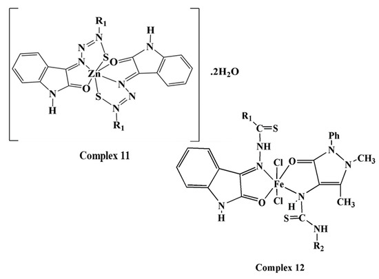
Figure 1.
Chemical structures of metal complexes 2–12.

Table 1.
Analytical and physical data of the ligand 1 and its metal complexes 2–11.
2.1. 1H-NMR Spectra
The 1H-NMR spectra of the ligand 1 and the zinc complex 11 are illustrated in the Supplementary Materials. The data shows three separate singlets at 12.819, 11.219 and 10.115 ppm, which can be attributed to the thiosemicarbazide N-N(2)H, the indole N-H moiety and the thiosemicabazide CS-N(4)-H, respectively [17]. Multiple signals appear in the 7.702–6.876 ppm region, and these can be attributed to the aromatic protons of the indole and phenyl rings of antipyrine. The other signals for the ligand at 3.068 (s, 3H) and 2.135 (s, 3H) correspond to the N-CH3 and C-CH3 of the antipyrine moiety, respectively [15]. The 1H-NMR spectrum of complex 11 does not show a signal corresponding to N-N(2)H of thiosemicarbazide, suggesting the loss of the thiol proton, and the binding ligand in its thiol form. The spectrum displays signals attributable to the indole N-H moiety, the CS-N(4)-H of thiosemicabazide and all the other characteristic signals at the same positions as those of the free ligand.
2.2. Mass Spectra
The mass spectra of ligand 1 and complexes 7, 8, 11, and 12 are shown in the Supplementary Materials. The molecular ion peak at m/z 406.27 amu in the spectrum of the ligand 1 is consistent with the molecular weight of its proposed structure. Moreover, the fragmentation pattern shows ion peaks at m/z 363.47, 291.15, 276.68, 261.69, 223.71, 204.07, 203.30, 145.64, 130.57, 118.18 and 76.3 amu corresponding to C19H17N5OS, C13H17N5OS, C12H13N5OS, C12H13N4OS, C9H10N4OS, C9H6N3OS, C11H13N3O, C8H5N2O, C8H5NO, C7H5NO and C6H5 fragments, respectively.
The mas spectrum of complex 7 (F.W. = C40H38N12O6S2Co2Cl2) shows a molecular ion peak at m/z 999.5 amu, consistent with a molecular weight of 998.8 g/mol, which is the molecular weight of the complex after the loss of one molecule of hydration water (F.W. = C40H34N12O4S2Co2Cl2). The spectrophotometer at hand cannot detect molecular ions with molecular weights greater than 1000 g/mol. The fragment ion peaks at m/z 868.9 and 463.9 amu correspond to the formulas C40H34N12O4S2Co and C20H17N6O2Co.
The mass spectrum of complex 8 (C22H30N6O9SCo, F.W. = 612.5) displays a molecular ion peak at m/z 612.85 (100%) amu, confirming the suggested structure of the complex. Moreover, the spectrum shows several peaks, including peaks at m/z 576.23 (94.07), 561.30 (61.93), 541.85 (6.86), and 523.22 (39.20), corresponding to the formula C22H26N6O7SCo, C22H24N6O6SCo, C22H22N6O5SCo, and C22H20N6O4SCo, respectively, due to the sequential loss of two molecules of water of hydration, another water of hydration, a final water of hydration, and one coordinated water molecule, respectively.
The mass spectrum of complex 11 (C40H38N12O6S2Zn, F.W. = 911.50) displays a molecular ion peak at m/z 911.95 (1.19), consistent with the suggested structure.The mass spectrum of complex 12 (C40H38N12O5S2FeCl3, F.W. = 992.50) exhibits a molecular ion peak at m/z 992.50 amu, confirming the putative molecular weight of the complex. Moreover, the spectrum shows several ion peaks, including peaks at 973.50 (1.90), 938.30 (12.96), 869.80 (8.27), and 464.40 (2.48) corresponding to the formula C40H36N12O4S2FeCl3, C40H38N12O5S2FeCl3, C40H38N12O5S2FeCl2, C40H38N12O5S2Fe and C20H18N6O2SFe, which are attributable to the losses of one water of hydration, an ionisable chloride, two coordinated chloro ligands and one ligand, respectively.
2.3. The Molar Conductance
The molar conductance values of the metal complexes in DMF (10−3 M) were illustrated in Table 1. The results showed that none of the complexes except 12 are electrolytes [16], indicating that the anions in all these complexes are directly bound to the metal ion. Complex 12 gave a molar conductance of 74.5 Ω−1cm2mol−1 attributable to a 1:1 electrolyte, and indicating the presence of a chloride ion in the outer coordination sphere [18]. The high value of molar conductance of complex 6 may be associated with the partial displacement of the perchlorato ligand by DMF [15].
2.4. Infrared (IR) Spectra
The IR spectra of the ligand 1 and its metal complexes 2–12 were shown in Supplementary Materials. The most diagnostic infrared spectral bands are illustrated in Table 2; the infrared spectrum of the ligand 1 displays bands at 3442, 3348, 3287, 3251, and 3141 attributable to ν(N-H) absorptions characteristic of –NH groups [19,20]. The spectrum also displays bands at 1738, 1645, 1688, 1623 and 1595 cm−1. The first band can be assigned to ν(C=O) of the isatin moiety [19,20], and the second band can be assigned to ν(C=O) of the antipyrine moiety. The latter three bands are associated with ν(C=N) [21]. The appearance of multiple bands characteristic of ν(C=N) can be explained by the formation of tautomers secondary to the presence of the isatin carbonyl group and C=S with the adjacent –NH groups. The absorption band of ν(C=S) (thioamide IV) observed at 881 cm−1 [15] and a weak band observed at 2650 cm−1, assigned to ν(C-SH), supported the thione-thiol tautomerization expected from the C=S group with an adjacent proton [22].
Table 2.
IR spectral bands (cm−1) for ligand 1 and its metal complexes 2–12 and their assignments.
The binding mode of the ligand in each metal complex has been deduced by comparing the IR spectrum of the ligand with that of the corresponding metal complex. In the infrared spectra of all the complexes, the band characteristic of the ν(C=N) of the isatin moiety is shifted to lower wave numbers compared to that of the free ligand, indicating the coordination of C=N group with the metal via the nitrogen atom in all complexes. In the IR spectra of the metal complexes, except for those of 7 and 8, the bands corresponding to the C=O of the isatin moiety appeared at lower wavenumbers compared to that of the ligand as a result of the participation of the carbonyl oxygen atom of this group in the coordination. This band did not shift in the infrared spectra of complexes 7 and 8, indicating that C=O is not involved in the coordination.
In the infrared spectra of the complexes, except 9, 10 and 12, the absorption band is attributable to the ν(C=O) of the antipyrine moiety appeared at the same position as that of the ligand, indicating that this group only participates in coordination to the metal ion in complexes 9, 10 and 12. In the infrared spectra of all complexes, except 9 and 12, the band corresponding to ν(C=S) appeared at lower wavenumbers (808–848 cm−1) compared to that of the free ligand, indicating that the C-S group takes part in the coordination. The appearance of a band at 1622–1560 cm−1, which is assignable to υ(–N=C-S-), accompanies the shift in the ν(C-S) band. The appearance of the later band indicates the participation of the thiol form in coordination rather than the thion form, as suggested by the 1H-NMR spectrum of complex 11. The ν(C=S) bands in the infrared spectra of complexes 9 and 12 were at the same position as that of the free ligand, indicating that the C=S moiety does not participate in coordination in complexes 9 and 12.
Complexes 9 and 12 are composed of two ligand molecules that bind to one metal ion, and their infrared spectra showed that the two ligand molecules have different binding modes. In the infrared spectra of the complexes 9 and 12, each exhibits four bands at 1735–1737, 1700–1660, 1645 and 1620 cm−1, attributable to the uncoordinated ν(C=O) of the isatin moiety, the coordinated ν(C=O) of the isatin moiety, the uncoordinated ν(C=O) of the antipyrine moiety and the coordinated ν(C=O) of the antipyrine moiety, respectively. Moreover, each of the spectra of complexes 9 and 12 displays two bands corresponding to the ν(C=N) of the isatin moiety at 1595 and 1520 cm−1, which are assignable to the free uncoordinated C=N and the coordinated C=N, respectively. The above results illustrate that one of the ligand molecules behaves as a neutral, bidentate ligand and coordinates via the C=N and C=O groups of the isatin moiety and that the second molecule also behaves as a neutral, bidentate ligand but coordinates via the C=O and N-H groups of the antipyrine moiety.
The spectra of acetato complexes 5, 8 and 10 show two absorption bands at 1620–1615 and 1372–1333 cm−1, assigned to υ(C=O) and υ(C-O), respectively. The difference between these two bands is approximately 287−243 cm−1, which indicates that the acetate in these complexes coordinates to the metal ion as a monodentate ligand [23]. The monodentate coordination of a perchlorate ligand lowers the symmetry of the system (Td→C3v). The modes of the free perchlorate that were initially degenerated (υ3 and υ4) are both split into two bands [24]. The infrared spectrum of the perchlorate [Cu(ClO4)L]·2H2O complex showed that both the υ3 band and υ4 band are split into two bands, at 1118 and 1085 cm−1 and at 694 and 631 cm−1, respectively, indicating that the perchlorate coordinates to the metal ion as a monodentate ligand. The infrared spectrum of complex 4, [Cu(NO3)L]·H2O showed two bands at 1462 and 1382 cm−1, attributable to a monodentate nitrato ligand [25]. The infrared spectra of the hydrated complexes display a broad band at 3491–3388 cm−1 from ν(OH) of the water molecule [26]. The IR spectra of all the complexes display a new band at 591–498 cm−1, assigned to ν(M-N) [27,28,29], and the spectra of all the metal complexes except 7 and 8 show a new band at 693–589 cm−1, assigned to υ(M-O) [19,20,21,22,23,24,25,26,27,28,29].
2.5. Magnetic and Electronic Spectra
The room-temperature magnetic moments (μeff BM per metal atom) and electronic spectral bands for the metal complexes in the solid state are described in Table 3. The data showed that the electronic absorption spectrum of ligand 1 exhibits bands at 268 and 280 nm, attributable to intra-ligand π→π* electronic transitions [15,30]. These bands do not change significantly upon complex formation [31].
Table 3.
The electronic absorption spectral bands (nm) and magnetic moments of ligand 1 and its metal complexes 2–12.
The somewhat broad band at 369 nm is assignable to the intra-ligand n→π* electronic transitions associated with the combination of the azomethine function of the thiosemicabazone, the thione function of the thiosemicabazone and the heterocyclic moiety [15,32]. In the spectra of the metal complexes, this band generally shifts to a higher energy as a result of the participation of the azomethine and/or thiol groups in coordination [33,34]. In the spectra of the metal complexes, new bands were observed in the 357–530 nm region due to S(π)→metal(II,III) charge-transfer processes [35,36], and the spectrum of bromo complex 3 showed a Br→metal(II) charge-transfer band [37]. Cl→metal(II,III) [38,39,40] and O→metal(II,III) [35] charge-transfer bands were generally observed at approximately 333 nm and were obscured by the stronger intra-ligand transfer bands. Copper(II) complexes 2–6 showed magnetic moments of 1.76–1.87 BM. These values are close to the spin-only value for one unpaired spin (~1.73 BM) and indicate that there is no molecular association between the copper(II) ions in the square planar environment [34,41,42]. The electronic absorption spectra of Cu(II) complexes 2–5 showed two or three bands at 870–660, 740–631 and 675–580 nm, assignable to the 2B1g(dx2−y2)→2A1g(dz2), 2B1g(dx2−y2)→2B2g(dxy) (υ2) and 2B1g(dx2−y2)→2Eg(dxz,dyz) (υ3) transitions, suggesting a square planar geometry around the copper(II) ion. Complex 6 displays a broad band at 790 nm that could be attributed to the abovementioned transitions in the ligands in a square planar geometry around the copper(II) ion [43,44,45].
Cobalt(II) complex 7 gave a magnetic moment of 3.1 BM, which is considerably less than what is expected for high-spin tetrahedral cobalt(II). This unexpected value could be explained by the occurrence of antiferromagnetic exchange through the chloro bridge linking the two cobalt(II) ions [46,47]. Complex 8 gave a magnetic moment of 3.7 BM per metal ion, which is consistent with high-spin tetrahedral cobalt(II) complexes [48]. The electronic spectra of complexes 7 and 8 displayed a broad band with several features at approximately 675 nm, which is attributable to the 4A2→4T1(P) (v3) transition in a tetrahedral geometry around the Co(II) ion [15].
The nickel(II) complexes were found to be paramagnetic, which ruled out square planar configurations. The magnetic moments of nickel(II) complexes 9 and 10 are 2.98 and 3.06 BM, respectively, corresponding to two unpaired electrons [49], suggesting geometries other than square planar. The electronic spectra of Ni(II) complexes 9 and 10 showed bands at 950, 690-675 nm and 560−567 nm, assignable to the 3A2g(F)→3T2g(F)(υ1), 3A2g(F)→3T1g(F)(υ2) and 3A2g(F)→3T1g(P) transitions, respectively, in an octahedral environment [50,51,52]. The υ2/υ1 ratios for these complexes are 1.41 and 1.37, which are below the usual range of 1.5–1.75. These ratios suggest a tetragonally distorted octahedral geometry around the nickel(II) ions [51,53].
Iron(III) complex 12 gave a magnetic moment of 5.95 BM per metal ion, corresponding to a high-spin d5 configuration. The electronic spectrum of complex 12 showed strong bands at 391, 450, 530 and 584 nm. The first three bands can be assigned to ligand-to-metal charge-transfer (LMCT) processes. The last band at 584 nm is broad and of relatively high intensity. The relatively high intensity of this band can be attributed to overlap with a low-lying ligand charge-transfer band. This is because in the case of iron(III), the d-d transitions are considered forbidden because they take place between ions with different multiplicities; examples of such transitions include the 6A1g→4T1g (G), 6A1g→4T2g (G) and 6A1g→4Eg (G) or 4Eg(D) transitions in tetragonally distorted octahedral Fe(III) environments [54]. The electronic spectrum of the zinc(II) complex showed bands at 396, 440, 491, and 520(sh), which can be attributed to ligand-to-metal (L→M) charge-transfer processes.
2.6. EPR Spectra of the Copper(II) Complexes
The EPR spectra of the Cu(II) complexes are illustrated in the Supplementary Materials. The EPR data of Cu(II) complexes 2–6 in the polycrystalline state at 298 K are listed in Table 4. The EPR spectra of the Cu(II) complexes have low g1 and gav values, indicating that the bonds have considerable covalent characteristics [18,55]. The EPR spectra of the Cu(II) complexes other than complex 5 ([Cu(L)(OAc)]·2H2O) show rhombic-type distortion with three g values, g1 = 2.189, 2.198, 2.287, and 2.25; g2 = 2.121, 2.079, 2.121, and 2.12; and g3 = 2.057, 1.989, 1.977, and 2.027, respectively. The rhombic symmetry of these complexes is attributable to the bulkiness of the ligand and to the unequal bond lengths of the four Cu-S, Cu-N, Cu-O, and Cu-X bonds (X=Cl, Br, NO3 or ClO4) [55]. The g tensors, which follow g1 ˃ g2 ˃ g3 ˃ 2, are indicative of complexes with a d(x2−y2) ground state. This result is supported by the value of R (R = (g2 − g1/(g3 − g2) < 1 (= 0.94 − 0.72)), confirming a d(x2−y2) ground state with covalent bonding character [56,57].
Table 4.
EPR spectral assignments and bonding parameters for Cu(II) complexes in polycrystalline state at 298 K.
The EPR spectral data of complex 5 demonstrate anisotropic signals with g‖ > g⊥ > 2.0023, which are indicative of a d(x2−y2) ground state with axial symmetry, resulting in a 2B1g ground state that is known for Cu2+ complexes [58,59]. These g values are consistent with Cu2+-complex 5 having a square planar geometry [60,61]. The geometric parameter G, which is a measure of the exchange interaction between the copper centres in the solid state, was calculated using the equation G = (g‖ − 2.0023)/(g⊥ − 2.0023) for axial and rhombic compounds, g‖ = g1 and g⊥ = g2 + g3/2. If G ˃ 4, the exchange interaction is negligible, and if it is less than 4, considerable exchange interactions exist [58,62]. Complexes 3, 4, and 5 have G values ˃ 4, indicating that exchange interactions are negligible, while for complexes 2 and 6, G ˂ 4, suggesting considerable exchange interactions between the Cu(II) centres. Kivelson and Neiman reported that the g‖-values of Cu2+ complexes can be used to determine the nature of the copper-ligand bonding. If the g‖-value is smaller than 2.3, the environment is essentially covalent, while if the value is larger than 2.3, the environment is essentially ionic [63]. For all the synthesized complexes, g‖ < 2.3, indicating that there is considerable covalent character in the copper-ligand bonds.
According to [45,64,65], the orbital reduction factor (k) and its parallel (k‖) and perpendicular (k⊥) components were calculated according to the following equations: k‖2 = (g‖ − 2.0023)∆2/8λ0, k⊥2 = (g⊥ − 2.0023) ∆1/2 λ0, k2 = (K‖2 + 2⊥2)/3 where λo is the spin orbit coupling (−828 cm−1) of the free Cu(II) ion, and Δ1 and Δ2 are the electronic transitions 2B1g→2B2g (dx2−y2→dxy) and 2B1g→2Eg (dx2−y2→dxzdyz), respectively. The calculated k‖2, k⊥2 and k2 values in Table 1 showed that for complexes 3–6, k‖ ˃ k⊥, indicating the presence of significant out-of-plane π-bonding [66] and confirming that 2B1g is the ground state for these complexes. For complex 2, k‖ ˂ k⊥, indicating the presence of significant in-plane π-bonding [67]. Moreover, when the environment is ionic, k = 1, but if this value is less than 1, the environment is covalent. These compounds showed k values lower than one, which is strong evidence of covalent character, which is in agreement with the results obtained from the g‖ values [65,68]. None of the solid-state EPR spectra of the complexes displayed hyperfine structure, confirming the presence of intermolecular interactions, but these interactions were not strong enough to overcome the dipolar interactions [69].
2.7. Thermal Analysis
The TGA spectra of metal complexes are shown in the Supplementary Materials. The TGA results of solid complexes 2, 3, 5 and 7–12 are listed in Table 5. The results show an agreement with the formulae suggested by the analytical, spectral and magnetic data. A general decomposition pattern, in which the complexes decompose in two or three stages, was observed. The first stage is the loss of waters by hydration at 29–160 °C; the second stage of decomposition corresponds to the loss of coordinated water molecules at 80–175 °C; complex 8 showed a third decomposition stage that is a result of overlapping processes including the loss of Cl−, Br− or OAc− anions; the loss the ligands to form metal oxides or metal sulfides mixed with carbon residues; and the oxidation of carbon to carbon dioxide leaving metal oxides or sulfides.
Table 5.
Thermal (TGA) data for metal complexes 2, 3, 5, and 7–12.
2.8. Biological Activities
2.8.1. Effect of Ligand 1 and Its Metal Complexes 2, 7 and 9 on Solid Tumor Volume
Mice bearing solid tumors were treated SC with 0.2 mM of ligand 1 and its metal complexes 2, 7 and 9. The treatment was started seven days after EAC implementation in mice and was continued for 14 consecutive days. Tumor volume was measured after 24 h from the last dose of treatment. As shown in Table 6, treatment of solid tumor-bearing mice with the ligand 1 resulted in a significant reduction in tumor volume with inhibition of (36.6%) as compared with tumor-bearing mice treated with DMSO. Metal complexes 2, 7 and 9 exhibited potent effects on reduction of the volume of solid tumor. The recorded inhibition percents were 66.5%, 70.37% and 75.53%, respectively, as compared with tumor-bearing mice treated with the vehicle.
Table 6.
Effect of ligand 1 and its metal complexes 2, 7 and 9 on the volume of solid tumor.
2.8.2. Effect of Ligand 1 and Its Metal Complexes 2, 7 and 9 on the Biochemical Analysis of Serum
As shown in Table 7, EAC cells induced a significant reduction in albumin and glucose and a significant elevation in alanine aminotransferase (ALT) and aspartate aminotransferase (AST) levels (p < 0.001) in comparison with normal mice (normal control group). Conversly, ligand 1 and its metal complexes 2, 7 and 9 exhibited significant increase of albumin and glucose levels and decrease ALT and AST levels relative to tumor-bearing mice treated with DMSO (Table 7).
Table 7.
Effect of ligand 1 and its metal complexes 2, 7 and 9 on serum biochemical parameters of mice bearing solid tumor.
An increase in glucose catabolism and a decrease in glucose synthesis in the presence of cancer cells were reported [70,71]. Therefore, the decrease in serum glucose in mice bearing solid tumor may be due to the tumors exhibit high rate of glycolysis. This is a way of stimulating the body into a constant state of gluconeogenesis which is accompanied with the reduction in serum albumin [72,73]. Treatment of tumor-bearing mice with ligand 1 and its metal complexes, especially complex 7, restored serum glucose, albumim, ALT and AST levels towards the standard levels when compared with the tumor-bearing mice treated with the vehicle.
2.8.3. Effect of Ligand 1 and Its Metal Complexes 2, 7 and 9 on the Expression of Vascular Endothelial Growth Factor (VEGF) in Tumor and Liver Tissues of EAC Mice
VEGF is a significant angiogenic factor released during the tumor formation to stimulate the angiogenesis process. Usage of VEGF antagonists is one of the essential anti-angiogenic therapies for cancer treatment [74]. Therefore, the effect of ligand 1 and its metal complexes 2, 7 and 9 on VEGF expression was studied in tumor and liver tissues of EAC mice. Results as presented in Figure 2A,B illustrated that VEGF is highly expressed in tumor and liver tissues of mice bearing solid tumor compared to normal mice.

Figure 2.
Effect of ligand 1 and its metal complexes 2, 7 and 9 on vascular endothelial growth factor (VEGF) expression in tumor tissues (A) and in liver tissues (B) of Ehrlich ascites carcinoma (EAC)-bearing mice. VEGF expression was determined by immunohostochemistry assay. VEGF expression was expressed as labeling index percentage. Data are expressed as mean ± SEM. Significantly (## p < 0.01) different from the normal mice and significantly (** p < 0.01) different from mice bearing solid tumor treated with vehicle (DMSO).
The increasing in the VEGF expression level in mice bearing solid tumors was related to the presence of EAC cells. This finding is consistent with previously reported results which concluded that EAC cells has been found to exhibit strong VEGF expression level [75]. Moreover, cancer usually enhances VEGF expression through tumor aggression, therefore, VEGF is involved in cancer pathology [76]. Conversely, the treatment with ligand 1 and its metal complexes 2, 7 and 9 downregulated the expression of VEGF in tumor (Figure 2A) and in liver tissues (Figure 2B) of mice bearing solid tumor in comparison to mice bearing solid tumor treated with DMSO. Complex 9 exhibited the most potent anti-VEGF activity among the tested compounds. These results revealed that the anti-tumor activities of the tested compounds might be due to their ability to inhibit VEGF.
2.8.4. Effect of Ligand 1 and Its Metal Complexes 2, 7 and 9 on the Expression of Cysteine Aspartyl-Specific Protease-7 (Caspase-7) in Tumor and Liver Tissues of EAC Mice
Apoptosis is a programmed cell death that is caused by caspases that target cysteine aspartyl residues in target proteins [77]. Defects in apoptosis mechanisms can induce tumor pathogenesis [78], for example, the activation of caspases by the signal transduction pathway can lead to irreversible apoptosis through protein degradation [79]. Therefore, it is important to design and develop new agents that act as caspases activators to treat cancers [80,81]. Consequently, the effect of ligand 1 and its metal complexe 2, 7 and 9 on the stimulation of caspase-7 was examined in tumor and liver tissues of EAC mice.
As shown in Figure 3A,B, the induction of solid tumors is associated with low expression of caspase-7 in tumor and liver tissues of mice bearing solid tumor compared to normal mice. Our finding was compatible with the reported data demonstrating the declined expression of caspase-3 in mice bearing EAC cells [82]. On the other hand, the treatment with ligand 1 and its metal complexes 2, 7 and 9 upregulated the expression of caspase-7 in tumor (Figure 3A) and in liver tissues (Figure 3B) of mice bearing EAC tumor in comparison to mice bearing solid tumor treated with DMSO.

Figure 3.
Effect of ligand 1 and its metal complexes 2, 7 and 9 on cysteine aspartyl-specific protease-7 (caspase-7) expression in tumor tissues (A) and in liver tissues (B) of Ehrlich ascites carcinoma (EAC)-bearing mice. Caspase-7 expression was determined by immunohistochemistry assay. Caspase-7 expression was expressed as labeling index percentage. Data are expressed as mean ± SEM. Significantly (## p < 0.01) different from the normal mice and significantly (** p < 0.01 and *** p < 0.001) different from mice bearing solid tumor treated with vehicle (DMSO).
Complex 9 followed by complex 7 exhibited the most potent apoptotic activity among the tested compounds. Caspase-7 is a critical mediator of apoptosis and the overexpression of it induced apoptosis of cancer cells [83]. Therefore, these results revealed that the anti-tumor activities of the tested compounds might be due to their ability to stimulate caspase-7.
3. Materials and Methods
3.1. Chemistry
3.1.1. Preparation of N(4)-antipyrinylthiosemicarbazide
The preparation of S-methylantipyrine carbodithioate was illustrated in Scheme 1. In details, 4-Aminoantipyrine (40.6 g, 0.2 mol) was added in small portions to an aqueous KOH solution (11.2 g, 100 mL), and the mixture was maintained below 5 °C using ice. Refrigerator-cooled CS2 (11.36 cm3, 0.2 mol) was then added dropwise to the stirred solution over l h while the solution was maintained below 10 °C. To the resulting yellow solution, MeI (11.68 cm3, 0.2 mol) dropwise was added over 2 h while the temperature was maintained below 10 °C. During the addition of MeI, the yellow colour of the mixture gradually diminished, and a white solid powder was formed. The crystals of S-methyl-antipyrinecarbodithioate were collected by vacuum filtration and washed with cold H2O. Because of the noxious odour of this solid (m.p. 193 ± 2 °C), it was used immediately without purification.

Scheme 1.
Preparation of S-methylantipyrine carbodithioate.
The preparation of N(4)-antipyrinylthiosemicarbazide was presented in Scheme 2. S-Methylantipyrinecarbodithioate (29.3 g, 0.1 mol) dissolved in EtOH (30 mL) was added to hydrazine monohydrate (3.2 mL, 0.1 mol). The solution was heated at reflux until the evolution of methyl mercaptan was almost complete. The release of methyl mercaptan was detected based on the yellow colour it imparts to paper moistened with Pb(OAc)2 placed at the mouth of the condenser. The reaction time for this step was 8 h. The resulting white solid crystals, N(4)-antipyrinyl-thiosemicarbazide, were collected by vacuum filtration, washed with cold i-PrOH and dried (m.p. 203 ± 2 °C).
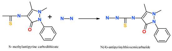
Scheme 2.
Preparation of N(4)-antipyrinylthiosemicarbazide.
3.1.2. Preparation of Isatin N(4)-antipyrine-thiosemicarbazide 1 (ligand)
Isatin N(4)-antipyrine-thiosemicarbazide 1 (ligand) was prepared by mixing equimolar amounts of isatin and N(4)-antipyrine-thiosemicarbazide in anhydrous EtOH with a few drops of concentrated H2SO4 at reflux (80 °C) for 2 h as shown in Scheme 3.

Scheme 3.
Preparation of Isatin N(4)-antipyrine-thiosemicarbazide 1 (ligand).
Ligand 1: Yield (87%), m.p. = 210–212 °C; color: yellow, Elemental analysis for C20H18N6O2S, (F.W. = 406.): Found (calcd) %C 59.14 (59.00), %H 5.15(4.43), %N 20.91(20.66), %S 7.85(7.87)); IR (KBr, cm−1), 3442, 3348, 3287, 3251, 3141ν(NH), 1738 ν(C=O)isatin, 1645 ν(C=O)antipyrine, 1688, 1623, 1595 ν(C=N), 881 ν(C=S); UV-Vis. (Nujol mulls) (nm) 268, 280, 369 nm, 1H-NMR (MHz, DMSO-d6, δ, ppm): 12. 819 (s, 1 H, thiosemicabazide N-NH), 11.219 (s, 1H, indole N-H), 10.115 (s, 1H, CS-N-H), 7.702–6.876. (m, multiple, H aromatic protons), 3.068 (s, 3 H, N-CH3), 2.135 (s, 3 H, CH3). GC–MS: m/z (relative abundance); 406.27, 363.47 (27.15), 291 (100), 276.68 (72.92), 261.69 (28.97), 223.71 (90.05), 204.07 (22.73), 203.3 (26.0), 145.64 (51.57), 130.57 (28.60), 118.18 (78.92), 67.30 (18.68).
3.1.3. Preparation of the Metal Complexes
The metal complexes of the ligand were prepared by mixing a hot ethanolic solution (30 mL) of the metal salt (CuCl2·2H2O, CuBr2, Cu(NO3)2·3H2O, Cu(OAc)2·H2O, Cu(ClO4)2, CoCl2·6H2O, Co(OAc)2·4H2O, NiCl2·6H2O, Ni(OAc)2·4H2O, ZnCl2 and FeCl3·6H2O) with the appropriate amount of a hot (75 °C) ethanolic solution of the ligand to form a 1:1 L/M (ligand/metal) solution. The reaction mixture was then refluxed for approximately 2 h. The obtained precipitates were separated by filtration, washed several times with ethanol and then diethyl ether, and then dried under vacuum over anhydrous CaCl2. Due to the poor solubility of these complexes in most organic solvents and water, all attempts to prepare a single crystal of the ligand or any of the metal complexes were unfortunately unsuccessful.
[Cu(L)Cl]·H2O (2): Yield (79%), m.p. = 238–240 °C; color: dark brown, ΛM: 10.0 ohm−1 cm2 mol−1, Elemental analysis for C20H19N6O3SCuCl., (F.W. = 522.6): Found (calcd) %C 46.0(46.0), %H 4.1(3.64), %N 16.3(16.10), %S 6.88(6.13): IR (KBr, cm−1), 3395 ν(H2O), 3161, 3100 ν(NH), 1702 ν(C=O)isatin, 1645 ν(C=O)antipyrine, 1616, 1539 ν(C=N), 808 ν(C=S), 632 ν(Cu-O), 498 ν(Cu-N) UV-Vis. (Nujol mulls) (nm) 310, 325 n→π*, 395, 460, 525 Charge transfer, 600, 672, 725 d→d, µeff (B.M) = 1.83.
[Cu(L)Br]·3H2O (3): Yield (89%), m.p. = 250 °C; color: brown, ΛM: 37.9 ohm−1 cm2 mol−1, Elemental analysis for, C20H23N6O5SCuBr, (F.W. = 602.5): Found (calcd) %C 38.81(39.83), %H 3.54(3.82), %N 15.02(13.94), %S 5.03(5.31) IR (KBr, cm−1), 3446 ν(H2O), 3106ν(NH), 1695 ν(C=O)isatin, 1643 ν(C=O)antipyrine, 1622, 1578 ν(C=N), 806 ν(C=S), 614 ν(Cu-O), 504 ν(Cu-N); UV-Vis. (Nujol mulls) (nm) 325 n→π*, 380, 445, 500 charge transfer, 580, 631, 660 d→d, µeff (B.M) = 1.81.
[Cu(L)(NO3)]·H2O (4): Yield (80%), m.p. = 247 °C; color: brown, ΛM: 34.3 ohm−1 cm2 mol−1, Elemental analysis for, C20H19N7O6SCu, (F.W. = 548.5): Found (calcd) %C 43.4(43.5), %H 3.29(3.50), %N 18.0(17.9), %S 6.0(5.8); IR (KBr, cm−1), 3425ν(H2O), 3166ν(NH), 1703 ν(C=O)isatin, 1645 ν(C=O)antipyrine, 1620, 1539 ν(C=N), 1382,1462 ν(NO3), 808 ν(C=S), 638 ν(Cu-O), 586 ν(Cu-N); UV-Vis. (Nujol mulls) (nm) 310 n→π*, 390, 455, 520, 574 charge transfer, 675, 720 d→d, µeff (B.M) = 1.78.
[Cu(L)OAc]·2H2O (5): Yield (74%), m.p. = 212–216 °C; color: brown, ΛM: 11.9 ohm−1 cm2 mol−1, Elemental analysis for, C22H24N6O6SCu, (F.W. = 563.5): Found (calcd) %C 46.12(46.90), %H 4.82(4.30), %N 15.13 (14.9), %S 5.1(5.7); IR (KBr, cm−1), 3417 ν(H2O), 3237ν(NH), 1680 ν(C=O)isatin, 1645 ν(C=O)antipyrine, 1560, 1525 ν(C=N), 1615, 1333 ν(OAc), 847 ν(C=S), 595 ν(Cu-O), 503 ν(Cu-N); UV-Vis. (Nujol mulls) (nm) 310 n→π*, 395, 430, 460, 560 charge transfer, 671,740(br), 870 d→d, µeff (B.M) = 1.79.
[Cu(L)ClO4]·2H2O (6): Yield (91%), m.p. = 250–252 °C; color: brown, ΛM: 45.50 ohm−1 cm2 mol−1; Elemental analysis for, C20H21N6O8SCuCl, (F.W. = 604.5): Found (calcd) %C 38.92(39.70), %H 3.75(3.48), %N 14.67(13.91), %S 5.18(5.29); IR (KBr, cm−1), 3428 ν(H2O), 3325, 3170ν(NH), 1628 ν(C=O)isatin, 1645 ν(C=O)antipyrine, 1568, 1516ν(C=N), 1118,1085, 694,631 ν(ClO4), 838 ν(C=S), 693 ν(Cu-O), 505 ν(Cu-N); UV-Vis. (Nujol mulls) (nm) 310 n→π*, 357, 390, 480(br) charge transfer, 790(br) d→d, µeff (B.M) = 1.76.
[Co(L)Cl]2·2H2O (7): Yield (66%), m.p. = 258–260 °C; color: brown, ΛM: 17.9 ohm−1 cm2 mol−1, Elemental analysis for C40H38N12O6S2Co2Cl2, (F.W. = 1034.8): Found (calcd) %C 47.0(46.38), %H 4.17(3.7), %N 17.0(16.21), %S .4(6.18), IR (KBr, cm−1), 3388 ν(H2O), 3150 ν(NH), 1734 ν(C=O)isatin, 1645 ν(C=O)antipyrine, 1600, 1515 ν(C=N), 841 ν(C=S), 585 ν(Co-N); GC–MS: m/z (relative abundance); 999.5 (22.46), 869.45 (15.13), 464.94 (4.00); UV-vis. (Nujol mulls) (nm) 318, 345 n→π*, 390, 450(br) charge transfer, 600, 675, 700 d→d, µeff (B.M) = 3.1.
[Co(L)OAc(H2O)]·4H2O (8): Yield (75%), m.p. ˃ 300 °C; color: brown, ΛM: 4.6 ohm−1 cm2 mol−1, Elemental analysis for C22H30N6O9SCo, (F.W. = 612.5): Found (calcd) %C 42.96(43.10), %H 5.3(4.89),%N 14.20(13.71), %S 4.87(5.22); IR (KBr, cm−1), 3491, 3422 ν(H2O), 3196ν(NH), 1734 ν(C=O)isatin, 1645 ν(C=O)antipyrine, 1590, 1545 ν(C=N), 1619, 1372 ν(OAc), 841 ν(C=S), 540 ν(Co-N); GC–MS: m/z (relative abundance); 612.85 (100), 576.23 (94.07, 561.30 (61.93), 541.85 (6.86), 523.22 (39.20); UV-Vis. (Nujol mulls) (nm) 320 n→π*, 390, 440(br), 530(br) charge transfer, 600, 675, 780d→d, µeff (B.M) = 3.7
[Ni(HL)2Cl2]·4H2O (9): Yield (84%), m.p. = 254–255 °C; color: brown, ΛM: 13.5 ohm−1 cm2 mol−1, Elemental analysis for C40H44N12O8S2 NiCl2., (F.W. = 1014.9): Found (calcd) %C 46.84(47.3), %H 4.94(4.33), %N 16.95(16.55), %S 5.89(6.30); IR (KBr, cm−1), 3441 ν(H2O), 3280 ν(NH), 1735, 1660 ν(C=O)isatin, 1645, 1620 ν(C=O)antipyrine, 1595(sh), 1520(sh) ν(C=N), 880 ν(C=S), 648 ν(Ni-O), 591 ν(Ni-N); UV-VIS. (Nujol mulls) (nm) 320 n→π*, 396, 420, 530 charge transfer, 560(s), 690(w), 950(br) d→d, µeff (B.M) = 2.98.
[Ni(L)OAc]2·4H2O (10): Yield (70%), m.p. ˃ 300 °C; color: brown, ΛM: 2.60 ohm−1 cm2 mol−1, Elemental analysis for C44H48N12O12S2 Ni2, (F.W. = 1017.4): Found (calcd) %C 47.76(47.24), %H 4.19(4.3), %N, 15. 60(15.03), %S 6.71(5.78); IR (KBr, cm−1), 3445 ν(H2O), 3310, 3200 ν(NH), 1664 ν(C=O)isatin, 1620 ν(C=O)antipyrine, 1580, 1540 ν(C=N), 1620, 1342 ν(OAc), 829 ν(C=S), 590 ν(Ni-O), 503 ν(Ni-N); UV-Vis. (Nujol mulls) (nm) 305, 315 n→π, 395, 450, 530 charge transfer, 567(s), 670(sh). 950(br) d→d, µeff (B.M) = 3.06.
[Zn(L)2]·2H2O (11): Yield (69%), m.p. ˃ 300 °C; color: dark yellow, ΛM: 1.40 ohm−1 cm2 mol−1, Elemental analysis for C40H38N12O6S2 Zn, (F.W. = 911.50): Found (calcd) %C 51.9(52.6), %H 3.7 (4.2), %N 18.8 (18.42), %S 6.64 (7.0); IR (KBr, cm−1 3413 ν(H2O), 3223ν(NH), 1700 ν(C=O)isatin, 1643 ν(C=O)antipyrine, 1609, 1570 ν(C=N), 815 ν(C=S), 612 ν(Zn-O), 580 ν(Zn-N): 1H-NMR (MHz, DMSO-d6, δ, ppm): 11.22 (s, 1H, indole N-H), 11.008 (s,1H, CS-N-H), 7.704–6.879. (m, multiple, C-H aromatic), 3.111 (s, 3 H, N-CH3), 2.174 (s, 3 H, CH3); GC–MS: m/z (relative abundance); 911.5 (1.19) UV-Vis. (Nujol mulls) (nm) 319 n→π, 396, 440, 491, 520(sh).
[Fe(HL)2Cl2]Cl·H2O (12): Yield (78%), m.p. = 210–212 °C; color: dark green, ΛM: 74.50 ohm−1 cm2 mol−1, Elemental analysis for C40H38N12O5S2FeCl3, (F.W. = 992.50): Found (calcd) %C 47.62(48.3), %H 3.9(3.8), %N 16.95 (16.91), %S 5.6 (6.44), IR (KBr, cm−1), 3429(s,br) ν(H2O), 3280 ν(NH), 1737, 1700 ν(C=O)isatin, 1645, 1621 ν(C=O)antipyrine, 1610, 1576 ν(C=N), 880 ν(C=S), 589 ν (Fe-O), 512 ν(Fe-N) GC–MS: m/z (relative abundance); 992.5 (11.39), 974.50 (1.90), 938.30 (12.96), 869.80 (8.27), 464.40 (2.48); UV-Vis. (Nujol mulls) (nm) 300, 340 n→π, 391, 450, 530(sh) charge transfer, 567(s), 584 d→d, µeff (B.M) = 5.95.
3.1.4. Instrumentation and Measurements
Elemental microanalyses [C, H, and N] were conducted in the Micro Analytical Unit. The IR spectra were acquired using KBr discs in a Spirit model FT-IR spectrophotometer (Shimadzu, Thermo Scientific, Glasgow, UK). The 1H-NMR spectra of the ligand and zinc complex were recorded in DMSO-d6 on a Gemini 200 NMR spectrophotometer (Varian, Palo Alto, CA, USA) at 300 MHz. Mass spectra were collected using a model ISQLT direct probe controller with a single quadrupole mass analyser (Thermo Scientific, Glasgow, UK) using Thermo X-calibur software (Xcalibur 4.1). Solid-state electronic spectra were recorded in Nujol mulls using a Shimadzu UV-Vis 1800 spectrophotometer (Thermo Scientific, Glasgow, UK). The molar conductance of a 1 × 10⁻³ M solution of the complexes in N,N-dimethylformamide (DMF) was measured at 25 °C with a Bibby MCl-type conductometer (Chelmsford, UK). The resistance was measured in ohms and the molar conductivities were calculated according to the following equation: ΛM = VxKxg/Mw* Ω where ΛM = molar conductivity (Ω-1cm2mol-1), V = volume of the complex solution (mL), K = cell constant (0.92 cm-1), Mw = molecular weight of the complex, g = weight of the complex (g), and Ω = resistance (Ω).
The solid-state electron paramagnetic resonance (EPR) spectra of the copper(II) complexes were recorded with an X-band EMX spectrometer (Bruker, Berlin, Germany) using a standard rectangular cavity of ER 4102 operating at 9.5 GHz with 100 kHz modulation at 298 K. Diphenyl picryl hydrazide (DPPH) was used as a g-marker to calibrate the spectra. The thermal analyses (TGA) were carried out in air using a Shimadzu DT-30 thermal analyser. Magnetic susceptibilities were measured at 27 °C using the modified Gouy method with a Johnson Matthey balance and were calculated using the following equation:
3.2. Biological Evalution
3.2.1. Preparation of Solutions of the Complexes
Stock solutions (1 mM) of the newly synthesized complexes were prepared by dissolving each complex in dimethylsulfoxide (DMSO)/H2O, and the solutions were stored at 4 °C. Only ligand 1 and complexes 2, 7 and 9 were successfully solubilized, allowing their antitumor activities to be determined.
3.2.2. Experimental Animals
Adult female Swiss albino mice (6–7 weeks old), weighing 20–22 g, were purchased and housed under standard ventilation and light cycle conditions. They were given food and water ad libitum. The current study protocol was approved by the Ethical Committee for Laboratory Animals of Science Faculty Menoufia University (No.: ECLA-SFMU-18015).
3.2.3. Tumor Cell Line
The Ehrlich ascites carcinoma (EAC) cell line was purchased from the National Cancer Institute (NCI, Cairo, Egypt). The EAC cell line was maintained in the peritoneal cavity of adult female Swiss albino mice by serial intraperitoneal transplantation of 2.5 × 10⁶ cells weekly.
3.2.4. Induction of Solid Tumor and Animals Grouping
The solid tumor was induced by subcutaneously (SC) injecting 1 × 10⁻⁶ EAC cells into the right thigh of the lower limb of mice [84,85]. Seven days after tumor inoculation, the tumor volume became palpable. The mice were randomly divided in separate cages into six groups:
- Group 1 (normal control): mice without any treatment.
- Group 2 (vehicle group): mice bearing solid tumors treated with DMSO/H2O.
- Group 3: mice bearing solid tumor and treated subcutaneously with 0.2 mM of ligand 1.
- Group 4: mice bearing solid tumor and treated subcutaneously with 0.2 mM of complexe 2.
- Group 5: mice bearing solid tumor and treated subcutaneously with 0.2 mM of complexe 7.
- Group 6: mice bearing solid tumor and treated subcutaneously with 0.2 mM of complexe 9.
The doses were selected based on the LD50 values of the complexes (data not shown). The treatment was started seven days after EAC implementation and was continued for 14 consecutive days.
3.2.5. Blood and Tissue Samples Collection
At the end of the experiment, mice were sacrificed. Blood samples were collected from jugular vein puncture into chilled non-heparinized tubes, which were centrifuged at 40× g for 10 min at 4 °C. The sera were frozen at −20 °C for future measurements. The livers and solid tumors were removed and cut into pieces, and the individual pieces were fixed in paraffin blocks for immunohistochemical assays.
3.2.6. Assessment of Tumor Volume (TV)
After 24 h from the last dose of treatment, mice were sacrificed and the solid tumors were carefully removed. The volume of the tumors was assessed [86] using the following formula: TV (mm3) = 4π (A/2)2 × (B/2), where (A) is the minor tumor axis and (B) is the major tumor axis.
3.2.7. Biochemical Analysis of the Serum
To determine the effect of ligand 1 and its metal complexes 2, 7 and 9 on the serum biochemical profile. The levels of alanine transaminase (ALT), aspartate transaminase (AST), albumin and glucose were determined using a Beckman autoanalyzer apparatus, Brea, CA, USA).
3.2.8. Immunohistochemical Examination of VEGF and Caspase-7 in Solid Tumors and Liver Tissues
The effect of ligand 1 and its metal complexes 2, 7 and 9 on the expression of VEGF and caspase-7 in solid tumor tissues and in liver tissues of mice bearing solid tumors was deremined by immunohistochemical methods. In details, the sections of tumor and liver tissues in paraffin were dewaxed, hydrated, and immersed in antigen retrieval solution (EDTA solution, pH 8). The samples were treated with 0.3% hydrogen peroxide and protein block and then incubated with anti-VEGF (Santa Cruz, Dallas, TX, USA, 1:100 dilution) and anti-caspase-7 (Lab Vision, Thermo Fischer Scientific, Glasgow, UK; ready to use) at 4 °C overnight. The slides were rinsed three times with PBS and incubated with anti-mouse IgG secondary antibodies (EnVision + System HRP; Dako, SanJose, CA, USA) for 30 min at room temperature. The samples were visualized with diaminobenzidine commercial kits (Liquid DAB+Substrate Chromogen System; Dako) and finally counterstained with Mayer’s haematoxylin. In the negative control, the primary antibody was replaced with normal mouse serum. The positive cells were counted from seven high-power fields of 400×. The labelling index (%) was determined by dividing the number of positive cells by the total cells × 100 [87,88].
3.3. Statistical Analysis
Data are expressed as mean ± SEM and the statistical comparison between groups were analyzed using one-way ANOVA followed by Tukey post hoc test. A p of < 0.05 value was considered statistically significant.
4. Conclusions
In summary, new metal complexes derived from isatin-N-(4)antipyrinethiosemicarbazone ligand 1 were synthesized and characterized. Also, their potential anti-cancer activities against solid tumors induced by EAC cells in mice were investigated. The results revealed that the tested complexes significantly reduced the volume of solid tumors. This reduction was associated with an increase in the level of caspase-7 and a dramatic decrease in the VEGF level. The tested complexes are promising anti-cancer agents those exert their activities via the induction of apoptosis and the inhibition of angiogenesis, leading to reduction of the tumor size.
Supplementary Materials
The following are available online; 1H-NMR, Mass, IR, EPR, TGA, and UV-Visible spectra.
Author Contributions
Conceptualization, F.E.-S., B.E.-A. and T.S.; Formal analysis, B.E.-A. and F.E.-S.; Funding acquisition, S.A.M.K. and H.R.E.-S.; Methodology, B.E.-A., and G.S.; Visualization, B.E.-A. and F.E.-S.; Writing—original draft, B.E.-A., F.E.-S., and G.S.; Writing—review & editing, B.E.-A., G.S., S.A.M.K., and H.R.E.-S. All authors have read and approved the manuscript.
Funding
We are very grateful to the Swedish Research links grant 2016-05885 (VR for the years 2017–2019) for generious financial support.
Acknowledgments
H.R.E.-S. is very grateful to Jiangsu University, China for Foreign Expert fellowship.
Conflicts of Interest
The authors declare no conflict of interest.
References
- Siegel, R.L.; Miller, K.D.; Jemal, A. Cancer statistics, 2019. CA 2019, 69, 7–34. [Google Scholar] [CrossRef] [PubMed]
- Folkman, J. Angiogenesis in cancer, vascular, rheumatoid and other disease. Nat. Med. 1995, 1, 27–30. [Google Scholar] [CrossRef] [PubMed]
- Thapa, D.; Lee, J.S.; Heo, S.W.; Lee, Y.R.; Kang, K.W.; Kwak, M.K. Novel hexahydrocannabinol analogs as potential anti-cancer agents inhibit cell proliferation and tumor angiogenesis. Eur. J. Pharmacol. 2011, 650, 61–71. [Google Scholar] [CrossRef] [PubMed]
- El-Aarag, B.Y.; Kasai, T.; Zahran, M.A.; Zakhary, N.I.; Shigehiro, T.; Sekhar, S.C.; Agwa, H.S.; Mizutani, A.; Murakami, H.; Kakuta, H.; et al. In vitro anti-proliferative and anti-angiogenic activities of thalidomide dithiocarbamate analogs. Int. Immunopharmacol. 2014, 21, 283–292. [Google Scholar] [CrossRef] [PubMed]
- El-Aarag, B.; Kasai, T.; Masuda, J.; Zahran, M.; Agwa, H.; Seno, M. Anticancer effects of novel thalidomide analogs in A549 cells through inhibition of vascular endothelial growth factor and matrix metalloproteinase-2. Biomed. Pharmacother. 2017, 85, 549–555. [Google Scholar] [CrossRef] [PubMed]
- Zahran, M.; Agwa, H.; Osman, A.; Hammad, S.; El-Aarag, B.; Ismail, N.; Salem, T.; Gamal-Eldeen, A. Synthesis and biological evaluation of phthalimide dithiocarbamate and dithioate derivatives as anti-proliferative and anti-angiogenic agents-I. Eur. J. Chem. 2017, 8, 391–399. [Google Scholar] [CrossRef][Green Version]
- Kamal, A.; Kumar, G.B.; Nayak, V.L.; Reddy, V.S.; Shaik, A.B.; Reddy, M.K. Design, synthesis and biological evaluation of imidazopyridine/imidazopyrimidine-benzimidazole conjugates as potential anticancer agents. Med. Chem. Commun. 2015, 6, 606–612. [Google Scholar] [CrossRef]
- Yang, L.L.; Lee, C.Y.; Yen, K.Y. Induction of apoptosis by hydrolyzable tannins from Eugenia jambos L. on human leukemia cells. Cancer Lett. 2000, 157, 65–75. [Google Scholar] [CrossRef]
- Rosu, T.; Negoiu, M.; Pasculescu, S.; Pahontu, E.; Poirier, D.; Gulea, A. Metal-based biologically active agents: Synthesis, characterization, antibacterial and antileukemia activity evaluation of Cu(II), V(IV) and Ni(II) complexes with antipyrine-derived compounds. Eur. J. Med. Chem. 2009, 45, 774–781. [Google Scholar] [CrossRef]
- Leovac, V.M.; Bogdanovic, G.A.; Jovanovic, L.S.; Joksovic, L.C.; Markovic, I.; Joksovic, M.D. Synthesis, characterization and antitumor activity of polymeric copper(II) complexes with thiosemicarbazones of 3-methyl-5-oxo-1-phenyl-3-pyrazolin-4-carboxaldehyde and 5-oxo-3-phenyl-3-pyrazolin-4-carboxaldehyde. J. Inorg. Biochem. 2011, 105, 1413–1421. [Google Scholar] [CrossRef] [PubMed]
- Refata, M.S.; El-Metwaly, N.M. El-Metwaly, Spectral, thermal and biological studies of Mn(II) and Cu(II) complexes with two thiosemicarbazide derivatives. Spectrochim. Acta Part A 2012, 92, 336–346. [Google Scholar] [CrossRef] [PubMed]
- Mendes, I.C.; Moreira, J.P.; Mangrich, A.S.; Balena, S.P.; Rodrigues, B.L.; Beraldo, H. Coordination to copper (II) strongly enhances the in vitro antimicrobial activity of pyridine-derived N(4)-tolyl thiosemicarbazones. Polyhedron 2007, 26, 3263–3270. [Google Scholar] [CrossRef]
- Vinuelas-Zahinos, E.; Maldonado-Rogado, M.A.; Luna-Giles, F.; Barros-Garcia, F.J. Coordination behaviour of Schiff base 2-acetyl-2-thiazoline hydrazone (ATH) towards cobalt(II), nickel(II) and copper(II). Polyhedron 2008, 27, 879–886. [Google Scholar] [CrossRef]
- Bharti, N.; Shailendra, Y.; Sharma, S.; Naqvi, F.; Azam, A. New palladium(II) complexes of 5-nitrothiophene-2-carboxaldehyde thiosemicarbazones. synthesis, spectral studies and in vitro anti-amoebic activity. Bioorg. Med. Chem. 2003, 11, 2923–2929. [Google Scholar] [CrossRef]
- El-Sawaf, A.K.; West, D.X.; El-Saied, F.A.; El-Bahnasawy, R.M. Synthesis, magnetic and spectral studies of iron(III), cobalt(II,III), nickel(II), copper(II) and zinc(II) complexes of 4–formylantipyrine N(4)-antipyrinylthiosemicarbazone. Transit. Met. Chem. 1998, 23, 649–655. [Google Scholar] [CrossRef]
- El-Sawaf, A.K.; West, D.X.; El-Saied, F.A.; El-Bahnasawy, R.M. Copper(II) complexes of 4-formylantipyrine N(4)-substitutedthiosemicarbazones. Transit. Met. Chem. 1998, 22, 360–365. [Google Scholar] [CrossRef]
- Ali, A.Q.; Teoh, S.G.; Salhin, A.; Eltayeb, N.E.; Ahamed, M.B.K.; Majid, A.A. Synthesis of isatin thiosemicarbazones derivatives: In vitro anti-cancer, DNA binding and cleavage activities. Spectrochim. Acta A 2014, 125, 440–448. [Google Scholar] [CrossRef] [PubMed]
- El-Saied, F.A.; El-Enein, S.A.A.; Emam, S.M.; El-Shater, H.A. Synthesis and characterization of Cu(II), Ni(II), Co(II), Mn(II), Zn(II), Ru(III), Hf(IV) and ZrO(II) complexes of 2-thiophenylidene-N-4-methoxy anilinoacetohydrazone. Polish J. Chem. 2009, 83, 1871–1883. [Google Scholar]
- Detrov, I.; Grupce, O.; Stafilov, T. The N-H stretching region of some imides and thioimides. Mol. Struct. 1986, 142, 275–278. [Google Scholar]
- Castineiras, A.; Gomez, M.C.; Sevillano, P. 2-(2,3-Dihydro-1,3-benzothiazol-2-yl)phenyl(diphenyl) phosphine oxide, Synthesis and characterization by IR and NMR spectroscopy and X-ray diffractometry. Mol. Struct. 2000, 554, 301–306. [Google Scholar] [CrossRef]
- Prasad, R.N.; Mathur, M.; Upadhayay, A. Synthesis and spectroscopic studies of Cr (III), Fe (III) and Co (II) complexes of hexaazamacrocycles. Indian Chem. Soc. 2007, 84, 1202–1204. [Google Scholar]
- Husain, K.; Bhat, A.; Azam, A. New Pd(II) complexes of the synthesized 1-N-substituted thiosemicarbazones of 3-indole carboxaldehyde: Characterization and antiamoebic assessment against E. histolytica. J. Med. Chem. 2008, 43, 2016–2028. [Google Scholar] [CrossRef] [PubMed]
- Mikuriya, M.; Okawa, H.; Kida, S. Binuclear Metal Complexes. XXXV. Synthesis and Magnetism of Binuclear Copper(II) Complexes with 3-[2-(Dialkylamino)ethylthio]-1-propanols. Bull. Chem. Soc. 1980, 53, 3717–3718. [Google Scholar] [CrossRef]
- Rreti, C.; Tosr, G. Complexes of Co alt (II), Nickel (II) and Copper Acetates, Perchlorates, and Tetrafluoroborates with Heterocyclic Ligands Containing VA and VIA group Donor Atoms. Can. J. chem. 1975, 53, 177–182. [Google Scholar]
- Jain, S.K.; Garg, B.S.; Bhoon, Y.K. Iron(III) complexes of 2-acetylpyridine-4-phenyl-3-thiosemicarbazones: Magnetic, E.s.r. and spectral studies. Transit. Met. Chem. 1986, 11, 89–95. [Google Scholar] [CrossRef]
- Maurya, R.C.; Rajput, S. Neutral dioxovanadium(V) complexes of biomimetic hydrazones ONO donor ligands of bioinorganic and medicinal relevance: Synthesis via air oxidation of bis(acetylaceto-nato)oxovanadium(IV), characterization, biological activity and 3D molecular modeling. J. Mol. Struct. 2007, 833, 133–144. [Google Scholar] [CrossRef]
- Sallam, S.A.; Orabi, A.S.; El-Shetary, B.A.; Lentz, A. Copper, nickel and cobalt complexes of Schiff-bases derived from β-diketones. Transit. Met. Chem. 2002, 27, 447–453. [Google Scholar] [CrossRef]
- Tossidis, A.; Bolos, C.A. Monohalogenobenzoylhydrazones II. Synthesis and Study of Ti(IV) Complexes with Monochlorobenzoylhydrazones of 2-Furaldehyde, 2-Thiophenaldehyde, 2-Pyrrolaldehyde and Di-2-pyridylketone as Ligands. Inorg. Chim. Acta 1986, 112, 93–97. [Google Scholar] [CrossRef]
- Ainscough, E.W.; Brodie, A.M.; Dobbs, A.J.; Ranford, J.D.; Waters, J.M. Antitumour copper(II) salicylaldehyde benzoylhydrazone (H2sb) complexes: Physicochemical properties and the single-crystal X-ray structures of [{Cu(H2sb)(CCl3CO2)2}2] and [{Cu(Hsb) (ClO4) (C2H5OH)}2] and the related salicylaldehyde acetylhydrazone (H2sa) complex, [Cu(Hsa)Cl(H2O)]·H2O. Inorg. Chim. Acta 1998, 267, 27–38. [Google Scholar]
- El-Sawaf, A.K.; Nassar, A.A.; El-Samanody, E. Synthesis, magnetic, spectral and biological studies of copper (II) complexes of 4-benzoyl-3-methyl-1-phenyl-2-pyrazolin-5-one N(4)-substituted thiosemicarbazones. Sci. J. Chem. 2014, 2, 17–26. [Google Scholar] [CrossRef][Green Version]
- Nair, M.K.M.; Radhakrishnan, P.K. Iodide Complexes of Yttrium and Lanthanides with 4-N-(4′-Antipyrylmethylidene) aminoantipyrine. Synth. React. Inorg. Met. Org. Chem. 1996, 26, 529–541. [Google Scholar] [CrossRef]
- Afrasiabi, Z.; Sinn, E.; Padhye, S.; Dutta, S.; Padhye, S.; Newton, C.; Anson, C.E.; Powell, A.K. Transition metal complexes of phenanthrenequinone thiosemicarbazone as potential anticancer agents: Synthesis, structure, spectroscopy, electrochemistry and in vitro anticancer activity against human breast cancer cellline, T47D. J. Inorg. Biochem. 2003, 95, 306–314. [Google Scholar] [CrossRef]
- Fouda, M.F.R.; Abd-Elzaher, M.M.; Shakdofa, M.M.; El-Saied, F.A.; Ayad, M.I.; el Tabl, A.S. Synthesis and Characterization of a Hydrazone Ligand Containing Antipyrine and its Transition Metal Complexes. J. Coord. Chem. 2008, 61, 1983–1996. [Google Scholar] [CrossRef]
- Lever, A.B.P. Electronic Spectra of Some Transition Metal Complexes: Derivation of Dq and B. J. Chem. Educ. 1968, 45, 711–712. [Google Scholar] [CrossRef]
- Shebl, M.; El-ghamry, M.A.; Khalil, S.M.; kishk, M.A. Mono- and binuclear copper(II) complexes of new hydrazone ligands derived from 4,6-diacetylresorcinol: Synthesis, spectral studies and antimicrobial activity. Spectrochim. Acta A Mol. Biomol. Spectrosc. 2014, 21, 232–241. [Google Scholar] [CrossRef] [PubMed]
- Downes, J.M.; Whelan, J.; Bosnich, B. Biological analogs, Spectroscopic characteristics of mercapto- and disulfide-copper(II) coordination in relation to type I proteins. Inorg. Chem. 1981, 20, 1081–1086. [Google Scholar] [CrossRef]
- Okawa, H.; Nakamoto, M.; Lzumitani, T.; Kida, S. Syntheses and Configurations of Copper(II) and Nickel(II) Complexes of trans-1,2-Dicyano-1,2-cyclohexanediamine. Bull. Chem. Soc. 1982, 55, 2671–2672. [Google Scholar] [CrossRef]
- Suzuki, M.; Kanatomi, H.; Koyama, H.; Murase, I. Copper(I) and Copper(II) Complexes of Tripod Ligands, Tris(2-alkylthioethyl)amine. Bull. Chem. Soc. 1980, 53, 1961–1964. [Google Scholar] [CrossRef]
- Kovala-Dermertzi, D.; Tsangaris, J.M.; Hadjiliadis, N. Complexes of aminobenzylamines. Part I. Complexes of O-aminobenzylamine with copper(II), cobalt(II) and nickel(II). Transit. Met. Chem. 1983, 8, 140–146. [Google Scholar] [CrossRef]
- Suzuki, M.; Demura, H.K.Y.; Murase, I. Syntheses and Properties of Dinuclear Copper(II) Complexes with 2,6-Bis[bis(2-pyridylmethyl)aminomethyl]-4-methylphenol. Bull. Chem. Soc. 1984, 57, 1003–1007. [Google Scholar] [CrossRef]
- El-Tabl, A.S.; El-Bahnasawy, R.M.; Hamdy, A.E.D. Synthesis, magnetic, spectral and antimicrobial studies on metal complexes of 4- methylphenylaminoacetoisatin hydrazine. J. Chem. Res. 2009, 11, 659–664. [Google Scholar]
- Chohan, Z.H.; Pervez, H.; Khan, K.M.; Supuran, C.T. Metal binding and antibacterial activity of ciprofloxacin complexes. J. Enzym. Inhib. Med. Chem. 2005, 20, 303–307. [Google Scholar] [CrossRef] [PubMed]
- Lever, A.B.P. Inorganic Electronic Spectroscopy, Studies in Physical and Theoretical Chemistry; Elsevier Science Ltd.: Amsterdam, The Netherlands, 1984. [Google Scholar]
- Matovic, Z.D.; Miletic, V.D.; Samardzic, G.; Pelosi, G.; Ianelli, S.; Trifunovic, S. Square-planar copper(II) complexes with a tetradentate amido-carboxylate ligands. Crystal structure of Na2[Cu(obap)]2⋅2H2O. Strain analysis and spectral assignments of complexes. Inorg. Chim. Acta 2005, 358, 3135–3144. [Google Scholar] [CrossRef]
- Mangalam, N.A.; Kurup, M.R.P. Versatile binding properties of a di-2-pyridyl ketone nicotinoylhydrazone ligand: Crystal structure of a Cu(II) complex. Spectrochim. Acta Part A Mol. Biomol. Spectrosc. 2011, 78, 926–934. [Google Scholar] [CrossRef] [PubMed]
- Aly, M.M.; Imam, S.M. Characterization of Copper(II), nickel(II), cobalt(II) and palladium(II) complexes of vicinal oxime-imine ligands; induced chelate isomerism in the same molecule of the nickel(II) complex. Mon. Chem. Chem. Mon. 1995, 126, 173–185. [Google Scholar] [CrossRef]
- el Saied, F.A.; Shakdofa, M.M.E.; el Tabl, A.S.; Elzaher, M.M.A. Coordination behaviour of N′1,N′4-bis((1, 5-dimethyl-3-oxo-2-phenyl-2,3-dihydro-1H-pyrazol-4-yl)methylene) succinohydrazide toward transition metal ions and their antimicrobial activities. Main Group Chem. 2014, 13, 87–99. [Google Scholar]
- Pahonțu, E.; Ilieș, D.C.; Shova, S.; Oprean, C.; Păunescu, V.; Olaru, O.T.; Rădulescu, F.Ș.; Gulea, A.; Roșu, T.; Drăgănescu, D. Synthesis, Characterization, Antimicrobial and Antiproliferative Activity Evaluation of Cu(II), Co(II), Zn(II), Ni(II) and Pt(II) Complexes with Isoniazid-Derived Compound. Molecules 2017, 22, 650. [Google Scholar]
- Earnshaw, A. Introduction to Magneto-Chemistry; Academic Press: New York, NY, USA, 1968. [Google Scholar]
- Chandra, S.; Sharma, K.K. Cobalt(II) and nickel(II) complexes of some nitrogen-oxygen and nitrogen-sulphur ligands. Transit. Met. Chem. 1984, 9, 1–3. [Google Scholar] [CrossRef]
- El-Tabl, A.S.; El-Saied, F.A.; Al-Hakimi, A.N. Synthesis, spectroscopic investigation and biological activity of metal complexes with ONO trifunctionalalized hydrazone ligand. Transit. Met. Chem. 2007, 32, 689–701. [Google Scholar] [CrossRef]
- Fouda, M.F.R.; Abd-El-zaher, M.M.; Shakdofa, M.M.; El-Saied, F.A.; Ayad, M.I.; El-Tabl, A.S. Synthesis and characterization of transition metal complexes of N′-[(1, 5-dimethyl-3-oxo-2-phenyl-2, 3-dihydro-1H-pyrazol-4-yl) methylene] thiophene-2-carbohydrazide. Transit. Met. Chem. 2008, 33, 219–228. [Google Scholar] [CrossRef]
- Al-Tabl, A.S.; AbouEl-Enein, S.A. Reactivity of the new potentially binucleating ligand, 2-(acetichydrazido-N-methylidene-α-naphthol)-benzothiazol, towards manganese (II), nickel (II), cobalt (II), copper (II) and zinc (II) salts. J. Coord. Chem 2004, 57, 281–294. [Google Scholar] [CrossRef]
- El-Reash, G.M.A.; Ibrahim, K.M.; Bekheit, M.M. Structural studies of 4(phenyl or benzamidos)-1-cyclohexa-none-3- thiosemicarbazone complexes. Transit. Met. Chem. 1990, 15, 148–151. [Google Scholar]
- El-Saied, F.A.; El-Asmy, A.A.; Kaminsky, W.; West, D.X. Spectral and structural studies of cobalt(II,III), nickel(II), and copper(II) complexes of dehydroacetic acid N4-dialkyl- and 3-azacyclothiosemicarbazones. Transit. Met. Chem. 2003, 28, 954–960. [Google Scholar] [CrossRef]
- El-Tabl, A.S.; Shakdofa, M.M.E.; El-Seidy, A.M.A.; Al-Hakimi, A.N. Synthesis, characterization and antifungal activity of metal complexes of 2-(5-((2-chlorophenyl) diazenyl)-2-hydroxybenzylidene) hydrazinecarbothioamide. Phosphorus Sulfur Silicon Relat. Elem. 2012, 187, 1312–1323. [Google Scholar] [CrossRef]
- Kasumov, V.T.; Köksal, F. Synthesis, ESR, UV-Visible and reactivity studies of new bis(N-dimethoxyaniline-3,5-(t)Bu2-salicylaldiminato)copper(II) complexes. Spectrochim. Acta A Mol. Biomol. Spectrosc. 2012, 98, 207–214. [Google Scholar] [CrossRef] [PubMed]
- Hathaway, B.J.; Billing, D.E. The electronic properties and stereochemistry of mono-nuclear complexes of the copper(II) ion. Coord. Chem. Rev. 1970, 5, 1949. [Google Scholar] [CrossRef]
- Zaky, R.R.; Ibrahim, K.M.; Gabr, I.M. Bivalent transition metal complexes of o-hydroxyacetophenone [N-(3-hydroxy-2-naphthoyl)] hydrazone: Spectroscopic, antibacterial, antifungal activity and thermogravimetric studies. Spectrochim. Acta A Mol. Biomol. Spectrosc. 2011, 81, 28–34. [Google Scholar] [CrossRef] [PubMed]
- Brown, D.R.; West, D.X. An ESR study of addition species formed between bis(2-thiopyridine N-oxide)Cu(II) and heterocyclic and aliphatic amines. J. Inorg. Nucl. Chem. 1981, 43, 1017–1021. [Google Scholar] [CrossRef]
- Nagashri, K.; Joseph, J.; Dhanaraj, C.J. Copper (II) complexes of hydroxyflavone derivatives as potential bioactive molecule to combat antioxidants: Synthesis, characterization and pharmacological activities. Appl. Organomet. Chem. 2011, 25, 704–717. [Google Scholar] [CrossRef]
- Proctor, I.M.; Hathaway, B.J.; Nicholis, P. The electronic properties and stereochemistry of the copper(II) ion. Part I. Bis(ethylenediamine)copper(II) complexes. J. Chem. Soc. A 1968, 1678–1684. [Google Scholar] [CrossRef]
- Kivelson, D.; Neiman, R. ESR Studies on the Bonding in Copper Complexes. J. Chem. Phys. 2004, 35, 149. [Google Scholar] [CrossRef]
- Shauib, N.M.; Elassar, A.Z.A.; El-Dissouky, A. Synthesis and spectroscopic characterization of copper(II) complexes with the polydentate chelating ligand 4,4’-[1,4-phenylenedi(nitrilo)dipente-2-one. Spectrochim. Acta Part A Mol. Biomol. Spectrosc. 2006, 63, 714–722. [Google Scholar] [CrossRef] [PubMed]
- Ray, R.K.; Kauffmann, G.B. An EPR study of copper(II)-substituted biguanide complexes. Part III. Inorg. Chim. Acta 1990, 174, 237–244. [Google Scholar] [CrossRef]
- Hathaway, B.J. A new look at the stereochemistry and electronic properties of complexes of the copper(II) ion. Complex Chem. 1984, 57, 55–118. [Google Scholar]
- Sakaguchi, U.; Adison, A.W. Spectroscopic and redox studies of some copper(II) complexes with biomimetic donor atoms: Implications for protein copper centres. J. Chem. Soc. Dalton Trans. 1979, 4, 600–608. [Google Scholar] [CrossRef]
- El-Tabl, A.S. Synthesis and spectral studies on mononuclear copper (II) complexes of isonitrosoacetylacetone ligand. Polish J. Chem. 1997, 71, 1213–1222. [Google Scholar]
- Mahajian, M.; Saxena, K.N.; Saxen, P.C. ESR study of Cu(II) complexes. Inorg. Nucl.Chem. 1981, 43, 2148–2149. [Google Scholar] [CrossRef]
- Oscar, C. Biopharmacological studies on certain new anticancer drugs. J. Biochem. 1988, 110, 701–707. [Google Scholar]
- Tagmizyan, W.A. Metabolism of impaired glucose in patients with neoplasia. Am. Biol. 1990, 2, 53–58. [Google Scholar]
- Silverstein, H.; Dervot, K.; Oscar, D. Studies on carbohydrate metabolism and different types of tumors bearing animals. Lancet 1988, 22, 40–45. [Google Scholar]
- Kind, P.R.N.; Gordon, M.; Laverick, M.; Nias, A.H.; Slavin, B.M. The effect of C3H mouse mammary tumor on the levels of serum and urine analytes in vivo. Br. J. Cancer 1985, 52, 607–612. [Google Scholar] [CrossRef] [PubMed]
- Parveen, A.; Subedi, L.; Kim, H.W.; Khan, Z.; Zahra, Z.; Farooqi, M.Q.; Kim, S.Y. Phytochemicals Targeting VEGF and VEGF-Related Multifactors as Anticancer Therapy. J. Clin. Med. 2019, 8, 350. [Google Scholar] [CrossRef] [PubMed]
- Abd-Alhaseeb, M.M.; Zaitone, S.A.; Abou-El-Ela, S.H.; Moustafa, Y.M. Olmesartan Potentiates the Anti-Angiogenic Effect of Sorafenib in Mice Bearing Ehrlich’s Ascites Carcinoma: Role of Angiotensin (1–7). PLoS ONE 2014, 9, e85891. [Google Scholar] [CrossRef] [PubMed]
- Liang, Y.; Brekken, R.A.; Hyder, S.M. Vascular endothelial growth factor induces proliferation of breast cancer cells and inhibits the anti-proliferative activity of anti-hormones. Endocr. Relat. Cancer 2006, 13, 905–919. [Google Scholar] [CrossRef] [PubMed]
- Kumar, S.; Cieplak, P. Effect of phosphorylation and single nucleotide polymorphisms on caspase substrates processing. Apoptosis 2018, 23, 194–200. [Google Scholar] [CrossRef] [PubMed]
- Hassan, M.; Watari, H.; AbuAlmaaty, A.; Ohba, Y.; Sakuragi, N. Apoptosis and Molecular Targeting Therapy in Cancer. BioMed Res. Int. 2014, 2014, 150845. [Google Scholar] [CrossRef] [PubMed]
- Cao, Z.; Yang, Q.; Yin, H.; Qi, Q.; Li, H.; Sun, G.; Wang, H.; Liu, W.; Li, J. Peroxynitrite induces apoptosis of mouse cochlear hair cells via a Caspase-independent pathway in vitro. Apoptosis 2017, 22, 1419–1430. [Google Scholar] [CrossRef] [PubMed]
- Vance, N.R.; Gakhar, L.; Spies, M.A. Allosteric Tuning of Caspase-7: A Fragment-Based Drug Discovery Approach. Angew. Chem. Int. Ed. Engl. 2017, 56, 14443–14447. [Google Scholar] [CrossRef] [PubMed]
- Zahran, M.A.-H.; El-Aarag, B.; Mehany, A.B.M.; Belal, A.; Younes, A.S. Design, synthesis, biological evaluations, molecular docking, and in vivo studies of novel phthalimide analogs. Arch. Pharm. Chem. Life Sci. 2018, 351, 1–12. [Google Scholar] [CrossRef] [PubMed]
- Salem, M.L.; Shoukry, N.M.; Teleb, W.K.; Abdel-Daim, M.M.; Abdel-Rahman, M.A. In vitro and in vivo antitumor effects of the Egyptian scorpion Androctonus amoreuxi venom in an Ehrlich ascites tumor model. SpringerPlus 2016, 5, 570. [Google Scholar] [CrossRef]
- Marcelli, M.; Cunningham, G.R.; Walkup, M.; He, Z.; Sturgis, L.; Kagan, C.; Mannucci, R.; Nicoletti, I.; Teng, B.; Denner, L. Signaling Pathway Activated during Apoptosis of the Prostate Cancer Cell Line LNCaP: Overexpression of Caspase-7 as a New Gene Therapy Strategy for Prostate Cancer. Cancer Res. 1999, 59, 382–390. [Google Scholar] [PubMed]
- Guirgis, A.A.; Zahran, M.; Mohamed, A.S.; Talaat, R.M.; Abdou, B.Y.; Agwa, H.S. Effect of thalidomide dithiocarbamate analogs on the intercellular adhesion molecule-1 expression. Int. Immunopharmacol. 2010, 10, 805–811. [Google Scholar] [CrossRef] [PubMed]
- Zahra, M.H.; Salem, T.A.; El-Aarag, B.; Yosri, N.; EL-Ghlban, S.; Zaki, K.; Marei, A.H.; Abd El-Wahed, A.; Saeed, A.; Khatib, A.; et al. Alpinia zerumbet (Pers.): Food and Medicinal Plant with Potential In Vitro and In Vivo Anti-Cancer Activities. Molecules 2019, 24, 2495. [Google Scholar] [CrossRef] [PubMed]
- Papadopoulos, D.; Kimler, B.F.; Estes, N.C.; Durham, F.J. Growth delay effect of combined interstitial hyperthermia and brachytherapy in a rat solid tumor model. Anticancer Res. 1989, 9, 45–47. [Google Scholar] [PubMed]
- El-Aarag, B.; Hussein, W.; Ibrahim, W.; Zahran, M. Thymoquinone Improves Anti-Diabetic Activity of Metformin in Streptozotocin-Induced Diabetic Male Rats. J. Diabetes Metab. 2017, 8, 780. [Google Scholar]
- Chen, J.; Stavro, P.M.; Thompson, L.U. Dietary Flaxseed Inhibits Human Breast Cancer Growth and Metastasis and Downregulates Expression of Insulin-Like Growth Factor and Epidermal Growth Factor Receptor. Nutr. Cancer 2002, 43, 187–192. [Google Scholar] [CrossRef] [PubMed]
Sample Availability: Samples of the compounds are available from the authors. |


© 2019 by the authors. Licensee MDPI, Basel, Switzerland. This article is an open access article distributed under the terms and conditions of the Creative Commons Attribution (CC BY) license (http://creativecommons.org/licenses/by/4.0/).