Abstract
The aryloxypyrazole structure is present in a number of bioactive molecules. Four 1,5-diaryl-3-oxypyrazoles containing benzoyl (I), thiazolidinethione (II and III) or per-O-acetylated glucopyranosyl (IV) moieties were characterized by single-crystal X-ray diffraction. Compounds I and II crystallize in a triclinic P-1 system, whereas III and IV crystallize in an orthorhombic Pbca and a monoclinic P21 space groups, respectively. The dihedral angles between the two benzene rings of the pyrazole are 61.33° (I), 62.87° (II), 57.09° (III) and 70.25° (IV). The structures were stabilized by classical intra- (C-H···S for II and III, C-H···O for IV) and intermolecular (C-H···O for I and IV) H-bonds, as well as intermolecular C-H···π stacking interactions. The theoretical FTIR results showed good agreement with the experimental data. Compounds IV, II and III showed moderate fungicidal activity against Sclerotinia sclerotiorum and Gibberella zeae. The structure-activity relationships were discussed.
1. Introduction
Since the discovery of the fungicide pyraclostrobin by BASF scientists [1,2,3], aryloxypyrazoles have attracted enormous attention due to their diverse bioactivities in fungicide [4,5], insecticide [6], and herbicide [7]. We have also devoted considerable effort to develop this series of fungicide, and found several 1,5-diaryl-3-oxypyrazoles containing alkyloxyacetate, heterocycle, glucopyranosyl or benzoyl moieties with fungicidal activity [8,9,10,11,12]. However, their detailed structural properties and structure-activity relationship have not been reported.
In this paper, we report the crystal structures and FTIR spectra of four 1,5-diaryl-3-oxypyrazoles bearing benzoyl (I), thiazolidinethione (II and III) or per-O-acetylated glucopyranosyl (IV) moieties (Figure 1). Meanwhile, their in vitro fungicidal activity against Sclerotinia sclerotiorum and Gibberella zeae has been investigated, and their structure-activity relationshipe were also discussed.
Figure 1.
The four 1,5-diaryl-3-oxypyrazoles I, II, III, and IV.
2. Results and Discussion
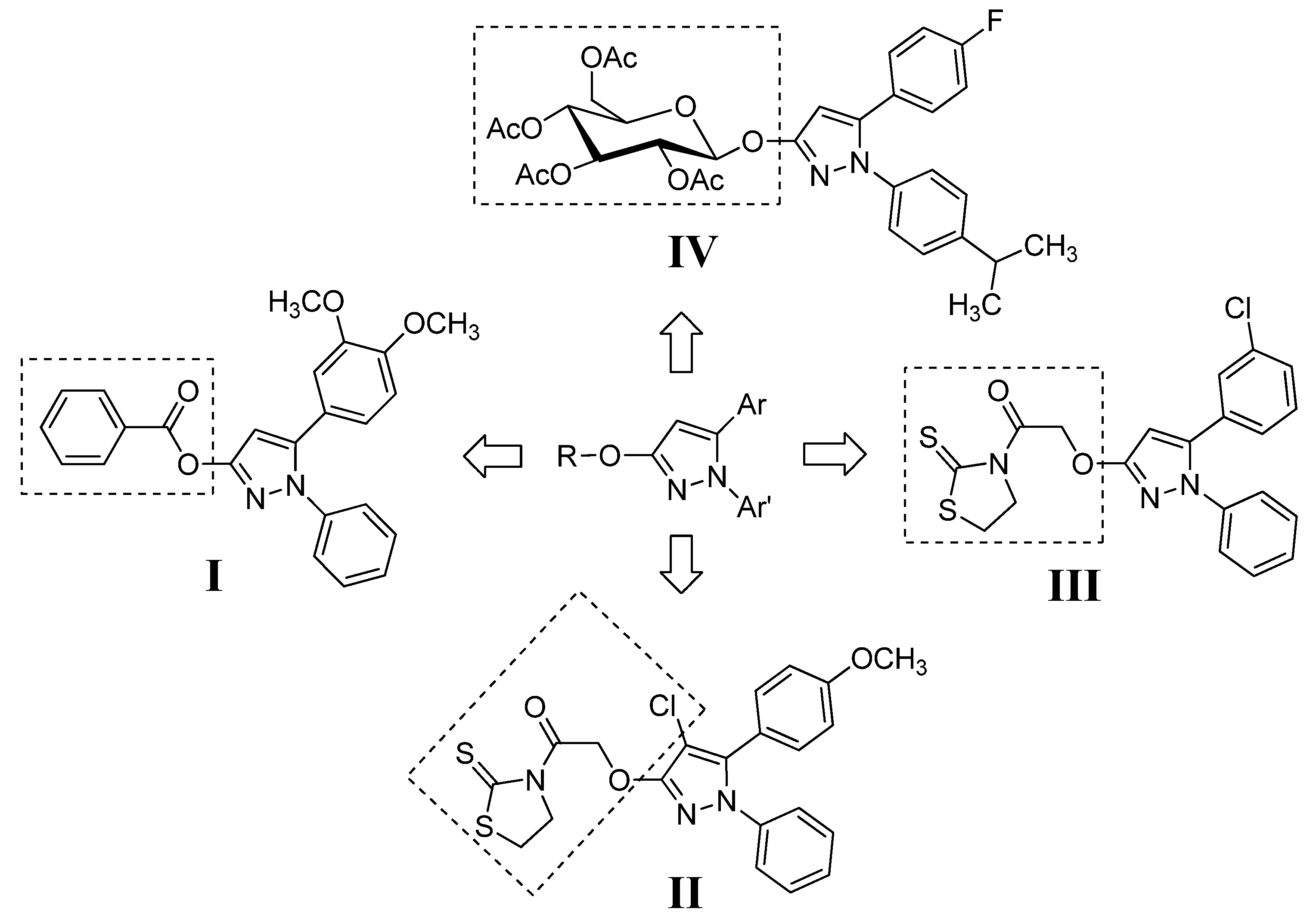
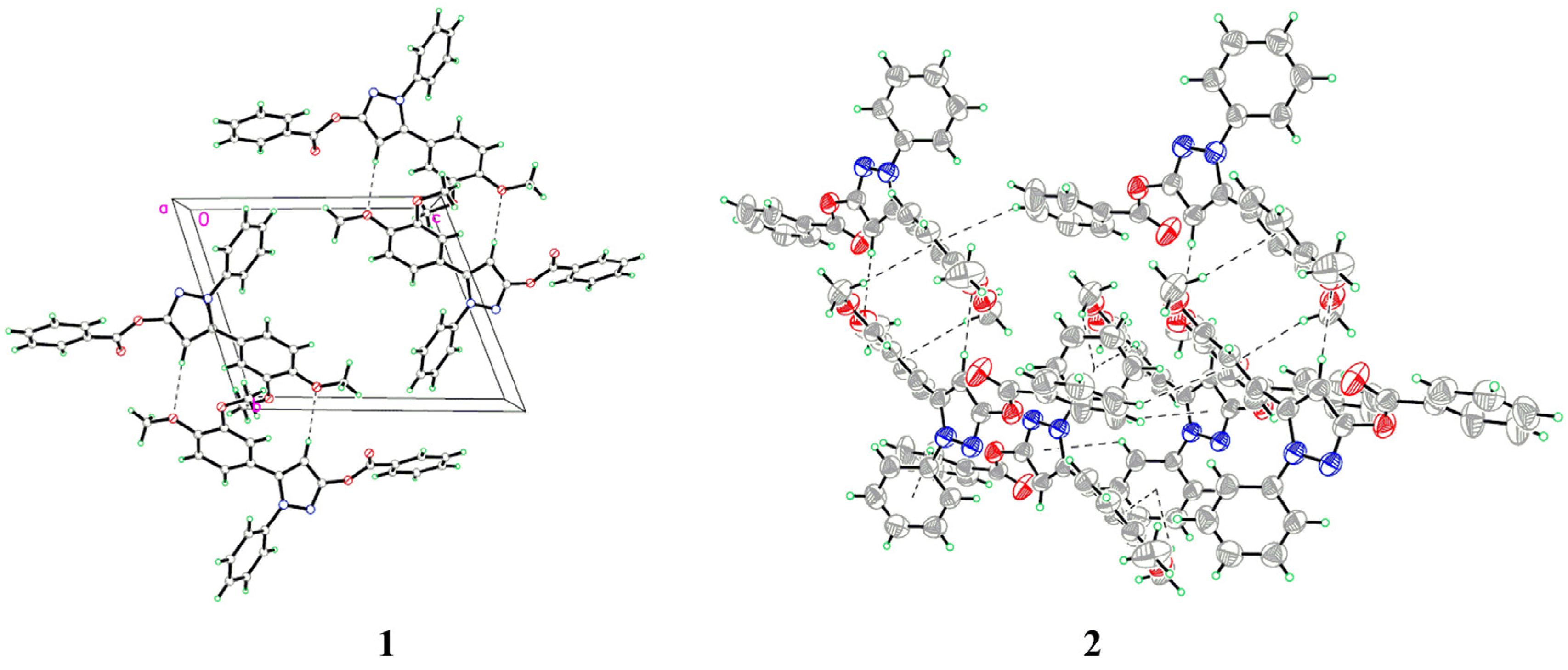
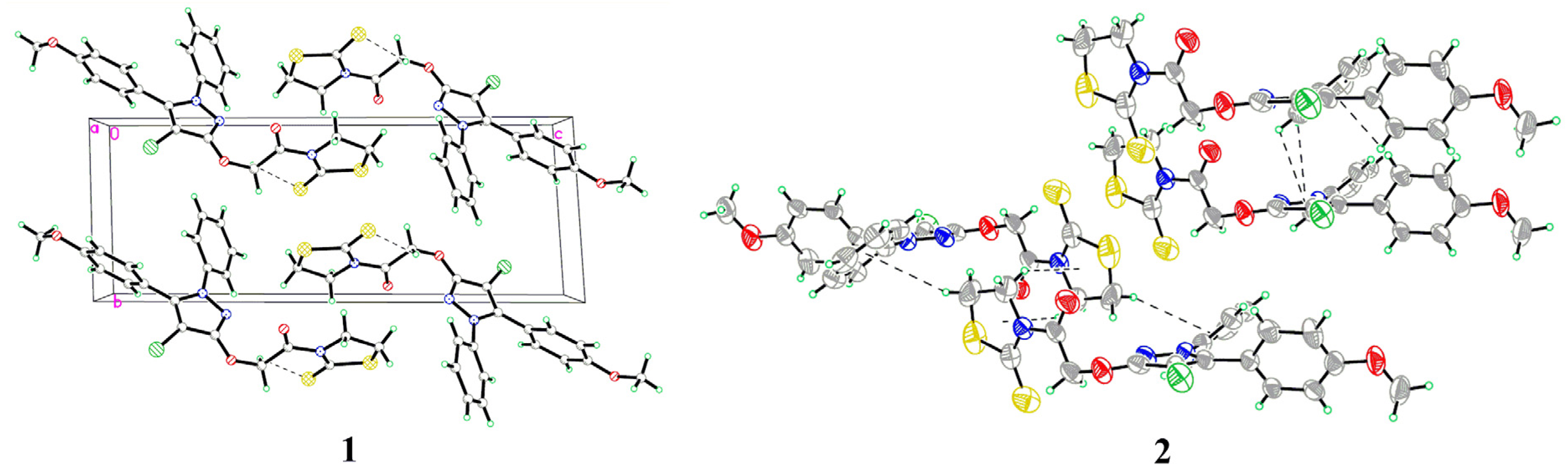
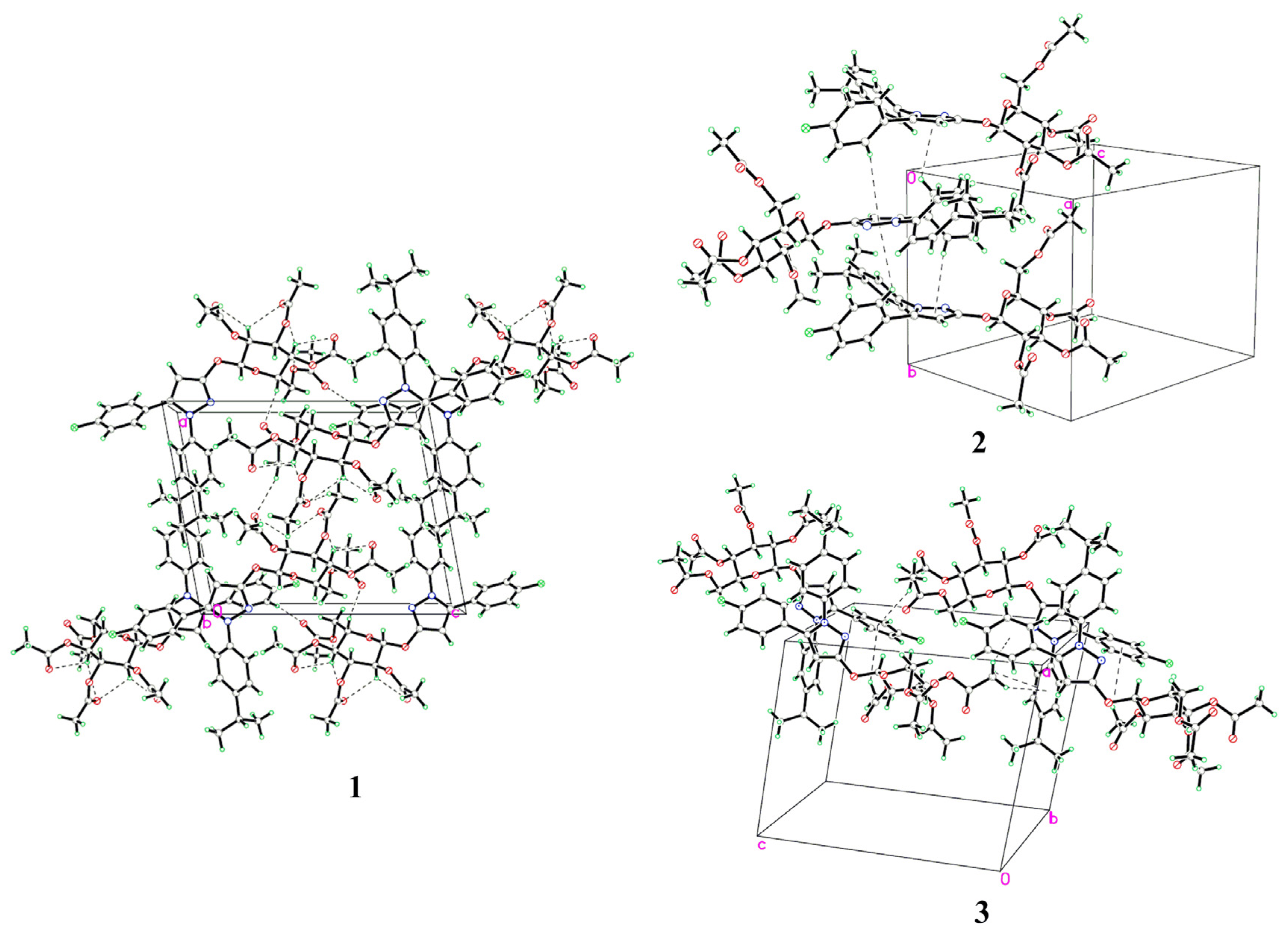
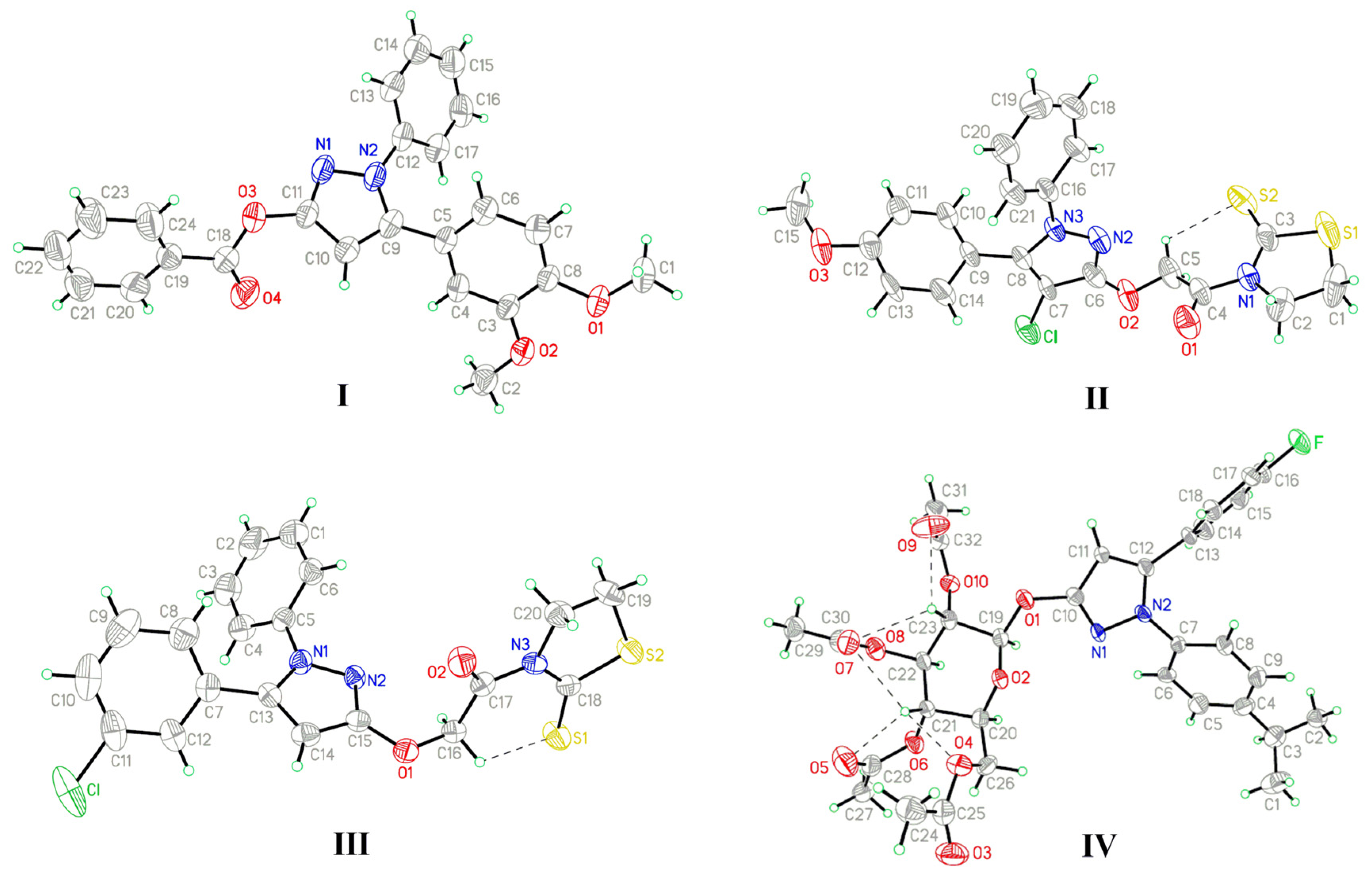
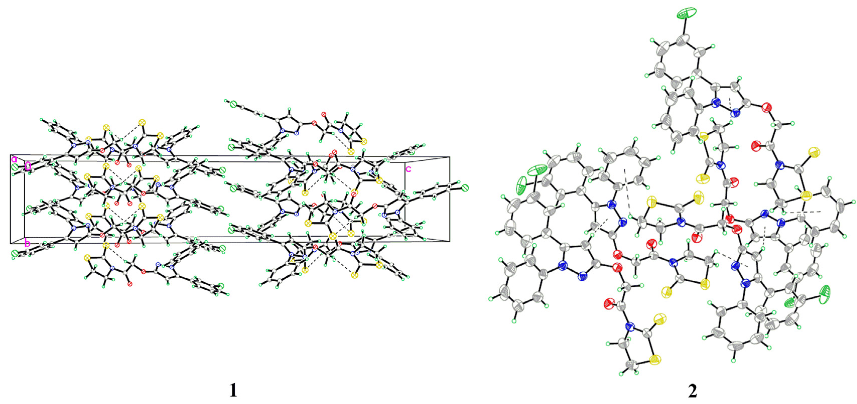
2.1. Structural Description
The detailed crystal and structure refinement data of I–IV are listed in Table 1. Compounds I and II crystallize in a triclinic p-1, whereas III and IV crystallize in an orthorhombic Pbca and a monoclinic P21 space groups, respectively. Selected bond lengths and angles/torsion angles for I–IV are given in Table 2. The bond lengths of N1-C4 (1.417(7) Å) and N3-C17 (1.412(6) Å) in II and III are longer than normal N-C amide bond (1.325-1.352 Å) [13]. The C7-Cl bond length in II is 1.732(6) Å, which is similar to the aryl-Cl value of 1.730(7) Å (C11-Cl) in III. The C16-F bond length in IV is 1.380(7) Å, representing a typical alkyl-F bond, and the value is similar to those (1.371(3) Å) reported for other related derivatives [14]. The bond angle of C9-C10-C11 in I is 104.2(3)°, whereas the corresponding bond angles in II–IV are 106.3(5)° (C6-C7-C8), 104.9(4)° (C13-C14-C15), and 104.4(5)° (C10-C11-C12), respectively. These values are similar to the typical angle values of five-membered rings (108.0°).
Table 1.
Crystal data and structure refinements for I, II, III and IV.
| I | II | III | IV | |
|---|---|---|---|---|
| CCDC | 906894 | 819778 | 918082 | 925316 |
| Empirical formula | C24H20N2O4 | C21H18ClN3O3S2 | C20H16ClN3O2S2 | C32H35FN2O10 |
| Formula weight | 400.42 | 459.95 | 429.93 | 626.62 |
| Temperature | 293(2) | 293(2) | 293(2) | 293(2) |
| Wavelength (Å) | 0.71073 | 0.71073 | 0.71073 | 0.71073 |
| Crystal system | Triclinic | Triclinic | Orthorhombic | Monoclinic |
| Space group | P-1 | P-1 | Pbca | P21 |
| a (Å) | 9.3040(19) | 6.0180(12) | 13.698(3) | 11.838(2) |
| b (Å) | 10.507(2) | 8.2370(16) | 7.6260(15) | 9.4500(19) |
| c (Å) | 12.075(2) | 21.600(4) | 38.908(8) | 14.514(3) |
| α (°) | 68.70(3) | 87.08(3) | 90.00 | 90.00 |
| β (°) | 81.40(3) | 87.46(3) | 90.00 | 100.41(3) |
| γ (°) | 67.99(3) | 82.30(3) | 90.00 | 90.00 |
| Volume (Å3) | 1,019.5(4) | 1,058.9(4) | 4,064.4(14) | 1,596.9(6) |
| Z | 2 | 2 | 8 | 2 |
| Calculated density (Mg/m3) | 1.304 | 1.443 | 1.405 | 1.303 |
| Absorption coefficient (mm−1) | 0.090 | 0.406 | 0.414 | 0.101 |
| F(000) | 420 | 476 | 1,776 | 660 |
| Crystal size (mm) | 0.10 × 0.20 × 0.30 | 0.05 × 0.05 × 0.10 | 0.10 × 0.20 × 0.30 | 0.10 × 0.20 × 0.30 |
| θ range for data collection(°) | 1.81–25.36 | 1.89–25.28 | 1.05–25.28 | 1.43–25.25 |
| Index ranges | 0 ≤ h ≤ 11 −11 ≤ k ≤ 12 −14 ≤ l ≤ 14 | 0 ≤ h ≤ 7 −9 ≤ k ≤ 9 −25 ≤ l ≤ 25 | 0 ≤ h ≤ 16 0 ≤ k ≤ 9 0 ≤ l ≤ 46 | −14 ≤ h ≤ 13 0 ≤ k ≤ 11 0 ≤ l ≤ 17 |
| Reflections collected | 4002 | 4239 | 3688 | 3091 |
| Independent reflections [R(int)] | 3750 [R(int) = 0.045] | 3843 [R(int) = 0.061] | 3688 [R(int) = 0.034] | 3091 [R(int) = 0.052] |
| Max. and min. transmission | 0.9911 and 0.9736 | 0.9800 and 0.9605 | 0.9597 and 0.8857 | 0.9900 and 0.9704 |
| Refinement method on F2 | Full-matrix least-squares | Full-matrix least-squares | Full-matrix least-squares | Full-matrix least-squares |
| Data/restraints/parameters | 3750/0/271 | 3843/0/272 | 3688/0/253 | 3091/7/388 |
| Goodness-of-fit on F2 | 1.005 | 1.002 | 1.032 | 1.004 |
| Final R indices [I > 2σ(I)]; R1, wR2 | R1 = 0.0688 wR2 = 0.1508 | R1 = 0.0854 wR2 = 0.1592 | R1 = 0.0740 wR2 = 0.1616 | R1 = 0.0656 wR2 = 0.1567 |
| R1, wR2 (all data) | R1 = 0.1452 wR2 = 0.1835 | R1 = 0.1766 wR2 = 0.1897 | R1 = 0.1359 wR2 = 0.1922 | R1 = 0.1000 wR2 = 0.1798 |
| Largest diff. peak and hole (e·Å−3) | 0.149 and −0.169 | 0.451 and −0.266 | 0.325 and −0.358 | 0.226 and −0.577 |
Table 2.
Selected bond lengths (Å), bond angles/torsion angles (°) for I, II, III, and IV.
| Comp. | Bond lengths (Å) | X-ray | Bond angles/Torsion angles (°) | X-ray |
|---|---|---|---|---|
| I | C11-O3 | 1.391(4) | C9-C10-C11 | 104.2(3) |
| N1-N2 | 1.374(3) | N2-N1-C11-O3 | 173.5(3) | |
| C5-C9 | 1.472(4) | N1-N2-C9-C5 | −172.8(3) | |
| O3-C18 | 1.360(4) | C1-O1-C8-C7 | 5.0(5) | |
| C2-O2-C3-C4 | −2.7(5) | |||
| II | C6-O2 | 1.326(8) | C6-C7-C8 | 106.3(5) |
| N1-C4 | 1.417(7) | N3-N2-C6-O2 | −179.7(6) | |
| C7-Cl | 1.732(6) | N2-N3-C8-C9 | −176.1(6) | |
| N2-N3 | 1.357(6) | C15-O3-C12-C11 | 1.1(11) | |
| C8-C9 | 1.479(8) | C4-N1-C3-S2 | −1.0(10) | |
| O2-C5 | 1.414(7) | |||
| III | C15-O1 | 1.351(5) | C13-C14-C15 | 104.9(4) |
| N3-C17 | 1.412(6) | N1-N2-C15-O1 | 178.1(4) | |
| C11-Cl | 1.730(7) | N2-N1-C13-C7 | 177.8(4) | |
| N1-N2 | 1.365(5) | C17-N3-C18-S1 | 9.4(7) | |
| C7-C13 | 1.477(7) | |||
| O1-C16 | 1.424(5) | |||
| IV | C10-O1 | 1.392(6) | C10-C11-C12 | 104.4(5) |
| C16-F | 1.380(7) | N2-N1-C10-O1 | 177.4(6) | |
| N1-N2 | 1.388(5) | N1-N2-C12-C13 | −176.3(6) | |
| C12-C13 | 1.499(7) | O1-C19-O2-C20 | −173.8(4) | |
| O1-C19 | 1.404(6) | O2-C19-C23-O10 | −174.2(4) | |
| C21-C20-C26-O4 | −61.0(6) | |||
| C19-O1-C10-C11 | 160.7(6) |
X-ray diffraction studies indicated that the dihedral angles between the C-linked benzene ring A and N-linked benzene ring B of I, II, III, and IV are 61.33°, 62.87°, 57.09°, and 70.25°, respectively. Rings A, B and benzoyloxy ring C in I are twisted 50.95°, 40.62°, and 64.18°, respectively, from the plane of the pyrazole ring. The dihedral angle between rings A and C is 32.20°, whereas the corresponding angle between rings B and C is 47.99°. The N2-N1-C11-O3 and N1-N2-C9-C5 torsion angles are 173.5(3)° and −172.8(3)°, whereas the corresponding angles in II, III, and IV are −179.7(6)° (N3-N2-C6-O2) and −176.1(6)° (N2-N3-C8-C9), 178.1(4)° (N1-N2-C15-O1) and 177.8(4)° (N2-N1-C13-C7), 177.4(6)° (N2-N1-C10-O1) and −176.3(6)° (N1-N2-C12-C13), respectively. The methoxyl groups in I and II are almost co-planar with the benzene ring. The torsion angles of C1-O1-C8-C7, C2-O2-C3-C4 and C15-O3-C12-C11 are found to be 5.0(5)°, −2.7(5)°, and 1.1(11)°, respectively, which are consistent with the literature value of 174.9(4)° [15]. Rings A, B and thiazolidine-2-thione ring D in II are twisted 64.12°, 38.34°, and 79.22°, respectively, from the plane of the pyrazole ring, whereas the corresponding angles in III are 45.00°, 50.49°, and 69.94°. Rings D in II and III both adopt envelope conformation, with the C1 and C19 atoms displaced by 0.240 Å and 0.368 Å from the plane of the other ring atoms. The torsion angle of C4-N1-C3-S2 in II is −1.0(10)°, whereas the corresponding torsion angle in III is 9.4(7)° (C17-N3-C18-S1). Rings A and B in IV are twisted 59.92° and 43.65°, respectively, from the plane of the pyrazole ring. An ORTEP view of IV reveals that the saccharide moiety in IV is a glucopyranose ring in the usual 4C1 conformation. The anomeric center of the saccharide has the β configuration, which is also confirmed from the torsion angles of O1-C19-O2-C20 −173.8(4)° and O2-C19-C23-O10 −174.2(4)°. The torsion angle C21-C20-C26-O4 of −61.0(6)° indicates the acetyl group attached to the primary hydroxyl group is in the gt position, which is known to be the favored orientation for a glucopyranose. The pyrazole ring is nearly coplanar with the anomeric C19 atom by making a torsion angle of C19-O1-C10-C11 160.7(6)°, and this orientation facilitates the delocalization of electrons from the lone-pair orbitals of O1 with the π orbitals of the pyrazole ring.
The atom numbering schemes and arrangements for I-IV are shown in Figure 2. The molecules are organized in the crystal lattice by classical intramolecular (C-H···S for II and III, and C-H···O for IV) and intermolecular (C-H···O for I and IV) H-bonds (Table 3a), as well as intermolecular C-H···π stacking interactions (Table 3b). For I (Figure 3), there is an intermolecular C-H···O H-bond, between the pyrazole hydrogen and the oxygen atom of the methoxyl group, as well as six C-H···π interactions, between the ring B hydrogen (H17A) and the center of the pyrazole ring (C17-H17A···Cg1), the methoxyl hydrogen (H2B), ring A hydrogen (H4A), or phenyl hydrogen (H24A) and the center of the ring B (C2-H2B···Cg2, C4-H4A···Cg2, and C24-H24A···Cg2), as well as the phenyl hydrogen (H22A) or the methoxyl hydrogen (H2C) to the center of the ring A (C22-H22A···Cg3 and C2-H2C···Cg3), to form a three-dimensional network. For II (Figure 4), the intramolecular C5-H5A···S2 H-bond results in the formation of one non-planar pseudo ring (C5/H5A/S2/C3/N1/C4), with H5A atom displaced by 0.580 Å from the plane of the other ring atoms. Five C-H···π interactions, between the methylene hydrogen (H2C) and the center of the ring D (C2-H2C···Cg4), the methylene hydrogen (H1A) or the phenyl hydrogen (H14A) and the center of the ring B (C1-H1A···Cg2 and C14-H14A···Cg2), and the phenyl hydrogens (H17A and H18A) and the center of the pyrazole ring (C17-H17A···Cg1 and C18-H18A···Cg1), form dimers. For III (Figure 5), the structure is stabilized by the intramolecular C-H···S H-bond, between the methylene hydrogen and the sulfur atom of the ring D to form a non-planar pseudo ring (H16B/C16/C17/N3/C18/S1) bearing envelope conformation, with H16B atom displaced by 0.604 Å from the plane of the other atoms, and three C-H···π interactions, one is between the methylene hydrogen (H19B) and the center of the ring B (C19-H19B···Cg2), and the others are from the pyrazole hydrogen (H14A) or the methylene hydrogen (H19C) to the center of the pyrazole ring (C14-H14A···Cg1 and C19-H19C···Cg1). For IV (Figure 6), five intra- and four intermolecular H-bonds, and six C-H···π interactions, between the phenyl hydrogens (H14A and H18A) to the center of the pyrazole ring (C14-H14A···Cg1 and C18-H18A···Cg1), and the methyl hydrogens (H27A-C and H31C) to the centers of the rings A or B (C27-H27A···Cg2, C27-H27C···Cg2, C27-H27B···Cg3, and C31-H31C···Cg3), reinforce the crystal packing.
Table 3.
Parameters (Å, °) for the intra- and intermolecular interactions in I, II, III, and IV.
| Comp. | D-H…A | D-H | H···A | D···A | D-H···A |
|---|---|---|---|---|---|
| (a) Intermolecular and intramolecular hydrogen bond | |||||
| I | C10-H10A···O1 a | 0.9300 | 2.5100 | 3.442(5) | 175.00 |
| II | C5-H5A···S2 | 0.9700 | 2.5900 | 3.033(8) | 108.00 |
| III | C16-H16B···S1 | 0.9700 | 2.5800 | 3.094(5) | 113.00 |
| IV | C21-H21A···O4 | 0.9800 | 2.5300 | 2.897(8) | 102.00 |
| C21-H21A···O5 | 0.9800 | 2.2700 | 2.645(8) | 102.00 | |
| C21-H21A···O7 | 0.9800 | 2.4000 | 2.975(8) | 117.00 | |
| C23-H23A···O7 | 0.9800 | 2.4000 | 2.958(7) | 116.00 | |
| C23-H23A···O9 | 0.9800 | 2.2600 | 2.659(9) | 103.00 | |
| C17-H17A···O3 b | 0.9300 | 2.5200 | 3.440(10) | 170.00 | |
| C20-H20A···O3 c | 0.9800 | 2.4100 | 3.363(9) | 163.00 | |
| C24-H24B···O9 d | 0.9600 | 2.5100 | 3.375(11) | 150.00 | |
| C29-H29C···O7 e | 0.9600 | 2.4200 | 3.320(10) | 155.00 | |
| Comp. | C-H···Cg | C-H | H···Cg | C···Cg | C-H···Cg |
| (b) C-H…π interactions | |||||
| I | C17-H17A···Cg1 f | 0.9300 | 3.2873 | 3.900(4) | 125.39 |
| C2-H2B···Cg2 f | 0.9600 | 3.1975 | 4.050(5) | 148.87 | |
| C4-H4A···Cg2 f | 0.9300 | 3.3208 | 4.113(4) | 144.40 | |
| C24-H24A···Cg2 g | 0.9300 | 3.3983 | 4.143(5) | 138.64 | |
| C2-H2C···Cg3 a | 0.9600 | 2.9016 | 3.706(5) | 142.02 | |
| C22-H22A···Cg3 h | 0.9300 | 3.1317 | 3.866(6) | 137.15 | |
| II | C2-H2C···Cg4 i | 0.9700 | 3.0259 | 3.812(8) | 139.03 |
| C1-H1A···Cg2 i | 0.9700 | 3.2864 | 3.927(8) | 125.25 | |
| C14-H14A···Cg2 j | 0.9300 | 3.0647 | 3.906(7) | 151.31 | |
| C17-H17A···Cg1 k | 0.9300 | 3.2738 | 3.550(7) | 99.63 | |
| C18-H18A···Cg1 k | 0.9300 | 3.0448 | 3.418(9) | 105.87 | |
| III | C19-H19B···Cg2 l | 0.9700 | 3.3425 | 4.032(6) | 129.74 |
| C14-H14A···Cg1 m | 0.9300 | 2.8623 | 3.725(6) | 154.68 | |
| C19-H19C···Cg1 n | 0.9700 | 3.3978 | 3.940(6) | 117.46 | |
| IV | C14-H14A···Cg1 o | 0.9300 | 3.2004 | 3.780(9) | 122.32 |
| C18-H18A···Cg1 p | 0.9300 | 3.3184 | 3.865(7) | 119.69 | |
| C27-H27A···Cg2 c | 0.9600 | 3.0282 | 3.571(9) | 117.24 | |
| C27-H27C···Cg2 c | 0.9600 | 3.2317 | 3.571(9) | 102.94 | |
| C27-H27B···Cg3 q | 0.9600 | 2.7786 | 3.647(9) | 150.82 | |
| C31-H31C···Cg3 o | 0.9600 | 3.3687 | 4.192(9) | 144.97 | |
Symmetry codes: a 1-x, 2-y, −z; b x, y, −1+z; c 1-x, 1/2+y, 1-z; d 2-x, −1/2+y, 1-z; e 2-x, 1/2+y, 1-z; f 1-x, 1-y, −z; g 2-x, 1-y, −z; h 1+x, y, −1+z; i 1-x, -y, 1-z; j 1+x, y, z; k −1+x, y, z; l 2-x, 1/2+y, 1/2-z; m 3/2-x, 1/2+y, z; n 2-x, −1/2+y, 1/2-z; o 1-x, 1/2+y, −z; p 1-x, −1/2+y, −z; q x, y, 1+z; Cg1, Cg2, Cg3, and Cg4 are the centroids of the pyrazole ring, N-linked benzene ring B, C-linked benzene ring A, and thiazolidine-2-thione ring D, respectively.
Figure 2.
X-ray crystal structures of I, II, III, and IV.
Figure 3.
(1) A packing diagram of I; (2) Intermolecular C-H···π interactions of I.
Figure 4.
(1) A packing diagram of II; (2) Intermolecular C-H···π interactions of II.
Figure 5.
(1) A packing diagram of III; (2) Intermolecular C-H···π interactions of III.
Figure 6.
(1) A packing diagram of IV; (2) Intermolecular C-H···π (pyrazole) interactions of IV; (3) Intermolecular C-H···π (phenyl) interactions of IV.
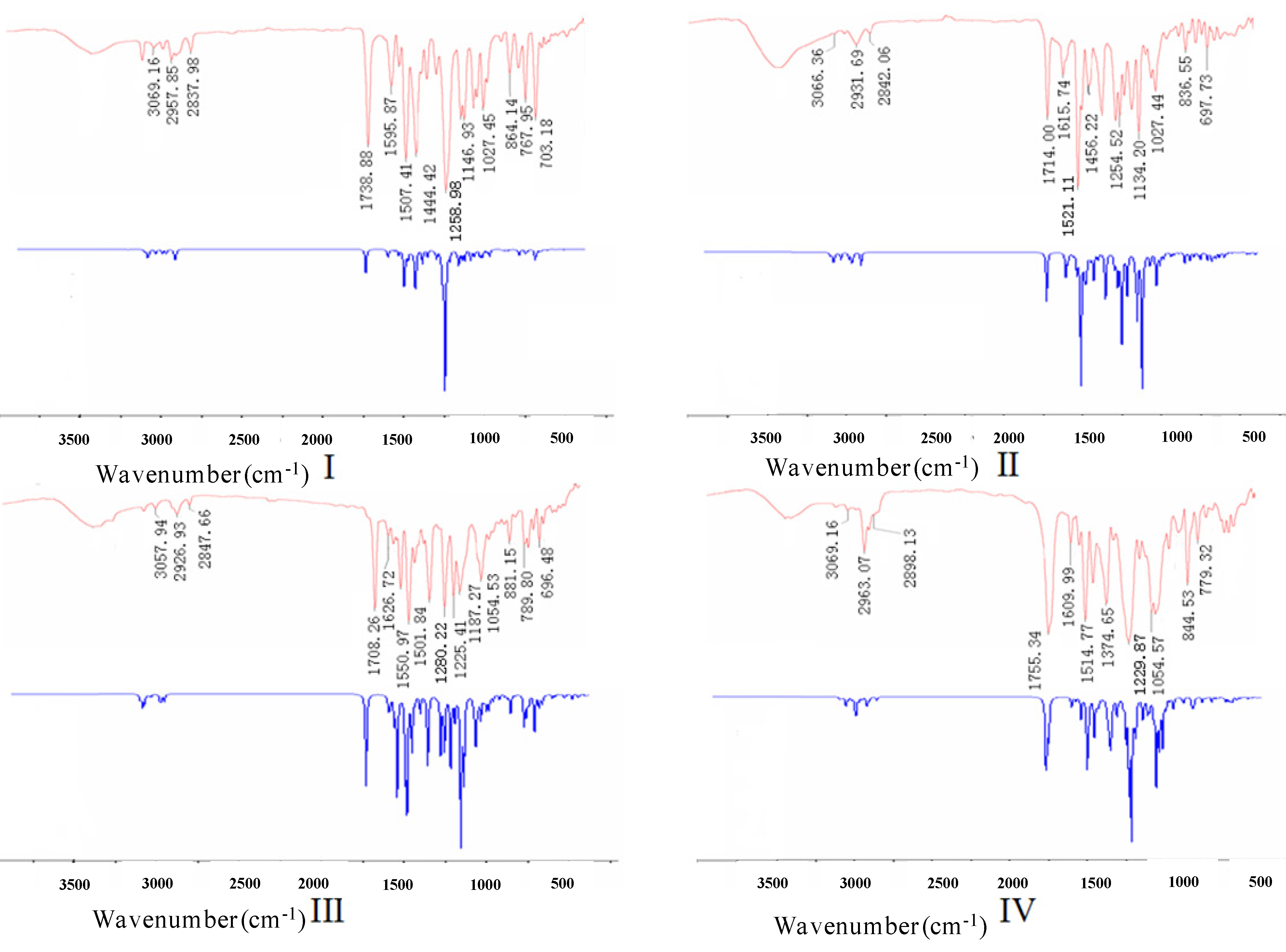
2.2. Experimental and Theoretical FTIR Results
The experimental and theoretical FTIR spectra for I–IV are shown in Figure 7, where the intensity is plotted against the vibrational frequencies. The primary vibrational frequencies with assignments are listed in Table 4. The main signals are grouped in three regions, including the ranges of 2800–3200 cm−1, 1000–1800 cm−1 and 500–1000 cm−1. The second region shows strongly mixed vibrational bands. The C=O stretching vibrations of I–IV were found at 1739 cm−1, 1714 cm−1, 1708 cm−1, and 1755 cm−1, whereas they were calculated at 1737 cm−1, 1718 cm−1, 1711 cm−1, and 1752 cm−1, respectively. The experimental and theoretical C=C stretching vibrations of the phenyl rings were found in the region of 1440–1630 cm−1, which were consistent with the literature value of 1,430–1,625 cm−1 [16]. The bands observed in the region of 1000–1300 cm−1 corresponded to the symmetric and asymmetric C-O stretching vibrations. The region below 1000 cm−1 exhibited the out of plane bending C-H vibrations of the aromatic rings, and the region in 3000–3200 cm−1 was the characteristic absorption of the aromatic C-H stretching vibrations. The C-Cl stretching vibration in II was consistent with the aromatic C-Cl stretching vibration in III. The experimental C-F stretching vibration at 1330 cm−1 in IV was consistent with its theoretical value of 1332 cm−1. These results indicated that the observed and calculated FTIR data were in good agreement with each other.
Table 4.
Primary vibrations of experimental and theoretical FTIR for I, II, III, and IV (cm−1).
| Vibration | I | II | III | IV | ||||
|---|---|---|---|---|---|---|---|---|
| Exp. | B3LYP/6-31G * | Exp. | B3LYP/6-31G * | Exp. | B3LYP/6-31G * | Exp. | B3LYP/6-31G * | |
| ν =CH | 3069 | 3090 | 3066 | 3076 | 3058 | 3073 | 3069 | 3068 |
| νC-H | 2958 | 2953 | 2932 | 2935 | 2927 | 2936 | 2963 | 2958 |
| 2838 | 2842 | 2842 | 2847 | 2848 | 2845 | 2898 | 2896 | |
| νC=O | 1739 | 1737 | 1714 | 1718 | 1708 | 1711 | 1755 | 1752 |
| νC=C | 1596 | 1593 | 1616 | 1624 | 1627 | 1623 | 1610 | 1603 |
| 1549 | 1548 | 1521 | 1521 | 1595 | 1604 | 1556 | 1555 | |
| 1507 | 1509 | 1456 | 1458 | 1551 | 1552 | 1515 | 1514 | |
| 1444 | 1440 | 1502 | 1508 | 1466 | 1463 | |||
| 1466 | 1459 | |||||||
| νC-O | 1259 | 1264 | 1280 | 1267 | 1280 | 1266 | 1230 | 1234 |
| 1167 | 1168 | 1255 | 1267 | 1225 | 1240 | 1161 | 1163 | |
| 1147 | 1130 | 1226 | 1222 | 1187 | 1183 | 1080 | 1081 | |
| 1088 | 1095 | 1179 | 1185 | 1054 | 1053 | 1055 | 1054 | |
| 1066 | 1071 | 1134 | 1147 | |||||
| 1027 | 1020 | 1027 | 1024 | |||||
| γ=C-H | 864 | 869 | 837 | 828 | 881 | 888 | 845 | 846 |
| 811 | 807 | 773 | 782 | 790 | 787 | 779 | 770 | |
| 768 | 764 | 698 | 695 | 767 | 774 | |||
| 703 | 703 | 696 | 701 | |||||
| 672 | 676 | |||||||
| νC-Cl | 734 | 736 | 734 | 737 | ||||
| νC-F | 1330 | 1332 | ||||||
Figure 7.
Experimental (above) and theoretical (below) FTIR spectra for I, II, III, and IV.
2.3. Fungicidal Activity
Compounds I–IV were evaluated for in vitro fungicidal activity against Sclerotinia sclerotiorum and Gibberella zeae, at a dosage of 10 μg/mL. As can be seen in Table 5, for Sclerotinia sclerotiorum, Compound IV (29%) possessing a per-O-acetylated glucopyranosyl moiety, displayed better activity than I (0%), II (21%), and III (14%).
Table 5.
In vitro fungicidal activities of I, II, III, and IV (% inhibition).
| Comp. | X | Y | Inhibition rate (%) | |
|---|---|---|---|---|
| S. sclerotiorum | G. zeae | |||
| I | 3,4-(OCH3)2 | H | 0 | 4 |
| II | p-OMe | H | 21 | 32 |
| III | m-Cl | H | 14 | 29 |
| IV | p-F | p-Me2CH | 29 | 11 |
The reason was speculated that the glucopyranosyl moiety could form more H-bonds (Figure 2) to improve the hydrophilicity of molecule, which could balance the HLB value and then increase the systemic of molecule within plant. Within the series of thiazolidinethione derivatives, compound II (21%, 32%) with an electron-withdrawing chloro group on the pyrazole ring displayed better fungicidal activity against the two fungi than III (14%, 29%), and both compounds showed better inhibitory activity against Gibberella zeae than I (4%) and IV (11%). The results might imply that the introduction of the heterocycle moiety by full consideration of the electronic effects was important for improving its fungicidal activity. However, compound I containing a benzoyloxy moiety showed the worst activity (0, 4%), which indicated switching the C3-substituent of the pyrazole ring from thiazolidinethione to benzoyloxy moiety had no effective impact on the inhibition rates. The structure-activity relationship revealed that the improvement of bioactivity might require a reasonable design of molecules (e.g., considering H-bonds effect and heterocycle) and full consideration of the electronic effects of electron-withdrawing groups to balance the HLB value and enhance the systemic of the whole molecule.
3. Experimental
3.1. General Information
Melting points were measured on an X-4 microscope electrothermal apparatus (Taike, Nanjing, China) and were uncorrected. 1H-NMR spectra were recorded on a Bruker spectrometer (Bruker, Leipzig, Germany) at 300 or 500 MHz using CDCl3 or DMSO-d6 as solvent, with tetramethylsilane as an internal standard. Elemental analyses were performed on a Flash EA-1112 elemental analyzer (Thermo, Illinois, USA). FTIR spectra of compounds I–IV were recorded in the region of 4000–400 cm−1 on a Nicolet 380 FT-IR spectrophotometer (Thermo, Waltham, MA, USA) using the KBr pellet technique.
3.2. Synthesis and Characterization
Compounds I–IV were prepared from 1,5-diaryl-1H-pyrazol-3-ols via a series of reactions that included substitution, hydrolysis, condensation, chlorination, glycosylation, or esterification [8,9,10,11,12].
3.2.1. Preparation of Compound I
To a solution of 5-(3,4-dimethoxyphenyl)-1-phenyl-1H-pyrazol-3-ol (0.59 g, 2.0 mmol) in CHCl3 (50 mL) was added Et3N (0.41 g, 4.0 mmol). The mixture was stirred for 10 min and benzoyl chloride (0.42 g, 3.0 mmol) was added. Then, the mixture was stirred at r.t. for 2 h. The solvent was removed under reduced pressure and the residue was recrystallized from ethanol to give the white solid product 5-(3,4-dimethoxyphenyl)-1-phenyl-3-benzoyloxy-1H-pyrazole (I). Yield: 87%; M.p. 133–134 °C; 1H-NMR (300 MHz, DMSO-d6) δ: 8.16 (d, J = 7.2 Hz, 2H, Ar-H), 7.79 (t, J = 7.3 Hz, 1H, Ar-H), 7.65 (t, J = 7.3 Hz, 2H, Ar-H), 7.48–7.31 (m, 5H, Ar-H), 7.00–6.80 (m, 3H, Ar-H), 6.67 (s, 1H, CH), 3.76 (s, 3H, OCH3), 3.57 (s, 3H, OCH3); Anal. Calcd for C24H20N2O4: C 71.99, H 5.03, N 7.00; found C 71.75, H 5.01, N 7.03.
3.2.2. Preparation of Compound II
SOCl2 (5 mL) was put into a round-bottom flask and then 2-((5-(4-methoxyphenyl)-1-phenyl-1H-pyrazol-3-yl)oxy)-1-(2-thioxothiazolidin-3-yl)ethanone (0.43 g, 1.0 mmol) as well as catalytic amount of DMF (0.1 mmol) was added. The mixture was heated under reflux for 4 h. Then excess SOCl2 was evaporated under reduced pressure and H2O (200 mL) was added with good stirring. The precipitate was filtered off, washed with water and dried. It was then purified by flash column chromatography (eluent: ethyl acetate/petroleum ether, 1:6 v/v) to afford the yellow solid product 2-((4-chloro-5-(4-methoxyphenyl)-1-phenyl-1H-pyrazol-3-yl)oxy)-1-(2-thioxothiazolid-in-3-yl)ethanone (II). Yield: 65%; M.p. 193–194 °C; 1H-NMR (500 MHz, CDCl3) δ: 7.26–7.15 (m, 7H, Ar-H), 6.88 (d, J = 8.8 Hz, 2H, Ar-H), 5.78 (s, 2H, CH2), 4.61 (t, J = 7.6 Hz, 2H, CH2), 3.82 (s, 3H, OCH3), 3.38 (t, J = 7.6 Hz, 2H, CH2); Anal. Calcd for C21H18ClN3O3S2: C 54.84, H 3.94, N 9.14; found C 54.95, H 3.95, N 9.17.
3.2.3. Preparation of Compound III
2-((5-(3-Chlorophenyl)-1-phenyl-1H-pyrazol-3-yl)oxy)acetic acid (0.33 g, 1.0 mmol) was dissolved in a solution of DCC (0.22 g, 1.05 mmol) in CH2Cl2 (50 mL), and the mixture was stirred at 0 °C for 1 h. Then, thiazolidine-2-thione (0.12 g, 1.0 mmol) and DMAP (0.01 g, 0.1 mmol) was added. The solution was stirred at 0 °C for 2 h and then at room temperature for 12 h. The white precipitate was filtered off and the solvent was evaporated under reduced pressure. The residue was purified by flash column chromatography over silica gel eluting with petroleum ether/ethyl acetate 3:1 to gain the the yellow solid product 2-((5-(3-chlorophenyl)-1-phenyl-1H-pyrazol-3-yl)oxy)-1-(2-thioxothiazolidin-3-yl)ethanone (III). Yield: 73%; M.p. 143–144 °C; 1H-NMR (500 MHz, CDCl3) δ: 7.30–7.05 (m, 9H, Ar-H), 6.05 (s, 1H, CH), 5.72 (s, 2H, CH2), 4.60 (t, J = 7.6 Hz, 2H, CH2), 3.37 (t, J = 7.6 Hz, 2H, CH2); Anal. Calcd for C20H16ClN3O2S2: C 55.87, H 3.75, N 9.77; found C 55.78, H 3.74, N 9.80.
3.2.4. Preparation of Compound IV
A mixture of CHCl3 (20 mL), Bu4N+Br− (0.23 g, 0.7 mmol), and H2O (2 mL) was heated to 55 °C, and then a solution of 1-(4-fluorophenyl)-5-(4-isopropylphenyl)-1H-pyrazol-3-ol (0.30 g, 1.0 mmol) in CHCl3 (15 mL) and 5% aqueous NaOH (6 mL) were added dropwise. The pH value was adjusted to 8–10, and acetobromo-α-D-glucose (0.54 g, 1.3 mmol) was added under vigorously stirring. The mixture was stirred at 55 °C for another 4 h, and then left to cool to room temperature. The organic layer was separated, washed with 5% aqueous NaOH, and dried. Then, the solvent was removed in vacuo and the residue was recrystallized from ethanol to give the white solid product 3-(2′,3′,4′,6′-tetra-O-acetyl-β-D-glucopyranosyloxy)-1-(4-isopropylphenyl)-5-(4-fluorophenyl)-1H-pyrazole (IV). Yield: 46%; M.p. 151–152 °C. 1H-NMR (500 MHz, CDCl3) δ: 7.26–6.97 (m, 8H, Ar-H), 5.99 (s, 1H, CH), 5.67 (d, J = 7.7 Hz, 1H, H1′), 5.32–5.26 (m, 2H, H2′, H3′), 5.19 (t, J = 9.5 Hz, 1H, H4′), 4.28 (dd, J = 4.7, 12.4 Hz, 1H, H6′b), 4.18 (dd, J = 2.4, 12.4 Hz, 1H, H6′a), 3.91–3.88 (m, 1H, H5′), 2.93–2.87 (m, 1H, CH), 2.05, 2.04, 2.03, 2.01 (4× s, 12H, 4× COCH3), 1.23 (d, J = 6.9 Hz, 6H, CH3); Anal. Calcd for C32H35FN2O10: C 61.34, H 5.63, N 4.47; found C 61.52, H 5.61, N 4.45.
3.3. X-ray Crystallography
Suitable crystals of I–IV were obtained by slow evaporation of ethyl acetate solutions at r.t. Crystal data were performed on a Nonius CAD-4 diffractometer (Enraf-Nonius, Rotterdam, The Netherlands) by using MoKα (λ = 0.71073 Å) irradiation. All of the structures were solved by direct methods using SHELXS-97 and refined by full-matrix least-squares on F2 for all data using SHELXL-97 [17]. All non-H-atoms were refined anisotropically, and H-atoms were introduced at calculated positions. The isotropic temperature factors were fixed to 1.2 times (1.5 times for methyl group) the equivalent isotropic displacement parameters of the C-atom the H-atom is attached to CCDC-906894 (I), CCDC-819778 (II), CCDC-918082 (III) and CCDC-925316 (IV) contain the supplementary crystallographic data for this paper. These data can be obtained free of charge via http://www.ccdc.cam.ac.uk/conts/retrieving.html (or from the CCDC, 12 Union Road, Cambridge CB2 1EZ, UK; Fax: +44 1223 336033; E-mail: deposit@ccdc.cam.ac.uk).
3.4. FTIR Spectra
The structures in the ground state (in vacuo) were optimized by the Gaussian 03 program using the B3LYP (DFT) method with the 6-31G (d) basis set [18,19,20]. The initial configurations for calculation were constructed according to the X-ray data. Frequency calculations at the same levels of theory revealed no imaginary frequencies, indicating that the B3LYP/6-31G (d) method was the optimal one in our system.
3.5. Fungicidal Activity Assays
The in vitro fungicidal activity of compounds I–IV against Sclerotinia sclerotiorum and Gibberella zeae was investigated at a dosage of 10 μg/mL, according to a reported method [10]. The fungi were obtained from Jiangsu Pesticide Research Institute Co., Ltd., Nanjing, China. The tested compounds I–IV were dissolved in acetone and added to a sterile agarized Czapek-Dox medium at 45 °C. In preliminary screenings, the compounds were used in a concentration of 10 μg/mL. The control sample contained only one equivalent of acetone. The media were poured onto 8-cm Petri dishes (10 mL for each dish) and after 2 days inoculated with 5-mm PDA discs of overgrown mycelium. In the case of Sclerotinia sclerotiorum, the medium was inoculated by a prick of laboratory needle containing fungus spores. The Petri dishes were incubated at r.t. in the dark. After 4 days, the diameters of the inoculation of the cultures were measured. The percentage inhibition of fungal growth was determined by comparison between the development of fungi colonies on media containing compounds and on the control. Three replicates of each test were carried out.
4. Conclusions
Four 1,5-diaryl-3-oxypyrazoles containing benzoyl (I), thiazolidinethione (II and III) or per-O-acetylated glucopyranosyl (IV) moieties have been analyzed by X-ray diffraction. The molecules were stabilized by classical intra- and intermolecular H-bonds, as well as intermolecular C-H···π stacking interactions. Compound IV with more H-bonds in the crystal displayed better activity (29%) against Sclerotinia sclerotiorum than I (0%), II (21%), and III (14%). Compound II (21%, 32%) showed better fungicidal activity against the two fungi than III (14%, 29%), and both II and III exhibited better inhibitory activity against Gibberella zeae than I (4%) and IV (11%). The structure-activity relationship revealed that the improvement of bioactivity might require full consideration of H-bonds effect, heterocycle, and electronic effects of electron-withdrawing groups to balance the HLB value and enhance the systemic of molecule.
Acknowledgments
This work was supported by the Project of Food Fast Detection Technology (BM2012026), the supporting project of the Twelfth Five Year Plan of China (2012BAK17B09), the Science Foundation for Young Scholars of Jiangsu Province of China (BK20130749), and the Youths Foundation of Southeast University ChengXian College (7303600001).
Conflicts of Interest
The authors declare no conflict of interest.
References
- Stierl, R.; Scherer, M.; Schrof, W.; Butterfield, E.J. Activity of the New BASF Strobilurin Fungicide, BAS 500F, Against Plasmopara viticola on Grapes. In The BCPC Conference: Pests and Diseases; Proceedings of the International Conference Held at the Brighton Hilton Metropole Hotel, Brighton, UK, 13–16 November 2000; pp. 261–266.
- Ammermann, E.; Lorenz, G.; Schelberger, K.; Mueller, B.; Kirstgen, R.; Sauter, H. BAS 500F: The new broad-spectrum strobilurine fungicide. In Proceedings of the International Conference Held at the Brighton Hilton Metropole Hotel, Brighton, UK, 13–16 November 2000; pp. 541–548.
- Stierl, R.; Merk, M.; Schrof, W.; Butterfield, E.J. Activity of the new BASF strobilurin fungicide, BAS 500F, against Septoria tritici on wheat. In Proceedings of the International Conference Held at the Brighton Hilton Metropole Hotel, Brighton, UK, 13–16 November 2000; pp. 859–864.
- Li, Y.; Liu, R.; Yan, Z.; Zhang, X.; Zhu, H. Synthesis, crystal structure and fungicidal activities of new type oxazolidinone-based strobilurin analogues. Bull. Korean Chem. Soc. 2010, 31, 1–7. [Google Scholar]
- Zhu, H.; Shi, H.; Jia, H.; Li, Y.; Song, G.; Liu, H.; Sun, Y.; Wang, J. Pyrazoleoxy Acetic Acid Compounds, Preparation Method and Use. Chinese Patent CN 101284815 A, 15 October 2008. [Google Scholar]
- Konno, T.; Kuriyama, K.; Hamaguchi, H.; Kajihara, O. Fenpyroximate (NNI 850), a new acaricide. Brighton Crop Prot. Conf. Pests Dis. 1990, 1, 71–78. [Google Scholar]
- Miura, Y.; Mabuchi, T.; Kajioka, M.; Yanai, I. 3-(Substituted Phenyl) Pyrazole Derivatives, Salts Thereof, Herbicides Therefrom, and Process for Producing Said Derivatives or Salts. Eur. Pat 0361114 A1, 4 April 1990. [Google Scholar]
- Liu, Y.; Shi, H.; Li, Y.; Zhu, H. Synthesis, crystal structure and fungicidal activity of novel 1,5-diaryl-1H-pyrazol-3-oxyacetate derivatives. J. Heterocycl. Chem. 2010, 47, 897–902. [Google Scholar] [CrossRef]
- Liu, Y.; He, G.; Kai, C.; Li, Y.; Zhu, H. Synthesis, crystal structure, and fungicidal activity of novel 1,5-diaryl-1H-pyrazol-3-oxy derivatives containing oxyacetic acid or oxy(2-thioxothiazolidin-3-yl) ethanone moieties. J. Heterocycl. Chem. 2012, 49, 1370–1375. [Google Scholar] [CrossRef]
- Liu, Y.; Shi, H.; He, G.; Song, G.; Zhu, H. Synthesis, crystal Structures, and fungicidal activity of novel 1,5-diaryl-3-(glucopyranosyloxy)-1H-pyrazoles. Helv. Chim. Acta 2012, 95, 1645–1656. [Google Scholar] [CrossRef]
- Liu, Y.Y.; Li, Y.; Zhu, H.J.; Zhang, Z.; Xu, G.H.; Chen, N.Q. Benzoyloxy Pyrazol Compounds, Preparation Method and Use. CN 102993099, 27 March 2013. [Google Scholar]
- Liu, Y.; He, G.; Chen, K.; Jin, Y.; Li, Y.; Zhu, H. DMF-Catalyzed direct and regioselective C–H functionalization: Electrophilic/nucleophilic 4-halogenation of 3-oxypyrazoles. Eur. J. Org. Chem. 2011, 27, 5323–5330. [Google Scholar]
- Goodman, M.; Ganis, P.; Avitabile, G.; Migdal, S. Solid-state conformation of amide groups. Crystal structures of N-ethyl-N-p-nitrophenylcarbamoyl chloride and of N-phenylurethane. J. Am. Chem. Soc. 1971, 93, 3328–3331. [Google Scholar] [CrossRef]
- Boxer, M.B.; Akakura, M.; Yamamoto, H. Ketone super silyl enol ethers in sequential reactions: Diastereoselective generation of tertiary carbinols in one pot. J. Am. Chem. Soc. 2008, 130, 1580–1582. [Google Scholar] [CrossRef]
- Jia, H.; Li, Y.; Liu, Y.; Liu, S.; Zhu, H. 1-(4-Isopropylphenyl)-5-(4-methoxyphenyl) pyrazolidin-3-one. Acta Cryst. 2008, 64, o855. [Google Scholar]
- Varsanyi, G. Assignments for Vibrational Spectra of Seven Hundred Benzene Derivatives; Hilger, A., Ed.; Wiley: New York, NY, USA, 1974. [Google Scholar]
- Sheldrick, G.M. A short history of SHELX. Acta Crystallogr. Sect. A 2008, 64, 112–122. [Google Scholar] [CrossRef]
- Li, Y.; Liu, Y.; Wang, H.; Xiong, X.; Wei, P.; Li, F. ynthesis, crystal structure, vibration spectral, and DFT studies of 4-aminoantipyrine and its derivatives. Molecules 2013, 18, 877–893. [Google Scholar] [CrossRef]
- Li, Y.; Zhang, H.; Liu, Y.; Li, F.; Liu, X. Synthesis, characterization, and quantum chemical calculation studies on 3-(3-nitrophenylsulfonyl)aniline. J. Mol. Struct. 2011, 997, 110–116. [Google Scholar] [CrossRef]
- Li, Y.; Yang, M.; Liu, Y.; Wei, R.; Liu, X.; Li, F. Synthesis, characterization and structural aspects of new haptens for PAHs. J. Mol. Struct. 2011, 987, 206–213. [Google Scholar] [CrossRef]
- Sample Availability: Samples of the compounds are available from the authors.
© 2014 by the authors. Licensee MDPI, Basel, Switzerland. This article is an open access article distributed under the terms and conditions of the Creative Commons Attribution license ( http://creativecommons.org/licenses/by/3.0/).