Topic Editors

Department of Cranio- and Maxillofacial Surgery, Hospital of the University of Regensburg, Franz-Josef-Strauß-Allee 11, 93053 Regensburg, Germany
Dr. Miroslaw J Szczepanski
Department of Biochemistry, Medical University of Warsaw, Banacha 1 Str., 02-097 Warsaw, Poland

The Role of Extracellular Vesicles as Modulators of the Tumor Microenvironment

Abstract submission deadline
closed (31 December 2024)
Manuscript submission deadline
30 June 2025
Viewed by
8052

Topic Information

Dear Colleagues,

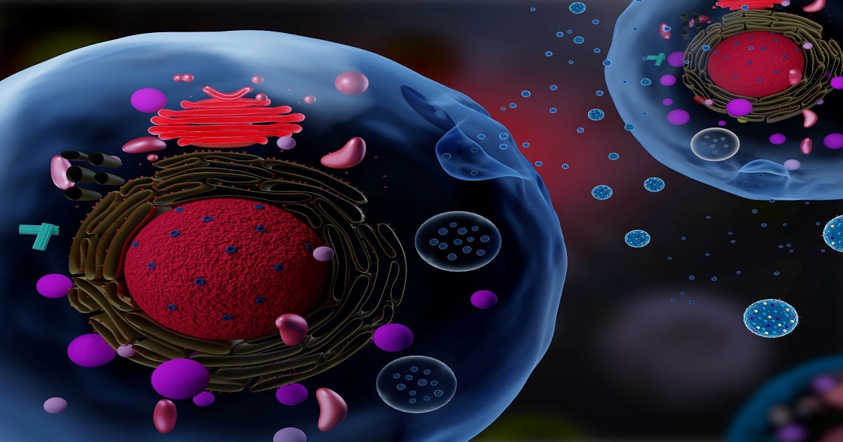
The tumor microenvironment (TME) is a complex and dynamic ecosystem that influences tumor progression, invasion, metastasis, and treatment response. It is composed of cancer cells, stromal cells, immune cells, and various components of the extracellular matrix and recent research indicates that these components communicate with each other via extracellular vesicles (EVs). EVs are small membrane-bound vesicles released by almost all cells, including cancer cells, and are known to carry various bioactive molecules, such as proteins, nucleic acids, and lipids. This topic is dedicated to the emerging understanding of how EVs contribute to the modulation of the TME. Hereby, we focus on a variety of EV-related aspects, including, but not limited to, EVs as modulators of intercellular communication, the role of EVs in remodeling the extracellular matrix, the immunomodulatory effects of EVs, and EVs as biomarkers and therapeutic targets. We hope that this topic will provide further insights into the complex role of EVs in modulating the TME and will accelerate the development of novel EV-based therapeutic strategies.

Dr. Nils Ludwig
Dr. Miroslaw J Szczepanski
Topic Editors

Keywords

  • extracellular vesicles
  • exosomes
  • tumor microenvironment
  • intercellular communication
  • immunomodulation

Participating Journals

Journal Name Impact Factor CiteScore Launched Year First Decision (median) APC
Biomolecules
biomolecules
4.8 9.4 2011 18.4 Days CHF 2700 Submit
Current Issues in Molecular Biology
cimb
2.8 2.9 1999 15.8 Days CHF 2200 Submit
Scientia Pharmaceutica
scipharm
2.3 4.6 1930 26.1 Days CHF 1000 Submit
Cancers
cancers
4.5 8.0 2009 17.4 Days CHF 2900 Submit
Current Oncology
curroncol
2.8 3.3 1994 19.8 Days CHF 2200 Submit
Cells
cells
5.1 9.9 2012 17 Days CHF 2700 Submit

Preprints.org is a multidisciplinary platform offering a preprint service designed to facilitate the early sharing of your research. It supports and empowers your research journey from the very beginning.

MDPI Topics is collaborating with Preprints.org and has established a direct connection between MDPI journals and the platform. Authors are encouraged to take advantage of this opportunity by posting their preprints at Preprints.org prior to publication:

  1. Share your research immediately: disseminate your ideas prior to publication and establish priority for your work.
  2. Safeguard your intellectual contribution: Protect your ideas with a time-stamped preprint that serves as proof of your research timeline.
  3. Boost visibility and impact: Increase the reach and influence of your research by making it accessible to a global audience.
  4. Gain early feedback: Receive valuable input and insights from peers before submitting to a journal.
  5. Ensure broad indexing: Web of Science (Preprint Citation Index), Google Scholar, Crossref, SHARE, PrePubMed, Scilit and Europe PMC.

Published Papers (4 papers)

Order results
Result details
Journals
Select all
Export citation of selected articles as:
34 pages, 1842 KiB  
Review
Cell Progression and Survival Functions of Enzymes Secreted in Extracellular Vesicles Associated with Breast and Prostate Cancers
by Cosmos Ifeanyi Onyiba, Niwasini Krishna Kumar, Christopher J. Scarlett and Judith Weidenhofer
Cells 2025, 14(7), 468; https://doi.org/10.3390/cells14070468 - 21 Mar 2025
Viewed by 1412
Abstract
Extracellular vesicles (EVs) are membrane-bound cargoes secreted by normal and pathological cells. Through their protein, nucleic acid, and lipid cargoes, EVs mediate several cellular processes, such as cell–cell communication, cell development, immune response, and tissue repair. Most importantly, through their enzyme cargo, EVs [...] Read more.
Extracellular vesicles (EVs) are membrane-bound cargoes secreted by normal and pathological cells. Through their protein, nucleic acid, and lipid cargoes, EVs mediate several cellular processes, such as cell–cell communication, cell development, immune response, and tissue repair. Most importantly, through their enzyme cargo, EVs mediate pathophysiological processes, including the pathogenesis of cancer. In this review, we enumerate several enzymes secreted in EVs (EV enzyme cargo) from cells and patient clinical samples of breast and prostate cancers and detail their contributions to the progression and survival of both cancers. Findings in this review reveal that the EV enzyme cargo could exert cell progression functions via adhesion, proliferation, migration, invasion, and metastasis. The EV enzyme cargo might also influence cell survival functions of chemoresistance, radioresistance, angiogenesis, cell death inhibition, cell colony formation, and immune evasion. While the current literature provides evidence of the possible contributions of the EV enzyme cargo to the progression and survival mechanisms of breast and prostate cancers, future studies are required to validate that these effects are modified by EVs and provide insights into the clinical applications of the EV enzyme cargo in breast and prostate cancer. Full article
Show Figures

Figure 1

33 pages, 55731 KiB  
Article
Extracellular Signaling Molecules from Adipose-Derived Stem Cells and Ovarian Cancer Cells Induce a Hybrid Epithelial-Mesenchymal Phenotype in a Bidirectional Interaction
by Vinícius Augusto Simão, Juliana Ferreira Floriano, Roberta Carvalho Cesário, Karolina da Silva Tonon, Larissa Ragozo Cardoso de Oliveira, Flávia Karina Delella, Fausto Almeida, Lucilene Delazari dos Santos, Fábio Rodrigues Ferreira Seiva, Débora Aparecida Pires de Campos Zuccari, João Tadeu Ribeiro-Paes, Russel J. Reiter and Luiz Gustavo de Almeida Chuffa
Cells 2025, 14(5), 374; https://doi.org/10.3390/cells14050374 - 4 Mar 2025
Viewed by 2141
Abstract
Ovarian cancer (OC) is characterized by high mortality rates due to late diagnosis, recurrence, and metastasis. Here, we show that extracellular signaling molecules secreted by adipose-derived mesenchymal stem cells (ASCs) and OC cells—either in the conditioned medium (CM) or within small extracellular vesicles [...] Read more.
Ovarian cancer (OC) is characterized by high mortality rates due to late diagnosis, recurrence, and metastasis. Here, we show that extracellular signaling molecules secreted by adipose-derived mesenchymal stem cells (ASCs) and OC cells—either in the conditioned medium (CM) or within small extracellular vesicles (sEVs)—modulate cellular responses and drive OC progression. ASC-derived sEVs and CM secretome promoted OC cell colony formation, invasion, and migration while upregulating tumor-associated signaling pathways, including TGFβ/Smad, p38MAPK/ERK1/2, Wnt/β-catenin, and MMP-9. Additionally, OC-derived sEVs and CM induced a pro-tumorigenic phenotype in ASCs, enhancing their invasiveness and expression of tumor-associated factors. Notably, both ASCs and OC cells exhibited increased expression of E-cadherin and Snail/Slug proteins, key markers of epithelial/mesenchymal hybrid phenotype, enhancing cellular plasticity and metastatic potential. We also demonstrated that these cellular features are, at least in part, due to the presence of tumor-supportive molecules such as TNF-α, Tenascin-C, MMP-2, and SDF-1α in the CM secretome of ASCs and OC cells. In silico analyses linked these molecular changes to poor prognostic outcomes in OC patients. These findings highlight the critical role of sEVs and tumor/stem cell-derived secretome in OC progression through bidirectional interactions that impact cellular behavior and phenotypic transitions. We suggest that targeting EV-mediated communication could improve therapeutic strategies and patient outcomes. Full article
Show Figures

Graphical abstract

12 pages, 1884 KiB  
Article
Macrophage Phenotype Induced by Circulating Small Extracellular Vesicles from Women with Endometriosis
by María Angeles Martínez-Zamora, Olga Armengol-Badia, Lara Quintas-Marquès, Francisco Carmona and Daniel Closa
Biomolecules 2024, 14(7), 737; https://doi.org/10.3390/biom14070737 - 21 Jun 2024
Cited by 2 | Viewed by 1771
Abstract
Evidence suggests that immune system dysfunction and macrophages are involved in the disease establishment and progression of endometriosis. Among the factors involved in this alteration in macrophage activity, Small Extracellular Vesicles (sEVs) have been described to play a role favoring the switch to [...] Read more.
Evidence suggests that immune system dysfunction and macrophages are involved in the disease establishment and progression of endometriosis. Among the factors involved in this alteration in macrophage activity, Small Extracellular Vesicles (sEVs) have been described to play a role favoring the switch to a specific phenotype with controversial results. This study aims to investigate the potential effect of circulating sEVs in the plasma of well-characterized patients with endometriosis on the polarization of macrophages. sEVs were isolated from the plasma of patients diagnosed with endometriosis confirmed by histopathological analysis. Two groups of patients were recruited: the endometriosis group consisted of patients diagnosed with endometriosis by imaging testing (gynecological ultrasonography and/or magnetic resonance imaging), confirmed by histopathologic study (n = 12), and the control group included patients who underwent laparoscopy for tubal sterilization without presurgical suspicion of endometriosis and without endometriosis or signs of any inflammatory pelvic condition during surgery (n = 12). Human THP1 monocytic cells were differentiated into macrophages, and the effect of sEVs on cell uptake and macrophage polarization was evaluated by fluorescent labeling and measurement of the IL1B, TNF, ARG1, and MRC1 expression, respectively. Although no changes in cell uptake were detected, sEVs from endometriosis induced a polarization of macrophages toward an M2 phenotype, characterized by lower IL1B and TNF expression and a tendency to increase MRC1 and ARG1 levels. When macrophages were stimulated with lipopolysaccharides, less activation was also detected after treatment with endometriosis sEVs. Finally, endometriosis sEVs also induced the expression of the nuclear receptor peroxisome proliferator-activated receptor-gamma (PPARG); however, treatment with rosiglitazone, a PPARG agonist, had no effect on the change in macrophage phenotype. We conclude that circulating sEVs in women with endometriosis have a certain capacity to shift the activation state of macrophages toward an M2 phenotype, but this does not modify the uptake level or the response to PPARG ligands. Full article
Show Figures

Figure 1

13 pages, 4045 KiB  
Article
Biological Activities of Citrus-Derived Extracellular Vesicles on Human Cells: The Role of Preservation
by Theodora Karamanidou, Konstantinos Krommydas, Maria Karanikou, Dimitrios Tsamos, Konstantinos Michalakis, Dimitris Kletsas, Alexander Tsouknidas and Harris Pratsinis
Curr. Issues Mol. Biol. 2024, 46(6), 5812-5824; https://doi.org/10.3390/cimb46060347 - 11 Jun 2024
Cited by 1 | Viewed by 1570
Abstract
Extracellular vesicles (EVs) have been identified as important mediators for cell-to-cell communication. Citrus-based EVs in particular offer an excellent platform for nutraceutical delivery systems, as their endemic cargo includes micronutrients (e.g., ascorbic acid), which contribute to their antioxidant capacity. Despite being extensively investigated [...] Read more.
Extracellular vesicles (EVs) have been identified as important mediators for cell-to-cell communication. Citrus-based EVs in particular offer an excellent platform for nutraceutical delivery systems, as their endemic cargo includes micronutrients (e.g., ascorbic acid), which contribute to their antioxidant capacity. Despite being extensively investigated as to their therapeutic and diagnostic potential, their cargo is inherently unstable and thus directly affected by their storage and preservation. In this study, EVs were isolated from citrus fruit using tangential flow filtration and evaluated for their physicochemical characteristics, antioxidant activity and effects on human cells. To assess how their isolation and preservation methods affect these properties, the EVs were tested immediately after isolation (from fresh and freeze-thawed juices) or following freeze-drying. A measurable biological effect of cryoprotection on citrus-derived EVs was evident, whether during or after isolation. This was more pronounced in the cell-based assays, ranging from −4% to +32% in human skin fibroblast proliferation. Nevertheless, the effects on human cancer cells varied depending on the cell line. Although these results should be considered preliminary observations, subject to further investigation, it is safe to state that any type of preservation is expected to impact the EVs’ biological activity. Full article
Show Figures

Graphical abstract

Back to TopTop