In the original publication [], there was a mistake in Figure 1 and Figure 4 as published. In Collage 1B, the original images for the ‘Col 1’ control group and the upper 30-week group in Figure 1 do not depict the bile duct. Additionally, the lower 52-week ‘Col 1’ group contained a technical error and has been replaced with a representative image. In Collage 4B, the original ‘SMAD’ control group in Figure 4 did not clearly show the positive staining in the portal triad areas. The corrected Figure 1 and Figure 4 are shown below. The authors state that the scientific conclusions are unaffected. This correction was approved by the Academic Editor. The original publication has also been updated.
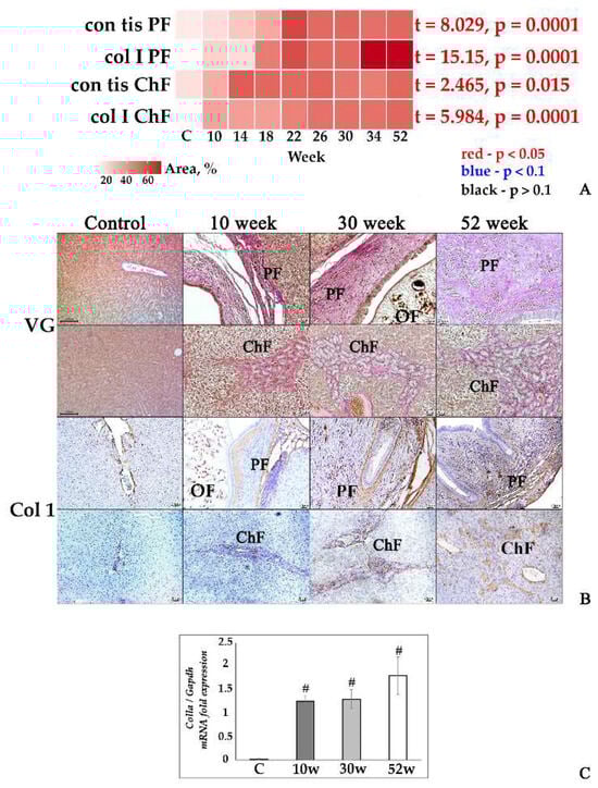
Figure 1.
Fibrotic changes in the liver tissue of O. felineus-infected Syrian hamsters. (A) Heat map progression of periductal fibrosis (con_tis_PF) and cholangiofibrosis (con_tis_ChF), and the amount of collagen 1a+ fibers in the liver of infected animals (col_I_PF and col_I_ChF, respectively); (B) histopathological changes in Syrian hamster liver, Van Gieson staining (VG), and IHC analysis for collagen 1a (Col1); (C) Col1a gene was normalized to average Gapdh expression. C—control, 10 w, 30 w, 52 w—week p.i. Data are presented as mean ± SEM, # p ≤ 0.05, as compared to the control group.
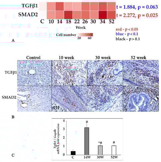
Figure 4.
TGFβ1/SMAD2 signaling pathway in the liver tissue of O. felineus-infected Syrian hamsters. (A) Heat map of changes in TGF-β1- and SMAD2-positive cells in the course of infection; (B) TGF-β1- and SMAD2-positive cells (marked by an asterisk) in the dynamics of infection, IHC study, ×400 magnification; (C) Tgfb1 gene was normalized to average Gapdh expression. C—control, 10 w, 30 w, 52 w—week p.i. Data are presented as mean ± SEM, # p ≤ 0.05, as compared to the control group, * p ≤ 0.05, as compared to the previous period of investigation.
Reference
- Kovner, A.; Zaparina, O.; Kapushchak, Y.; Minkova, G.; Mordvinov, V.; Pakharukova, M. Jagged-1/Notch Pathway and Key Transient Markers Involved in Biliary Fibrosis during Opisthorchis felineus Infection. Trop. Med. Infect. Dis. 2022, 7, 364. [Google Scholar] [CrossRef] [PubMed]
Disclaimer/Publisher’s Note: The statements, opinions and data contained in all publications are solely those of the individual author(s) and contributor(s) and not of MDPI and/or the editor(s). MDPI and/or the editor(s) disclaim responsibility for any injury to people or property resulting from any ideas, methods, instructions or products referred to in the content. |
© 2025 by the authors. Licensee MDPI, Basel, Switzerland. This article is an open access article distributed under the terms and conditions of the Creative Commons Attribution (CC BY) license (https://creativecommons.org/licenses/by/4.0/).