Bacterial Enterotoxins (Closed)

A topical collection in Toxins (ISSN 2072-6651). This collection belongs to the section "Bacterial Toxins".

Viewed by 40509

Editor


E-Mail Website
Collection Editor
Department of Food Technology, Food Safety and Health, Faculty of Bioscience Engineering, Ghent University (UGent), Coupure Links 657, 9000 Ghent, Belgium
Interests: Bacillus cereus enterotoxins; emetic toxin; cereulide; Staphylococcal enterotoxins; Clostridium botulinum neurotoxins; Clostridium perfringens enterotoxin; shiga toxin; food safety; beauvericin; enniatins; food safety in Serbia

Topical Collection Information

Dear Colleagues,

Bacterial enterotoxins, in the broadest sense, are proteins or peptides that exert their effect on intestines, causing a plethora of gastrointestinal disease manifestations. Enterotoxins are produced by both gram-positive and gram-negative pathogens, both in food and in host’s gastro intestinal tract. In Europe, data collected by Rapid and Alert System for Food and Feed safety about 10% of all outbreaks is found to be caused by bacterial toxins, and only toxins taken here into account were those of Bacillus, Clostridium, and Staphylococcus, which were found in almost all types of food. In USA and UK, enterotoxins produced by C. perfringens are even reported as the top two causative agents of foodborne bacterial poisoning. Bacterial enterotoxins are for many foodborne pathogens the major virulence factor responsible for the symptoms of food poisoning. This is the case among others for Bacillus cereus, Clostridium perfringens, enterotoxigenic Escherichia coli, and Staphylococcus aureus. Many pathogens produce different toxins, and often one strain can produce multiple toxins. The path from the presence of genes encoding for toxin production and actual protein expression, or non-ribosomal peptide synthesis, is not always straight forward, and final toxin production depends on many factors. Understanding these factors is detrimental to food safety assurance. Also, there is great need for a better understanding of the threat to public health from exposure to a single toxin arising from multiple pathways or to multiple toxins that have the same mechanism of toxicity. The aim of the current Special Issue is to gather the most recent cutting-edge research on the topic of bacterial enterotoxins, including, but not limited to, in-vitro, in-vivo and in-silico toxicity, their roles in host-pathogen interactions, the mode and mechanisms of action, the regulation of production, the structure-activity relationship, the exposure assessment, and the detection and control relevant to food safety. This Special Issue aims to become a reference for a new body of knowledge on bacterial toxins, from microbial genes to the human gut.

Prof. Dr. Andreja Rajkovic
Collection Editor

Manuscript Submission Information

Manuscripts should be submitted online at www.mdpi.com by registering and logging in to this website. Once you are registered, click here to go to the submission form. Manuscripts can be submitted until the deadline. All submissions that pass pre-check are peer-reviewed. Accepted papers will be published continuously in the journal (as soon as accepted) and will be listed together on the collection website. Research articles, review articles as well as short communications are invited. For planned papers, a title and short abstract (about 100 words) can be sent to the Editorial Office for announcement on this website.

Submitted manuscripts should not have been published previously, nor be under consideration for publication elsewhere (except conference proceedings papers). All manuscripts are thoroughly refereed through a double-blind peer-review process. A guide for authors and other relevant information for submission of manuscripts is available on the Instructions for Authors page. Toxins is an international peer-reviewed open access monthly journal published by MDPI.

Please visit the Instructions for Authors page before submitting a manuscript. The Article Processing Charge (APC) for publication in this open access journal is 2700 CHF (Swiss Francs). Submitted papers should be well formatted and use good English. Authors may use MDPI's English editing service prior to publication or during author revisions.

Keywords

  • host–pathogen interaction
  • food safety
  • toxin
  • expression
  • exposure
  • toxicity

Published Papers (8 papers)

2022

Jump to: 2021, 2020, 2019, 2018

23 pages, 5258 KiB  
Article
In-Depth Characterization of a Re-Engineered Cholera Toxin Manufacturing Process Using Growth-Decoupled Production in Escherichia coli
by Natalia Danielewicz, Wenyue Dai, Francesca Rosato, Michael E. Webb, Gerald Striedner, Winfried Römer, W. Bruce Turnbull and Juergen Mairhofer
Toxins 2022, 14(6), 396; https://doi.org/10.3390/toxins14060396 - 8 Jun 2022
Cited by 3 | Viewed by 3765
Abstract
Non-toxic derivatives of the cholera toxin are extensively used in neuroscience, as neuronal tracers to reveal the location of cells in the central nervous system. They are, also, being developed as vaccine components and drug-delivery vehicles. Production of cholera-toxin derivatives is often non-reproducible; [...] Read more.
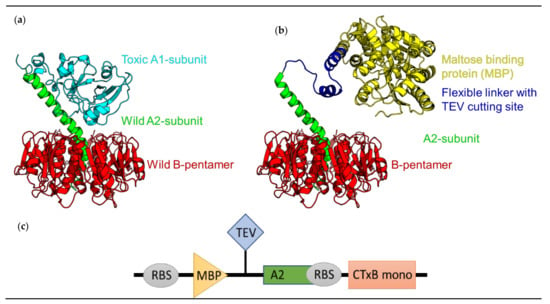
Non-toxic derivatives of the cholera toxin are extensively used in neuroscience, as neuronal tracers to reveal the location of cells in the central nervous system. They are, also, being developed as vaccine components and drug-delivery vehicles. Production of cholera-toxin derivatives is often non-reproducible; the quality and quantity require extensive fine-tuning to produce them in lab-scale settings. In our studies, we seek a resolution to this problem, by expanding the molecular toolbox of the Escherichia coli expression system with suitable production, purification, and offline analytics, to critically assess the quality of a probe or drug delivery, based on a non-toxic derivative of the cholera toxin. We present a re-engineered Cholera Toxin Complex (rCTC), wherein its toxic A1 domain was replaced with Maltose Binding Protein (MBP), as a model for an rCTC-based targeted-delivery vehicle. Here, we were able to improve the rCTC production by 11-fold (168 mg/L vs. 15 mg/L), in comparison to a host/vector combination that has been previously used (BL21(DE3) pTRBAB5-G1S). This 11-fold increase in the rCTC production capability was achieved by (1) substantial vector backbone modifications, (2) using Escherichia coli strains capable of growth-decoupling (V strains), (3) implementing a well-tuned fed-batch production protocol at a 1 L scale, and (4) testing the stability of the purified product. By an in-depth characterization of the production process, we revealed that secretion of rCTC across the E. coli Outer Membrane (OM) is processed by the Type II secretion-system general secretory pathway (gsp-operon) and that cholera toxin B-pentamerization is, likely, the rate-limiting step in complex formation. Upon successful manufacturing, we have validated the biological activity of rCTC, by measuring its binding affinity to its carbohydrate receptor GM1 oligosaccharide (Kd = 40 nM), or binding to Jurkat cells (93 pM) and delivering the cargo (MBP) in a retrograde fashion to the cell. Full article
Show Figures

Figure 1

2021

Jump to: 2022, 2020, 2019, 2018

17 pages, 2365 KiB  
Article
The Pore-Forming Hemolysin BL Enterotoxin from Bacillus cereus: Subunit Interactions in Cell-Free Systems
by Franziska Ramm, Marlitt Stech, Anne Zemella, Hendrik Frentzel and Stefan Kubick
Toxins 2021, 13(11), 807; https://doi.org/10.3390/toxins13110807 - 15 Nov 2021
Cited by 3 | Viewed by 3545
Abstract
The tripartite enterotoxin Hemolysin BL (Hbl) has been widely characterized as a hemolytic and cytotoxic virulence factor involved in foodborne diarrheal illness caused by Bacillus cereus. Previous studies have described the formation of the Hbl complex and aimed to identify the toxin’s [...] Read more.
The tripartite enterotoxin Hemolysin BL (Hbl) has been widely characterized as a hemolytic and cytotoxic virulence factor involved in foodborne diarrheal illness caused by Bacillus cereus. Previous studies have described the formation of the Hbl complex and aimed to identify the toxin’s mode of action. In this study, we analyzed the assembly of Hbl out of its three individual subunits L1, L2 and B in a soluble as well as a putative membrane bound composition using a Chinese hamster ovary (CHO) cell-free system. Subunits were either coexpressed or synthesized individually in separate cell-free reactions and mixed together afterwards. Hemolytic activity of cell-free synthesized subunits was demonstrated on 5% sheep blood agar and identified both synthesis procedures, coexpression as well as individual synthesis of each subunit, as functional for the synthesis of an active Hbl complex. Hbl’s ability to perforate cell membranes was evaluated using a propidium iodide uptake assay. These data suggested that coexpressed Hbl subunits augmented cytotoxic activity with increasing concentrations. Further, a pre-pore-complex of L1-L2 showed cytotoxic effects suggesting the possibility of an interaction between the cell membrane and the pre-pore-complex. Overall, this study shows that cell-free protein synthesis is a fast and efficient way to study the assembly of multiple protein subunits in soluble as well as vesicular fractions. Full article
Show Figures

Graphical abstract

19 pages, 6212 KiB  
Article
Cellular Activity of Salmonella Typhimurium ArtAB Toxin and Its Receptor-Binding Subunit
by Elise Overgaard, Brad Morris, Omid Mohammad Mousa, Emily Price, Adriana Rodriguez, Leyla Cufurovic, Richard S. Beard and Juliette K. Tinker
Toxins 2021, 13(9), 599; https://doi.org/10.3390/toxins13090599 - 27 Aug 2021
Cited by 2 | Viewed by 3465
Abstract
Salmonellosis is among the most reported foodborne illnesses in the United States. The Salmonellaenterica Typhimurium DT104 phage type, which is associated with multidrug-resistant disease in humans and animals, possesses an ADP-ribosylating toxin called ArtAB. Full-length artAB has been found on a [...] Read more.
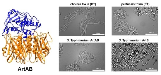
Salmonellosis is among the most reported foodborne illnesses in the United States. The Salmonellaenterica Typhimurium DT104 phage type, which is associated with multidrug-resistant disease in humans and animals, possesses an ADP-ribosylating toxin called ArtAB. Full-length artAB has been found on a number of broad-host-range non-typhoidal Salmonella species and serovars. ArtAB is also homologous to many AB5 toxins from diverse Gram-negative pathogens, including cholera toxin (CT) and pertussis toxin (PT), and may be involved in Salmonella pathogenesis, however, in vitro cellular toxicity of ArtAB has not been characterized. artAB was cloned into E. coli and initially isolated using a histidine tag (ArtABHIS) and nickel chromatography. ArtABHIS was found to bind to African green monkey kidney epithelial (Vero) cells using confocal microscopy and to interact with glycans present on fetuin and monosialotetrahexosylganglioside (GM1) using ELISA. Untagged, or native, holotoxin (ArtAB), and the pentameric receptor-binding subunit (ArtB) were purified from E. coli using fetuin and d-galactose affinity chromatography. ArtAB and ArtB metabolic and cytotoxic activities were determined using Vero and Chinese hamster ovary (CHO) epithelial cells. Vero cells were more sensitive to ArtAB, however, incubation with both cell types revealed only partial cytotoxicity over 72 h, similar to that induced by CT. ArtAB induced a distinctive clustering phenotype on CHO cells over 72 h, similar to PT, and an elongated phenotype on Vero cells, similar to CT. The ArtB binding subunit alone also had a cytotoxic effect on CHO cells and induced morphological rounding. Results indicate that this toxin induces distinctive cellular outcomes. Continued biological characterization of ArtAB will advance efforts to prevent disease caused by non-typhoidal Salmonella. Full article
Show Figures

Graphical abstract

2020

Jump to: 2022, 2021, 2019, 2018

12 pages, 879 KiB  
Article
Nature-Identical Compounds and Organic Acids Reduce E. coli K88 Growth and Virulence Gene Expression In Vitro
by Andrea Bonetti, Benedetta Tugnoli, Barbara Rossi, Giulia Giovagnoni, Andrea Piva and Ester Grilli
Toxins 2020, 12(8), 468; https://doi.org/10.3390/toxins12080468 - 23 Jul 2020
Cited by 20 | Viewed by 4356
Abstract
Post-weaning diarrhoea (PWD) is one of the long-standing challenges in pig husbandry. Due to the risks of resistance caused by antibiotics (AB) misuse, conventional treatments against Escherichia coli K88 (E. coli K88), the PWD etiological agent, urgently need to be replaced. Organic [...] Read more.
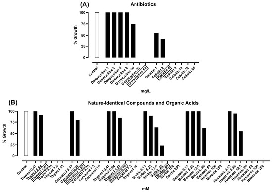
Post-weaning diarrhoea (PWD) is one of the long-standing challenges in pig husbandry. Due to the risks of resistance caused by antibiotics (AB) misuse, conventional treatments against Escherichia coli K88 (E. coli K88), the PWD etiological agent, urgently need to be replaced. Organic acids (OA) and nature-identical compounds (NIC) are currently finding a central role in infection management thanks to their recognized antimicrobial activity. This study investigated the susceptibility of an E. coli K88 field strain to a wide panel of AB, NIC, and OA. Secondly, we evaluated the ability of sub-lethal doses of the most active compounds to modulate the expression of E. coli K88 virulence genes. Results showed that the bacterial strain was resistant to many of the tested antibiotics, but an antimicrobial action was registered for selected NIC and OA. The quantitative PCR analysis revealed that thymol, carvacrol, eugenol, and benzoic acid were able to downregulate (p < 0.05) the expression of bacterial genes related to motility, adhesion to enterocytes, heat-labile (LT) and heat-stable (ST) toxin secretion, quorum sensing, and biofilm formation. Therefore, this study demonstrated that selected OA and NIC not only control E. coli K88 growth but also modulate the expression of many virulence genes at sub-lethal doses, thus offering new insights on their mechanism of action and suggesting a powerful tool to manage PWD. Full article
Show Figures

Figure 1

2019

Jump to: 2022, 2021, 2020, 2018

15 pages, 6868 KiB  
Article
Repeated Oral Administration of a KDEL-Tagged Recombinant Cholera Toxin B Subunit Effectively Mitigates DSS Colitis despite a Robust Immunogenic Response
by Joshua M. Royal, Micaela A. Reeves and Nobuyuki Matoba
Toxins 2019, 11(12), 678; https://doi.org/10.3390/toxins11120678 - 20 Nov 2019
Cited by 14 | Viewed by 4021
Abstract
Cholera toxin B subunit (CTB), a non-toxic homopentameric component of Vibrio cholerae holotoxin, is an oral cholera vaccine antigen that induces an anti-toxin antibody response. Recently, we demonstrated that a recombinant CTB variant with a Lys-Asp-Glu-Leu (KDEL) endoplasmic reticulum retention motif (CTB-KDEL) exhibits [...] Read more.
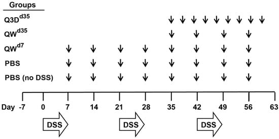
Cholera toxin B subunit (CTB), a non-toxic homopentameric component of Vibrio cholerae holotoxin, is an oral cholera vaccine antigen that induces an anti-toxin antibody response. Recently, we demonstrated that a recombinant CTB variant with a Lys-Asp-Glu-Leu (KDEL) endoplasmic reticulum retention motif (CTB-KDEL) exhibits colon mucosal healing effects that have therapeutic implications for inflammatory bowel disease (IBD). Herein, we investigated the feasibility of CTB-KDEL for the treatment of chronic colitis. We found that weekly oral administration of CTB-KDEL, dosed before or after the onset of chronic colitis, induced by repeated dextran sodium sulfate (DSS) exposure, could significantly reduce disease activity index scores, intestinal permeability, inflammation, and histological signs of chronicity. To address the consequences of immunogenicity, mice (C57BL/6 or C3H/HeJ strains) were pre-exposed to CTB-KDEL then subjected to DSS colitis and CTB-KDEL treatment. While the pre-dosing of CTB-KDEL elicited high-titer anti-drug antibodies (ADAs) of the immunoglobin A (IgA) isotype in the intestine of C57BL/6 mice, the therapeutic effects of CTB-KDEL were similar to those observed in C3H/HeJ mice, which showed minimal ADAs under the same experimental conditions. Thus, the immunogenicity of CTB-KDEL does not seem to impede the protein’s mucosal healing efficacy. These results support the development of CTB-KDEL for IBD therapy. Full article
Show Figures

Figure 1

9 pages, 581 KiB  
Article
Genotypes, Enterotoxin Gene Profiles, and Antimicrobial Resistance of Staphylococcus aureus Associated with Foodborne Outbreaks in Hangzhou, China
by Qi Chen and Sangma Xie
Toxins 2019, 11(6), 307; https://doi.org/10.3390/toxins11060307 - 29 May 2019
Cited by 38 | Viewed by 4211
Abstract
Staphylococcal food poisoning is an illness caused by the consumption of food that contains sufficient amounts of one or more enterotoxins. In the present study, a total of 37 S. aureus isolates were recovered from leftover food, swabs from a kitchen environment, and [...] Read more.
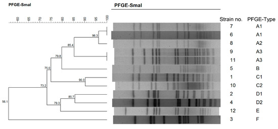
Staphylococcal food poisoning is an illness caused by the consumption of food that contains sufficient amounts of one or more enterotoxins. In the present study, a total of 37 S. aureus isolates were recovered from leftover food, swabs from a kitchen environment, and patient feces associated with four foodborne outbreaks that occurred in Hangzhou, southeast China, and were characterized by multilocus sequence typing (MLST), spa typing, pulse-field gel electrophoresis (PFGE), and antimicrobial susceptibility. Classical enterotoxin and enterotoxin-like genes were profiled by PCR analysis. ST6-t304 was the most common clone (40.54%), followed by ST2315-t11687 (32.43%). Six clusters (A to F) were divided based on PFGE patterns, and Clusters A and C were the most common types, constituting 86.49% of all isolates. Moreover, sea was the most frequently identified enterotoxin gene (81.08%), followed by the combination of seg–sei–selm–seln–sleo–selu and sec–sell (each 48.65%). Five isolates also harbored the exotoxin cluster sed–selj–ser. In addition, resistance to penicillin (97.30%), erythromycin (37.85), tetracycline (32.43%), clindamycin, gentamicin, and sulfamethoxazole (each 10.81%) was observed. Our research demonstrated the link between leftover foods and patients by molecular typing and detecting the profiles of enterotoxin or enterotoxin-like genes in human and food isolates. S. aureus maintains an extensive repertoire of enterotoxins and drug resistance genes that could cause potential health threats to consumers. Full article
Show Figures

Figure 1

2018

Jump to: 2022, 2021, 2020, 2019

11 pages, 2432 KiB  
Article
Characterization of A Staphylococcal Food Poisoning Outbreak in A Workplace Canteen during the Post-Earthquake Reconstruction of Central Italy
by Fabrizia Guidi, Anna Duranti, Silvia Gallina, Yacine Nia, Annalisa Petruzzelli, Angelo Romano, Valeria Travaglini, Alberto Olivastri, Vincenzo Calvaresi, Lucia Decastelli and Giuliana Blasi
Toxins 2018, 10(12), 523; https://doi.org/10.3390/toxins10120523 - 6 Dec 2018
Cited by 25 | Viewed by 5652
Abstract
In summer 2017, a foodborne outbreak occurred in Central Italy, involving 26 workers employed in the post-earthquake reconstruction. After eating a meal provided by a catering service, they manifested gastrointestinal symptoms; 23 of them were hospitalized. The retrospective cohort study indicated the pasta [...] Read more.
In summer 2017, a foodborne outbreak occurred in Central Italy, involving 26 workers employed in the post-earthquake reconstruction. After eating a meal provided by a catering service, they manifested gastrointestinal symptoms; 23 of them were hospitalized. The retrospective cohort study indicated the pasta salad as the most likely vehicle of poisoning. Foods, environmental samples, and food handlers’ nasal swabs were collected. Bacillus cereus (Bc) and coagulase-positive staphylococci (CPS) including S. aureus, together with their toxins, were the targets of the analysis. CPS, detected in all the leftovers, exceeded 105 CFU/g in the pasta salad, in which we found Staphylococcal Enterotoxins (SEs) (0.033 ng SEA/g; 0.052 ng SED/g). None of the environmental and human swabs showed contamination. We characterized 23 S. aureus from foods. They all belonged to the human biotype, showed the same toxigenic profile (sea, sed, sej, and ser genes), and had the same Pulsed Field Gel Electrophoresis (PFGE) pattern; none of them harbored mecA or mupA genes. We also detected Bc contamination in the pasta salad but none of the isolates harbored the ces gene for the emetic toxin cereulide. The EU Reference Laboratory for CPS confirmed the case as a strong-evidence outbreak caused by the ingestion of SEs produced by a single strain of S. aureus carried by the same human source. This outbreak was successfully investigated despite the emergency situation in which it occurred. Full article
Show Figures

Figure 1

15 pages, 1792 KiB  
Article
Oxygen Consumption Rate Analysis of Mitochondrial Dysfunction Caused by Bacillus cereus Cereulide in Caco-2 and HepG2 Cells
by Marlies Decleer, Jelena Jovanovic, Anita Vakula, Bozidar Udovicki, Rock-Seth E. K. Agoua, Annemieke Madder, Sarah De Saeger and Andreja Rajkovic
Toxins 2018, 10(7), 266; https://doi.org/10.3390/toxins10070266 - 2 Jul 2018
Cited by 47 | Viewed by 8698
Abstract
The emetic syndrome of Bacillus cereus is a food intoxication caused by cereulide (CER) and manifested by emesis, nausea and in most severe cases with liver failure. While acute effects have been studied in the aftermath of food intoxication, an exposure to low [...] Read more.
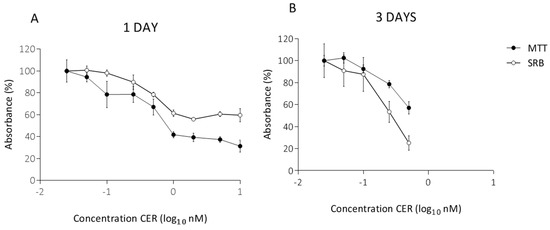
The emetic syndrome of Bacillus cereus is a food intoxication caused by cereulide (CER) and manifested by emesis, nausea and in most severe cases with liver failure. While acute effects have been studied in the aftermath of food intoxication, an exposure to low doses of cereulide might cause unnoticed damages to the intestines and liver. The toxicity which relies on the mitochondrial dysfunction was assessed on Caco-2 and HepG2 cells after exposure of one, three and ten days to a range of low doses of cereulide. Oxygen consumption rate analyses were used to study the impact of low doses of CER on the bioenergetics functions of undifferentiated Caco-2 and HepG2 cells using Seahorse XF extracellular flux analyzer. Both Caco-2 and HepG2 cells experienced measurable mitochondrial impairment after prolonged exposure of 10 days to 0.25 nM of cereulide. Observed mitochondrial dysfunction was greatly reflected in reduction of maximal cell respiration. At 0.50 nM CER, mitochondrial respiration was almost completely shut down, especially in HepG2 cells. These results corresponded with a severe reduction in the amount of cells and an altered morphology, observed by microscopic examination of the cells. Accurate and robust quantification of basal respiration, ATP production, proton leak, maximal respiration, spare respiratory capacity, and non-mitochondrial respiration allowed better understanding of the effects of cereulide in underlying respiratory malfunctions in low-dose exposure. Full article
Show Figures

Figure 1

Back to TopTop