


Pathophysiology 2026, 33(3), 45; https://doi.org/10.3390/pathophysiology33030045 - 3 Jul 2026
Abstract
Pancreatic β-cells maintain glucose homeostasis through tightly regulated insulin biosynthesis, storage, and secretion. To prevent pathological accumulation of excess or aging secretory granules (SGs), β-cells use crinophagy, a selective lysosomal degradation pathway in which mature insulin-containing granules fuse directly with lysosomes to form
[...] Read more.
Pancreatic β-cells maintain glucose homeostasis through tightly regulated insulin biosynthesis, storage, and secretion. To prevent pathological accumulation of excess or aging secretory granules (SGs), β-cells use crinophagy, a selective lysosomal degradation pathway in which mature insulin-containing granules fuse directly with lysosomes to form hybrid organelles termed crinosomes. Crinophagy was historically considered a simple mechanism for discarding obsolete, aged SGs. The acidic, protease-rich environment of crinosomes is proposed to generate unconventional insulin-derived epitopes through cathepsin-mediated proteolysis and transpeptidation reactions. These cryptic epitopes, which include hybrid insulin peptides (HIPs) resulting from the covalent fusion of insulin fragments with peptides from co-resident granule proteins, are largely absent from the thymic epitope repertoire. This creates a “peripheral–thymic mismatch” that allows autoreactive CD4+ T cells to escape central tolerance, ultimately driving β-cell destruction in type 1 diabetes (T1D). Recent studies demonstrate that pharmacological or genetic inhibition of crinophagy reduces crinosome abundance, narrows the pathogenic epitope repertoire, and delays the onset of diabetes in preclinical models. In type 2 diabetes (T2D), a related pathway termed stress-induced nascent granule degradation (SINGD) diverts newly synthesized insulin granules to lysosomes under glucolipotoxic conditions, contributing to insulin depletion and progressive β-cell failure. This review summarizes the current understanding of the molecular mechanisms behind crinophagy. It discusses its two main functions: maintaining physiological quality control and generating pathological antigens. Additionally, the review explores how crinophagy interacts with other cellular stress pathways and highlights new therapeutic strategies aimed at targeting this process to protect pancreatic β-cell function and potentially prevent or delay diabetes.
Full article
(This article belongs to the Section Cellular and Molecular Mechanisms)
►
Show Figures
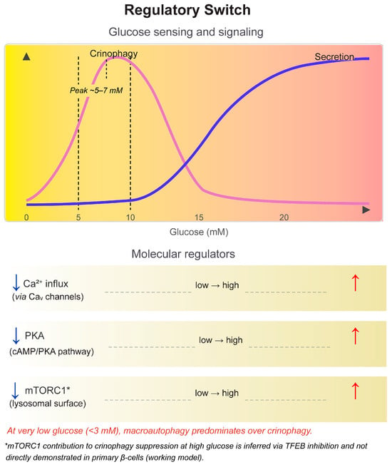
Figure 1


