



Journal Menu
► ▼ Journal Menu-
- Nanomaterials Home
- Aims & Scope
- Editorial Board
- Reviewer Board
- Topical Advisory Panel
- Early Career Editorial Board
- Instructions for Authors
- Special Issues
- Topics
- Sections & Collections
- Article Processing Charge
- Indexing & Archiving
- Editor’s Choice Articles
- Most Cited & Viewed
- Journal Statistics
- Journal History
- Journal Awards
- Society Collaborations
- Conferences
- Editorial Office
Journal Browser
► ▼ Journal BrowserNeed Help?
Announcements
28 May 2026
Nanomaterials | Behind the Paper: Enzyme-Responsive Gadolinium Oxide Nanoparticles for Cancer Theranostics
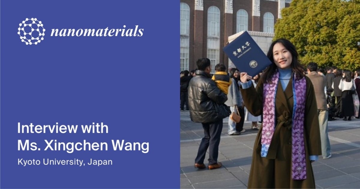
In this Behind the Paper interview, we speak with Ms. Xingchen Wang, first author of a recent article published in Nanomaterials (ISSN: 2079-4991). Ms. Wang shares her perspective on the motivation behind the study, the design of the nanoparticle platform, the role of MMP-responsive drug release, and potential next steps for dual-imaging-guided cancer nanomedicine.
About the Paper:
“Biocompatible Gadolinium Oxide Nanoparticles Incorporated Doxorubicin Enables Magnetic Resonance and Photoacoustic Dual Imaging for Cancer Theranostics”
by Xingchen Wang, Yuta Imai, Yu Kimura, Risako Miura, Hirohiko Imai and Teruyuki Kondo
Nanomaterials 2026, 16(6), 343; https://doi.org/10.3390/nano16060343
About the Author:
Ms. Xingchen Wang is a PhD candidate at the Graduate School of Engineering, Kyoto University. Her doctoral dissertation focuses on the fusion of nanotechnology and cancer theranostics. She specializes in the development of highly biocompatible gelatin-coated Gd2O3 nanoparticles for target drug delivery and dual modality imaging. She uses enzyme-responsive nanomaterials to bridge the gap between accurate diagnosis and effective treatment. She has successfully designed and developed several nanoparticle systems that utilize tumor-specific microenvironments for controlled drug release. She hopes to contribute to research on nanoparticles in engineering and further their application to medical and clinical contexts.
The following is an interview with Ms. Wang:
1. Could you briefly introduce the background of this study and explain what inspired your team to develop this gadolinium oxide nanoparticle platform for cancer theranostics?
Our team has years of research enhancing the capabilities of MRI contrast agents. Through the development of Gd nanomaterials, we have accumulated a wide range of experience, including Gd dendrimers and Gd2O3 nanoparticles. The novelty of the nano-system is that the control of size enables the ability to increase the contrast of MRI images, in vivo biological distribution dynamics, and tumor passive accumulation. Under such a background, we aimed to expand their role beyond the diagnosis of Gd nanomaterials to integrated cancer theranostics
By developing a multifunctional nano-particle platform that combines dual MRI/photoacoustic imaging and enzyme-response drug supply, we aimed to realize more accurate tumor visualization along with selective therapeutic activation in the tumor microenvironment.
2. What do you consider the main novelty of this work compared with previous gadolinium-based theranostic nanoparticles and dual MRI/photoacoustic imaging probes?
The key feature of our platform is the integration of drug release and dual MRI/photoacoustic imaging in response to tumor microenvironments. MRI provides the entire body with anatomical information about deep tissues, while photoacoustic imaging provides high spatial/temporal resolution of the specific body. By combining these modalities, our platform enables much more reliable and accurate tumor detection than using either modality alone. Furthermore, the nanoparticles are passively accumulated in the tumor through a tumor-specific EPR effect, and the gelatin on the surface of the nanoparticles is hydrolyzed by MMP-2/9 enzyme activity and anti-cancer drugs are released.
3. Your study uses MMP-responsive degradation of succinylated PEG-gelatin to trigger doxorubicin release. Could you explain why this enzyme-responsive mechanism is advantageous for selective tumor therapy?
The major advantage of our MMP-compatible design is that drug release in the tumor microenvironment is selective. MMP-2 and MMP-9 are highly secreted in various invasive solid tumors but only show extremely low activity in normal tissues. This biological differentiation allows the autonomous release of preferential anti-cancer agents to the tumor site, significantly reducing the toxicity to normal tissues. As a result, it is possible to minimize the side effects of anti-cancer drugs on normal cells.
Another significant advantage is that this mechanism continues to support visualization capabilities in MR/PA dual imaging. In vivo experiments demonstrated that tumor reduction could be tracked using these two imaging modalities, a result that would be beneficial for optimizing treatment.
4. What was the main challenge in preparing or evaluating the SPG–DOX–Gd nanoparticles, and how did your team address it?
The main challenge was to attain both sufficient stability in the physiological environment and efficient responsiveness in the tumor site. If the nanoparticle structure is too unstable, drug leaks occur before they accumulate in the tumor. Conversely, if the structure is overly stable, the drug release at the tumor site becomes inefficient. To cope with this problem, the composition and chemical modification of the gelatin shell were carefully optimized. Accordingly, the optimization process of selecting surface coatings that respond to tumor-related MMP enzymes has succeeded in establishing a nanoparticle system that efficiently releases the drug only at the tumor site, while maintaining stability under physiological conditions.
5. Could you share what made Nanomaterials a suitable venue for this work, and how you found the journal’s editorial communication, peer-review, and publication process?
We decided to submit our research because our research content was fully consistent with the Special Issue “Nanomaterials in Medicine and Healthcare” of Nanomaterials. In other words, the development of theranostic probes based on a multifunctional nano-materials platform, as well as the study on dual imaging and drug delivery, conforms to the scope of the studies covered by this journal. The peer review process was very professional, and the peer reviewers provided valuable suggestions and were able to have sufficient scientific discussions. As a result, they helped to significantly improve the quality of our manuscript. Consequently, we appreciate that our research results were shared with the scientific community in a timely manner.
