Deep Learning in Bioinformatics and Biomedicine

A special issue of Applied Sciences (ISSN 2076-3417). This special issue belongs to the section "Applied Biosciences and Bioengineering".

Deadline for manuscript submissions: closed (20 October 2022) | Viewed by 7142

Special Issue Editor


E-Mail Website
Guest Editor
Bioinformatics Lab, Riga Stradins University, Dzirciema Iela 16, LV-1007 Riga, Latvia
Interests: computational analysis and integration of genome; transcriptome; microbiome and other omics data with clinical and lifestyle information

Special Issue Information

Dear Colleagues,

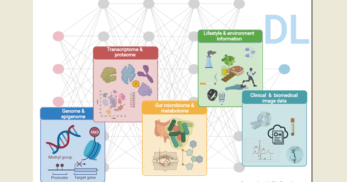
The Special Issue on The Deep Learning in Bioinformatics and Biomedicine is dedicated to discussing recent developments in the field of deep learning (DL) and its applications in bioinformatics and biomedicine ranging from the analyses of biomedical images to omics (genome, transcriptome, proteome, metabolome, and microbiome) and healthcare data.

DL is a subfield of machine learning, concerned with algorithms that are neural implementations with multiple processing layers, inspired by the structure and function of the human brain to learn and make intelligent decisions. DL approaches are designed for supervised classification, requiring input–output mapping that has to be learned from large amounts of labeled training data and requires the power of parallel and distributed computing. With the advent of the big data era, fueled by the rapid development of high-throughput technologies and digitalization of health records, exponentially increasing volumes of complex data are accumulating and becoming widely available, and their transformation into valuable knowledge is urgently required, especially in the context of personalized medicine and disease risk prediction to improve therapies for patients.

DL is believed to have the necessary transformative impact for the field and has recently caught the interest of both academia and industry, with the number of publications describing its application in bioinformatics and biomedicine steadily increasing, especially in the last 5 years. DL has been applied for a broad range of tasks, mostly the analysis of medical images, e.g., for the detection of cancer metastases. In genomics, DL has been used to prioritize potential disease-causing genetic variants and for the splice junction prediction. DL has also been recognized as more suitable to deal with electronic health records, which contain individual’s entire medical history as a series of multimodal data. Overall, the capabilities of DL for bioinformatics and biomedicine are not yet fully exploited, and future progress is expected to come, for example, from improving the encoding and learning from the raw data instead of using hand-crafted features, requiring domain expertise.

In this Special Issue, we invite submissions presenting or exploring cutting-edge research, recent advances, and innovative ideas in the area of DL for the fields of bioinformatics and biomedicine. Studies from both academia and industry analyzing biomedical images, omics (genome, transcriptome, proteome, metabolome, and microbiome) and/or healthcare data are welcome, as well as comprehensive review papers elaborating on the topic.

Dr. Baiba Vilne
Guest Editor

Manuscript Submission Information

Manuscripts should be submitted online at www.mdpi.com by registering and logging in to this website. Once you are registered, click here to go to the submission form. Manuscripts can be submitted until the deadline. All submissions that pass pre-check are peer-reviewed. Accepted papers will be published continuously in the journal (as soon as accepted) and will be listed together on the special issue website. Research articles, review articles as well as short communications are invited. For planned papers, a title and short abstract (about 100 words) can be sent to the Editorial Office for announcement on this website.

Submitted manuscripts should not have been published previously, nor be under consideration for publication elsewhere (except conference proceedings papers). All manuscripts are thoroughly refereed through a single-blind peer-review process. A guide for authors and other relevant information for submission of manuscripts is available on the Instructions for Authors page. Applied Sciences is an international peer-reviewed open access semimonthly journal published by MDPI.

Please visit the Instructions for Authors page before submitting a manuscript. The Article Processing Charge (APC) for publication in this open access journal is 2400 CHF (Swiss Francs). Submitted papers should be well formatted and use good English. Authors may use MDPI's English editing service prior to publication or during author revisions.

Keywords

  • machine learning
  • deep learning
  • big data
  • biomedicine
  • bioinformatics
  • biomedical imaging
  • (multi)-omics
  • genomics
  • transcriptomics
  • epigenomics
  • proteomics
  • metabolomics
  • metagenomics
  • health-care data
  • disease risk prediction
  • personalized medicine

Published Papers (3 papers)

Order results
Result details
Select all
Export citation of selected articles as:

Research

17 pages, 2809 KiB  
Article
Advanced Analysis of 3D Kinect Data: Supervised Classification of Facial Nerve Function via Parallel Convolutional Neural Networks
by Mohsen Shayestegan, Jan Kohout, Karel Štícha and Jan Mareš
Appl. Sci. 2022, 12(12), 5902; https://doi.org/10.3390/app12125902 - 9 Jun 2022
Cited by 7 | Viewed by 1574
Abstract
In this paper, we designed a methodology to classify facial nerve function after head and neck surgery. It is important to be able to observe the rehabilitation process objectively after a specific brain surgery, when patients are often affected by face palsy. The [...] Read more.
In this paper, we designed a methodology to classify facial nerve function after head and neck surgery. It is important to be able to observe the rehabilitation process objectively after a specific brain surgery, when patients are often affected by face palsy. The dataset that is used for classification problems in this study only contains 236 measurements of 127 patients of complex observations using the most commonly used House–Brackmann (HB) scale, which is based on the subjective opinion of the physician. Although there are several traditional evaluation methods for measuring facial paralysis, they still suffer from ignoring facial movement information. This plays an important role in the analysis of facial paralysis and limits the selection of useful facial features for the evaluation of facial paralysis. In this paper, we present a triple-path convolutional neural network (TPCNN) to evaluate the problem of mimetic muscle rehabilitation, which is observed by a Kinect stereovision camera. A system consisting of three modules for facial landmark measure computation and facial paralysis classification based on a parallel convolutional neural network structure is used to quantitatively assess the classification of facial nerve paralysis by considering facial features based on the region and the temporal variation of facial landmark sequences. The proposed deep network analyzes both the global and local facial movement features of a patient’s face. These extracted high-level representations are then fused for the final evaluation of facial paralysis. The experimental results have verified the better performance of TPCNN compared to state-of-the-art deep learning networks. Full article
(This article belongs to the Special Issue Deep Learning in Bioinformatics and Biomedicine)
Show Figures

Figure 1

22 pages, 7702 KiB  
Article
Synthetic Data Generation for the Development of 2D Gel Electrophoresis Protein Spot Models
by Dalius Matuzevičius
Appl. Sci. 2022, 12(9), 4393; https://doi.org/10.3390/app12094393 - 27 Apr 2022
Cited by 5 | Viewed by 1756
Abstract
Two-dimensional electrophoresis gels (2DE, 2DEG) are the result of the procedure of separating, based on two molecular properties, a protein mixture on gel. Separated similar proteins concentrate in groups, and these groups appear as dark spots in the captured gel image. Gel images [...] Read more.
Two-dimensional electrophoresis gels (2DE, 2DEG) are the result of the procedure of separating, based on two molecular properties, a protein mixture on gel. Separated similar proteins concentrate in groups, and these groups appear as dark spots in the captured gel image. Gel images are analyzed to detect distinct spots and determine their peak intensity, background, integrated intensity, and other attributes of interest. One of the approaches to parameterizing the protein spots is spot modeling. Spot parameters of interest are obtained after the spot is approximated by a mathematical model. The development of the modeling algorithm requires a rich, diverse, representative dataset. The primary goal of this research is to develop a method for generating a synthetic protein spot dataset that can be used to develop 2DEG image analysis algorithms. The secondary objective is to evaluate the usefulness of the created dataset by developing a neural-network-based protein spot reconstruction algorithm that provides parameterization and denoising functionalities. In this research, a spot modeling algorithm based on autoencoders is developed using only the created synthetic dataset. The algorithm is evaluated on real and synthetic data. Evaluation results show that the created synthetic dataset is effective for the development of protein spot models. The developed algorithm outperformed all baseline algorithms in all experimental cases. Full article
(This article belongs to the Special Issue Deep Learning in Bioinformatics and Biomedicine)
Show Figures

Figure 1

14 pages, 10843 KiB  
Article
iEnhancer-Deep: A Computational Predictor for Enhancer Sites and Their Strength Using Deep Learning
by Haider Kamran, Muhammad Tahir, Hilal Tayara and Kil To Chong
Appl. Sci. 2022, 12(4), 2120; https://doi.org/10.3390/app12042120 - 17 Feb 2022
Cited by 9 | Viewed by 2137
Abstract
Enhancers are short motifs that contain high position variability and free scattering. Identifying these non-coding DNA fragments and their strength is vital because they play an important role in the control of gene regulation. Enhancer identification is more complicated than other genetic factors [...] Read more.
Enhancers are short motifs that contain high position variability and free scattering. Identifying these non-coding DNA fragments and their strength is vital because they play an important role in the control of gene regulation. Enhancer identification is more complicated than other genetic factors due to free scattering and their very high amount of locational variation. To classify this biological difficulty, several computational tools in bioinformatics have been created over the last few years as current learning models are still lacking. To overcome these limitations, we introduce iEnhancer-Deep, a deep learning-based framework that uses One-Hot Encoding and a convolutional neural network for model construction, primarily for the identification of enhancers and secondarily for the classification of their strength. Parallels between the iEnhancer-Deep and existing state-of-the-art methodologies were drawn to evaluate the performance of the proposed model. Furthermore, a cross-species test was carried out to assess the generalizability of the proposed model. In general, the results show that the proposed model produced comparable results with the state-of-the-art models. Full article
(This article belongs to the Special Issue Deep Learning in Bioinformatics and Biomedicine)
Show Figures

Figure 1

Back to TopTop