Abstract
The use of metallic nanoparticles in engineering and biomedicine disciplines has gained considerable attention. Scientists are exploring new synthesis protocols of these substances considering their small size and lucrative antimicrobial potential. Among the most economical techniques of synthesis of metallic nanoparticles via chemical routes, which includes the use of chemicals as metal reducing agents, is considered to generate nanoparticles possessing toxicity and biological risk. This limitation of chemically synthesized nanoparticles has engendered the exploration for the ecofriendly synthesis process. Biological or green synthesis approaches have emerged as an effective solution to address the limitations of conventionally synthesized nanoparticles. Nanoparticles synthesized via biological entities obtained from plant extracts exhibit superior effect in comparison to chemical methods. Recently, conifer extracts have been found to be effective in synthesizing metallic nanoparticles through a highly regulated process. The current review highlights the importance of conifers and its extracts in synthesis of metallic nanoparticles. It also discusses the different applications of the conifer extract mediated metallic nanoparticles.
1. Introduction
Nanobiotechnology has had an enormous impact on all life forms, which has intrigued researchers currently [1]. Richard Feynman a physicist, in the year 1959 described the theoretical concept of miniaturization with hidden hints for nanotechnology for the first time, which implies a technological advancement utilizing materials with dimensions of about 1–100 nm [2,3]. Nanoparticles (NPs) have diverse applications in agriculture, biomedicine as antimicrobial agents, catalysis, biolabeling, sensors, electronics, fiber optics and other areas [4]. NPs bear properties that are different as compared to their bulk materials due to variation in their shapes, particle size and increased surface area. Studies on biomedical science invigorate interest in identifying the application of NPs in varied fields. The NPs can be synthesized, and stabilizing formulation can be developed through chemical and physical synthesis methods [5]. However, the use of environment-corrosive chemicals in the former synthesis process has ecodeterring potentials and can lead to the generation of toxic byproducts. Therefore, the desirability of the green synthesis approaches for NPs involves superior control over no or decreased use of ecotoxic chemicals [6]. Nanoparticles of desired size shape and functionality can be generated through two different fundamental approaches, viz., bottom-up and top-down methods [7]. Previously, nanomaterials/nanoparticles were synthesized using physical forces/agencies, such as ball milling, etching, lithography and sputtering [8].
The continuous developments in the timber industry have compelled scientists to opt for biological approaches to protect the environment. Generally, the natural ecofriendly synthesis of NPs involves the utilization of plant extracts and microbes [9,10,11,12]. These NPs can be prepared under different synthesis conditions and characterized by various analytical techniques [13]. The NPs derived from plant extracts dominate over NPs synthesized from microorganisms as the former is synthesized via a single-step, non-hazardous method. This type of synthesis is cost-effective and eco-friendly [7]. The conifer trees produce a broad spectrum of secondary metabolites, including polyphenols and terpenes, to overcome stress conditions [14]. These secondary metabolites have the potential to reduce metal ions into NPs [7]. This review intends to summarize the literature related to metallic NPs synthesized via conifers extracts, characteristics of the synthesized NPs and their biological properties like anticancer, antioxidant, antimicrobial, etc.
2. Importance of Conifers
Conifers are evergreen woody gymnosperms characterized by single veined leaves in the form of blade, scales and needle, and unisexual female and male cones having bract scales [15]. The conifers encompass two formerly known classes or orders viz Coniferopsida (Coniferales) and Taxopsida (Taxales) but exclude other gymnosperms like Cycads, Ephedras, Gnetums and two unique gymnosperms Welwitschia and Ginkgo. There are eight families: Araucariaceae, Cupressaceae, Cephalotaxaceae, Pinaceae, Phyllocladaceae, Podocarpaceae, Sciadopityaceae and Taxaceae [16]. Conifers have been stated as the reservoir of different natural products like essential oils (EOs), gums, resins and terpenes [17].
Conifer trees are abundantly found all over the world and have highly diverse natural compounds. Secondary metabolites such as phenols, flavonoids, alkaloids and terpenes found different part such as in leaves, bark, stem, seed and root tissues show structural and chemical diversity [14]. Therefore, conifers are considered as an essential source of biologically active agents possessing important medicinal attributes that have been utilized for the treatment of various ailments since ancient times [18,19]. These natural substances are known to exhibit the potential to influence the growth of insects and plants [20]. Natural compounds as EOs and extracts of genera Pinus, Cedrus, Juniperus, Taxus, Picea and Abies are effective against diseases such as cancer, diabetes, asthma, liver and kidney disorders, cardiovascular-related problems and many other bacterial and fungal infections [18]. The species, Araucaria angustifolia and A. excelsa, have ethno pharmaceutical values due to the presence of bioflavonoids, lignans while A. araucana is rich in terpenes [21]. It has been reported that in vitro and in vivo studies of an extract compound from A. angustifolia, A. bidwillii, Cupressus sempervirens, Podocarpus spp. and Thuja occidentalis possess biological activities like allelopathic, antibacterial, antidiabetic, antidepressant, antiedematogenic, anti-inflammatory, antioxidant, antiproliferative, antiviral, cytotoxic and entomotoxic, neuroprotective activities, gastro protective activity, hypocholesterolemic and antifeedant activities [20,21,22,23,24,25]. Acetone, dichloromethane, methanol and ethyl acetate extracts of leaves and cones of C. sempervirens var. horizantalis (CSH) and var. pyramidalis (CSP) exhibit inhibitory activity against tyrosinase (TYRO), butyrylcholinesterase (BChE) and acetylcholinesterase (AChE) enzymes [25].
Thuja (Esberitox) has been used for treating the common cold and many bacterial infections. In various in vivo and in vitro test models, Thuja immunopharmacological potential has also been studied [26]. In Korea, Torreya nucifera is known for its ethnopharmacological use against constipation, diabetes, hemorrhoids and infection caused by helminths. Additionally, in Asia, T. nucifera reported as having antioxidant activity (L.) seeds are used in treating various diseases [27,28]. Amentoflavone, a biflavonoid compound naturally occurring in Podocarpaceae and Cupressaceae shows good anti-inflammation, antidiabetes, antioxidation and antisenescence effects on many essential processes of the central nervous and cardiovascular system [29]. Reports suggest that Taxus yunnanensis extracts and their purified compounds displayed cytotoxicity against HeLa, MCF-7, HT1080 and K562 cell lines. Many researchers have found that the extracts of T. yunnanensis can be used as an antiosteoporotic, anti-inflammatory and hypoglycemic agent for diabetes [30]. As per the literature, it has been found that conifers are abundantly found and have much importance in our daily life [14]. So, now a day’s keeping these things in mind, new research of interest is growing to develop more uses of conifers by deriving metallic NPs, which helps in dealing problems, diseases related to human beings plants pathogens agriculture, promoting plant growth by enhancing the quality of soil and industrial uses [31,32,33,34].
3. Green Synthesis of Nanoparticles Mediated by Conifers Extract Synthesis Mechanism, Characterization
Plants are considered as bioreactors for the synthesis of metallic NPs. Different physical and chemical approaches are used to synthesize metal NPs, which allow one to obtain particles with the desired characteristics [35]. Conifers exhibit unique metal NPs synthesis properties (Figure 1). Green-synthesis of metallic NPs is an efficient, trouble-free, economical and environmental-friendly biological synthesis approach [36]. Metallic NPs include both types of pure metals and metal oxides, which have various applications [37]. The critical factor, which needs to be considered during the synthesis of NPs, is the selection of a solvent medium, which needs to be a non-toxic material for NPs stabilization and an ecofriendly reducing agent [38].
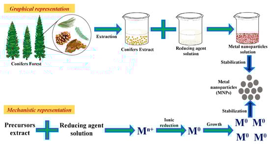
Figure 1.
Graphical overview of the green synthesis protocol using conifer extracts for synthesizing metallic nanoparticles.
3.1. Mechanism
For the synthesis of metallic NPs from conifers, plant samples were collected, washed and dried to form a powder. The powdered sample was mixed with 250 mL double distilled water and boiled for 5 min. The heated materials were filtered and centrifuged to obtain a clear extract. The metallic salt solution was added dropwise into extract under sonication and then incubated for different time intervals (10–120 min), pH ranges (7–11) and temperatures. The color change indicates the formation of NPs due to the reduction of metal ions into metal atoms by excitation of surface plasmon vibrations [30].
Additionally, compounds such as polyphenolics, peptides, vitamins, sugars and water present in extracts of conifers are effective for synthesizing NPs [39,40,41,42,43,44]. Interestingly, plant metal bioaccumulation studies have shown that metals are usually aggregated in the form of NPs. The polyphenol compounds are responsible for the reduction process by the abstraction of hydrogen [35]. Further, these compounds also act as capping agents to avert the coalescence of colloidal particles through the interplay of electrostatic forces [45]. The metal ion reduction was monitored by measuring the absorbance from 300 to 600 nm using UV–vis spectrophotometer to find the absorbance peak. The solution was then centrifuged, and the pellet was dried by lyophilization [30].
3.2. Characterization Techniques
Metallic NPs synthesized from various conifers extracts had diverse shape, size and surface area; and were categorized using different approaches as shown in Table 1. The synthesized metallic NPs structural composition, size, shape and crystal phase were deduced using energy-dispersive X-ray spectroscopy (EDX), FTIR, SEM, HRTEM, UV–Vis [46], XRD, atomic force microscopy (AFM) and dynamic light scattering (DLS) [47,48]. The range of absorption of the UV spectra from wavelength 300 to 800 nm illustrates the existence of several metallic NPs of varying size, i.e., from 6 to 200 nm [30,47]. DLS analysis entails estimation of the size of synthesized NPs and the quantification of the charges on the surface of the NPs. The composition of the element is determined through the EDX analysis [49]. XRD is functionalized to recognize crystallite size. FT-IR spectroscopy detects the surface residues and the functional groups such as flavonoids, hydroxyls and phenols, which bond with the surface of the NPs throughout the process of the synthesis for an effective reduction and stabilization [50].

Table 1.
Green synthesis of metallic nanoparticles synthesized from conifer extracts and their characterization through different analytical techniques.
4. Types of Nanoparticles
4.1. Metal Nanoparticles
4.1.1. Silver Nanoparticles
In nanotechnology, silver nanoparticles (AgNPs) are well-known. They have gained significant attention because of their extraordinary antimicrobial ability against several microorganisms and role in combating the drug resistance menace in multidrug resistant microbes. These properties of AgNPs can be effectively utilized in food packaging, agriculture for eradication/ management of crop diseases, biomedicine, drug resistivity and pharmacology to formulate novel drug carrier systems and in decontamination of water [76,77]. AgNPs derived MNPs from various conifers, and their applications are in Table 2.
Table 2.
Agricultural and biomedical applications of conifer plant extract derived metal (Ag and Au) nanoparticles.
Synthesis of gold and silver NPs from different plant organ extracts such as bark, needle, cone, callus and berry of a variety of coniferous plant species are shown in Table 1. Due to better adaptability and easy down streaming to nanosystems, AgNPs synthesis supports better control on the shape and dimensions of NPs [78]. In the presence of aqueous medium and oxygen, silver atoms found in Ag2O NPs release Ag+ ions. Ag+ ions are a potent oxidant, which can oxidize microbial biomolecules non-specifically. In water-treatment systems, this potential makes AgNPs, suitable alternative disinfectants [33]. However, Das et al., in 2018 reported in their studies that AgNPs are toxic to numerous organisms, mammalian cells and humans [79]. The novel AgNPs synthesis via two-step procedure initiates via the reduction of Ag+ ions to Ag, followed by aggregation and stabilization of nanoparticles [80]. The agricultural and biomedical applications of AgNPs obtained from different coniferous plants are shown in Table 2.
4.1.2. Gold
Gold nanoparticles (AuNPs) display many unique biological, favorable chemical and optical properties, which make them safe for more comprehensive applications [82]. Due to their significant biocompatibility, Au NPs are widely reported in different varieties of conifers, as shown in Table 1. Many studies proved that catalytic, optical, physical and thermal properties of AuNPs depend on their shape and size, which has drawn attention towards the development of an optimized protocol for the synthesis of monodisperse AuNPs formulation. Currently, biological synthesis approach involving plant extracts without the use of toxic chemicals during synthesis has gained enormous attention due to the benefit of avoidance or minimization of adverse impact on the application [83]. Synthesis of gold NPs from different types of extracts of various conifers such as Pinus, Taxus, Juniperus, Abies and Thuja has been reported [54,73]. AuNPs synthesized from plant extracts exhibit several advantages with primary includes the biocompatible or less-toxic nature of the generated NPs. Moreover, the therapeutic potential of drugs can be potentially enhanced by significantly reducing the particle size to the nanoscale. Use of such formulations will enhance the interaction of the active drug molecule with a specific protein and increases the efficiency of the formulated drug [84]. High purity AuNPs can be utilized for anticancer applications and also have the potential to bind to the human serum albumin (HSA) protein [85]. Application of gold NPs synthesized from various conifers is shown in Table 2.
4.2. Nanoparticles of Metallic Oxides
Nanoparticles of zinc oxide (ZnO NPs) are non-toxic, biosafe, ecofriendly and have an easy amendable nature that makes them the targeted candidate for biological applications [86]. Zn NPs derived from plant extract have been applied for varied biomedical benefits such as the treatment of chronic disorders like cancers, and diabetes, as a potent antimicrobial agent to kill pathogenic bacteria, fungi and for use as a photocatalyst for decontamination of contaminant dyes from water and sediments [72,87,88]. Additionally, studies suggested that Zn NPs have significance in eradicating hardy, persistent aquatic weed resistant to chemical and physical means of eradication. Taxus baccata extracts have been used to synthesize Zn NPs. UV–Vis spectrophotometry analysis of the T. baccata extracts derived Zn NPs exhibited in the range of 250–400 nm. Zn NPs obtained were crystalline and hexagonal shaped, showing an average particle size of 20–25 nm [72].
Copper nanoparticles (CuNPs) have unique properties like high chemical reactivity, mechanical strength and antimicrobial potential, which made them useful for diverse applications. Additionally, due to a high surface area–volume ratio, CuNPs can easily interact and react with other NPs [89]. Studies revealed excellent antibacterial activity of Cu-NPs derived from P. merkusii flower extract against B. subtilis and E. coli as compared to Ag-NPs [53].
Iron nanoparticles (FeNPs) have unique biological and physicochemical characteristics, which make it the most applicable nanostructures in a different field of science. Additionally, FeNPs are biocompatible and easy to handle and have sturdy magnetic properties. FeNPs synthesized from P. eldarica needle extract had a size range of 8–34 nm [61]. FeNPs has transpired antibacterial characteristics against bacterial strains like B. subtilis, S. aureus, E. coli, P. aeruginosa, K. pneumonia and Streptococcus pyogenes [33]. In environmental sciences, FeNPs have been used both as a catalyst and nanosorbent [90]. Additionally, they are efficient in removing and detoxifying chemical toxic agents and organic pollutants like arsenic, chromium, carbon monoxide, mercury and toxic ions [91,92,93,94]. Additionally, FeNPs have been stated to be operative as a Fenton-like catalyst for the removal of contaminant toxic dyes from aqueous environments [95]. Metal derived NPs and their application in are presented in Figure 2.
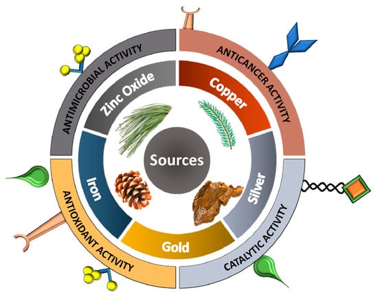
Figure 2.
Graphical illustration of conifer sources used for the production of metallic nanoparticles with potential biological activities.
5. Conifer-Derived Metallic Nanoparticles Potential Applications
5.1. Biomedical Applications
5.1.1. Antimicrobial Action
Metal NPs synthesized from different conifer extracts possess good antibacterial, antifungal and insecticidal potentials. Plant extracts show mild activity against some Gram-positive and Gram-negative bacteria and fungi. At the same time, MNPs synthesized from plant extracts are known to exhibit high potential against yeast and Gram-positive and Gram-negative bacteria. It also has been reported that MNPs derived from conifers found in high altitude show good fungal and bacterial inhibitory activity [65].
Mechanism of Action Towards Microbes
Despite several approaches, technologies and formulations had been made over the years; still, the precise mechanism of action of MNPs toward bacteria and fungi has not been fully understood. However, several reports are available in the literature, which comes in favor that the mechanism of action of MNPs in bacteria and fungi is almost similar. The antimicrobial action of MNPs is related to the following four-step mechanism; (a) direct contact with the cell membrane, (b) destabilization of the cell membrane, change in its permeability and release of metal ions, (c) production of reactive oxygen species (ROS) and free radicals and (d) signal transduction pathways modulation (Figure 3) [96].
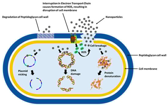
Figure 3.
Graphical illustration of the antimicrobial mechanism of nanoparticles.
Direct Contact with the Cell Membrane
The size and zeta potential of the MNPs influence their adhesion on the bacterial surface. Depending on the technique used for their fabrication, MNPs may possess positive, negative or neutral charges. Since both bacteria and fungi have a partial negative charge on their surfaces, positively charged MNPs gets strongly attracted to the negatively charged cell surface, resulting in improved antimicrobial activity as compared to negatively charged or neutral MNPs [97].
Destabilization of the Cell Membrane, Changes in Its Permeability and Metal Ions Release
After adhesion, MNPs with smaller size penetrate directly into the cell, whereas larger NPs are retained outside the cell surface. In both cases, MNPs continue to release their ions and destabilize cell membrane. Cell wall destabilization causes bacterial permeability, allowing entry of large-sized NPs into the cell. Inside the cell, improved production of ions leads to interaction with proteins, lipids, and DNA, resulting in cell dysfunction and cell death [98].
Production of Reactive Oxygen Species (ROS) and Free Radicals
Furthermore, MNPs are well known to produce ROS and free radicals such as H2O2, O2− and OH•. The ROS and free radicals are highly reactive species that are responsible for the production of extreme oxidative stress inside the cell resulting in cell apoptosis, proteins destruction, DNA damage and finally cell death [99].
Signal Transduction Pathways Modulation
It has been noticed that under normal conditions, ROS occurs naturally in microbes. Antioxidant enzymes such as glutathione (GSH), superoxide dismutase and catalase protect the microbes from the effects of ROS. These enzymes are efficient in eliminating some toxic species at a low concentration, but when the oxidative stress becomes high, they are rendered ineffective in neutralizing toxic species at a high concentration. At high concentration, oxidative species interact with respiratory proteins and inactivate these antioxidant enzymes because of their high affinity toward thiol, carboxyl and phosphates groups present in respiratory proteins [100].
Reported study on antimicrobial (bacteria and fungi) activity by different parts of the conifers derived MNPs were discussed. The antibacterial activity of Picea abies bark extract synthesized AgNPs was investigated in vitro against E. coli, K. pneumoniae and P. aeruginosa with an minimum inhibitory concentration (MIC) of 3.25, 3.25 and 7.5 mg/mL [101]. AgNPs synthesized from T. yunnanensis callus had a significant growth inhibitory activity against human pathogenic bacteria with an MIC of 8 for E. coli, 1 for B. subtilis, 1 for S. aureus and 4 μg/mL for S. paratyphi, however, the growth of bacteria was inhibited by AgNPs 2 µg/mL [30]. The MIC values shown by AgNPs obtained from the cone extract of Pinus thunbergii were low for the Gram-negative bacterial strains, Xanthomonas oryzae, Burkholderia glumae and Pseudomonas syringae were 11.9 and 6 μL/mL respectively while the MIC for the Gram-positive Bacillus megaterium and B. thuringiensis were 11.9 μL/mL [81]. The Pinus densiflora cone extract mediated AgNPs at a working concentration of 40 µg per mL effectively inhibited skin pathogen bacteria such as Brevibacterium linens (zone of inhibition: 7 mm) compared to commercial AgNPs Propionibacterium acnes (14 mm), Staphylococcus epidermidis (10 mm) and B. cereus (9 mm) [56]. The antibacterial activity was evaluated on S. aureus by aqueous extract, and copper NPs derived from the P. merkusii cone. The bacterial growth–inhibition was recorded to be substantially high (29.67 ± 0.01 mm) at 100% concentration [53]. The researcher advocated the use of this formulation as a natural fungicide. AgNPs mediated from the gum of A. heterophylla have been reported to be effective against Gram-positive bacteria Streptococcus sp. [48].
The zone of inhibition of AgNPs by leaves extract of C. torulosa against B. subtilis was observed and found to be 5 mm for 50 µL, 6 mm for 100 µL, 6 mm for 150 µL and 3 mm for 200 µL whereas Salmonella enterica showed 6 mm for 50 µL, 7 mm for 100 µL, 8 mm for 150 µL and 10 mm for 200 µL. AgNP’s showed the zone of inhibition against P. aeruginosa as 4 mm for 50 µL, 6 mm for 100 µL, 6 mm for 150 µL and 6 mm for 200 µL [64]. Few researchers have stated that Juniperus procera; obtained from a high altitude possess both antibacterial and antifungal activity [102]. J. procera leaf extract derived AgNPs showed antibacterial and antifungal activity against B. subtilis, K. pneumonia, Micrococcus luteus, Proteus mirabilis and C. albicans with the formation of inhibition zones of 28 ± 1.2, 18 ± 0.9, 28 ± 1.1, 29 ± 1.3 and 24 ± 0.1.2 mm respectively [65]. Studies reported that T. occidentalis mediated AgNPs exhibited an inhibitory effect on B. subtilis, Listeria monocytogenes, P. aeruginosa, Salmonella typhimurium and S. aureus at 5–10 g/mL concentrations. The AgNPs biosynthesized by using T. occidentalis (L.) leaves extract exhibits improved inhibitory activity against microbes [70]. The AgNPs concentration 30 µg/mL made an inhibitory growth zone for B. subtilis (15.6 mm), E. coli (16 mm) and Pseudomonas putida (16.3 mm). Besides, at a concentration of 10 µg/mL growth of all the fungal strains used in the study was considerably reduced with the medium showing no signs of proliferation. The study showed that these biosynthesized AgNPs could serve as antibiotic agents and can be used in combination with other antibiotics [66]. Studies suggested that the leaves extract of T. occidentalis synthesized AgNPs showed significant larvicidal activity against the Culex quinquefasciatus mosquito with an LC50 value of 39.90 ppm and LC90 value of 129.48 ppm. Additionally, as it is dose-dependent, the increase in concentration from 100 to 300 ppm increased the mortality rate to 100% after 72 h of treatment [68]. Pine pollen derived AgNPs efficiently exhibited quantitative inhibition and disruption of growth of fungus Neofusicoccum parvum [103]. AuNPs synthesized from P. kesiya pollen exhibited fungicidal activity against Candida albicans, at 500 μg/mL concentration with 17.51 zones of inhibition [73]. The AgNPs generated from the P. wallichiana stem displayed good antibacterial activity than antifungal activity against Acinetobacter baumannii, E. coli, Morganella morganii, Proteus vulgaris, P. aeruginosa and S. aureus. A. baumannii showed activity 60% with minimal inhibitory concentration (MIC) of 2.36 mg/mL and minimal bactericidal concentration (MBC) of 5.0 mg/mL. Additionally, AgNPs showed moderate antifungal potential against Aspergillus niger (40%), Penicillium notatum (45%) and P. chrysogenum (50%), whereas it displayed minimum activity against A. parasiticus (13%), Hemimycena pseudocrispula (30%) and Verticillium longisporum (21%) [74].
5.1.2. Anticancer
According to the WHO (World Health Organization) report of 2018, cancer has emerged as a leading cause of death, accounting for an estimated death of 9.6 million people globally. In males, cancers such as colorectal, liver, lung, prostate and stomach are predominant while cervical, breast, colorectal, lung, and thyroid cancer are most common in females [104]. Different types of natural bioactive compounds present in plants are believed to have medicinal value and are potential candidates to develop anticancer drugs. Various classes of heterocyclic compounds (alkaloids and flavonoids) have been isolated from several conifer plants showing cytotoxic efficacy against multiple types of cancerous cells both in vitro and in vivo [74,105]. The field of nanotechnology holds the potential to transform cancer diagnostic methods and therapeutic technologies [106].
Mechanism behind anticancer activity
In literature, numerous reports are available describing the mechanism behind the action of MNPs toward cancer cells (Figure 4). However, the precise mechanism of anticancer activity is till not well understood. It is suggested that the interaction between MNPs and cancer cells may take place in various ways like electrostatic interaction between the cell surface and metallic nanoparticles, nanoparticles captured by cell receptors and internalization of nanoparticles via endocytosis. It is well known that MNPs can easily generate reactive species inside the cell system leading to DNA damage, mitochondrial disruption, protein oxidation and, finally, cell death [106,107]. Additionally, the ability of MNPs to generate the hyperthermia effect inside the cancer cell functions as a drug carrier and regulate pH-dependent release makes it a superior candidate for anticancer activity [108].
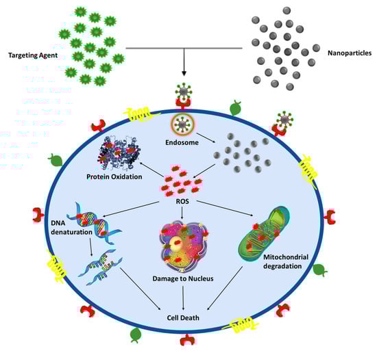
Figure 4.
Graphical illustration of the anticancer mechanism of nanoparticles.
Cancer is a lethal threat to humankind [109,110]. Regrettably, despite the success in designing various anticancer therapeutics, this disease remains relentless because of the toxic nature of the drugs towards normal cells. Therefore, the treatment of cancer with the help of NPs can be an effective approach. The nanosize of NPs makes them distinctive and reconcilable for penetration within the cells. This attribute of NPs contributes to its efficacy against tumor cells even at a low dosage while also reducing the toxic effects to the adjacent normal cells [101]. NPs derived using conifer extracts have gained significant attention due to their adaptable properties and easy synthesizing procedure [78].
The pine barks extract derived AuNPs have shown concentration-dependent cytotoxicity towards non-malignant HEK293 kidney cells and malignant A549 lung cells with a decrease in cell viability to 23% and 35% respectively. Additionally, it also increases the death rate of lung cancer cells and reduces the toxic effects on non-malignant human embryonic kidney cells. Whereas, pine bark derived oleamide capped AuNPs have been reported to decrease the viability of the cancerous cells with an insignificant cytotoxic effect on non-malignant cells. Thus, the pine extracts mediated AuNPs exhibited the potential of anticancer agents [85]. Studies have unveiled that Pinus sylvestris bark extract contains high amounts of phenols and shows cytotoxic effects towards HeLa cells by inducing apoptosis after 48 h of exposure [111]. Additionally, the biosynthesized AgNPs from callus of T. yunnanensis showed significant inhibition in a dose-dependent manner in four human malignant cells. For the A549 cell line, the half maximal inhibitory concentration i.e., IC50 of AgNPs was recorded to be 40.3 µg/mL while it was reported to be 42.2 µg/mL for the MCF-7cell line, which indicated a moderate cytotoxic effect on both the cell lines [30]. On the increase in concentration from 0.2 to 1 mg/mL of biologically synthesized AgNPs using the gum of A. heterophylla has been reported to reduce the cell viability rate of the MCF 7 cell line [48]. ZnNPs synthesized from T. baccata showed a cytotoxicity effect on the breast cancer cell line. Zn has the efficiency to prevent cell change to cancer cells. The MTT test by the MCF-7 cell line proved that the anticancer activity of Zn nanoparticles from T. baccata leaves ethanolic and alcoholic extract was expected to be higher than that of extract alone as the ZnNPs concentration up to 50 mM decreased cell viability (24.83%) with a maximum cell inhibition of 75.17% [72]. It has been found that AgNPs synthesized from the aqueous extract of T. baccata needles having an IC50 value of 0.25 µg mL-1 showed significant mortality of malignant cells of the MCF-7 cell line after 48 h of exposure. The cytotoxic effect of NPs substantially improved with the increase in dosage and incubation time with the killing of more than 50% of the cells after exposure for 72 h [34]. Additionally, it has been reported that the T. baccata needles ethanolic extract synthesized AuNPs displayed gradually higher anticancer activity on all the three cancer cell lines, i.e., MCF7, HeLa and Caov-4 in contrast to the aqueous extracts derived AuNPs [59].
T. occidentalis leaves mediated AgNPs have also been accorded for anticancer potential against HeLa, MDA-MB 231, MCF 7 and KB cell lines at a dosage of 6.25–50 g/mL. AgNPs exhibited 45% cell death in peripheral blood mononuclear cells (PBMCs), which is significantly less than the cytotoxicity exhibited in the anticancer study with an equal dose. Cytotoxicity of Thuja extracted AgNPs was significantly much higher in cancer cells compared to other normal cells [70]. The MTT assay showed that the antiproliferative action of the T. occidentalis leaves AgNPs against the HeLa cell line is dose-dependent. However, with the increase in AgNPs concentration, there was a gradual appearance of toxicity. Furthermore, Coincubation of the AgNPs with the concentration of 50 g/mL results in the reduction of 70% viability of the cancerous cells. Whereas, treating by AgNPs up to a 50 g/mL concentration on L929 cells showed no statistically significant change in cell proliferation. However, on the elevation of AgNPs concentration to 65 g/mL showed a significant reduction in cell viability [69].
5.1.3. Antioxidant
Reactive oxygen species (ROS) such as superoxide dismutase, hydrogen peroxides and hydrogen radicals are highly reactive and toxic species, overproduction of which may cause damage to lipids, DNA, carbohydrate, proteins and build oxidative stress, thereby resulting in the formation of different diseases [112]. Antioxidants molecules have emerged as potential candidates that scavenge these free radicals and ROS and prevent associated problems [113]. MNPs act as an antioxidant agent via two possible mechanisms; (1) via single electron transfer and (2) hydrogen transfer. Single-electron transfer refers to the reduction of oxidative compounds by an electron donation, whereas hydrogen transfer includes total oxyradical scavenging using a hydrogen atom [114,115]. During the oxidation process, the generation of free radicals takes place in the cells. The formation of free radicals disturbs the normal cellular processes and causes cellular damage (Figure 5). Antioxidants are required to prevent cellular, and DNA damage and oxidative stress besides help prevent chronic disorders such as cancer, malignant transformations and heart diseases [116].
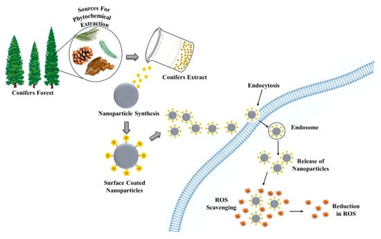
Figure 5.
Graphical illustration of the antioxidant mechanism of nanoparticles.
The AgNPs mediated from P. abies stem bark extract possessed free radical scavenging activity against both 2, 20-azino-bis (3-ethylbenzothiazoline-6-sulphonic acid (ABTS) and DPPH radicals [51]. The study revealed that instead of metal oxides, the noble metal NPs (AgNPs and AuNPs) are strong scavengers of superoxide ions, nitric oxide radicals, DPPH and hydroxyl radicals. The biosynthesized AgNPs from the P. wallichiana stem has been stated to have the proficient antioxidant potential of 70.25 ± 0.56% and 61.77 ± 0.828% at a concentration of 400 and 500 µg/mL respectively [74].
5.2. Other Activities
Different development parameters like biomass accumulation, growth, germination and biochemical activities have been evaluated in plants, which shows varied effects of these metallic NPs in diverse plants [37]. Zero valent iron coated silver nanoparticles (ZVI@AgNPs derived using leaf extract of C. sempervirens have been reported to show significant catalytic potential as they can remove 98.5% initial dye in 4 h [33]. Decolorization of Congo red, Orange G and Rhodamine B dyes have been reported by use of AgNPs derived using Cupressus torulosa D.Don leaf extract [64]. Green synthesized MNPs derived using T. occidentalis leaves (GSNPs) have demonstrated considerable improvement in plant growth and soil quality. Total nitrogen, and nitrate leaching assays, revealed that GSNPs reduces the leaching of nitrate from the soil, which substantially improves the total nitrogen (N) content. The GSNPs amended soil has shown an adequate yield of Proteus vulgaris. Moreover, high chlorophyll content and enzyme activation have been recorded in GSNPs exposed plants. Of GSNP a 50 mg kg−1 concentration has been claimed to be advantageous for improving plant growth and soil quality. However, a higher dosage of green synthesized nanoparticles (GSNPs) might be detrimental to plants. T. occidentalis-derived NPs have been reported to enhance the N availability in soil, thus significantly advancing the nitrogen-driven growth potential of crop plants by acting as growth promoters [79]. Pine needle extract mediated palladium nanoparticles (PdNPs) have been developed in which palladium/residue of the pine needle (Pd/RPN) are reused as a catalyst. The Pd/RPN nanocomposite shows high catalytic activity for the Suzuki coupling reaction. Moreover, the catalyst can be easily recovered through centrifugation and reutilized for at least six-folds without losing its viability [32].
Clotting of blood within the blood vessels often leads to thrombotic diseases such as myocardial infarction. Diverse agents are employed to dissolve blood clots in vessels, which may otherwise have serious and fatal consequences. Studies have reported that P. wallichiana stem mediated AgNPs shows effective thrombolytic activity in contrast to the crude extract. The thrombolytic potential of crude leaves extract, and AgNPs exclusively were 15.9% and 25.8%, respectively [74]. The P. wallichiana stem mediated AgNPs have also shown a significant antipyretic effect in mice after 1, 2 and 3 h of administration. AgNPs cause the reduction in temperature (36.40 ± 3.11°C at 10 mg/kg) and bodyweight of mice in contrast to paracetamol (50 mg/kg) [74].
6. Conclusions and Future Outlook
Nature has an ingenious way to synthesize highly effective miniature functionalized materials. Additionally, the increasing interest towards green chemistry and its utilization for synthesizing metallic nanoparticles lead an aspiration to develop an economical and environment-friendly approach. The primary benefit of synthesizing these metallic nanoparticles with conifer plant extract is that it is cost-effective, economical, energy-efficient and healthier, as it protects the environment and human health by producing less waste and safe products. Besides, the synthesis of metallic nanoparticles from conifer extracts has an advantage over other biological entities such as microbes, which require maintenance of culture and consume a lot of time. Moreover, microbes also lose their ability to synthesize nanoparticles with time. Hence, metallic nanoparticles synthesized via plant extract will have an immense impact in the coming future. Green synthesized metallic nanoparticles have significant features of nanotechnology with unmatched applications.
Various reports have been recorded about the synthesis of metallic nanoparticles using conifer extracts. However, there is a need to explore commercial, economic and ecofriendly approaches. Moreover, reproducibility of NPs in a high amount also poses a challenge in the green synthesis of conifer-derived MNPs. It has been observed that there is substantial variation in the composition of conifer extracts of the same species when procured from different regions, which is an attribute for the different result during in vitro evaluation. Moreover, subsequent interaction with metal ions is believed to contribute to the variability in size and shape of nanoparticles. This imposes a major challenge for the synthesis of metallic nanoparticles with the help of conifer extracts as the stabilizing and reducing agent. Therefore, the selection among plants with the season in which the biological material is collected is an essential requirement aiming the reproducibility of the synthesis. Additionally, in MNP synthesis, the reaction time and temperature influence the shape and size of synthesized MNPs. Many studies do not report the time of the year when the plant material for the MNPs synthesis is collected, the conditions of growth wherein the plant was cultivated or collected, and the quantification of major metabolites present in the extract. All that information must be considered essential for the reproducibility of the process and to understand the mechanisms culminating in the formation of MNPs. Furthermore, there is a need to identify biomolecules responsible for the synthesis of metallic nanoparticles and develop a single-step method to surpass the above-discussed challenges and pave the way for new opportunities for green chemistry to create ecofriendly metallic nanoparticles.
Author Contributions
Conceptualization P.B., R.S., and K.K.; Manuscript writing, K.B. and D.S.D.; Manuscript editing, A.K., E.N., S.B., A.S., S.T., C.C., and R.V.; Critical revising, D.K., P.B., R.S., and K.K. All authors have read and agreed to the published version of the manuscript.
Funding
This research was funded by the University of Hradec Kralove (Faculty of Science VT2019-2021). This study was partially supported by grants from the Ministry of Health of the Czech Republic (FN HK 00179906).
Conflicts of Interest
The authors declare no conflict of interest.
Abbreviations
| A549 | Human Lung Carcinoma Epithelial Cells |
| ABTS | 2,2’-azino-bis(3-ethylbenzothiazoline-6-sulfonic acid) |
| AChE | Acetylcholinesterase |
| AFM | Atomic force microscopy |
| Ag+ | Silver Ions |
| AgNPs | Silver Nanoparticles |
| ATR-FTIR | Attenuated total reflection-Fourier-transform infrared spectroscopy |
| AuNPs | Gold Nanoparticles |
| BChE | Butyrylcholinesterase |
| Caov-4 | Human Ovarian Cancer Cell Line |
| CSH | Cupressus Sempervirens Var. Horizantalis |
| CSP | Cupressus Sempervirens Var. Pyramidalis |
| CuNPs | Copper Nanoparticles |
| DLS | Dynamic Light Scattering |
| DNA | Deoxyribonucleic Acid |
| DPPH | 2,2,1-diphenyl-1-picrylhydrazyl |
| EDX | Energy-dispersive X-ray spectroscopy |
| Eos | Essential Oils |
| FeNPs | Iron Nanoparticles |
| FESEM | Field Emission Electron Microscope |
| FT-IR | Fourier-Transform Infrared Spectroscopy |
| GSH | Glutathione |
| GSNPs | Green Synthesized Nanoparticles |
| H2O2 | Hydrogen peroxide |
| HEK293 | Human Embryonic Kidney Cell Line |
| HeLa | Human Cells Derived From Cervical Cancer Cell Line |
| HRTEM | High-Resolution Transmission Electron Microscopy |
| HSA | Human Serum Albumin |
| HT1080 | Human Fibrosarcoma Cell Line |
| IC50 | Half Maximal Inhibitory Concentration |
| K562 | Immortalised Myelogenous Leukemia Cell Line |
| KB | Ubiquitous KERATIN-Forming Tumour Cell Line Hela |
| L929 | Mouse Fibroblast Cell Line |
| LS174T | Human Caucasian colon adenocarcinoma cell line |
| MDA-MB231 | Epithelial, Human Breast Cancer Cell Line |
| MCF-7 | Breast Cancer Cell Line |
| MIC | Minimum Inhibitory Concentration |
| MNPs | Metal Nanoparticles |
| MTT | 3-(4,5-dimethylthiazol-2-yl)-2,5-diphenyltetrazolium bromide |
| ND | Not determined |
| NPs | Nanoparticles |
| NS | Not Specified |
| O2− | Superoxide Ion |
| OH• | Hydroxyl Radical |
| PBMCs | Peripheral Blood Mononuclear Cells |
| Pd/RPN | Palladium/Residue of the Pine Needle |
| ROS | Reactive Oxygen Species |
| RT | Room Temperature |
| SEM | Scanning Electron Microscopy |
| SMMC-7721 | Hepatocellular Carcinoma Cell Line |
| TEM | Transmission Electron Microscopy |
| TYRO | Tyrosinase |
| UV–Vis | Ultraviolet-Visible Spectroscopy |
| WHO | World Health Organization |
| XRD | X-Ray Powder Diffraction |
| ZnO NPs | Zinc Oxide Nanoparticles |
| ZVI@AgNPs | Zero Valent Iron Coated Silver Nanoparticles |
References
- Bhattacharyya, D.; Singh, S.; Satnalika, N.; Khandelwal, A.; Jeon, S.-H. Nanotechnology, big things from a tiny world: A review. Int. J. u-e-Serv. Sci. Technol. 2009, 2, 29–38. [Google Scholar]
- Baker, S.; Satish, S. Endophytes: Toward a vision in synthesis of nanoparticle for future therapeutic agents. Int. J. Bio-Inorg. Hybd. Nanomat. 2012, 1, 67–77. [Google Scholar]
- Sarmast, M.K.; Salehi, H. Silver Nanoparticles: An Influential Element in Plant Nanobiotechnology. Mol. Biotechnol. 2016, 58, 441–449. [Google Scholar] [CrossRef] [PubMed]
- Salam, H.A.; Rajiv, P.; Kamaraj, M.; Jagadeeswaran, P.; Gunalan, S.; Sivaraj, R. Plants: Green route for nanoparticle synthesis. Int. J. Biol. Sci. 2012, 1, 85–90. [Google Scholar]
- Iravani, S. Green synthesis of metal nanoparticles using plants. Green Chem. 2011, 13, 2638–2650. [Google Scholar] [CrossRef]
- Ahmed, S.; Ahmad, M.; Swami, B.L.; Ikram, S. A review on plants extract mediated synthesis of silver nanoparticles for antimicrobial applications: A green expertise. J. Adv. Res. 2016, 7, 17–28. [Google Scholar] [CrossRef]
- Singh, J.; Dutta, T.; Kim, K.-H.; Rawat, M.; Samddar, P.; Kumar, P. ‘Green’ synthesis of metals and their oxide nanoparticles: Applications for environmental remediation. J. Nanobiotechnology 2018, 16, 1–24. [Google Scholar] [CrossRef]
- Cao, G. Nanostructures & Nanomaterials: Synthesis, Properties & Applications; Imperial College Press: London, UK, 2004; ISBN 1860944809. [Google Scholar]
- Kavitha, K.S.; Baker, S.; Rakshith, D.; Kavitha, H.U.; Yashwantha Rao, H.C.; Harini, B.P.; Satish, S. Plants as green source towards synthesis of nanoparticles. Int. Res. J. Biol. Sci. 2013, 2, 66–76. [Google Scholar]
- Akhtar, M.S.; Panwar, J.; Yun, Y.-S. Biogenic Synthesis of Metallic Nanoparticles by Plant Extracts. ACS Sustain. Chem. Eng. 2013, 1, 591–602. [Google Scholar] [CrossRef]
- Kumar, H.; Bhardwaj, K.; Sharma, R.; Nepovimova, E.; Kuca, K.; Dhanjal, D.S.; Verma, R.; Bhardwaj, P.; Sharma, S.; Kumar, D. Fruit and Vegetable Peels: Utilization of High Value Horticultural Waste in Novel Industrial Applications. Molecules 2020, 25, 2812. [Google Scholar] [CrossRef]
- Mehta, M.; Sharma, P.; Kaur, S.; Dhanjal, D.S.; Singh, B.; Vyas, M.; Gupta, G.; Chellappan, D.K.; Nammi, S.; Singh, T.G.; et al. Plant-based drug delivery systems in respiratory diseases. In Targeting Chronic Inflammatory Lung Diseases Using Advanced Drug Delivery Systems; Academic Press: Cambrige, MA, USA, 2020; pp. 517–539. [Google Scholar]
- Ndeh, N.T.; Maensiri, S.; Maensiri, D. The effect of green synthesized gold nanoparticles on rice germination and roots. Adv. Nat. Sci. Nanosci. Nanotechnol. 2017, 8, 035008. [Google Scholar] [CrossRef]
- Kopaczyk, J.M.; Warguła, J.; Jelonek, T. The variability of terpenes in conifers under developmental and environmental stimuli. Environ. Exp. Bot. 2020, 104197. [Google Scholar] [CrossRef]
- Gernandt, D.; Willyard, A.; Syring, J.; Liston, A. The Conifers (Pinophyta). Genet. Genom. Breed. Conifers 2011, 29–67. [Google Scholar] [CrossRef]
- Farjon, A. Coniferous Trees. In Forests and Forest Plants-Vol II; Owens, J.N., Lund, H.G., Eds.; Eolss Publisher Co. Ltd.: Oxford, UK, 2009; pp. 39–58. [Google Scholar]
- Mourey, A.; Canillac, N. Anti-Listeria monocytogenes activity of essential oils components of conifers. Food Control. 2002, 13, 289–292. [Google Scholar] [CrossRef]
- Bhardwaj, K.; Islam, M.T.; Jayasena, V.; Sharma, B.; Sharma, S.; Sharma, P.; Kuča, K.; Bhardwaj, P. Review on essential oils, chemical composition, extraction, and utilization of some conifers in Northwestern Himalayas. Phytotherapy Res. 2020, 34, 2889–2910. [Google Scholar] [CrossRef] [PubMed]
- Bhardwaj, K.; Bhardwaj, P.; Kaur, S. Medicinal Value of Secondary Metabolites of Pines grown in Himalayan Region of India. Res. J. Biotech. 2020, 15, 131–140. [Google Scholar]
- Abdillahi, H.; Stafford, G.; Finnie, J.; Van Staden, J. Ethnobotany, phytochemistry and pharmacology of Podocarpus sensu latissimo (s.l.). South Afr. J. Bot. 2010, 76, 1–24. [Google Scholar] [CrossRef]
- Aslam, M.S.; Choudhary, B.; Uzair, M.; Ijaz, A. Phytochemical and Ethno-Pharmacological Review of the Genus Araucaria—Review. Trop. J. Pharm. Res. 2013, 12, 651–659. [Google Scholar] [CrossRef]
- Kumar, B.; Rani, R.; Das, S.; Das, S. Phytoconstituents and therapeutic potential of Thuja occidentalis. Res. J. Pharm. Biol. Chem. Sci. 2012, 3, 354–362. [Google Scholar]
- Tumen, I.; Deniz, F.S.S.; Orhan, I.E. Evaluation of possible in vitro neurobiological effects of two varieties of Cupressus sempervirens (Mediterranean cypress) through their antioxidant and enzyme inhibition actions. Turk. J. Biochem. 2012, 37, 5–13. [Google Scholar] [CrossRef]
- Branco, C.S.; Rodrigues, T.S. Chemical Constituents and Biological Activities of Araucaria angustifolia (Bertol.) O. Kuntze: A Review. J. Org. Inorg. Chem. 2016, 2, 1–10. [Google Scholar] [CrossRef]
- Al-Snafi, A.E. Medical importance of Cupressus sempervirens—A review. IOSR J. Pharm. 2016, 6, 66–76. [Google Scholar]
- Naser, B.; Bodinet, C.; Tegtmeier, M.; Lindequist, U. Thuja occidentalis (Arbor vitae): A review of its pharmaceutical, pharmacological and clinical properties. Evid. Based Complement. Altern. Med. 2005, 2, 67–78. [Google Scholar] [CrossRef] [PubMed]
- Kim, S.H.; Park, J.G.; Hong, Y.D.; Kim, E.; Baik, K.-S.; Yoon, D.H.; Kim, S.; Lee, M.-N.; Rho, H.S.; Shin, S.S.; et al. Src/Syk/IRAK1-targeted anti-inflammatory action of Torreya nucifera butanol fraction in lipopolysaccharide-activated RAW264.7 cells. J. Ethnopharmacol. 2016, 188, 167–176. [Google Scholar] [CrossRef]
- Lee, W.S.; Kim, J.-R.; Han, J.-M.; Jang, K.C.; Sok, D.-E.; Jeong, T.-S. Antioxidant Activities of Abietane Diterpenoids Isolated fromTorreya nuciferaLeaves. J. Agric. Food Chem. 2006, 54, 5369–5374. [Google Scholar] [CrossRef]
- Yu, S.; Yan, H.; Zhang, L.; Shan, M.; Chen, P.-D.; Ding, A.; Li, S.F.Y. A Review on the Phytochemistry, Pharmacology, and Pharmacokinetics of Amentoflavone, a Naturally-Occurring Biflavonoid. Molecules 2017, 22, 299. [Google Scholar] [CrossRef]
- Xia, Q.H.; Ma, Y.J.; Wang, J.W. Biosynthesis of Silver Nanoparticles Using Taxus yunnanensis Callus and Their Antibacterial Activity and Cytotoxicity in Human Cancer Cells. Nanomaterials 2016, 6, 160. [Google Scholar] [CrossRef]
- Ali, A.; Ahmed, T.; Wu, W.; Hossain, A.; Hafeez, R.; Masum, M.I.; Wang, Y.; An, Q.; Sun, G.; Li, B. Advancements in Plant and Microbe-Based Synthesis of Metallic Nanoparticles and Their Antimicrobial Activity against Plant Pathogens. Nanomater. 2020, 10, 1146. [Google Scholar] [CrossRef]
- Liu, G.; Bai, X.; Lv, H. Green synthesis of supported palladium nanoparticles employing pine needles as reducing agent and carrier: New reusable heterogeneous catalyst in the Suzuki coupling reaction. Appl. Organomet. Chem. 2016, 31, e3587. [Google Scholar] [CrossRef]
- Taghizadeh, S.-M.; Berenjian, A.; Taghizadeh, S.; Ghasemi, Y.; Taherpour, A.; Sarmah, A.K.; Ebrahiminezhad, A. One-put green synthesis of multifunctional silver iron core-shell nanostructure with antimicrobial and catalytic properties. Ind. Crop. Prod. 2019, 130, 230–236. [Google Scholar] [CrossRef]
- Kajani, A.A.; Bordbar, A.-K.; Esfahani, S.H.Z.; Khosropour, A.R.; Razmjou, A. Green synthesis of anisotropic silver nanoparticles with potent anticancer activity using Taxus baccata extract. RSC Adv. 2014, 4, 61394–61403. [Google Scholar] [CrossRef]
- Makarov, V.V.; Love, A.J.; Sinitsyna, O.V.; Makarova, S.S.; Yaminsky, I.V.; Taliansky, M.E.; Kalinina, N.O. “Green” Nanotechnologies: Synthesis of Metal Nanoparticles Using Plants. Acta Nat. 2014, 6, 35–44. [Google Scholar] [CrossRef]
- Maurya, S.; Bhardwaj, A.K.; Gupta, K.K.; Agarwal, S.; Kushwaha, A.; Vk, C.; Pathak, R.K.; Gopal, R.; Uttam, K.N.; Singh, A.K. Green synthesis of silver nanoparticles using Pleurotus and its bactericidal activity. Cell. Mol. Biol. 2016, 62, 131. [Google Scholar]
- Mohamed, M.S.; Kumar, D.S. Effect of Nanoparticles on Plants with Regard to Physiological Attributes. In Plant Nanotechnology; Springer Science and Business Media LLC: Cham, Switzerland, 2016; pp. 119–153. [Google Scholar]
- Baruwati, B.; Varma, R.S. High Value Products from Waste: Grape Pomace Extract A Three-in-One Package for the Synthesis of Metal Nanoparticles. ChemSusChem 2009, 2, 1041–1044. [Google Scholar] [CrossRef] [PubMed]
- Nadagouda, M.N.; Varma, R.S. A Greener Synthesis of Core (Fe, Cu)-Shell (Au, Pt, Pd, and Ag) Nanocrystals Using Aqueous Vitamin C. Cryst. Growth Des. 2007, 7, 2582–2587. [Google Scholar] [CrossRef]
- Mallikarjuna, N.N.; Varma, R.S. Microwave-Assisted Shape-Controlled Bulk Synthesis of Noble Nanocrystals and Their Catalytic Properties. Cryst. Growth Des. 2007, 7, 686–690. [Google Scholar] [CrossRef]
- Nadagouda, M.N.; Varma, R.S. Green synthesis of silver and palladium nanoparticles at room temperature using coffee and tea extract. Green Chem. 2008, 10, 859–862. [Google Scholar] [CrossRef]
- Baruwati, B.; Nadagouda, M.N.; Varma, R.S. Bulk Synthesis of Monodisperse Ferrite Nanoparticles at Water−Organic Interfaces under Conventional and Microwave Hydrothermal Treatment and Their Surface Functionalization. J. Phys. Chem. C 2008, 112, 18399–18404. [Google Scholar] [CrossRef]
- Baruwati, B.; Polshettiwar, V.; Varma, R.S. Glutathione promoted expeditious green synthesis of silver nanoparticles in water using microwaves. Green Chem. 2009, 11, 926–930. [Google Scholar] [CrossRef]
- Polshettiwar, V.; Baruwati, B.; Varma, R.S. Self-Assembly of Metal Oxides into Three-Dimensional Nanostructures: Synthesis and Application in Catalysis. ACS Nano 2009, 3, 728–736. [Google Scholar] [CrossRef]
- Lukman, A.I.; Gong, B.; Marjo, C.E.; Roessner, U.; Harris, A.T. Facile synthesis, stabilization, and anti-bacterial performance of discrete Ag nanoparticles using Medicago sativa seed exudates. J. Colloid Interface Sci. 2011, 353, 433–444. [Google Scholar] [CrossRef] [PubMed]
- Al-Dhafri, K.; Ching, C.L.; Philip, K. Phyto-synthesis of silver nanoparticles and its bioactivity response towards nosocomial bacterial pathogens. Biocatal. Agric. Biotechnol. 2019, 18, 101075. [Google Scholar] [CrossRef]
- Wu, T.; Duan, X.; Hu, C.; Wu, C.; Chen, X.; Huang, J.; Liu, J.; Cui, S. Synthesis and characterization of gold nanoparticles from Abies spectabilis extract and its anticancer activity on bladder cancer T24 cells. Artif. Cells Nanomed. Biotechnol. 2019, 47, 512–523. [Google Scholar] [CrossRef] [PubMed]
- Samrot, A.V.; Saipriya, C.; Angalene, J.L.A.; Roshini, S.M.; Cypriyana, P.J.J.; Saigeetha, S.; Raji, P.; Kumar, S.S. Evaluation of Nanotoxicity of Araucaria heterophylla Gum Derived Green Synthesized Silver Nanoparticles on Eudrilus eugeniae and Danio rerio. J. Clust. Sci. 2019, 30, 1017–1024. [Google Scholar] [CrossRef]
- Jiang, J.; Oberdörster, G.; Biswas, P. Characterization of size, surface charge, and agglomeration state of nanoparticle dispersions for toxicological studies. J. Nanoparticle Res. 2009, 11, 77–89. [Google Scholar] [CrossRef]
- Kumar, H.; Bhardwaj, K.; Kuča, K.; Kalia, A.; Nepovimova, E.; Verma, R.; Kumar, D. Flower-Based Green Synthesis of Metallic Nanoparticles: Applications beyond Fragrance. Nanomaterials 2020, 10, 766. [Google Scholar] [CrossRef] [PubMed]
- Tanase, C.; Berta, L.; Coman, N.-A.; Roșca, I.; Man, A.; Toma, F.; Mocan, A.; Nicolescu, A.; Jakab-Farkas, L.; Biró, D.; et al. Antibacterial and Antioxidant Potential of Silver Nanoparticles Biosynthesized Using the Spruce Bark Extract. Nanomaterials 2019, 9, 1541. [Google Scholar] [CrossRef]
- Iravani, S.; Zolfaghari, B. Green Synthesis of Silver Nanoparticles UsingPinus eldaricaBark Extract. BioMed Res. Int. 2013, 2013, 1–5. [Google Scholar] [CrossRef]
- Masruri, M.; Pangestin, D.N.; Ulfa, S.M.; Riyanto, S.; Srihardyastutie, A.; Rahman, M.F. A Potent Staphylococcus Aureus Growth Inhibitor Of A Dried Flower Extract Of Pinus Merkusii Jungh & De Vriese And Copper Nanoparticle. IOP Conf. Series: Mater. Sci. Eng. 2018, 299, 12072. [Google Scholar] [CrossRef]
- Mariychuk, R.; Fejer, J.; Porubska, J.; Grishchenko, L.M.; Lisnyak, V.V. Green synthesis and characterization of gold triangular nanoprisms using extract of Juniperus communis L. Appl. Nanosci. 2020, 10, 2835–2841. [Google Scholar] [CrossRef]
- Prashanth, S.; Menaka, I.; Muthezhilan, R.; Sharma, N.K. Synthesis of plant-mediated silver nano particles using medicinal plant extract and evaluation of its anti microbial activities. Int. J. Eng. Sci. Technol. 2011, 3, 6235–6250. [Google Scholar]
- Velmurugan, P.; Park, J.-H.; Lee, S.-M.; Jang, J.S.; Lee, K.-J.; Han, S.-S.; Lee, S.-H.; Cho, M.; Oh, B.-T. Synthesis and characterization of nanosilver with antibacterial properties using Pinus densiflora young cone extract. J. Photochem. Photobiol. B: Biol. 2015, 147, 63–68. [Google Scholar] [CrossRef] [PubMed]
- Azkiya, N.I.; Masruri, M.; Ulfa, S.M. Green Synthesis of Silver Nanoparticles using Extract ofPinus merkusiiJungh & De Vriese Cone Flower. IOP Conf. Series: Mater. Sci. Eng. 2018, 299, 12070. [Google Scholar] [CrossRef]
- Samrot, A.V.; Angalene, J.L.A.; Roshini, S.M.; Raji, P.; Stefi, S.M.; Preethi, R.; Selvarani, A.J.; Madankumar, A. Bioactivity and Heavy Metal Removal Using Plant Gum Mediated Green Synthesized Silver Nanoparticles. J. Clust. Sci. 2019, 30, 1599–1610. [Google Scholar] [CrossRef]
- Kajani, A.A.; Bordbar, A.-K.; Esfahani, S.H.Z.; Razmjou, A. Gold nanoparticles as potent anticancer agent: Green synthesis, characterization, and in vitro study. RSC Adv. 2016, 6, 63973–63983. [Google Scholar] [CrossRef]
- Noruzi, M.; Zare, D.; Davoodi, D. A rapid biosynthesis route for the preparation of gold nanoparticles by aqueous extract of cypress leaves at room temperature. Spectrochim. Acta Part A: Mol. Biomol. Spectrosc. 2012, 94, 84–88. [Google Scholar] [CrossRef]
- Kheshtzar, R.; Berenjian, A.; Taghizadeh, S.-M.; Ghasemi, Y.; Asad, A.G.; Ebrahiminezhad, A. Optimization of reaction parameters for the green synthesis of zero valent iron nanoparticles using pine tree needles. Green Process. Synth. 2019, 8, 846–855. [Google Scholar] [CrossRef]
- Hernández, L.G.; Islas, D.A.; Guerrero, M.U.F.; Ortega, P.A.R.; Lechuga, L.G. Use of Extract of Cupressus Goveniana for Synthesis and Stabilization of Nanoparticles Silver. In Proceedings of the TMS 2015 144th Annual Meeting & Exhibition; Springer Science and Business Media LLC: Cham, Switzerland, 2015; pp. 1105–1112. [Google Scholar]
- Ebrahiminezhad, A.; Taghizadeh, S.; Ghasemi, Y. Green Synthesis of Silver Nanoparticles using Mediterranean Cypress (Cupressus sempervirens) Leaf Extract. Am. J. Biochem. Biotechnol. 2017, 13, 1–6. [Google Scholar] [CrossRef]
- Rajput, K.; Bhatt, A.; Agrawal, P.K. Plant mediated biosynthesis, characterization and application of silver nanoparticles by leaves extract of cupressus torulosa. Int. J. Adv. Res. 2016, 4, 1199–1207. [Google Scholar] [CrossRef]
- Ibrahim, E.H.; Kilany, M.; Ghramh, H.A.; Khan, K.A.; Islam, S.U. Cellular proliferation/cytotoxicity and antimicrobial potentials of green synthesized silver nanoparticles (AgNPs) using Juniperus procera. Saudi J. Biol. Sci. 2019, 26, 1689–1694. [Google Scholar] [CrossRef]
- Kanawaria, S.K.; Sankhla, A.; Jatav, P.K.; Yadav, R.S.; Verma, K.S.; Velraj, P.; Kachhwaha, S.; Kothari, S.L. Rapid biosynthesis and characterization of silver nanoparticles: An assessment of antibacterial and antimycotic activity. Appl. Phys. A 2018, 124, 320. [Google Scholar] [CrossRef]
- Bhor, G.L.; Kharate, S.; Nikam, S.; Kulkarni, V.D. Synthesis of Silver Nanoparticles using thuja leaf extract. Res. J. Mater. Sci. 2016, 4, 4–6. [Google Scholar]
- Riat, A.K.; Geyi, D.; Rafi, M.; Kaur, G. Efficacy of Thuja occidentalis plant mediated synthesis of Silver nanoparticles against Culex quinquefasciatus Larvae. Res. J. Pharm. Technol. 2018, 11, 4981. [Google Scholar] [CrossRef]
- Barua, S.; Konwarh, R.; Bhattacharya, S.S.; Das, P.; Devi, K.S.P.; Maiti, T.K.; Mandal, M.; Karak, N. Non-hazardous anticancerous and antibacterial colloidal ‘green’silver nanoparticles. Colloids Surf. B. Biointerfaces 2013, 105, 37–42. [Google Scholar] [CrossRef]
- Barua, S.; Banerjee, P.P.; Sadhu, A.; Sengupta, A.; Chatterjee, S.; Sarkar, S.; Barman, S.; Chattopadhyay, A.; Bhattacharya, S.; Mondal, N.C.; et al. Silver Nanoparticles as Antibacterial and Anticancer Materials Against Human Breast, Cervical and Oral Cancer Cells. J. Nanosci. Nanotechnol. 2017, 17, 968–976. [Google Scholar] [CrossRef]
- Kalpana, D.; Han, J.H.; Park, W.S.; Lee, S.M.; Wahab, R.; Lee, Y.S. Green biosynthesis of silver nanoparticles using Torreya nucifera and their antibacterial activity. Arab. J. Chem. 2019, 12, 1722–1732. [Google Scholar] [CrossRef]
- Sarli, S.; Ghasemi, N. Optimization of biosynthesized Zn nanoparticles by poisonous Taxus baccata leaves extract and evaluation of their effect on the bacterias and MCF-7 cancer cells. Eurasian Chem. Commun. 2020, 2, 302–318. [Google Scholar] [CrossRef]
- Fernando, S.I.D.; Judan-Cruz, K.G.; De Guia, A.C.M. Biologically synthesized gold nanoparticles (Aunp) using pine (Pinus kesiya) pollen extract show antifungal activity against Candida albicans. Int. J. Agric. Technol. 2017, 13, 2615–2622. [Google Scholar]
- Khan, N.; Khan, I.; Nadhman, A.; Azam, S.; Ullah, I.; Ahmad, F.; Khan, H.A. Pinus wallichiana-synthesized silver nanoparticles as biomedical agents: In-vitro and in-vivo approach. Green Chem. Lett. Rev. 2020, 13, 69–82. [Google Scholar] [CrossRef]
- Das, S.; Das, J.; Samadder, A.; Bhattacharyya, S.S.; Das, D.; Khuda-Bukhsh, A.R. Biosynthesized silver nanoparticles by ethanolic extracts of Phytolacca decandra, Gelsemium sempervirens, Hydrastis canadensis and Thuja occidentalis induce differential cytotoxicity through G2/M arrest in A375 cells. Colloids Surfaces B: Biointerfaces 2013, 101, 325–336. [Google Scholar] [CrossRef]
- Deshmukh, S.; Patil, S.; Mullani, S.; Delekar, S.D. Silver nanoparticles as an effective disinfectant: A review. Mater. Sci. Eng. C 2019, 97, 954–965. [Google Scholar] [CrossRef] [PubMed]
- Anand, R.; Bhagat, M. Silver nanoparticles (AgNPs): As nanopesticides and nanofertilizers. MOJ Biol. Med. 2019, 4, 19–20. [Google Scholar]
- Srikar, S.K.; Giri, D.D.; Pal, D.B.; Mishra, P.K.; Upadhyay, S.N. Green Synthesis of Silver Nanoparticles: A Review. Green Sustain. Chem. 2016, 6, 34–56. [Google Scholar] [CrossRef]
- Das, P.; Barua, S.; Sarkar, S.; Karak, N.; Bhattacharyya, P.; Raza, N.; Kim, K.-H.; Bhattacharya, S.S. Plant extract–mediated green silver nanoparticles: Efficacy as soil conditioner and plant growth promoter. J. Hazard. Mater. 2018, 346, 62–72. [Google Scholar] [CrossRef] [PubMed]
- Mashwani, Z.-U.-R.; Khan, M.A.; Khan, T.; Nadhman, A. Applications of plant terpenoids in the synthesis of colloidal silver nanoparticles. Adv. Colloid Interface Sci. 2016, 234, 132–141. [Google Scholar] [CrossRef] [PubMed]
- Khullar, P.; Goshisht, M.K.; Moudgil, L.; Singh, G.; Mandial, D.; Kumar, H.; Ahluwalia, G.K.; Bakshi, M.S. Mode of Protein Complexes on Gold Nanoparticles Surface: Synthesis and Characterization of Biomaterials for Hemocompatibility and Preferential DNA Complexation. ACS Sustain. Chem. Eng. 2016, 5, 1082–1093. [Google Scholar] [CrossRef]
- Lazarides, A.; Kelly, K.L.; Jensen, T.; Schatz, G. Optical properties of metal nanoparticles and nanoparticle aggregates important in biosensors. J. Mol. Struct. Theochem 2000, 529, 59–63. [Google Scholar] [CrossRef]
- Usman, A.I.; Aziz, A.A.; Abu Noqta, O. Application of Green Synthesis of Gold Nanoparticles: A Review. J. Teknol. 2018, 81, 1–5. [Google Scholar] [CrossRef]
- Anand, K.; Rajamanikandan, R.; Sharma, A.S.; Ilanchelian, M.; Khan, F.I.; Tiloke, C.; Katari, N.K.; Boomi, P.; Balakumar, C.; Saravanan, M.; et al. Human serum albumin interaction, in silico and anticancer evaluation of Pine-Gold nanoparticles. Process Biochem. 2020, 89, 98–109. [Google Scholar] [CrossRef]
- Velmurugan, P.; Lee, S.-M.; Iydroose, M.; Lee, K.-J.; Oh, B.-T. Pine cone-mediated green synthesis of silver nanoparticles and their antibacterial activity against agricultural pathogens. Appl. Microbiol. Biotechnol. 2013, 97, 361–368. [Google Scholar] [CrossRef]
- Jamdagni, P.; Khatri, P.; Rana, J. Green synthesis of zinc oxide nanoparticles using flower extract of Nyctanthes arbor-tristis and their antifungal activity. J. King Saud Univ. Sci. 2018, 30, 168–175. [Google Scholar] [CrossRef]
- Bala, N.; Saha, S.K.; Chakraborty, M.; Maiti, M.K.; Das, S.K.; Basu, R.; Nandy, P. Green synthesis of zinc oxide nanoparticles using Hibiscus subdariffa leaf extract: Effect of temperature on synthesis, anti-bacterial activity and anti-diabetic activity. RSC Adv. 2015, 5, 4993–5003. [Google Scholar] [CrossRef]
- Chen, L.; Batjikh, I.; Hurh, J.; Han, Y.; Huo, Y.; Ali, H.; Li, J.F.; Rupa, E.J.; Ahn, J.C.; Mathiyalagan, R.; et al. Green synthesis of zinc oxide nanoparticles from root extract of Scutellaria baicalensis and its photocatalytic degradation activity using methylene blue. Optik 2019, 184, 324–329. [Google Scholar] [CrossRef]
- Suárez-Cerda, J.; Espinoza-Gómez, H.; Alonso-Núñez, G.; Rivero, I.A.; Gochi-Ponce, Y.; Flores-López, L.Z. A green synthesis of copper nanoparticles using native cyclodextrins as stabilizing agents. J. Saudi Chem. Soc. 2017, 21, 341–348. [Google Scholar] [CrossRef]
- Xu, P.; Zeng, G.; Huang, D.L.; Feng, C.L.; Hu, S.; Zhao, M.H.; Lai, C.; Wei, Z.; Huang, C.; Xie, G.X.; et al. Use of iron oxide nanomaterials in wastewater treatment: A review. Sci. Total. Environ. 2012, 424, 1–10. [Google Scholar] [CrossRef] [PubMed]
- Mak, S.-Y.; Chen, D.-H. Fast adsorption of methylene blue on polyacrylic acid-bound iron oxide magnetic nanoparticles. Dye. Pigment. 2004, 61, 93–98. [Google Scholar] [CrossRef]
- Sylvester, P.; Westerhoff, P.; Möller, T.; Badruzzaman, M.; Boyd, O. A Hybrid Sorbent Utilizing Nanoparticles of Hydrous Iron Oxide for Arsenic Removal from Drinking Water. Environ. Eng. Sci. 2007, 24, 104–112. [Google Scholar] [CrossRef]
- Parham, H.; Zargar, B.; Shiralipour, R. Fast and efficient removal of mercury from water samples using magnetic iron oxide nanoparticles modified with 2-mercaptobenzothiazole. J. Hazard. Mater. 2012, 205, 94–100. [Google Scholar] [CrossRef]
- Zargar, B.; Parham, H.; Hatamie, A. Fast removal and recovery of amaranth by modified iron oxide magnetic nanoparticles. Chemosphere 2009, 76, 554–557. [Google Scholar] [CrossRef]
- Shahwan, T.; Abu-Sirriah, S.; Nairat, M.; Boyacı, E.; Eroğlu, A.E.; Scott, T.B.; Hallam, K.R. Green synthesis of iron nanoparticles and their application as a Fenton-like catalyst for the degradation of aqueous cationic and anionic dyes. Chem. Eng. J. 2011, 172, 258–266. [Google Scholar] [CrossRef]
- Dakal, T.C.; Kumar, A.; Majumdar, R.S.; Yadav, V. Mechanistic Basis of Antimicrobial Actions of Silver Nanoparticles. Front. Microbiol. 2016, 7, 1831. [Google Scholar] [CrossRef] [PubMed]
- Abbaszadegan, A.; Ghahramani, Y.; Gholami, A.; Hemmateenejad, B.; Dorostkar, S.; Nabavizadeh, M.; Sharghi, H. The Effect of Charge at the Surface of Silver Nanoparticles on Antimicrobial Activity against Gram-Positive and Gram-Negative Bacteria: A Preliminary Study. J. Nanomater. 2015, 2015, 1–8. [Google Scholar] [CrossRef]
- Losasso, C.; Belluco, S.; Cibin, V.; Zavagnin, P.; Mičetić, I.; Gallocchio, F.; Zanella, M.; Bregoli, L.; Biancotto, G.; Ricci, A. Antibacterial activity of silver nanoparticles: Sensitivity of different Salmonella serovars. Front. Microbiol. 2014, 5, 227. [Google Scholar] [CrossRef] [PubMed]
- Qing, Y.; Cheng, L.; Li, R.; Liu, G.; Zhang, Y.; Tang, X.; Wang, J.; Liu, H.; Qin, Y. Potential antibacterial mechanism of silver nanoparticles and the optimization of orthopedic implants by advanced modification technologies. Int. J. Nanomed. 2018, 13, 3311–3327. [Google Scholar] [CrossRef]
- Gordon, O.; Slenters, T.V.; Brunetto, P.S.; Villaruz, A.E.; Sturdevant, D.E.; Otto, M.; Landmann, R.; Fromm, K.M. Silver Coordination Polymers for Prevention of Implant Infection: Thiol Interaction, Impact on Respiratory Chain Enzymes, and Hydroxyl Radical Induction. Antimicrob. Agents Chemother. 2010, 54, 4208–4218. [Google Scholar] [CrossRef]
- Husen, A. Medicinal Plant Product-Based Fabrication Nanoparticles (Au and Ag) and Their Anticancer Effects; CRC Press: Boca Raton, FL, USA, 2019; pp. 133–147. [Google Scholar]
- Gherbawy, Y.A.; Elhariry, H.M. Endophytic fungi associated with high-altitude Juniperus trees and their antimicrobial activities. Plant Biosyst. 2016, 150, 131–140. [Google Scholar] [CrossRef]
- Khatami, M.; Mortazavi, S.M.; Kishani-Farahani, Z.; Amini, A.; Amini, E.; Heli, H. Biosynthesis of Silver Nanoparticles Using Pine Pollen and Evaluation of the Antifungal Efficiency. Iran. J. Biotechnol. 2017, 15, 95–101. [Google Scholar] [CrossRef]
- Siegel, R.L.; Miller, K.D.; Jemal, A. Cancer statistics, 2019. CA Cancer J. Clin. 2019, 69, 7–34. [Google Scholar] [CrossRef]
- Kumar, H.; Bhardwaj, K.; Dhanjal, D.S.; Nepovimova, E.; Șen, F.; Regassa, H.; Singh, R.; Verma, R.; Kumar, V.; Kumar, D.; et al. Fruit Extract Mediated Green Synthesis of Metallic Nanoparticles: A New Avenue in Pomology Applications. Int. J. Mol. Sci. 2020, 21, 8458. [Google Scholar] [CrossRef]
- Rao, P.V.; Nallappan, D.; Madhavi, K.; Rahman, S.; Wei, L.J.; Gan, S.H. Phytochemicals and Biogenic Metallic Nanoparticles as Anticancer Agents. Oxidative Med. Cell. Longev. 2016, 2016, 1–15. [Google Scholar] [CrossRef]
- Patil, M.P.; Kim, G.-D. Eco-friendly approach for nanoparticles synthesis and mechanism behind antibacterial activity of silver and anticancer activity of gold nanoparticles. Appl. Microbiol. Biotechnol. 2017, 101, 79–92. [Google Scholar] [CrossRef] [PubMed]
- Conde, J.; Doria, G.; Baptista, P. Noble Metal Nanoparticles Applications in Cancer. J. Drug Deliv. 2011, 2012, 1–12. [Google Scholar] [CrossRef] [PubMed]
- Sharma, P.; Mehta, M.; Dhanjal, D.S.; Kaur, S.; Gupta, G.; Singh, H.; Thangavelu, L.; Kumar, S.R.; Tambuwala, M.; Bakshi, H.A.; et al. Emerging trends in the novel drug delivery approaches for the treatment of lung cancer. Chem. Interactions 2019, 309, 108720. [Google Scholar] [CrossRef] [PubMed]
- Mehta, M.; Dhanjal, D.S.; Paudel, K.R.; Singh, B.; Gupta, G.; RajeshKumar, S.; Thangavelu, L.; Tambuwala, M.; Bakshi, H.A.; Chellappan, D.K.; et al. Cellular signalling pathways mediating the pathogenesis of chronic inflammatory respiratory diseases: An update. Inflammopharmacology 2020, 28, 795–817. [Google Scholar] [CrossRef] [PubMed]
- Amalinei, R.L.M.; Trifan, A.; Cioanca, O.; Miron, S.D.; Mihai, C.T.; Rotinberg, P.; Miron, A. Polyphenol-rich extract from Pinus sylvestris L. bark--chemical and antitumor studies. Med Surg. J. 2014, 118, 551–557. [Google Scholar]
- Dhanjal, D.S.; Bhardwaj, S.; Sharma, R.; Bhardwaj, K.; Kumar, D.; Chopra, C.; Nepovimova, E.; Singh, R.; Kuca, K. Plant Fortification of the Diet for Anti-Ageing Effects: A Review. Nutrients 2020, 12, 3008. [Google Scholar] [CrossRef]
- Kumar, H.; Bhardwaj, K.; Nepovimova, E.; Kuca, K.; Dhanjal, D.S.; Bhardwaj, S.; Bhatia, S.K.; Verma, R.; Kumar, D. Antioxidant Functionalized Nanoparticles: A Combat against Oxidative Stress. Nanomaterials 2020, 10, 1334. [Google Scholar] [CrossRef]
- Bedlovičová, Z.; Strapáč, I.; Baláž, M.; Salayová, A. A Brief Overview on Antioxidant Activity Determination of Silver Nanoparticles. Molecules 2020, 25, 3191. [Google Scholar] [CrossRef]
- Roy, A.; Bulut, O.; Some, S.; Mandal, A.K.; Yilmaz, M.D. Green synthesis of silver nanoparticles: Biomolecule-nanoparticle organizations targeting antimicrobial activity. RSC Adv. 2019, 9, 2673–2702. [Google Scholar] [CrossRef]
- Watters, J.L.; A Satia, J.; Kupper, L.L.; A Swenberg, J.; Schroeder, J.C.; Switzer, B.R.; Florin, T.A.; Fryer, G.E.; Miyoshi, T.; Weitzman, M.; et al. Associations of Antioxidant Nutrients and Oxidative DNA Damage in Healthy African-American and White Adults. Cancer Epidemiol. Biomark. Prev. 2007, 16, 1428–1436. [Google Scholar] [CrossRef]
Publisher’s Note: MDPI stays neutral with regard to jurisdictional claims in published maps and institutional affiliations. |
© 2020 by the authors. Licensee MDPI, Basel, Switzerland. This article is an open access article distributed under the terms and conditions of the Creative Commons Attribution (CC BY) license (http://creativecommons.org/licenses/by/4.0/).