Recent Advances in Noninvasive Biosensors for Forensics, Biometrics, and Cybersecurity

Abstract
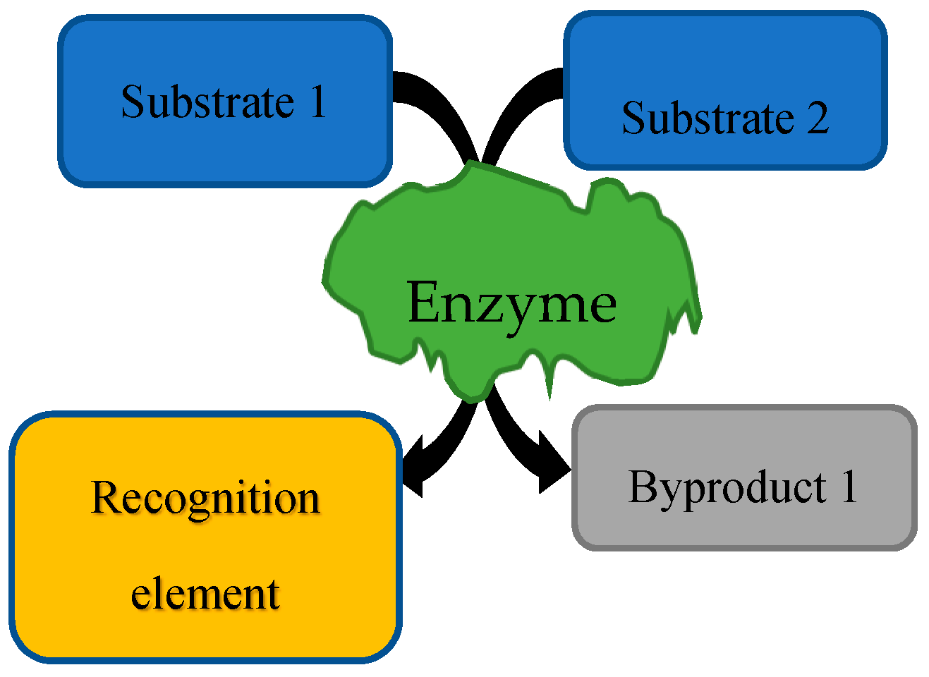
1. Background
- Forensics
- Biometrics
- Cybersecurity
2. Research
- Forensics
- Biometrics
- Cybersecurity
3. Conclusions
Funding
Conflicts of Interest
References
- D’Orazio, P. Biosensors in clinical chemistry—2011 update. Clin. Chim. Acta 2011, 412, 1749–1761. [Google Scholar] [CrossRef] [PubMed]
- Burcu Bahadir, E.; Kemal Sezgintürk, M. Applications of electrochemical immunosensors for early clinical diagnostics. Talanta 2015, 132, 162–174. [Google Scholar] [CrossRef] [PubMed]
- Bianchi, V.; Guerra, C.; De munari, I.; Ciampolini, P. Wearable sensors for behavioral assessment. Gerontechnology 2016. [Google Scholar] [CrossRef]
- Guilbault, G.G.; Palleschi, G.; Lubrano, G. Non-invasive biosensors in clinical analysis. Biosens. Bioelectron. 1995, 10, 379–392. [Google Scholar] [CrossRef]
- Guilbault, G.G.; Pravda, M.; Kreuzer, M.; O’Sullivan, C.K. Biosensors—42 Years and counting. Anal. Lett. 2004, 37, 1481–1496. [Google Scholar] [CrossRef]
- Ferapontova, E.E. DNA Electrochemistry and Electrochemical Sensors for Nucleic Acids. Annu. Rev. Anal. Chem. 2018, 11, 197–218. [Google Scholar] [CrossRef]
- Sanghavi, B.J.; Wolfbeis, O.S.; Hirsch, T.; Swami, N.S. Nanomaterial-based electrochemical sensing of neurological drugs and neurotransmitters. Microchim. Acta 2015, 182, 1–41. [Google Scholar] [CrossRef]
- Shen, Y.; Tran, T.T.; Modha, S.; Tsutsui, H.; Mulchandani, A. A paper-based chemiresistive biosensor employing single-walled carbon nanotubes for low-cost, point-of-care detection. Biosens. Bioelectron. 2019, 130, 367–373. [Google Scholar] [CrossRef]
- García-Arellano, H.; Fink, D.; Muñoz Hernández, G.; Vacík, J.; Hnatowicz, V.; Alfonta, L. Nuclear track-based biosensors with the enzyme laccase. Appl. Surf. Sci. 2014, 310, 66–76. [Google Scholar] [CrossRef]
- Tzouvadaki, I.; De Micheli, G.; Carrara, S. Memristive Biosensors for Ultrasensitive Diagnostics and Therapeutics. In Springer Series in Advanced Microelectronics; Springer: Singapore, 2020. [Google Scholar]
- Volkov, A.G.; Volkova-Gugeshashvili, M.I.; Osei, A.J. Plants as environmental biosensors: Non-invasive monitoring techniques. In Proceedings of the Appropriate Technologies for Environmental Protection in the Developing World—Selected Papers from ERTEP 2007, Ghana, Africa, 17–19 July 2007. [Google Scholar]
- Somerset, V. Environmental Biosensors; InTech: Rijeka, Croatia, 2011; ISBN 978-953-307-486-3. [Google Scholar]
- Van Dorst, B.; Mehta, J.; Bekaert, K.; Rouah-Martin, E.; De Coen, W.; Dubruel, P.; Blust, R.; Robbens, J. Recent advances in recognition elements of food and environmental biosensors: A review. Biosens. Bioelectron. 2010, 26, 1178–1194. [Google Scholar] [CrossRef]
- Bollella, P.; Hibino, Y.; Kano, K.; Gorton, L.; Antiochia, R. Highly Sensitive Membraneless Fructose Biosensor Based on Fructose Dehydrogenase Immobilized onto Aryl Thiol Modified Highly Porous Gold Electrode: Characterization and Application in Food Samples. Anal. Chem. 2018, 90, 12131–12136. [Google Scholar] [CrossRef] [PubMed]
- Bahadir, E.B.; Sezgintürk, M.K. Applications of commercial biosensors in clinical, food, environmental, and biothreat/biowarfare analyses. Anal. Biochem. 2015, 478, 107–120. [Google Scholar] [CrossRef]
- Alocilja, E.C.; Radke, S.M. Market analysis of biosensors for food safety. Biosens. Bioelectron. 2003, 18, 841–846. [Google Scholar] [CrossRef]
- Thakur, M.S.; Ragavan, K.V. Biosensors in food processing. J. Food Sci. Technol. 2013, 50, 625–641. [Google Scholar] [CrossRef] [PubMed]
- Siepenkoetter, T.; Salaj-Kosla, U.; Magner, E. The Immobilization of Fructose Dehydrogenase on Nanoporous Gold Electrodes for the Detection of Fructose. ChemElectroChem 2017, 4, 905–912. [Google Scholar] [CrossRef]
- Shi, H.; Zhao, H.; Liu, Y.; Gao, W.; Dou, S.C. Systematic analysis of a military wearable device based on a multi-level fusion framework: Research directions. Sensors (Switzerland) 2019, 19, 2651. [Google Scholar] [CrossRef] [PubMed]
- Pohanka, M.; Jun, D.; Kuca, K. Amperometric biosensors for real time assays of organophosphates. Sensors 2008, 8, 5303–5312. [Google Scholar] [CrossRef]
- Andrew, A.M. Unconventional computing. Kybernetes 2012, 41, 518–522. [Google Scholar] [CrossRef]
- Yamashita, B.; French, M.; Bleay, S.; Cantu, A.; Inlow, V.; Ramotowski, R.; Sears, V.; Wakefield, M. Latent Print Development-Chapter 7 Fingerprint Sourcebook. In The Fingerprint Sourcebook; U.S. Department of Justice: Washington, DC, USA, 2010. [Google Scholar]
- Francese, S.; Bradshaw, R.; Ferguson, L.S.; Wolstenholme, R.; Clench, M.R.; Bleay, S. Beyond the ridge pattern: Multi-informative analysis of latent fingermarks by MALDI mass spectrometry. Analyst 2013, 138, 4215–4228. [Google Scholar] [CrossRef]
- Thody, A.J.; Shuster, S. Control and function of sebaceous glands. Physiol. Rev. 1989, 59, 383–416. [Google Scholar] [CrossRef]
- Beskaravainy, P.M.; Molchanov, M.V.; Suslikov, A.V.; Paskevich, S.I.; Kutyshenko, V.P.; Vorob’ev, S.I. NMR study of human biological fluids for detection of pathologies. Biomeditsinskaya Khim. 2015, 61, 141–149. [Google Scholar] [CrossRef] [PubMed][Green Version]
- Hier, S.W.; Cornbleet, T.; Bergeim, O. The amino acids of human sweat. J. Biol. Chem. 1946, 166, 327–333. [Google Scholar] [PubMed]
- Coltman, C.A.; Rowe, N.J.; Atwell, R.J. The amino acid content of sweat in normal adults. Am. J. Clin. Nutr. 1966, 18, 373–378. [Google Scholar] [CrossRef] [PubMed]
- Croxton, R.S.; Baron, M.G.; Butler, D.; Kent, T.; Sears, V.G. Variation in amino acid and lipid composition of latent fingerprints. Forensic Sci. Int. 2010, 199, 93–102. [Google Scholar] [CrossRef]
- NIJ DNA Backlog. Available online: https://www.ncjrs.gov/App/Publications/abstract.aspx?ID=259066 (accessed on 15 September 2020).
- Hartzell-Baguley, B.; Hipp, R.E.; Morgan, N.R.; Morgan, S.L. Chemical composition of latent fingerprints by gas chromatography-mass spectrometry. An experiment for an instrumental analysis course. J. Chem. Educ. 2007, 84, 689. [Google Scholar] [CrossRef]
- Archer, N.E.; Charles, Y.; Elliott, J.A.; Jickells, S. Changes in the lipid composition of latent fingerprint residue with time after deposition on a surface. Forensic Sci. Int. 2005, 154, 224–239. [Google Scholar] [CrossRef]
- Jacob, S.; Jickells, S.; Wolff, K.; Smith, N. Drug Testing by Chemical Analysis of Fingerprint Deposits from Methadone- Maintained Opioid Dependent Patients Using UPLC-MS/MS. Drug Metab. Lett. 2008. [Google Scholar] [CrossRef]
- Goucher, E.; Kicman, A.; Smith, N.; Jickells, S. The detection and quantification of lorazepam and its 3-O-glucuronide in fingerprint deposits by LC-MS/MS. J. Sep. Sci. 2009. [Google Scholar] [CrossRef]
- Michalski, S.; Shaler, R.; Dorman, F.L. The Evaluation of Fatty Acid Ratios in Latent Fingermarks by Gas Chromatography/Mass Spectrometry (GC/MS) Analysis. J. Forensic Sci. 2013. [Google Scholar] [CrossRef]
- Antoine, K.M.; Mortazavi, S.; Miller, A.D.; Miller, L.M. Chemical differences are observed in children’s versus adults’ latent fingerprints as a function of time. J. Forensic Sci. 2010. [Google Scholar] [CrossRef]
- Ricci, C.; Phiriyavityopas, P.; Curum, N.; Chan, K.L.A.; Jickells, S.; Kazarian, S.G. Chemical imaging of latent fingerprint residues. Appl. Spectrosc. 2007. [Google Scholar] [CrossRef] [PubMed]
- Mou, Y.; Rabalais, J.W. Detection and Identification of Explosive Particles in Fingerprints Using Attenuated Total Reflection-Fourier Transform Infrared Spectromicroscopy. J. Forensic Sci. 2009. [Google Scholar] [CrossRef] [PubMed]
- Morelato, M.; Beavis, A.; Kirkbride, P.; Roux, C. Forensic applications of desorption electrospray ionisation mass spectrometry (DESI-MS). Forensic Sci. Int. 2013, 226, 10–21. [Google Scholar] [CrossRef] [PubMed]
- Ifa, D.R.; Jackson, A.U.; Paglia, G.; Cooks, R.G. Forensic applications of ambient ionization mass spectrometry. Anal. Bioanal. Chem. 2009, 394, 1995–2008. [Google Scholar] [CrossRef] [PubMed]
- Hazarika, P.; Jickells, S.M.; Wolff, K.; Russell, D.A. Multiplexed detection of metabolites of narcotic drugs from a single latent fingermark. Anal. Chem. 2010. [Google Scholar] [CrossRef] [PubMed]
- van Dam, A.; Aalders, M.C.G.; de Puit, M.; Gorré, S.M.; Irmak, D.; van Leeuwen, T.G.; Lambrechts, S.A.G. Immunolabeling and the compatibility with a variety of fingermark development techniques. Sci. Justice 2014. [Google Scholar] [CrossRef] [PubMed]
- Bradshaw, R.; Rao, W.; Wolstenholme, R.; Clench, M.R.; Bleay, S.; Francese, S. Separation of overlapping fingermarks by Matrix Assisted Laser Desorption Ionisation Mass Spectrometry Imaging. Forensic Sci. Int. 2012. [Google Scholar] [CrossRef]
- Wolstenholme, R.; Bradshaw, R.; Clench, M.R.; Francese, S. Study of latent fingermarks by matrix-assisted laser desorption/ionisation mass spectrometry imaging of endogenous lipids. Rapid Commun. Mass Spectrom. 2009. [Google Scholar] [CrossRef]
- Huynh, C.; Halámek, J. Trends in fingerprint analysis. TrAC—Trends Anal. Chem. 2016, 82, 328–336. [Google Scholar] [CrossRef]
- An, J.H.; Shin, K.J.; Yang, W.I.; Lee, H.Y. Body fluid identification in forensics. BMB Rep. 2012, 45, 545–553. [Google Scholar] [CrossRef]
- Gill, P.; Jeffreys, A.J.; Werrett, D.J. Forensic application of DNA “fingerprints”. Nature 1985. [Google Scholar] [CrossRef]
- Flight, C.; Jones, M.; Ballantyne, K.N. Determination of the maximum distance blood spatter travels from a vertical impact. Forensic Sci. Int. 2018. [Google Scholar] [CrossRef]
- Brettell, T.A.; Butler, J.M.; Saferstein, R. Forensic science. Anal. Chem. 2005, 77, 3839–3860. [Google Scholar] [CrossRef] [PubMed]
- Erbisti, P.C.F.; Gardner, R.M. Bloodstain Pattern Analysis with an Introduction to Crime Scene Reconstruction; CRC Press: Boca Raton, FL, USA, 2008. [Google Scholar]
- Elkins, K. Forensic DNA Biology, 3rd ed.; CRC Press: Boca Raton, FL, USA, 2013; ISBN 9780123945853. [Google Scholar]
- Bocklandt, S.; Lin, W.; Sehl, M.E.; Sánchez, F.J.; Sinsheimer, J.S.; Horvath, S.; Vilain, E. Epigenetic predictor of age. PLoS ONE 2011, 6, e41821. [Google Scholar] [CrossRef] [PubMed]
- Kramer, F.; Halámková, L.; Poghossian, A.; Schöning, M.J.; Katz, E.; Halámek, J. Biocatalytic analysis of biomarkers for forensic identification of ethnicity between Caucasian and African American groups. Analyst 2013, 138, 6251–6257. [Google Scholar] [CrossRef] [PubMed]
- Bakshi, S.; Halámková, L.; Halámek, J.; Katz, E. Biocatalytic analysis of biomarkers for forensic identification of gender. Analyst 2014, 139, 559–563. [Google Scholar] [CrossRef]
- Meissner, C.; Ritz-Timme, S. Molecular pathology and age estimation. Forensic Sci. Int. 2010, 203, 34–43. [Google Scholar] [CrossRef]
- Pizzamiglio, M.; Mameli, A.; Maugeri, G.; Garofano, L. Identifying the culprit from LCN DNA obtained from saliva and sweat traces linked to two different robberies and use of a database. Int. Congr. Ser. 2004. [Google Scholar] [CrossRef]
- Pizzamiglio, M.; Marino, A.; Portera, G.; My, D.; Bellino, C.; Garofano, L. Robotic DNA extraction system as a new way to process sweat traces rapidly and efficiently. Int. Congr. Ser. 2006. [Google Scholar] [CrossRef]
- Sikirzhytski, V.; Sikirzhytskaya, A.; Lednev, I.K. Multidimensional Raman spectroscopic signature of sweat and its potential application to forensic body fluid identification. Anal. Chim. Acta 2012. [Google Scholar] [CrossRef]
- Stouder, S.; Reubush, K.; Hobson, D.; Smith, J. Trace Evidence Scrapings: A Valuable Source of DNA? Forensic Sci. Commun. 2001, 4, 4. [Google Scholar]
- Verde, T.; Shephard, R.J.; Corey, P.; Moore, R. Sweat composition in exercise and in heat. J. Appl. Physiol. Respir. Environ. Exerc. Physiol. 1982. [Google Scholar] [CrossRef] [PubMed]
- Patterson, M.J.; Galloway, S.D.R.; Nimmo, M.A. Variations in regional sweat composition in normal human males. Exp. Physiol. 2000. [Google Scholar] [CrossRef] [PubMed]
- Costa, F.; Calloway, D.H.; Margen, S. Regional and total body sweat composition of men fed controlled diets. Am. J. Clin. Nutr. 1969. [Google Scholar] [CrossRef]
- Pinson, E.A. Evaporation from human skin with sweat glands inactivated. Am. J. Physiol. Content 1942. [Google Scholar] [CrossRef]
- ÅStrand, I. Lactate Content in Sweat. Acta Physiol. Scand. 1963. [Google Scholar] [CrossRef]
- Buono, M.J.; Lee, N.V.L.; Miller, P.W. The relationship between exercise intensity and the sweat lactate excretion rate. J. Physiol. Sci. 2010. [Google Scholar] [CrossRef]
- Kondoh, Y.; Kawase, M.; Ohmori, S. D-Lactate concentrations in blood, urine and sweat before and after exercise. Eur. J. Appl. Physiol. Occup. Physiol. 1992. [Google Scholar] [CrossRef]
- Derbyshire, P.J.; Barr, H.; Davis, F.; Higson, S.P.J. Lactate in human sweat: A critical review of research to the present day. J. Physiol. Sci. 2012, 62, 429–440. [Google Scholar] [CrossRef]
- Meyer, F.; Laitano, O.; Bar-Or, O.; McDougall, D.; Heingenhauser, G.J.F. Effect of age and gender on sweat lactate and ammonia concentrations during exercise in the heat. Braz. J. Med. Biol. Res. 2007. [Google Scholar] [CrossRef] [PubMed]
- Ghamouss, F.; Ledru, S.; Ruillé, N.; Lantier, F.; Boujtita, M. Bulk-modified modified screen-printing carbon electrodes with both lactate oxidase (LOD) and horseradish peroxide (HRP) for the determination of l-lactate in flow injection analysis mode. Anal. Chim. Acta 2006. [Google Scholar] [CrossRef] [PubMed]
- Ballesta Claver, J.; Valencia Mirón, M.C.; Capitán-Vallvey, L.F. Disposable electrochemiluminescent biosensor for lactate determination in saliva. Analyst 2009. [Google Scholar] [CrossRef] [PubMed]
- Jia, W.; Bandodkar, A.J.; Valdés-Ramírez, G.; Windmiller, J.R.; Yang, Z.; Ramírez, J.; Chan, G.; Wang, J. Electrochemical tattoo biosensors for real-time noninvasive lactate monitoring in human perspiration. Anal. Chem. 2013. [Google Scholar] [CrossRef] [PubMed]
- Bandodkar, A.J.; Molinnus, D.; Mirza, O.; Guinovart, T.; Windmiller, J.R.; Valdés-Ramírez, G.; Andrade, F.J.; Schöning, M.J.; Wang, J. Epidermal tattoo potentiometric sodium sensors with wireless signal transduction for continuous non-invasive sweat monitoring. Biosens. Bioelectron. 2014. [Google Scholar] [CrossRef] [PubMed]
- Buono, M.J. Sweat ethanol concentrations are highly correlated with co-existing blood values in humans. Exp. Physiol. 1999. [Google Scholar] [CrossRef]
- Nyman, E.; Palmlöv, A. The Elimination of Ethyl Alcohol in Sweat. Skand. Arch. Physiol. 1936. [Google Scholar] [CrossRef]
- Kim, J.; Jeerapan, I.; Imani, S.; Cho, T.N.; Bandodkar, A.; Cinti, S.; Mercier, P.P.; Wang, J. Noninvasive Alcohol Monitoring Using a Wearable Tattoo-Based Iontophoretic-Biosensing System. ACS Sens. 2016. [Google Scholar] [CrossRef]
- Swift, R. Transdermal alcohol measurement for estimation of blood alcohol concentration. Alcohol. Clin. Exp. Res. 2000, 24, 422–423. [Google Scholar] [CrossRef]
- Mishra, R.K.; Sempionatto, J.R.; Li, Z.; Brown, C.; Galdino, N.M.; Shah, R.; Liu, S.; Hubble, L.J.; Bagot, K.; Tapert, S.; et al. Simultaneous detection of salivary Δ9-tetrahydrocannabinol and alcohol using a Wearable Electrochemical Ring Sensor. Talanta 2020. [Google Scholar] [CrossRef]
- Teymourian, H.; Parrilla, M.; Sempionatto, J.R.; Montiel, N.F.; Barfidokht, A.; Van Echelpoel, R.; De Wael, K.; Wang, J. Wearable Electrochemical Sensors for the Monitoring and Screening of Drugs. ACS Sens. 2020. [Google Scholar] [CrossRef]
- Jeerapan, I.; Sempionatto, J.R.; Wang, J. On-Body Bioelectronics: Wearable Biofuel Cells for Bioenergy Harvesting and Self-Powered Biosensing. Adv. Funct. Mater. 2020. [Google Scholar] [CrossRef]
- Sempionatto, J.R.; Jeerapan, I.; Krishnan, S.; Wang, J. Wearable Chemical Sensors: Emerging Systems for On-Body Analytical Chemistry. Anal. Chem. 2019, 92, 378–396. [Google Scholar] [CrossRef] [PubMed]
- Mickelsen, O.; Keys, A. The composition of sweat, with special reference to vitamins. J. Biol. Chem. 1943, 149, 479–490. [Google Scholar]
- Lemonick, S. You Could Unlock Your Phone with Sweat. Forbes 2017. Available online: https://www.forbes.com/sites/samlemonick/2017/11/15/you-could-unlock-your-phone-with-sweat/#7225db7849ae (accessed on 15 September 2020).
- French, L. Virtual Case Notes: Sweat, Skin Secretions Could Contain Chemical ‘Password’ for Future Mobile Authentication. Forensic Magazine, 16 November 2017. [Google Scholar]
- Murphy, M. Sweat Could Soon Unlock Your Phone. Available online: https://nypost.com/2017/11/14/sweat-could-soon-unlock-your-phone/ (accessed on 15 September 2020).
- Nagamine, K.; Nomura, A.; Ichimura, Y.; Izawa, R.; Sasaki, S.; Furusawa, H.; Matsui, H.; Tokito, S. Printed organic transistor-based biosensors for non-invasive sweat analysis. Anal. Sci. 2020. [Google Scholar] [CrossRef] [PubMed]
- Bandodkar, A.J.; Jeang, W.J.; Ghaffari, R.; Rogers, J.A. Wearable Sensors for Biochemical Sweat Analysis. Annu. Rev. Anal. Chem. 2019, 12, 1–22. [Google Scholar] [CrossRef]
- Gao, W.; Nyein, H.Y.Y.; Shahpar, Z.; Tai, L.C.; Wu, E.; Bariya, M.; Ota, H.; Fahad, H.M.; Chen, K.; Javey, A. Wearable sweat biosensors. In Proceedings of the Technical Digest—International Electron Devices Meeting, IEDM, San Francisco, CA, USA, 3–7 December 2016. [Google Scholar]
- Bandodkar, A.J.; Wang, J. Non-invasive wearable electrochemical sensors: A review. Trends Biotechnol. 2014, 32, 363–371. [Google Scholar] [CrossRef]
- Heikenfeld, J. Non-invasive Analyte Access and Sensing through Eccrine Sweat: Challenges and Outlook circa 2016. Electroanalysis 2016, 28, 1242–1249. [Google Scholar] [CrossRef]
- Bandodkar, A.J.; Hung, V.W.S.; Jia, W.; Valdés-Ramírez, G.; Windmiller, J.R.; Martinez, A.G.; Ramírez, J.; Chan, G.; Kerman, K.; Wang, J. Tattoo-based potentiometric ion-selective sensors for epidermal pH monitoring. Analyst 2013. [Google Scholar] [CrossRef]
- Guinovart, T.; Bandodkar, A.J.; Windmiller, J.R.; Andrade, F.J.; Wang, J. A potentiometric tattoo sensor for monitoring ammonium in sweat. Analyst 2013. [Google Scholar] [CrossRef]
- He, X.; Xu, T.; Gu, Z.; Gao, W.; Xu, L.P.; Pan, T.; Zhang, X. Flexible and Superwettable Bands as a Platform toward Sweat Sampling and Sensing. Anal. Chem. 2019. [Google Scholar] [CrossRef]
- Hauke, A.; Oertel, S.; Knoke, L.; Fein, V.; Maier, C.; Brinkmann, F.; Jank, M.P.M. Screen-Printed Sensor for Low-Cost Chloride Analysis in Sweat for Rapid Diagnosis and Monitoring of Cystic Fibrosis. Biosensors 2020, 10, 123. [Google Scholar] [CrossRef]
- He, X.; Yang, S.; Pei, Q.; Song, Y.; Liu, C.; Xu, T.; Zhang, X. Integrated Smart Janus Textile Bands for Self-Pumping Sweat Sampling and Analysis. ACS Sens. 2020. [Google Scholar] [CrossRef] [PubMed]
- Oh, S.Y.; Hong, S.Y.; Jeong, Y.R.; Yun, J.; Park, H.; Jin, S.W.; Lee, G.; Oh, J.H.; Lee, H.; Lee, S.S.; et al. Skin-Attachable, Stretchable Electrochemical Sweat Sensor for Glucose and pH Detection. ACS Appl. Mater. Interfaces 2018. [Google Scholar] [CrossRef] [PubMed]
- Lee, H.; Choi, T.K.; Lee, Y.B.; Cho, H.R.; Ghaffari, R.; Wang, L.; Choi, H.J.; Chung, T.D.; Lu, N.; Hyeon, T.; et al. A graphene-based electrochemical device with thermoresponsive microneedles for diabetes monitoring and therapy. Nat. Nanotechnol. 2016. [Google Scholar] [CrossRef] [PubMed]
- Koh, A.; Kang, D.; Xue, Y.; Lee, S.; Pielak, R.M.; Kim, J.; Hwang, T.; Min, S.; Banks, A.; Bastien, P.; et al. A soft, wearable microfluidic device for the capture, storage, and colorimetric sensing of sweat. Sci. Transl. Med. 2016. [Google Scholar] [CrossRef]
- Khodagholy, D.; Curto, V.F.; Fraser, K.J.; Gurfinkel, M.; Byrne, R.; Diamond, D.; Malliaras, G.G.; Benito-Lopez, F.; Owens, R.M. Organic electrochemical transistor incorporating an ionogel as a solid state electrolyte for lactate sensing. J. Mater. Chem. 2012. [Google Scholar] [CrossRef]
- Emaminejad, S.; Gao, W.; Wu, E.; Davies, Z.A.; Nyein, H.Y.Y.; Challa, S.; Ryan, S.P.; Fahad, H.M.; Chen, K.; Shahpar, Z.; et al. Autonomous sweat extraction and analysis applied to cystic fibrosis and glucose monitoring using a fully integrated wearable platform. Proc. Natl. Acad. Sci. USA 2017. [Google Scholar] [CrossRef]
- Mark, H.; Harding, C.R. Amino acid composition, including key derivatives of eccrine sweat: Potential biomarkers of certain atopic skin conditions. Int. J. Cosmet. Sci. 2013. [Google Scholar] [CrossRef]
- Imani, S.; Bandodkar, A.J.; Mohan, A.M.V.; Kumar, R.; Yu, S.; Wang, J.; Mercier, P.P. A wearable chemical-electrophysiological hybrid biosensing system for real-time health and fitness monitoring. Nat. Commun. 2016. [Google Scholar] [CrossRef]
- Zhang, Y.; Guo, H.; Kim, S.B.; Wu, Y.; Ostojich, D.; Park, S.H.; Wang, X.; Weng, Z.; Li, R.; Bandodkar, A.J.; et al. Passive sweat collection and colorimetric analysis of biomarkers relevant to kidney disorders using a soft microfluidic system. Lab Chip 2019. [Google Scholar] [CrossRef]
- Gao, W.; Emaminejad, S.; Nyein, H.Y.Y.; Challa, S.; Chen, K.; Peck, A.; Fahad, H.M.; Ota, H.; Shiraki, H.; Kiriya, D.; et al. Fully integrated wearable sensor arrays for multiplexed in situ perspiration analysis. Nature 2016. [Google Scholar] [CrossRef] [PubMed]
- Schazmann, B.; Morris, D.; Slater, C.; Beirne, S.; Fay, C.; Reuveny, R.; Moyna, N.; Diamond, D. A wearable electrochemical sensor for the real-time measurement of sweat sodium concentration. Anal. Methods 2010. [Google Scholar] [CrossRef]
- Glennon, T.; O’Quigley, C.; McCaul, M.; Matzeu, G.; Beirne, S.; Wallace, G.G.; Stroiescu, F.; O’Mahoney, N.; White, P.; Diamond, D. ‘SWEATCH’: A Wearable Platform for Harvesting and Analysing Sweat Sodium Content. Electroanalysis 2016. [Google Scholar] [CrossRef]
- Tai, L.C.; Gao, W.; Chao, M.; Bariya, M.; Ngo, Q.P.; Shahpar, Z.; Nyein, H.Y.Y.; Park, H.; Sun, J.; Jung, Y.; et al. Methylxanthine Drug Monitoring with Wearable Sweat Sensors. Adv. Mater. 2018. [Google Scholar] [CrossRef] [PubMed]
- Tanenbaum, A.S.; Wetherall, D.J. Computer Networks. World Wide Web Internet Web Inf. Syst. 2011. [Google Scholar] [CrossRef]
- Schneier, B. Secrets and Lies: Digital Security in a Networked World; John Wiley & Sons, Inc.: New York, NY, USA, 2000. [Google Scholar]
- Kaufman, C.; Perlman, R.; Spencer, M. Network Security Private Communication in a Public World; Prentice Hall: Westford, MA, USA, 2002; ISBN 0-13-046019-2. [Google Scholar]
- Kishimura, A.; Yamashita, T.; Yamaguchi, K.; Aida, T. Rewritable phosphorescent paper by the control of competing kinetic and thermodynamic self-assembling events. Nat. Mater. 2005. [Google Scholar] [CrossRef]
- Mutai, T.; Satou, H.; Araki, K. Reproducible on-off switching of solid-state luminescence by controlling molecular packing through heat-mode interconversion. Nat. Mater. 2005. [Google Scholar] [CrossRef]
- Perruchas, S.; Goff, X.F.L.; Maron, S.; Maurin, I.; Guillen, F.; Garcia, A.; Gacoin, T.; Boilot, J.P. Mechanochromic and thermochromic luminescence of a copper iodide cluster. J. Am. Chem. Soc. 2010. [Google Scholar] [CrossRef]
- Yoon, S.J.; Chung, J.W.; Gierschner, J.; Kim, K.S.; Choi, M.G.; Kim, D.; Park, S.Y. Multistimuli two-color luminescence switching via different slip-stacking of highly fluorescent molecular sheets. J. Am. Chem. Soc. 2010. [Google Scholar] [CrossRef]
- Yan, D.; Lu, J.; Ma, J.; Wei, M.; Evans, D.G.; Duan, X. Reversibly thermochromic, fluorescent ultrathin films with a supramolecular architecture. Angew. Chem. Int. Ed. 2011. [Google Scholar] [CrossRef]
- Li, K.; Xiang, Y.; Wang, X.; Li, J.; Hu, R.; Tong, A.; Tang, B.Z. Reversible photochromic system based on rhodamine B salicylaldehyde hydrazone metal complex. J. Am. Chem. Soc. 2014. [Google Scholar] [CrossRef]
- Sun, H.; Liu, S.; Lin, W.; Zhang, K.Y.; Lv, W.; Huang, X.; Huo, F.; Yang, H.; Jenkins, G.; Zhao, Q.; et al. Smart responsive phosphorescent materials for data recording and security protection. Nat. Commun. 2014. [Google Scholar] [CrossRef] [PubMed]
- Wu, Y.; Xie, Y.; Zhang, Q.; Tian, H.; Zhu, W.; Li, A.D.Q. Quantitative photoswitching in bis(dithiazole)ethene enables modulation of light for encoding optical signals. Angew. Chem. Int. Ed. 2014, 53, 2090–2094. [Google Scholar] [CrossRef] [PubMed]
- Sarkar, T.; Selvakumar, K.; Motiei, L.; Margulies, D. Message in a molecule. Nat. Commun. 2016, 7, 1–9. [Google Scholar] [CrossRef] [PubMed]
- Ratner, T.; Reany, O.; Keinan, E. Encoding and processing of alphanumeric information by chemical mixtures. ChemPhysChem 2009, 10, 3303–3309. [Google Scholar] [CrossRef]
- Palacios, M.A.; Benito-Pena, E.; Manesse, M.; Mazzeo, A.D.; LaFratta, C.N.; Whitesides, G.M.; Walt, D.R. InfoBiology by printed arrays of microorganism colonies for timed and on-demand release of messages. Proc. Natl. Acad. Sci. USA 2011. [Google Scholar] [CrossRef]
- Kim, K.W.; Bocharova, V.; Halámek, J.; Oh, M.K.; Katz, E. Steganography and encrypting based on immunochemical systems. Biotechnol. Bioeng. 2011, 108, 1100–1107. [Google Scholar] [CrossRef]
- Shoshani, S.; Piran, R.; Arava, Y.; Keinan, E. A molecular cryptosystem for images by DNA computing. Angew. Chem. Int. Ed. 2012, 51, 2883–2887. [Google Scholar] [CrossRef]
- Poje, J.E.; Kastratovic, T.; Macdonald, A.R.; Guillermo, A.C.; Troetti, S.E.; Jabado, O.J.; Fanning, M.L.; Stefanovic, D.; Macdonald, J. Visual displays that directly interface and provide read-outs of molecular states via molecular graphics processing units. Angew. Chem. Int. Ed. 2014. [Google Scholar] [CrossRef]
- Ling, J.; Naren, G.; Kelly, J.; Moody, T.S.; De Silva, A.P. Building pH Sensors into Paper-Based Small-Molecular Logic Systems for Very Simple Detection of Edges of Objects. J. Am. Chem. Soc. 2015, 137, 3763–3766. [Google Scholar] [CrossRef]
- Ling, J.; Naren, G.; Kelly, J.; Fox, D.B.; Prasanna De Silva, A. Small molecular logic systems can draw the outlines of objects via edge visualization. Chem. Sci. 2015. [Google Scholar] [CrossRef] [PubMed]
- Zhang, Y.; Liu, X.; Sun, M. DNA based random key generation and management for OTP encryption. BioSystems 2017. [Google Scholar] [CrossRef] [PubMed]
- Clelland, C.T.; Risca, V.; Bancroft, C. Hiding Data in DNA Microdots. Nature 1999, 399, 533–534. [Google Scholar] [CrossRef] [PubMed]
- Lustgarten, O.; Carmieli, R.; Motiei, L.; Margulies, D. A Molecular Secret Sharing Scheme. Angew. Chem. Int. Ed. 2019, 58, 184–188. [Google Scholar] [CrossRef]
- Srilatha, N.; Murali, G. Fast three level DNA Cryptographic technique to provide better security. In Proceedings of the 2016 2nd International Conference on Applied and Theoretical Computing and Communication Technology, iCATccT 2016, Bangalore, India, 21–23 July 2016. [Google Scholar]
- Malathi, P.; Manoaj, M.; Manoj, R.; Raghavan, V.; Vinodhini, R.E. Highly Improved DNA Based Steganography. Procedia Comput. Sci. 2017, 115, 651–659. [Google Scholar]
- Cherian, A.; Raj, S.R.; Abraham, A. A Survey on different DNA Cryptographic Methods. Int. J. Sci. Res. (IJSR) 2013, 2, 167–169. [Google Scholar]
- Roy, P.; Dey, D.; De, D.; Sinha, S. DNA cryptography. In Cyber Security and Threats: Concepts, Methodologies, Tools, and Applications; IGI Global: Hershey, PA, USA, 2018; ISBN 9781522556350. [Google Scholar]
- Halvorsen, K.; Wong, W.P. Binary DNA Nanostructures for Data Encryption. PLoS ONE 2012, 7, e44212. [Google Scholar] [CrossRef]
- Marwan, S.; Shawish, A.; Nagaty, K. DNA-based cryptographic methods for data hiding in DNA media. BioSystems 2016, 150, 110–118. [Google Scholar] [CrossRef]
- Huynh, C.; Brunelle, E.; Halámková, L.; Agudelo, J.; Halámek, J. Forensic Identification of Gender from Fingerprints. Anal. Chem. 2015, 87, 11531–11536. [Google Scholar] [CrossRef]
- Brunelle, E.; Huynh, C.; Le, A.M.; Halámková, L.; Agudelo, J.; Halámek, J. New Horizons for Ninhydrin: Colorimetric Determination of Gender from Fingerprints. Anal. Chem. 2016. [Google Scholar] [CrossRef]
- Brunelle, E.; Le, A.M.; Huynh, C.; Wingfield, K.; Halámková, L.; Agudelo, J.; Halámek, J. Coomassie Brilliant Blue G-250 Dye: An Application for Forensic Fingerprint Analysis. Anal. Chem. 2017. [Google Scholar] [CrossRef] [PubMed]
- Brunelle, E.; Huynh, C.; Alin, E.; Eldridge, M.; Le, A.M.; Halámková, L.; Halámek, J. Fingerprint Analysis: Moving Toward Multiattribute Determination via Individual Markers. Anal. Chem. 2018. [Google Scholar] [CrossRef]
- Agudelo, J.; Huynh, C.; Halámek, J. Forensic determination of blood sample age using a bioaffinity-based assay. Analyst 2015, 140, 1411–1415. [Google Scholar] [CrossRef] [PubMed]
- Agudelo, J.; Halámková, L.; Brunelle, E.; Rodrigues, R.; Huynh, C.; Halámek, J. Ages at a Crime Scene: Simultaneous Estimation of the Time since Deposition and Age of Its Originator. Anal. Chem. 2016, 88, 6479–6484. [Google Scholar] [CrossRef] [PubMed]
- Huynh, C.; Brunelle, E.; Agudelo, J.; Halámek, J. Bioaffinity-based assay for the sensitive detection and discrimination of sweat aimed at forensic applications. Talanta 2017. [Google Scholar] [CrossRef]
- Hair, M.E.; Gerkman, R.; Mathis, A.I.; Halámková, L.; Halámek, J. Noninvasive Concept for Optical Ethanol Sensing on the Skin Surface with Camera-Based Quantification. Anal. Chem. 2019. [Google Scholar] [CrossRef] [PubMed]
- Brunelle, E.; Halámek, J. Biocomputing approach in forensic analysis. Int. J. Parallel Emergent Distrib. Syst. 2017. [Google Scholar] [CrossRef]
- Moore, S.; Stein, W.H. Photometric ninhydrin method for use in the chromatography of amino acids. J. Biol. Chem. 1948, 178, 367–388. [Google Scholar]
- Meyer, H. The ninhydrin reaction and its analytical applications. Biochem. J. 1957. [Google Scholar] [CrossRef]
- Walker, J.M. Protein Protocols Handbook; Humana Press: Totowa, NJ, USA, 2002. [Google Scholar]
- Halámek, J.; Bocharova, V.; Chinnapareddy, S.; Windmiller, J.R.; Strack, G.; Chuang, M.C.; Zhou, J.; Santhosh, P.; Ramirez, G.V.; Arugula, M.A.; et al. Multi-enzyme logic network architectures for assessing injuries: Digital processing of biomarkers. Mol. Biosyst. 2010, 6, 2554–2560. [Google Scholar] [CrossRef]
- Sakaguchi, S. A new method for the colorimetric determination of arginine. J. Biochem. 1950. [Google Scholar] [CrossRef]
- Gibson, L.E.; Cooke, R.E. A test for concentration of electrolytes in sweat in cystic fibrosis of the pancreas utilizing pilocarpine by iontophoresis. Pediatrics 1959, 23, 545–549. [Google Scholar] [PubMed]
- Hair, M.E.; Mathis, A.I.; Brunelle, E.K.; Halámková, L.; Halámek, J. Metabolite Biometrics for the Differentiation of Individuals. Anal. Chem. 2018, 90, 5322–5328. [Google Scholar] [CrossRef] [PubMed]
- Agudelo, J.; Privman, V.; Halámek, J. Promises and Challenges in Continuous Tracking Utilizing Amino Acids in Skin Secretions for Active Multi-Factor Biometric Authentication for Cybersecurity. ChemPhysChem 2017, 18, 1714–1720. [Google Scholar] [CrossRef]
- McGoldrick, L.K.; Weiss, E.A.; Halámek, J. Symmetric-Key Encryption Based on Bioaffinity Interactions. ACS Synth. Biol. 2019. [Google Scholar] [CrossRef]
- Park, S.K.; Miller, K.W. Random number generators: Good ones are hard to find. Commun. ACM 1988. [Google Scholar] [CrossRef]
- Leier, A.; Richter, C.; Banzhaf, W.; Rauhe, H. Cryptography with DNA binary strands. BioSystems 2000. [Google Scholar] [CrossRef]
- Cox, J.P.L. Long-term data storage in DNA. Trends Biotechnol. 2001, 19, 247–250. [Google Scholar] [CrossRef]
- Gahlaut, A.; Bharti, A.; Dogra, Y.; Singh, P. DNA based cryptography. In Communications in Computer and Information Science; Springer: Singapore, 2017; Volume 750. [Google Scholar]
- Cui, G.; Wang, Y.; Han, D.; Wang, Y.; Wang, Z.; Wu, Y. An encryption scheme based on DNA microdots technology. Commun. Comput. Inf. Sci. 2014. [Google Scholar] [CrossRef]
- Zhang, Q.; Guo, L.; Wei, X. Image encryption using DNA addition combining with chaotic maps. Math. Comput. Model. 2010. [Google Scholar] [CrossRef]


| Protocol | Description | Technique | Analyte | LOD/Range | Ref. |
|---|---|---|---|---|---|
| Tattoo | Wearable skin tattoo for wireless signal transduction | Potentiometry | Sodium | 0.1–100 mM | [71] |
| Tattoo | Wearable skin tattoo for pH monitoring | Potentiometry | pH | pH 3–7 | [89] |
| Tattoo | Wearable skin tattoo for lactate monitoring | Potentiometry | Lactate | 1–20 mM | [70] |
| Tattoo | Wearable skin tattoo for monitoring | Potentiometry | Ammonium | 10−4–0.1 M | [90] |
| Superwettable bands | Multiplex method for on-body sampling | Colorimetric | pH | pH 4.5–7 | [91] |
| chloride | 0–100 mM | ||||
| glucose | 0–15 mM | ||||
| calcium | 0–15 mM | ||||
| Screen-printed electrode | Monitoring of cystic fibrosis patients | Potentiometry | Chloride | 2.7 × 10⁻⁵ mol/L | [92] |
| Janus textile bands | Multiplex method for on body sampling | Potentiometry | Glucose | 18–40 µM | [93] |
| Lactate | 10 mM | ||||
| Potassium | 0.3–6.3 mM | ||||
| Sodium | 60 mM | ||||
| Wearable sensor | Stretchable, skin-attachable sweat sensor | Potentiometry, Carbon nanotubes, gold nanosheets | Glucose | 10.89 µA mM−1 cm−2 | [94] |
| pH | 71.44 mV pH−1 | ||||
| Graphene electrochemical | Diabetes monitoring | Gold-doped graphene | Glucose | 10 µM–0.7 mM | [95] |
| Microfluidic wearable sensor | Multiplex analysis for sensing in sweat | Colorimetric | Lactate | 0–100 mM | [96] |
| Chloride | 0–mM | ||||
| Creatine | 0–1000 µM | ||||
| pH | pH 5–8.5 | ||||
| Glucose | 0–25 mM | ||||
| liquigel | Organic electrochemical transistor | Transistor | Lactate | 0.3–1.3 mM | [97] |
| Direct iontophoresis | Sweat extraction and electrochemical analysis using smartphone | Potentiometry | Glucose | 0–100 µM | [98] |
| Chloride | 20–80 mM | ||||
| Free amino acid analysis | Eccrine sweat amino acid composition | Cation chromatography and amino acid analyzer, GC-MS | Amino acids | - | [99] |
| Wearable Sensor | Chemical electrocardiogram and simultaneous metabolite monitoring | Amperometry | Lactate | 0–28 mM | [100] |
| microfluidic | Sweat collection and analysis for kidney disorders | Colorimetric | Creatine | 0–0.5 mM | [101] |
| Urea | 0–250 mM | ||||
| pH | pH 5–7 | ||||
| Wearable sensor | Integrated multiplex array for sweat analysis | Amperometry | Sodium | 20–120 mM | [102] |
| Potassium | 2–16 mM | ||||
| Glucose | 0–200 µM | ||||
| lactate | 2–30 mM | ||||
| Wearable sensor | Monitoring for cystic fibrosis patients for sodium concentration | Potentiometry Atomic Absorption | Sodium | 20–100 mM | [103] |
| Watch sensor | Monitoring of sodium levels | Potentiometry | Sodium | 10−4 –10−1 M | [104] |
| Tattoo | Wearable skin tattoo for alcohol monitoring in sweat | Amperometry | Alcohol | 0–36 mM | [74] |
| Wearable sensor | Drug monitoring via differential pulse | Voltammetry | Caffeine | 0–40 x µM | [105] |
| Wearable sensor | Detection of THC and Alcohol | Voltammetry Amperometry | THC | 0.5 µM | [76] |
| Alcohol | 0.1–1 mM |
Publisher’s Note: MDPI stays neutral with regard to jurisdictional claims in published maps and institutional affiliations. |
© 2020 by the authors. Licensee MDPI, Basel, Switzerland. This article is an open access article distributed under the terms and conditions of the Creative Commons Attribution (CC BY) license (http://creativecommons.org/licenses/by/4.0/).
Share and Cite
McGoldrick, L.K.; Halámek, J. Recent Advances in Noninvasive Biosensors for Forensics, Biometrics, and Cybersecurity. Sensors 2020, 20, 5974. https://doi.org/10.3390/s20215974
McGoldrick LK, Halámek J. Recent Advances in Noninvasive Biosensors for Forensics, Biometrics, and Cybersecurity. Sensors. 2020; 20(21):5974. https://doi.org/10.3390/s20215974
Chicago/Turabian StyleMcGoldrick, Leif K., and Jan Halámek. 2020. "Recent Advances in Noninvasive Biosensors for Forensics, Biometrics, and Cybersecurity" Sensors 20, no. 21: 5974. https://doi.org/10.3390/s20215974
APA StyleMcGoldrick, L. K., & Halámek, J. (2020). Recent Advances in Noninvasive Biosensors for Forensics, Biometrics, and Cybersecurity. Sensors, 20(21), 5974. https://doi.org/10.3390/s20215974