Article

Immediately Placed Single Locking-Taper Implants in the Aesthetic Area of Upper Maxilla: A Short-Term Pilot Study

1
Dentistry and Maxillo-Facial Surgery Unit, Department of Surgery, Dentistry, Paediatrics and Gynaecology (DIPSCOMI), University of Verona, Piazzale L.A. Scuro 10, 37134 Verona, Italy
2
Dental Implant Unit, Research Department, Faculty of Dentistry, University of Cartagena, Cartagena 130001, Colombia
*
Authors to whom correspondence should be addressed.
Prosthesis 2025, 7(3), 60; https://doi.org/10.3390/prosthesis7030060
Submission received: 15 April 2025 / Revised: 13 May 2025 / Accepted: 23 May 2025 / Published: 27 May 2025
(This article belongs to the Section Prosthodontics)

Abstract

:
Background: As the rehabilitation of the upper anterior maxilla primarily requires high predictability of successful aesthetic outcomes, procedures of immediate implant placement are frequently employed. The aim of this pilot study was to retrospectively evaluate the short-term outcomes of a protocol of immediate implant placement in fresh extraction sockets, followed by immediate non-functional provisional restorations. Methods: Patients were treated for the replacement of maxillary central or lateral incisors, or cuspid teeth with a single-crown locking-taper implant. Clinical and photographic records were retrospectively compared between the teeth prior to extraction (T0) and restorations one year after prosthetic loading (T1). Outcomes were analyzed using the Pink Esthetic Score (PES), according to the patient’s phenotype (thin/thick), with or without the use of connective tissue graft (CTG). Results: The overall mean PES of 25 implants treated was 9.24 ± 2.36 at T0 and 9.60 ± 1.70 at T1. Comparison of groups between T0 and T1 revealed significant PES variations (p = 0.04), with the best and the worst scores, respectively, registered for thin + CTG group (from 7.50 ± 1.91 to 9.75 ± 2.87) and thin group (from 11.33 ± 2.33 to 10 ± 0.89); moderate increases were assessed for thick group (from 8.44 ± 2.40 to 9.44 ± 2.12) and thick + CTG group (from 9.50 ± 1.04 to 9.33 ± 0.81). Conclusions: Within the limits of a short-term analysis of a small number of patients, immediate implant rehabilitation for aesthetic areas of the upper maxilla can be assumed as a safe and predictable protocol. Concomitant use of CTG seems to provide beneficial effects in thin phenotypes, not any additional value in thick phenotypes.

1. Introduction

As widely reported [1], healing of post-extractive sites in the anterior maxillary areas may lead to significant loss of the buccal bone contour during the first months after tooth extraction. Because of the unpredictable dynamics of bone remodeling, the precise final position of the marginal bone level and crestal width is difficult to determine [2,3]. Protocol of immediate implant placement into fresh extraction sockets, followed likewise by immediate provisional restorations [4], showed to minimize the timing and number of procedures, avoiding temporary removable dentures, not easily accepted by patients [5,6].
Highly predictable outcomes of this treatment have demonstrated its validation over the years [7]. If defined criteria are observed, long-term satisfactory clinical outcomes can be thus obtained [8,9]: one of the most important prerequisites for adequate aesthetic results is the correct implant positioning in 3D-space dimensions [10], with consequent stability of level of gingival margin. Minimal vestibular gingival recession can be achieved only in the presence of sufficient facial bony height and thickness.
As grafting attempts for repairing bone defects simultaneously with immediate replacement of teeth are usually associated with poor clinical and aesthetic expectations [11,12], some authors have suggested hard tissue augmentation prior to implant placement [13]. Nevertheless, a “sub-optimal” implant placement clearly implies a risk of insufficient results: this procedure performed in post-extraction sites of aesthetic areas must be therefore considered with a high degree of complexity [14].
Furthermore, in the presence of implant-supported restorations characterized by micro-gaps or micro-movements at the implant-abutment interface [15], inadequate maintenance of biologic width usually leads to progressive bone loss and undesired facial bone resorption, with final gingival recession. In this proposal, a specific type of implant, characterized by a sealing locking-taper connection (impervious to bacterial leakage) and a plateau design (able to properly dissipate high lateral occlusal forces), can be placed sub-crestally, successfully dealing with disproportionate crown-to-implant ratios even in case of thin alveolar ridges [16], thus providing sufficient space for the interproximal papillae, essential for aesthetic restorations [17].
As the primary objectives of implant therapy of the anterior maxilla directly concern high predictability of successful aesthetic outcomes [11,18,19], the aim of this pilot clinical study was to retrospectively evaluate, one year after prosthetic loading (T1), the conditions of peri-implant soft tissues in 25 patients consecutively treated with a procedure of immediate implant placement into fresh extraction sockets, using locking-taper plateau-design implants, and with immediate non-functional provisional single-crown restorations. To allow a comprehensive aesthetic evaluation based on scientifically validated indexes, the Pink Esthetic Score (PES) proposed by Furhauser [20] was employed in the study. Despite that, as the surrounding conditions of a tooth which will be replaced by an implant are not always the same as those of the contralateral tooth [21,22], the present evaluation was conducted comparing the implant and the pre-existing situation (the extracted tooth as it was immediately before the extraction, T0).

2. Materials and Methods
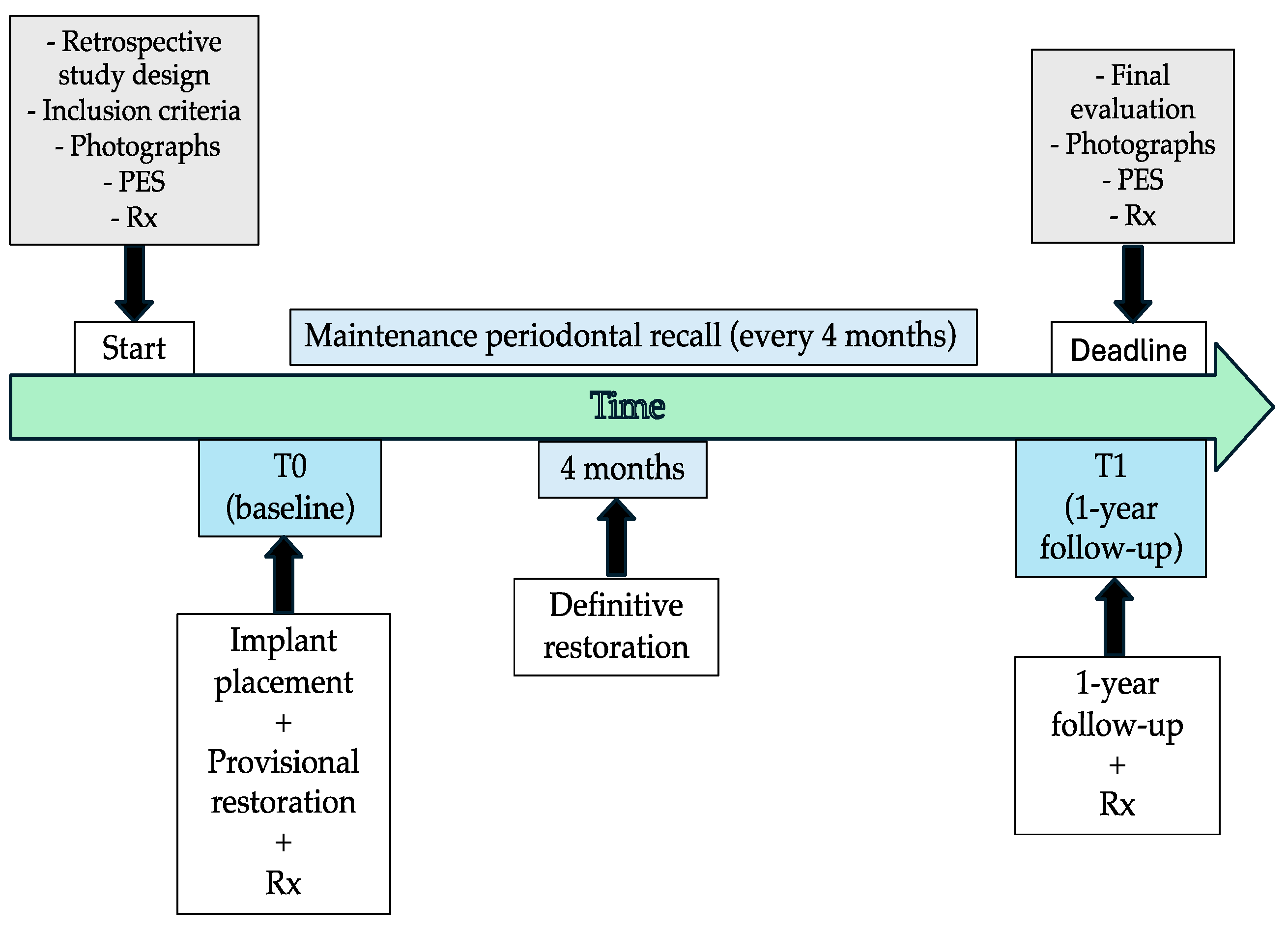
2.1. Study Design

A retrospective pilot study was conducted in 2025, on patients: (i) who had been referred and consecutively treated (between 2018 and 2023) at the Dental and Maxillo-Facial Surgery Clinic at the University of Verona, for the replacement of maxillary central or lateral incisors, or cuspid teeth; (ii) with implants placed at the time of extraction [23] and immediately non-functionally provisionally restored; (iii)with at least one year of follow-up.
The study was approved by the University Institutional Review Board (protocol code AESTETANTE, Prog. 1815CESC, 30 May 2018). The nature and aim of the study, together with the anonymity in the scientific use of data (also including the processing of sensitive data and pictures), were clearly presented in a written informed consent form, and signed by every patient. All procedures accorded with the Helsinki Declaration and good clinical practice guidelines for research on human beings.

2.2. Inclusion and Exclusion Criteria

Patients enrolled in the study matched the following inclusion criteria:
-
having had a single-tooth replacement with one single locking-taper plateau-design implant [16] in the aesthetic upper jaw, in the area between the upper right canine and the upper left canine;
-
being compliant with a regular maintenance program (professional oral hygiene sessions every 4 months) [16];
-
having complete clinical, photographic, and radiographic documentation.
Concerning exclusion criteria, patients were excluded from the retrospective evaluation if clinical, photographic, and radiographic data were incomplete.

2.3. Implant System

Regarding the abovementioned locking-taper implant system used in the study, it is characterized by
-
a plateau root-form design: the absence of micro-gaps and micro-movements confers greater mechanical stability to the implant-crown assembly and allows for minimal bone resorption [24];
-
a convergent crest module (Sloping Shoulder®): the implant shoulder gradually slopes inward and coronally toward the IAI, creating space for crestal bone even in the presence of narrow ridges or areas with limited restorative space;
-
a locking-taper screwless implant-abutment connection (IAI) [16]: when the conical shape abutment (1.5° angle) is activated by gentle tapping, a locking-taper occurs; specifically, the 2 surfaces of the same metal (Ti V 6 A14) rub together in such a way that the metals enter in intimate contact [25] to create a hermetic seal (cold fusion) that provides a frictional bacterial seal against microbial penetration or infiltration [26,27].

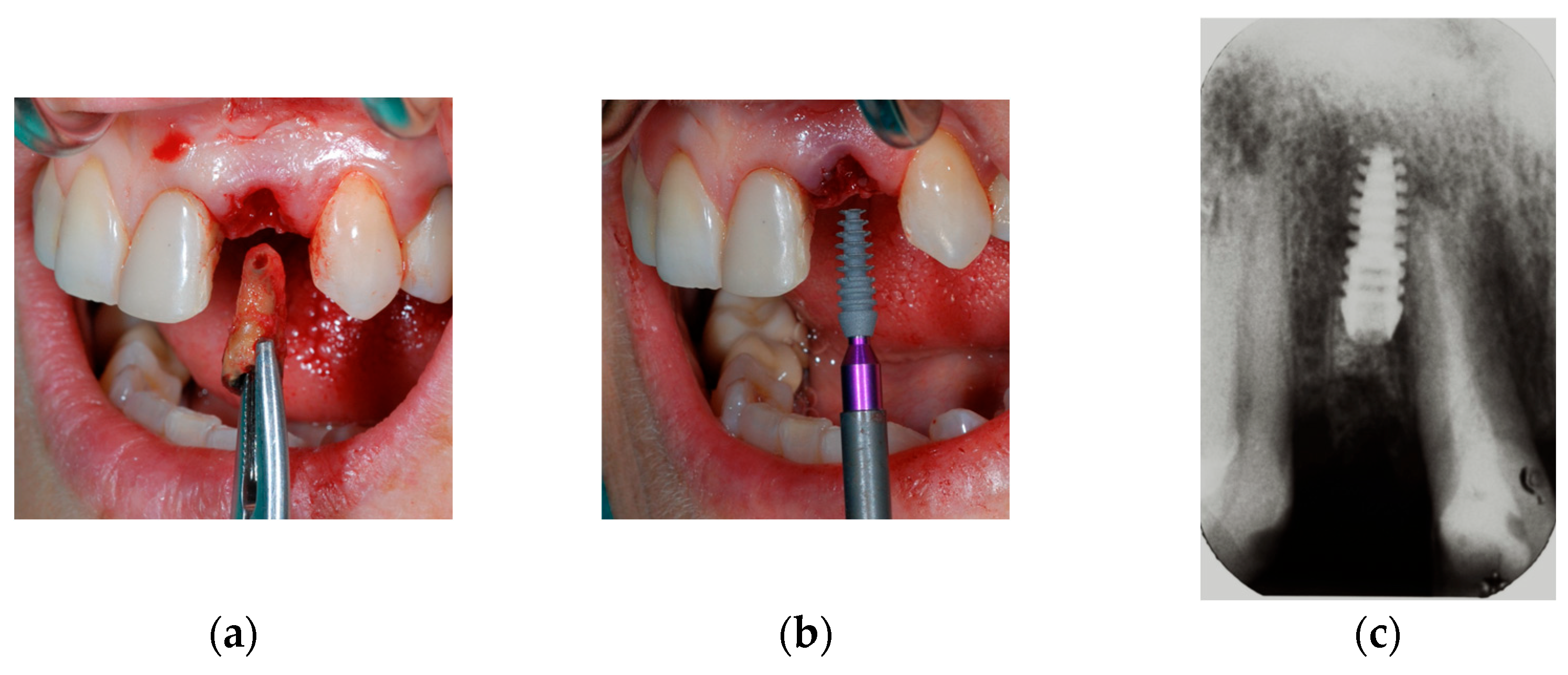
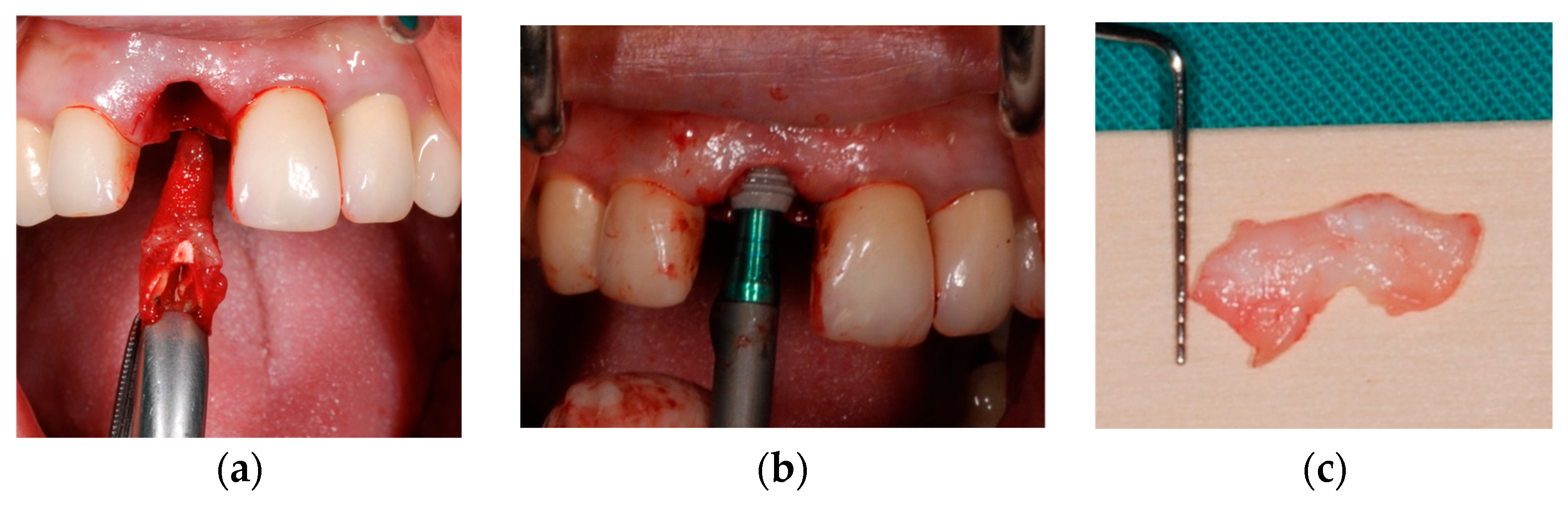
2.4. Surgical Protocol

Surgical and prosthetic procedures were conducted as previously described [16,17]. Briefly, tooth extraction was carried out avoiding damage to either buccal or palatal bone plates. Once the tooth was extracted, a pilot drill of 2.0 mm diameter was used to prepare for the reamers and to determine implant insertion depth. The depth of drilling was 2.0–3.0 mm deeper than that of the chosen implant. The socket was then expanded with reamers of increasing diameter, beginning with a 25 mm diameter without irrigation at a maximum of 50 rpm, until the desired diameter was reached. The implant was then positioned by tapping on the healing plug or directly into the implant well. Autogenous bone removed from the reamer burs and beta-tricalcium phosphate granules (SynthoGraft Pure Phase Beta-Tricalcium Phosphate®, Bicon Dental Implant, Boston, MA, USA) were used to fill the gaps between the implant and the residual bony walls. When deemed appropriate, a subepithelial connective tissue graft (CTG, thickness ≥ 1.5 mm) taken from the palate was placed: the need for concomitant use of CTG was examined by the surgeon considering its use in cases of elevated risk for mid-facial recession, that was in cases with <0.5 mm buccal bone thickness [7,10,28].

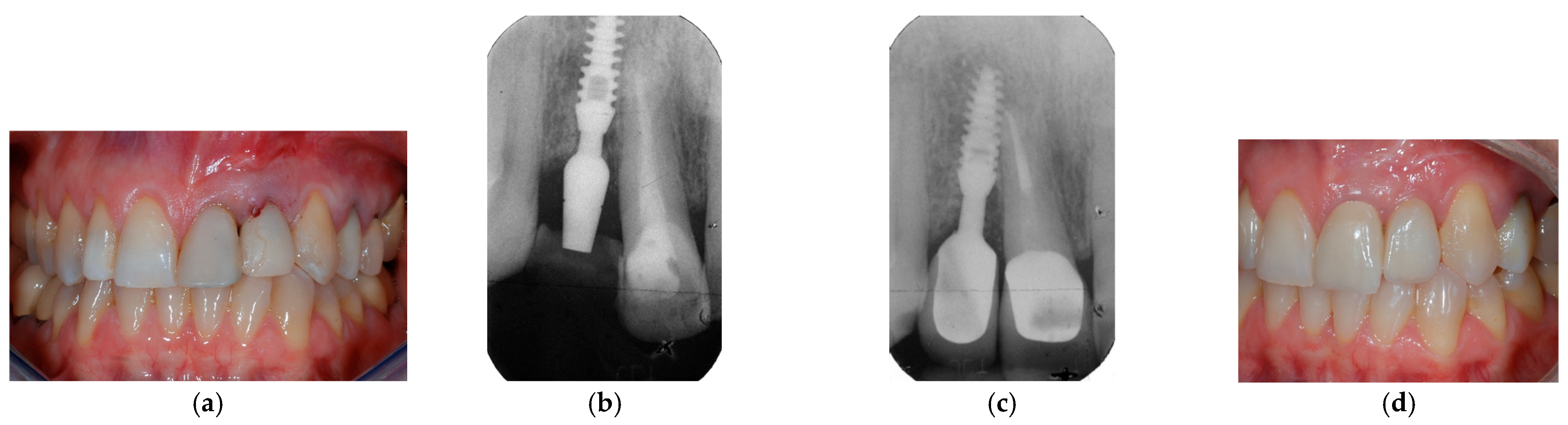
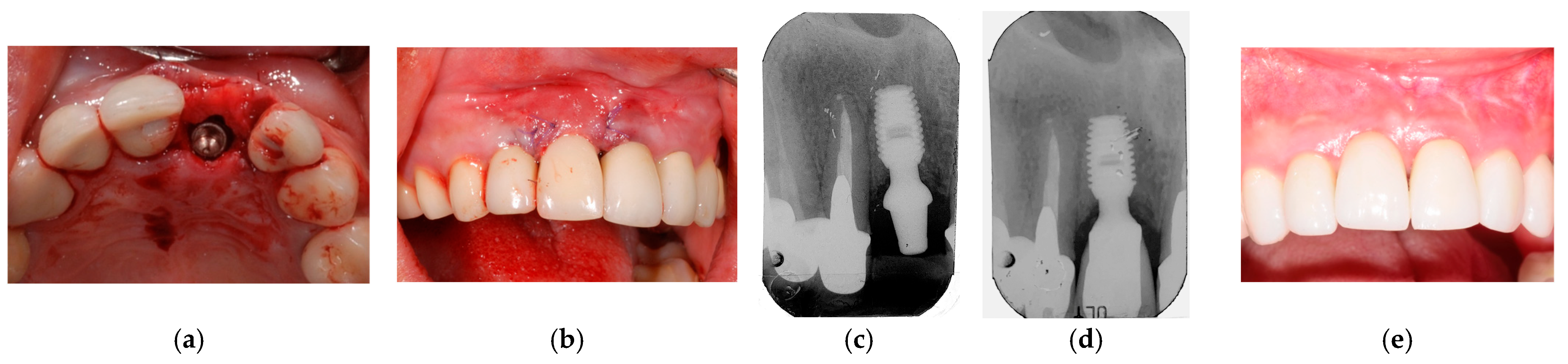
2.5. Prosthetic Protocol

The healing plug was there replaced with an appropriate temporary acrylic resin crown made by designing wax-up. The technique used for the restorations was the Integrated Abutment Crown (IAC) [29], in which crowns are conventionally fabricated but also extra-orally cemented to the abutment, excess cement is removed and then the one-piece abutment and crown, are inserted. The temporary crown was splinted with cold resin to adjacent elements to reduce micro-movements at the bone-implant interface [16]. The final restoration with a porcelain crown was placed after 4 months. Extra-oral cementation was carried out with adhesive resin (3M ESPE RelyX Unicem Self-Adhesive Universal Resin Cement®, Milan, Italy). This provided for control of any cement overflow; any excess was removed before placement of the crown-abutment complex. All crowns were inserted with a custom silicone jig to avoid any crown fracture with tipping.
Recall appointments were established to manage prosthetic complications as needed, and a maintenance program was designed to provide patients with a professional oral hygiene session every 4 months.

2.6. Photographic Measurements of Variations in (i) Papillae Height and (ii) Gingival Margin Position

Photographs were taken before tooth extraction (T0) and 1 year from loading time (T1), to compare variables related to soft tissues and prosthetic rehabilitations between the two time intervals. Non-standardized digital photographic records were obtained with a digital reflex (D80, Nikon, Tokyo, Japan) and a 105 mm lens (AF-S VR Micro-Nikkor 105 mm f/2.8G IF-ED, Nikon, Tokyo, Japan) with a ring flash (EM-140 DG, SIGMA, Kawasaki, Japan). Photographs were taken at, or slightly superior to, the occlusal plane of the study tooth, making sure that the papillae were fully and symmetrically represented to guarantee comparability. Photographs were transferred to a computer and settled to 8 × 6 cm dimensions. To correct any distortions, images were standardized to be overlapped. Four anatomical reference points were identified: the mucogingival line (MGJ), the tip of the mesial papilla (MP), the tip of the distal papilla (DP), the zenith of the gingival/mucosal margin (GM). A line passing through the medial point of the incisal edges of the adjacent natural teeth was used as a reference line (RL).
By measuring the vertical distance between the reference points and the RL, it was possible to obtain the following parameters at T0 and at T1:
-
Mesial papilla height: the distance of MP from RL;
-
Distal papilla height: the distance of DP from RL;
-
Recession (REC): the difference between the distance of GM from RL at T0 or at T1;
-
Keratinized tissue (KT) width: the distance of GM from MGJ.
Changes in these parameters between T0 and T1 were also calculated.
Measurements were performed by another operator, different from the clinicians who performed the surgical and prosthetic phases. Measurements were assessed to the nearest 0.01 mm with the aid of a software program (Rasband, W.S., ImageJ, U. S. National Institutes of Health, Bethesda, MD, USA, version 1.52a 2018), which used a standardized measuring tool based on the dimensions of a real probe present in one of the clinical images in conjunction with a magnification tool. Before the start of the study, this investigator was calibrated for adequate intra-examiner levels of accuracy and reproducibility in recording the clinical and aesthetic parameters. Three clinical cases were utilized for this purpose: duplicate measurements were collected with an interval of 24 h between the first and second recordings. The intra-class correlation coefficients, used as a measure of intra-examiner reproducibility, had to be greater than 0.8.

2.7. Aesthetic Outcomes in Relation to the Use of CTG and Patient’s Phenotype

Aesthetic outcomes regarding soft tissues were assessed at T0 and T1, respectively, on periodontal and peri-implant soft tissues by means of Pink Esthetic Score (PES) [20]. This index evaluates: (i) the degree of interproximal space-filling by the mesial papilla; (ii) the degree of interproximal space-filling by the distal papilla; (iii) the pattern of the gingival margin scalloping; (iv) the level of marginal gingival tissue; (v) the presence of the alveolar process; (vi) the color of marginal gingiva; (vii) the texture of marginal gingiva. The last five parameters are evaluated according to their harmony with the adjacent teeth. For each parameter, points between 0 and 2 were set, for a maximum total score equal to 14 points.
Periodontal soft tissues surrounding the tooth element prior to extraction (at T0) were categorized, according to the phenotype, as thin or thick (thin/thick). Data regarding phenotype at T0 were extracted from patients’ medical records, which reported the transparency of the periodontal probe assessed through the gingival margin during the gingival sulcus probing [30].
Evaluation of aesthetic outcomes both at T0 and T1 were related to the patient’s phenotype and to the presence or absence of CTG. These outcomes were finally analyzed according to the following groups of patients:
-
thin: thin phenotype without CTG;
-
thin + CTG: thin phenotype with CTG;
-
thick: thick phenotype without CTG;
-
thick + CTG: a thick phenotype with CTG.

2.8. Statistical Analysis

Microsoft Excel was used to create a database containing information on all participants of the study. Stata v.13.0 for Macintosh was used for all data analysis (StataCorp, College Station, TX, USA). The Shapiro–Wilk test was used to evaluate the normality assumptions for continuous data; mean and standard deviation were reported for normally distributed data, and median and interquartile range (iqr) otherwise. Absolute frequencies, percentages, and 95% confidence intervals were reported for categorical data. The comparison between the means of continuous variables in different times (T0, T1) was performed by using paired Student’s t-test or Wilcoxon matched-pairs signed-rank test. The comparison of the means among groups (thin, thick, thin + CTG, thick + CTG) was done using one-way analysis of variance (ANOVA), or Kruskal–Wallis equality-of-populations rank test. The level of significance was set at 0.05.

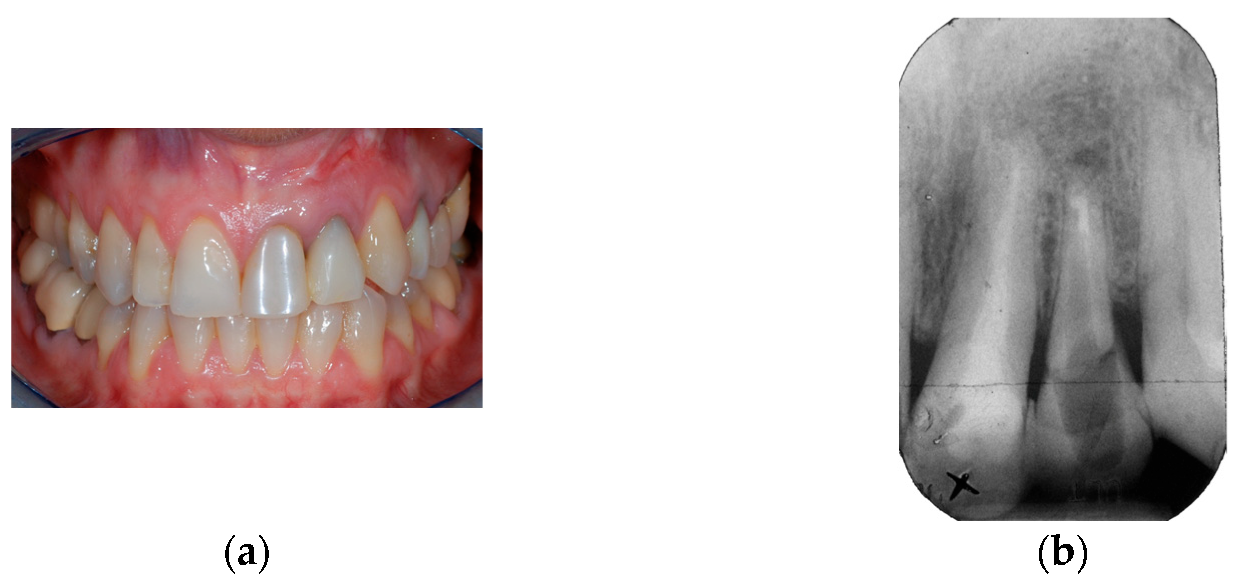
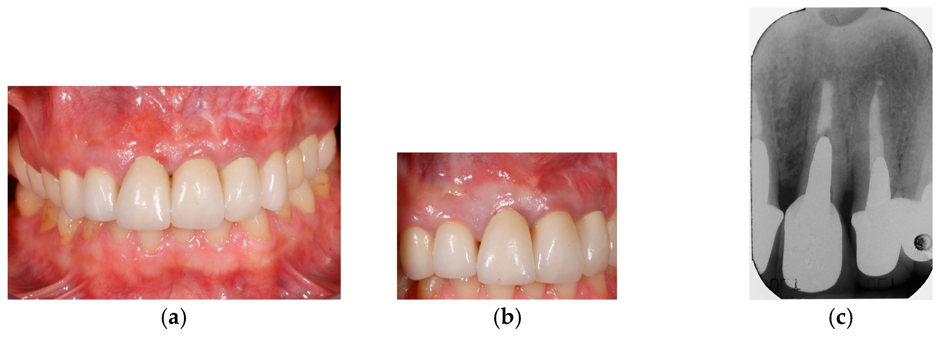
2.9. Clinical Cases and Study Diagram

Figure 1, Figure 2, Figure 3, Figure 4, Figure 5 and Figure 6 show two clinical cases, while Figure 7 reports a study diagram with all phases of the retrospective evaluation.

3. Results

A total of 25 patients (18 women and 7 men), for a total of 25 locking-taper implants placed in the anterior maxillary region, were analyzed in this study; among reasons for tooth loss, root fracture was the most frequent (see Table 1). Ten out of twenty-five implants received a CTG during surgical intervention: CTG was put, respectively, (see Table 1) in four and six patients with thin and thick phenotypes.
Prosthetic complications (splint fracture with consequent loss of abutment retention) were reported in 3 patients during the first 2 months post-implant placement, for an overall incidence of prosthetic complications of 12%: in these cases, the IAC crown was removed, disinfected, reinserted, and re-splinted.
At the average 1-year re-evaluation, the overall implant survival was 100% and no peri-implant infections were reported.
The overall PES values (see Table 2) were, respectively, 9.24 ± 2.36 at T0 and 9.60 ± 1.70 at T1, without statistically significant differences (p = 0.39). Even if no significant differences were generally found in comparison with the pre-existing teeth, evaluation of PES parameters around implants 1 year after surgery resulted in: (i) a less mesial and distal papilla interproximal space-filling; (ii) a more coronal position of the gingival margin; (iii) a slight aesthetic improvement of gingival contour, alveolar process, color, and texture.
Regarding specific changes in peri-implant soft tissue levels in comparison with the pre-existing teeth, mean variations in mesial and distal papilla heights were, respectively, 0.12 mm and 0.20 mm. The mean REC was 0.40 mm, while the KT shrinkage was 0.06 mm (see Table 3).
Comparison of groups between T0 and T1 revealed significant PES variations (p = 0.04), with the best and the worst scores, respectively, registered for thin + CTG group (from 7.50 ± 1.91 to 9.75 ± 2.87) and thin group (from 11.33 ± 2.33 to 10 ± 0.89); moderate increases were assessed for thick group (from 8.44 ± 2.40 to 9.44 ± 2.12) and thick + CTG group (from 9.50 ± 1.04 to 9.33 ± 0.81). Moreover, the great majority of the variables resulted in unvaried outcomes in the thick group and worsened in the thin group.
Most of the variables showed a great improvement, except for the distal papilla height, in the thin + CTG group. Papillae height, gingival level, and gingival contour revealed negative variations in the thick + CTG group.
Overall mean values of PES parameters and soft tissues were also specifically analyzed among groups, respectively, at T0 (see Table 4) and at T1 (see Table 5).
In patients with a thick phenotype, the use of a CTG led to better aesthetic results at T1 only in 33% of cases, differently from patients with a thin phenotype, which improved in 75% of cases. Plus, 52% and 64% of the implants did not show any changes, respectively, in mesial papilla and distal papilla heights. GM position was found as unvaried in 48% and improved in 12% of cases. REC was found in 40% of cases, most of them between 0 and 1 mm; extensive REC (≥2 mm) was observed in only one case, and even in this case, the implant abutment did not result in exposure. Regarding KT, its width decreased by 36%, remained unchanged in 48%, and increased in 16% of cases.

4. Discussion

In response to evolving social factors and more demanding patients looking for immediate definitive results, immediate implant placement after extraction, combined with immediate provisionalization, has become a growing tendency in implant dentistry. Nevertheless, this procedure may be considered with caution [31] because of the risk of recession of buccal soft tissues and loss of papillae heights [32,33] with consequent grey transparency of the implant surface, or finally even exposure of the titanium implant surface through the soft tissues surrounding the crown [3,30].
Up-to-date literature [3,4,6,12,14,30,34] presented this protocol with average values of 1.1 mm of facial gingival recession, and mean mesial/distal papilla reductions of, respectively, 0.2 mm and 0.9 mm in their clinical experience. Specifically, Kan et al. [4] reported, after 1 year of follow-up, a mean mid-facial gingival recession and mesial/distal papilla height reduction of, respectively, 0.55 mm, 0.53 mm, and 0.39 mm. De Rouck et al. [30] declared for the same parameters, respectively, values of 0.53 mm, 0.41 mm, and 0.31 mm. Evans et al. [3] described a change in crown height due to a marginal tissue recession of 0.9 mm. In the present study, after 1 year of follow-up (T1), the average mesial/distal papilla height reductions were, respectively, 0.12 mm and 0.2 mm, mean REC was 0.4 mm, while the KT shrinkage was 0.06 mm, with no statistically significant changes in soft tissues parameters occurring from baseline (T0).
Furthermore, the overall PES values were, respectively, 9.24 ± 2.36 at T0 and 9.60 ± 1.70 at T1, without statistically significant differences (p = 0.39) between time intervals, completely overlapping with results obtained by the study of Furhauser [20] (PES = 9.25). Even if no significant differences were generally found in comparison with the pre-existing teeth, evaluation of PES parameters around implants 1 year after surgery resulted in a less mesial and distal papilla interproximal space-filling, a more coronal position of the gingival margin, and slight aesthetic improvements of gingival contour, alveolar process, color, and texture.
It is well documented in the literature [4,32] that the presence of papilla is mostly related to the bone level of the adjacent teeth: on this basis, it has been postulated [30] that the chosen surgical technique and the implant design may represent negligible factors regarding the influence on final results. Nevertheless, the authors assume that specific features of an implant system, as shown in this pilot study, may play an adjunctive role in achieving satisfactory outcomes. In this proposal, the absence of micro-gaps, micro-movements [15], and cement remnants [35] at the level of the locking-taper connection, together with the presence of sloping shoulders in association with IAC [29], all represent advantages allowing for a sub-crestal implant placement of even more than 3 mm [36]. These advantages enable the use of these implants even in the case of thin alveolar ridges, without the risk of “saucerization” of the crestal bone, with consequent undesired soft tissue modifications [37,38].
Concerning papillae variations, 52% and 64% of the implants did not show any changes in mesial and distal heights. Appropriate management of soft tissues is evident as part of a proper preoperative assessment of cases, fundamental for the follow-up predictability of clinical outcomes. It can be thus underlined that the stability of mesial and distal papillae was assessed over time, independently from the immediate non-functional provisional single-crown restorations and the definitive ones. Accurate planning of prosthetic phases, with final provisionalization 4 months after implant placement, was not focused on the compensation of initial recession, but on the concept of appropriate soft tissue healing to ensure stable conditions in terms of both clinical and radiographic follow-up.
In confirmation of these satisfactory results, obtained through the use of a safe protocol with peculiar characteristics regarding the implant type, the level of GM was found unvaried in 48% and improved in 12% of cases. Recessions were found in 40% of cases, most of them between 0 and 1 mm; extensive recession (≥2 mm) was observed in only one case, and even in this case, the implant abutment did not result in exposure. Regarding KT, its width decreased by 36%, remained unchanged in 48%, and increased in 16% of the cases.
Considering that periodontal surgical techniques can change the phenotype from thin to thick, many authors [28,31,39,40] currently assume as essential, in case of aesthetic rehabilitations in patients with thin phenotype or insufficient buccal bone thickness [28], to associate a free CTG to implant placement, to avoid or minimize the risk of recession of peri-implant soft tissues. It is well known that, same as the periodontal phenotype, the peri-implant phenotype is site-specific and exposed to modifications of environmental factors [41] or surgical interventions [42]. In the case of surgical or restorative procedures, initial gingival thickness may predict the outcomes of root coverage procedures and restorative treatments [43]. While a similar phenomenon was observed for peri-implant mucosa by several authors using protocols of immediate implant placement and provisionalization [3,44,45,46], other randomized clinical trials [47,48,49] failed to observe any significant differences in soft tissue aesthetics following this procedure, irrespective of the gingival phenotype, or the eventual presence of a CTG graft.
In this study the potential of concomitant use of CTG was examined in relation to the elevated risk for mid-facial recession, that was in cases with <0.5 mm buccal bone thickness, mainly providing clinical advantages in patients with both thin and thick phenotypes. Comparison of groups between T0 and T1 revealed significant PES variations (p = 0.04), with the best and the worst ones, respectively, registered for thin + CTG group (from 7.50 ± 1.91 to 9.75 ± 2.87) and thin group (from 11.33 ± 2.33 to 10 ± 0.89); moderate increases were assessed for thick group (from 8.44 ± 2.40 to 9.44 ± 2.12) and thick + CTG group (from 9.50 ± 1.04 to 9.33 ± 0.81). Moreover, the great majority of the variables resulted in unvaried in the thick group, while worsened in the thin group. In the thin + CTG group, most of the variables showed a great improvement, with even 0.62 mm of KT gain. In the thick + CTG group, papillae height, gingival level, and gingival contour revealed negative variations. Regarding overall aesthetic improvement between T0 and T1, in patients with a thick phenotype, the use of a CTG led to better results at T1 only in 33% of cases, differently from patients with a thin phenotype, which improved in 75% of cases. Nevertheless, the presence of a thick phenotype resulted as a protective factor against soft tissue recession, confirming that tissue phenotype represents a critical variable, more than the type of implant treatment itself, in determining both the final PES score and clinical results. It may be of particular interest the finding that improvements found in the thick + CTG group were not as marked as in the thin + CTG group: a possible explanation for this issue [50] is that bilaminar CTG technique, in conjunction with immediate tooth replacement procedures, may be considered a sensitive procedure with inherent risks not to be overlooked. In light of these considerations, authors finally assume the use of CTG is appropriate unless necessary, as it can enhance the aesthetics of peri-implant tissues, but might be also associated with higher patient morbidity.
Looking at the study limitations, caution should be accounted for the retrospective approach in a Hospital setting and a small number of patients followed for a short-term follow-up: these main limits introduce biases for the interpretation of outcomes obtained from the hypothesis related to the predictability of the proposed implant therapy. On the other hand, the limited number of patients allowed a strict maintenance protocol [51] for plaque control and motivation, with great dedication by the University Dental Clinic. Another strength of the present investigation can be referred to the detailed analysis of clinical conditions according to patients’ phenotype and the use of CTG.
The present pilot retrospective study can be reported as a clinical experience of daily routine practice based on the findings of a minimally invasive approach, which aimed to address patients’ aesthetic expectations and ensure peri-implant health around a specific type of implant. Further assessment of clinical conditions in larger groups of patients with mid- and long-term follow-up is advisable for consistent evidence of this specific type of surgery.

5. Conclusions

The specific protocol of immediate implant rehabilitation for the aesthetic anterior areas of the upper maxilla can be assumed as a safe treatment. However, outcomes showed that peri-implant soft tissue aesthetics, independently from the design of the implant used, can be affected by (i) the patient’s phenotype at the time of the procedure; (ii) concomitant use of CTG, which demonstrated to provide beneficial effects in case of patients with thin phenotype, but did not provide any additional value in patients with thick one.
Potential clinical implications of these preliminary findings highlight the importance of a detailed pre-clinical evaluation of the patient’s phenotype characteristics to properly plan a surgical treatment in the aesthetic area of the upper maxilla.
The use of CTG is suggested in selected cases of post-extraction sites to adequately address aesthetic expectations and clinical outcomes at the same time, avoiding the risk of recession, especially in patients with thin phenotypes. The present study offers a valid option of treatment with a specific type of implant-supported restoration, showing predictable short-term results through a minimally invasive approach without the need for complex augmentation procedures, thus simplifying daily routine surgical practice.

Author Contributions

Conceptualization, G.L., M.M. and M.A.; methodology, A.S., A.Z., A.P. and P.F.; software, G.L. and A.S.; validation, E.M., M.G. and G.C.; formal analysis, A.S., A.Z., A.P. and P.F.; investigation, E.M., M.G. and G.C.; resources, G.L. and M.M.; data curation, G.L. and A.S.; writing—original draft preparation, G.L., A.S., A.Z. and A.P.; writing—review and editing, M.M., E.M., M.G., G.C., M.A. and P.F.; visualization, M.M., E.M., M.G., G.C., M.A. and P.F.; supervision, G.L., A.S., A.Z. and A.P.; project administration, G.L. All authors have read and agreed to the published version of the manuscript.

Funding

This research received no external funding.

Institutional Review Board Statement

The study was conducted in accordance with the Declaration of Helsinki, and approved by the Ethics Committee of the University of Verona (protocol code AESTETANTE, Prog. 1815CESC and date of approval 30 May 2018) for studies involving humans.

Informed Consent Statement

Informed consent was obtained from all subjects involved in the study.

Data Availability Statement

The data presented in this study are available on request from the corresponding authors.

Conflicts of Interest

The authors declare no conflicts of interest.

Abbreviations

The following abbreviations are used in this manuscript:
Pink Esthetic Score PES
connective tissue graft CTG
implant-abutment connection IAI
mucogingival line MGJ
mesial papilla MP
distal papilla DP
gingival/mucosal marginGM
reference line RL
recession REC
keratinized tissue KT

References

  1. Schropp, L.; Wenzel, A.; Kostopoulos, L.; Karring, T. Bone healing and soft tissue contour changes following single-tooth extraction: A clinical and radiographic 12-month prospective study. Int. J. Periodontics Restor. Dent. 2003, 23, 313–323. [Google Scholar]
  2. Araújo, M.G.; Silva, C.O.; Souza, A.B.; Sukekava, F. Socket healing with and without immediate implant placement. Periodontol. 2000 2019, 79, 168–177. [Google Scholar] [CrossRef] [PubMed]
  3. Evans, C.D.; Chen, S.T. Esthetic outcomes of immediate implant placements. Clin. Oral Implant. Res. 2008, 19, 73–80. [Google Scholar] [CrossRef] [PubMed]
  4. Kan, J.Y.; Rungcharassaeng, K.; Lozada, J. Immediate placement and provisionalization of maxillary anterior single implants: 1-year prospective study. Int. J. Oral Maxillofac. Implant. 2003, 18, 31–39. [Google Scholar]
  5. Rosenquist, B.; Grenthe, B. Immediate placement of implants into extraction sockets: Implant survival. Int. J. Oral Maxillofac. Implant. 1996, 11, 205–209. [Google Scholar] [CrossRef]
  6. Chen, S.T.; Wilson, T.G., Jr.; Hämmerle, C.H. Immediate or early placement of implants following tooth extraction: Review of biologic basis, clinical procedures, and outcomes. Int. J. Oral Maxillofac. Implant. 2004, 19, 12–25. [Google Scholar]
  7. Kan, J.Y.K.; Yin, S.; Rungcharassaeng, K.; Zucchelli, G.; Urban, I.; Lozada, J. Facial implant gingival level and thickness changes following maxillary anterior immediate tooth replacement with scarf-connective tissue graft: A 4–13-year retrospective study. J. Esthet. Restor. Dent. 2023, 35, 138–147. [Google Scholar] [CrossRef]
  8. Belser, U.C.; Bernard, J.P.; Buser, D. Implant-supported restorations in the anterior region: Prosthetic considerations. Pract. Periodontics Aesthet. Dent. 1996, 8, 875–883. [Google Scholar]
  9. Belser, U.C.; Buser, D.; Hess, D.; Schmid, B.; Bernard, J.P.; Lang, N.P. Aesthetic implant restorations in partially edentulous patients—A critical appraisal. Periodontol. 2000 1998, 17, 132–150. [Google Scholar] [CrossRef]
  10. Grunder, U.; Gracis, S.; Capelli, M. Influence of the 3-D bone-to-implant relationship on esthetics. Int. J. Periodontics Restor. Dent. 2005, 25, 113–119. [Google Scholar]
  11. Juodzbalys, G.; Wang, H.L. Soft and hard tissue assessment of immediate implant placement: A case series. Clin. Oral Implant. Res. 2007, 18, 237–243. [Google Scholar] [CrossRef] [PubMed]
  12. Chen, S.T.; Darby, I.B.; Adams, G.G.; Reynolds, E.C. A prospective clinical study of bone augmentation techniques at immediate implants. Clin. Oral Implant. Res. 2005, 16, 176–184. [Google Scholar] [CrossRef] [PubMed]
  13. Meijndert, L.; Meijer, H.J.; Stellingsma, K.; Stegenga, B.; Raghoebar, G.M. Evaluation of aesthetics of implant-supported single-tooth replacements using different bone augmentation procedures: A prospective randomized clinical study. Clin. Oral Implant. Res. 2007, 18, 715–719. [Google Scholar] [CrossRef]
  14. Cordaro, L. Implants for restoration of single tooth spaces in areas of high esthetic risk. In The SAC Classification in Implant Dentistry, 2nd ed.; Dawson, A., Martin, W., Polido, W.D., Eds.; Quintessence Publishing: Berlin, Germany, 2009; pp. 50–56. [Google Scholar]
  15. King, G.N.; Hermann, J.S.; Schoolfield, J.D.; Buser, D.; Cochran, D.L. Influence of the size of the microgap on crestal bone levels in non-submerged dental implants: A radiographic study in the canine mandible. J. Periodontol. 2002, 73, 1111–1117. [Google Scholar] [CrossRef] [PubMed]
  16. Lombardo, G.; Signoriello, A.; Marincola, M.; Liboni, P.; Bonfante, E.A.; Nocini, P.F. Survival rates of ultra-short (<6 mm) compared with short locking-taper implants supporting single crowns in posterior areas: A 5-year retrospective study. Clin. Implant. Dent. Relat. Res. 2021, 23, 904–919. [Google Scholar]
  17. Lombardo, G.; Corrocher, G.; Pighi, J.; Mascellaro, A.; Marincola, M.; Nocini, P.F. Esthetic Outcomes of Immediately Loaded Locking Taper Implants in the Anterior Maxilla: A Case Series Study. J. Oral Implantol. 2016, 42, 258–264. [Google Scholar] [CrossRef]
  18. Buser, D.; Chappuis, V.; Bornstein, M.M.; Wittneben, J.G.; Frei, M.; Belser, U.C. Long-term stability of contour augmentation with early implant placement following single tooth extraction in the esthetic zone: A prospective, cross-sectional study in 41 patients with a 5- to 9-year follow-up. J. Periodontol. 2013, 84, 1517–1527. [Google Scholar] [CrossRef]
  19. Belser, U.C.; Grütter, L.; Vailati, F.; Bornstein, M.M.; Weber, H.P.; Buser, D. Outcome evaluation of early placed maxillary anterior single-tooth implants using objective esthetic criteria: A cross-sectional, retrospective study in 45 patients with a 2- to 4-year follow-up using pink and white esthetic scores. J. Periodontol. 2009, 80, 140–151. [Google Scholar] [CrossRef]
  20. Fürhauser, R.; Florescu, D.; Benesch, T.; Haas, R.; Mailath, G.; Watzek, G. Evaluation of soft tissue around single-tooth implant crowns: The pink esthetic score. Clin. Oral Implant. Res. 2005, 16, 639–644. [Google Scholar] [CrossRef]
  21. Dursun, E.; Lin, G.H.; Taheri, S.; Chu, S.J.; Wang, H.L.; Tözüm, T.F. A Comparison of Esthetic Features of Pre-existing Natural Tooth Versus Post-Implant Restoration in the Esthetic Zone: A Retrospective 12-month Follow-up. Int. J. Oral Maxillofac. Implant. 2018, 33, 919–928. [Google Scholar] [CrossRef]
  22. Hartlev, J.; Kohberg, P.; Ahlmann, S.; Andersen, N.T.; Schou, S.; Isidor, F. Patient satisfaction and esthetic outcome after immediate placement and provisionalization of single-tooth implants involving a definitive individual abutment. Clin. Oral Implant. Res. 2014, 25, 1245–1250. [Google Scholar] [CrossRef] [PubMed]
  23. Hämmerle, C.H.; Chen, S.T.; Wilson, T.G., Jr. Consensus statements and recommended clinical procedures regarding the placement of implants in extraction sockets. Int. J. Oral Maxillofac. Implant. 2004, 19, 26–28. [Google Scholar]
  24. Chou, H.Y.; Romanas, G.; Müftü, A.; Müftü, S. Peri-implant bone remodeling around an extraction socket: Predictions of bone maintenance by finite element method. Int. J. Oral Maxillofac. Implant. 2012, 27, 39–48. [Google Scholar]
  25. Bozkaya, D.; Muftu, S. Mechanics of the tapered interference fit in dental implants. J. Biomech. 2003, 36, 1649–1658. [Google Scholar] [CrossRef]
  26. Dibart, S.; Warbington, M.; Su, M.F.; Skobe, Z. In vitro evaluation of the implant-abutment bacterial seal: The locking taper system. Int. J. Oral Maxillofac. Implant. 2005, 20, 732–737. [Google Scholar]
  27. Aloise, J.P.; Curcio, R.; Laporta, M.Z.; Rossi, L.; da Silva, A.M.; Rapoport, A. Microbial leakage through the implant-abutment interface of Morse taper implants in vitro. Clin. Oral Implant. Res. 2010, 21, 328–335. [Google Scholar] [CrossRef]
  28. Seyssens, L.; De Lat, L.; Cosyn, J. Immediate implant placement with or without connective tissue graft: A systematic review and meta-analysis. J. Clin. Periodontol. 2020, 48, 284–301. [Google Scholar] [CrossRef]
  29. Urdaneta, R.A.; Marincola, M. The Integrated Abutment Crownt, a screwless and cementless restoration for single-tooth implants: A report on a new technique. J. Prosthodont. 2007, 16, 311–318. [Google Scholar] [CrossRef]
  30. De Rouck, T.; Eghbali, R.; Collys, K.; De Bruyn, H.; Cosyn, J. The gingival biotype revisited: Transparency of the periodontal probe through the gingival margin as a method to discriminate thin from thick gingiva. J. Clin. Periodontol. 2009, 36, 428–433. [Google Scholar] [CrossRef]
  31. Levine, R.A.; Ganeles, J.; Gonzaga, L.; Kan, J.K.; Randel, H.; Evans, C.D.; Chen, S.T. 10 Keys for Successful Esthetic-Zone Single Immediate Implants. Compend. Contin. Educ. Dent. 2017, 38, 248–260. [Google Scholar]
  32. Choquet, V.; Hermans, M.; Adriaenssens, P.; Daelemans, P.; Tarnow, D.P.; Malevez, C. Clinical and radiographic evaluation of the papilla level adjacent to single-tooth dental implants. A retrospective study in the maxillary anterior region. J. Periodontol. 2001, 72, 1364–1371. [Google Scholar] [CrossRef] [PubMed]
  33. Kois, J.C.; Kan, J.Y. Predictable peri-implant gingival aesthetics: Surgical and prosthodontic rationales. Pract. Proced. Aesthet. Dent. 2001, 13, 691–698. [Google Scholar] [PubMed]
  34. Chang, M.; Wennström, J.L.; Odman, P.; Andersson, B. Implant supported single-tooth replacements compared to contralateral natural teeth. Crown and soft tissue dimensions. Clin. Oral Implant. Res. 1999, 10, 185–194. [Google Scholar] [CrossRef]
  35. Linkevicius, T.; Puisys, A.; Vindasiute, E.; Linkeviciene, L.; Apse, P. Does residual cement around implant-supported restorations cause peri-implant disease? A retrospective case analysis. Clin. Oral Implant. Res. 2013, 24, 1179–1184. [Google Scholar] [CrossRef]
  36. Lombardo, G.; Signoriello, A.; Marincola, M.; Nocini, P.F. Assessment of Peri-Implant Soft Tissues Conditions around Short and Ultra-Short Implant-Supported Single Crowns: A 3-Year Retrospective Study on Periodontally Healthy Patients and Patients with a History of Periodontal Disease. Int. J. Environ. Res. Public. Health 2020, 17, 9354. [Google Scholar] [CrossRef] [PubMed]
  37. De Santis, D.; Sinigaglia, S.; Pancera, P.; Faccioni, P.; Portelli, M.; Luciano, U.; Cosola, S.; Penarrocha, D.; Bertossi, D.; Nocini, R.; et al. An overview of socket preservation. J. Biol. Regul. Homeost. Agents 2019, 33, 55–59. [Google Scholar]
  38. Buser, D.; Martin, W.; Belser, U.C. Optimizing esthetics for implant restorations in the anterior maxilla: Anatomic and surgical considerations. Int. J. Oral Maxillofac. Implant. 2004, 19, 43–61. [Google Scholar]
  39. Jung, R.E.; Becker, K.; Bienz, S.P.; Dahlin, C.; Donos, N.; Hammächer, C.; Iglhaut, G.; Liñares, A.; Ortiz-Vigón, A.; Sanchez, N.; et al. Effect of peri-implant mucosal thickness on esthetic outcomes and the efficacy of soft tissue augmentation procedures: Consensus report of group 2 of the SEPA/DGI/OF workshop. Clin. Oral Implant. Res. 2022, 33, 100–108. [Google Scholar] [CrossRef] [PubMed]
  40. Tavelli, L.; Barootchi, S.; Avila-Ortiz, G.; Urban, I.A.; Giannobile, W.V.; Wang, H.L. Peri-implant soft tissue phenotype modification and its impact on peri-implant health: A systematic review and network meta-analysis. J. Periodontol. 2021, 92, 21–44. [Google Scholar] [CrossRef]
  41. Jepsen, S.; Caton, J.G.; Albandar, J.M.; Bissada, N.F.; Bouchard, P.; Cortellini, P.; Demirel, K.; de Sanctis, M.; Ercoli, C.; Fan, J.; et al. Periodontal manifestations of systemic diseases and developmental and acquired conditions: Consensus report of workgroup 3 of the 2017 world workshop on the classification of periodontal and peri-implant diseases and conditions. J. Periodontol. 2018, 89, 237–248. [Google Scholar] [CrossRef]
  42. Barootchi, S.; Tavelli, L.; Zucchelli, G.; Giannobile, W.V.; Wang, H.L. Gingival phenotype modification therapies on natural teeth: A network meta-analysis. J. Periodontol. 2020, 91, 1386–1399. [Google Scholar] [CrossRef] [PubMed]
  43. Cortellini, P.; Bissada, N.F. Mucogingival conditions in the natural dentition: Narrative review, case definitions, and diagnostic considerations. J. Periodontol. 2018, 89, 204–213. [Google Scholar] [CrossRef] [PubMed]
  44. Kan, J.Y.; Rungcharassaeng, K.; Lozada, J.L.; Zimmerman, G. Facial gingival tissue stability following immediate placement and provisionalization of maxillary anterior single implants: A 2- to 8-year follow-up. Int. J. Oral Maxillofac. Implant. 2011, 26, 179–187. [Google Scholar]
  45. Mailoa, J.; Arnett, M.; Chan, H.L.; George, F.M.; Kaigler, D.; Wang, H.L. The Association Between Buccal Mucosa Thickness and Periimplant Bone Loss and Attachment Loss: A Cross-Sectional Study. Implant. Dent. 2018, 27, 575–581. [Google Scholar] [CrossRef]
  46. Chen, S.T.; Darby, I.B.; Reynolds, E.C.; Clement, J.G. Immediate implant placement postextraction without flap elevation. J. Periodontol. 2009, 80, 163–172. [Google Scholar] [CrossRef]
  47. van Nimwegen, W.G.; Raghoebar, G.M.; Zuiderveld, E.G.; Jung, R.E.; Meijer, H.J.A.; Mühlemann, S. Immediate placement and provisionalization of implants in the aesthetic zone with or without a connective tissue graft: A 1-year randomized controlled trial and volumetric study. Clin. Oral Implants Res. 2018, 29, 671–678. [Google Scholar] [CrossRef]
  48. Ferrantino, L.; Camurati, A.; Gambino, P.; Marzolo, M.; Trisciuoglio, D.; Santoro, G.; Farina, V.; Fontana, F.; Asa’ad, F.; Simion, M. Aesthetic outcomes of nonfunctional immediately restored single post-extraction implants with and without connective tissue graft: A multicenter randomized controlled trial. Clin. Oral Implant. Res. 2021, 32, 684–694. [Google Scholar] [CrossRef] [PubMed]
  49. Zuiderveld, E.G.; Meijer, H.J.A.; den Hartog, L.; Vissink, A.; Raghoebar, G.M. Effect of connective tissue grafting on periimplant tissue in single immediate implant sites: A RCT. J. Clin. Periodontol. 2018, 45, 253–264. [Google Scholar] [CrossRef]
  50. Chung, S.; Rungcharassaeng, K.; Kan, J.Y.; Roe, P.; Lozada, J.L. Immediate single tooth replacement with subepithelial connective tissue graft using platform switching implants: A case series. J. Oral Implantol. 2011, 37, 559–569. [Google Scholar] [CrossRef]
  51. Ballini, A.; Di Cosola, M.; Saini, R.; Benincasa, C.; Aiello, E.; Marrelli, B.; Rajiv Saini, S.; Ceruso, F.M.; Nocini, R.; Topi, S.; et al. A Comparison of Manual Nylon Bristle Toothbrushes versus Thermoplastic Elastomer Toothbrushes in Terms of Cleaning Efficacy and the Biological Potential Role on Gingival Health. Appl. Sci. 2021, 11, 7180. [Google Scholar] [CrossRef]
Figure 1. Case 1—A healthy, non-smoker 48-year-old woman with thick gingival phenotype: preoperative facial view of the failing treated left lateral incisor; PES evaluation at T0 was 9.5 (a); Preoperative periapical radiograph of the site (b).
Prosthesis 07 00060 g001
Figure 2. Case 1—The fractured root was atraumatically removed without flap elevation (a); A 3.5 × 11 mm locking-taper implant was placed without the aid of surgical guide immediately into the socket. The buccal gap was filled with bone material and no CTG graft was used (b); Periapical radiograph immediately after implant placement (c).
Prosthesis 07 00060 g002
Figure 3. Case 1—The relined provisional crown was cemented onto the pre-fabricated temporary abutment and splinted to the neighboring teeth (a); Periapical radiograph immediately after provisional restoration (b); Periapical radiograph of the implant-supported rehabilitation 1-year after loading (c); Facial view a of the implant-supported rehabilitation at 1 year follow-up, showing 0.8 mm REC. PES evaluation at T1 was 10.5 (d).
Prosthesis 07 00060 g003
Figure 4. Case 2–A healthy, non-smoker 42-year-old woman, presenting a thin gingival phenotype (a); Preoperative facial view of the failing treated right central incisor. PES evaluation at T0 was 9 (b); Preoperative periapical radiograph of the site (c).
Prosthesis 07 00060 g004
Figure 5. Case 2—The fractured root was atraumatically removed without flap elevation (a); A 4.5 × 8 mm locking-taper implant was immediately free-hand placed into the socket. The buccal gap was filled with bone material (b); A CTG was harvested from the palate (c).
Prosthesis 07 00060 g005
Figure 6. Case 2—Occlusal view after implant placement with major part of the CTG sutured directly under the soft tissues, in an “envelope” previously created by an undermining partial thickness incision in the vestibular tissues surrounding the implants (a); Frontal view of the immediate non-functional provisional restoration. The relined provisional crown was cemented onto the pre-fabricated temporary abutment and splinted to the neighboring teeth (b); Periapical radiograph immediately after provisional restoration (c); Periapical radiograph of the implant-supported rehabilitation at 1-year follow-up after loading (d); Facial view at 1-year follow-up, showing the increased thickness of soft tissues and proper peri-implant gingival framework. PES evaluation after 1 year was 12 (e).
Prosthesis 07 00060 g006
Figure 7. Diagram of the study phases of the retrospective evaluation.
Prosthesis 07 00060 g007
Table 1. Overall characteristics of 25 implants treated. Variables are presented as n (%), with 95% C.I.
n%C.I.
Sex
male7220.13–0.49
female18720.50–0.86
Implant position
116240.10–0.45
127280.13–0.49
13140.01–0.26
215200.08–0.41
226240.10–0.45
Connective tissue graft (CTG)
no15600.38–0.77
yes10400.22–0.61
Gingival phenotype
thin10400.22–0.61
thick15600.38–0.77
Gingival phenotype combined with CTG
thin 6240.10–0.45
thick9360.18–0.57
thin + CTG4160.05–0.37
thick + CTG6240.10–0.45
Aetiology for tooth loss
agenesis140.01–0.26
root fracture12480.30–0.69
caries6240.10–0.45
non-treatable endodontic lesions5200.08–0.41
root resorption140.01–0.26
Table 2. Overall mean scores of PES parameters and soft tissues at T0 (tooth) and at T1 (implant at follow-up).
Mesial
Papilla
Distal
Papilla
Gingival LevelGingival ContourAlveolar ProcessColorTextureTotal PESKTGM
T01.12 ± 0.721.08 ± 0.701.32 ± 0.741.32 ± 0.691.32 ± 0.551.6 ± 0.571.48 ± 0.589.24 ± 2.363.82 ± 0.9211.08 ± 2.03
T11.00 ± 0.640.88 ± 0.661.20 ± 0.641.52 ± 0.581.60 ± 0.501.72 ± 0.451.68 ± 0.479.60 ± 1.703.76 ± 1.2611.48 ± 1.58
p value0.410.060.440.250.060.410.130.390.80.06
At each time interval, values for PES parameters (mesial papilla, distal papilla, gingival level, gingival contour, alveolar process, color, texture) and for total PES are presented in the score as (mean ± sd); values for keratinized tissue (KT) and gingival margin (GM) are presented in mm as (mean ± sd). The comparison between the means at two different times was performed by using a paired Student’s t-test.
Table 3. Mean variations (Δ) of PES parameters and soft tissues between T0 and T1: overall scores and scores according to groups of phenotype and CTG.
ΔMesial
Papilla
ΔDistal
Papilla
ΔGingival LevelΔGingival ContourΔAlveolar ProcessΔColorΔTextureΔTotal PESΔKTREC
Overall(-)0.12 (0.72)(-)0.20 (0.50)(-)0.12 (0.78)0.2 (0.86)0.28 (0.73)0.12 (0.72)0.20 (0.64)0.36 (2.07)(-)0.06 (1.17)0.40 (1.00)
thin(-)0.33 (0.51)(-)0.33 (0.51)(-)0.33 (1.03)0.16 (0.98)(-)0.16 (0.75)(-)0.16 (0.75)(-)0.16 (0.75)(-)1.33 (2.16)(-)0.50 (0.54)0.33 (1.36)
thick0.00 (0.86)0.00 (0.50)0.00 (0.70)0.33 (1.00)0.22 (0.66)0.22 (0.83)0.22 (0.66)1.00 (1.93)0.11 (1.53)0.33 (1.11)
thin + CTG0.25 (0.50)(-)0.25 (0.50)0.00 (0.81)0.50 (0.57)0.75 (0.95)0.50 (0.57)0.50 (0.57)2.25 (1.70)0.62 (0.47)0.50 (1.00)
thick + CTG(-)0.33 (0.81)(-)0.33 (0.51)(-)0.16 (0.75)(-)0.16 (0.75)0.50 (0.54)0.00 (0.63)0.33 (0.51)(-)0.16 (0.98)0.50 (1.22)0.50 (0.54)
p value0.430.530.920.610.280.450.430.04 *0.310.95
For each group, values for PES parameters (Δmesial papilla, Δdistal papilla, Δgingival level, Δgingival contour, Δalveolar process, Δcolor, Δtexture) and for Δtotal PES are presented in score as (median (iqr)).; values for ΔKT and recession (REC) are presented in mm as (median (iqr)). The comparison of the variations among groups was conducted using Kruskal–Wallis equality-of-populations rank test; * = statistically significant.
Table 4. Mean values of PES parameters and soft tissues at T0: scores according to groups of phenotype and CTG.
Mesial
Papilla
Distal
Papilla
Gingival LevelGingival ContourAlveolar ProcessColorTextureTotal PESKTGM
thin1.66 ± 0.511.66 ± 0.511.50 ± 0.541.33 ± 0.811.66 ± 0.511.66 ± 0.511.83 ± 0.4011.33 ± 2.334.00 ± 0.8911.33 ± 1.96
thick1.00 ± 0.700.77 ± 0.830.88 ± 0.921.22 ± 0.831.22 ± 0.441.66 ± 0.701.66 ± 0.508.44 ± 2.403.77 ± 0.6610.88 ± 2.02
thin + CTG0.75 ± 0.951.25 ± 0.501.50 ± 0.571.25 ± 0.501.00 ± 0.811.00 ± 0.010.75 ± 0.507.50 ± 1.914.00 ± 0.0111.75 ± 2.06
thick + CTG1.00 ± 0.630.83 ± 0.401.66 ± 0.511.50 ± 0.541.33 ± 0.511.83 ± 0.401.33 ± 0.519.50 ± 1.043.16 ± 1.1610.66 ± 2.50
p value0.180.060.180.90.270.130.01 *0.03 *0.080.85
Values for PES parameters and for total PES are presented in score as (mean ± sd); values for KT and GM are presented in mm as (mean ± sd). The comparison of the means among groups was conducted using a one-way analysis of variance (ANOVA); * = statistically significant.
Table 5. Mean values of PES parameters and soft tissues at T1: scores according to groups of phenotype and CTG.
Mesial
Papilla
Distal
Papilla
Gingival LevelGingival ContourAlveolar ProcessColorTextureTotal PESKTGM
thin 1.33 ± 0.811.33 ± 0.511.16 ± 0.751.50 ± 0.541.50 ± 0.541.50 ± 0.541.66 ± 0.5110.00 ± 0.893.50 ± 0.8311.66 ± 0.81
thick1.00 ± 0.500.77 ± 0.660.88 ± 0.601.55 ± 0.721.44 ± 0.521.88 ± 0.331.88 ± 0.339.44 ± 2.123.88 ± 1.6111.22 ± 1.48
thin + CTG1.00 ± 0.811.00 ± 0.811.50 ± 0.571.75 ± 0.501.75 ± 0.501.50 ± 0.571.25 ± 0.509.75 ± 2.874.62 ± 0.4712.25 ± 1.25
thick + CTG0.66 ± 0.510.50 ± 0.541.50 ± 0.541.33 ± 0.511.83 ± 0.401.83 ± 0.401.66 ± 0.519.33 ± 0.813.66 ± 1.6311.16 ± 2.48
p value0.380.160.240.760.450.280.170.910.920.7
Values for PES parameters and for total PES are presented in score as (mean ± sd); values for KT and GM are presented in mm as (mean ± sd). The comparison of the means among groups was conducted using one-way analysis of variance (ANOVA).
Disclaimer/Publisher’s Note: The statements, opinions and data contained in all publications are solely those of the individual author(s) and contributor(s) and not of MDPI and/or the editor(s). MDPI and/or the editor(s) disclaim responsibility for any injury to people or property resulting from any ideas, methods, instructions or products referred to in the content.

