Abstract
Biological clusters, encompassing proteins, nucleic acids, and lipids, represent functional assemblies that underpin cellular physiology and contribute to disease pathogenesis. Their detection and characterization remain technically challenging due to their multistep, heterogeneous, and often transient nature. Fluorescence correlation spectroscopy (FCS) has become a powerful tool for quantifying particle numbers, diffusion states, and brightness changes, thereby providing direct insights into finite molecular assemblies. Applications include diverse oligomers and complexes of proteins, lipids, and nucleic acids, underscoring both physiological and pathological relevance. Recent methodological extensions—including multi-color cross-correlation FCS, image- and super-resolution-based approaches, and brightness analyses—have expanded the capacity to resolve complex molecular interactions. Transient state (TRAST) monitoring provides additional sensitivity to photophysical state transitions of fluorophores and to their physicochemical environments. Looking ahead, integration with AI promises to lower technical barriers and accelerate broader adoption. This review highlights the conceptual framework, recent advances, and future opportunities of FCS in probing biological clusters and aggregates.
1. Introduction
Biomolecules form diverse supramolecular complexes and aggregates both inside and outside cells [1]. These assemblies have dual roles in cellular physiology: some are beneficial and indispensable for survival, whereas others are detrimental, undermining cellular integrity and contributing to disease pathology. Protein aggregates are widely recognized to be closely associated with the onset and progression of neurodegenerative diseases such as Alzheimer’s disease, Parkinson’s disease, and amyotrophic lateral sclerosis (ALS). In particular, disease-associated proteins, including amyloid-β (Aβ), α-synuclein, polyglutamine (polyQ), superoxide dismutase 1 (SOD1), and transactive response element DNA/RNA-binding protein 43 kDa (TDP-43), form soluble oligomers or fibrillar structures that are thought to act as major drivers of cytotoxicity, synaptic dysfunction, and ultimately neuronal death [2]. Thus, the formation of supramolecular complexes—including proteins, nucleic acids, and lipids—is a universal phenomenon directly tied to cellular functions and disease pathology. Understanding the causes of neurodegenerative diseases is an urgent challenge, not only in basic molecular medicine but also in pharmacology and clinical neurology. These studies are therefore important not only for pathology but also for drug discovery, mechanistic insights, and the development of preventive and therapeutic strategies. However, the formation of aggregates and assemblies is typically multistep and heterogeneous, and therefore, their detection through biochemical and physicochemical methodologies entails substantial methodological challenges. Furthermore, soluble aggregates and nascent assemblies are often transient and may exist at low concentrations, making them extremely challenging to detect.
Nucleic acids and lipids also form higher-order assemblies in cells. For example, RNA contributes to phase-separated structures such as cytoprotective stress granules and is suspected to be linked to protein aggregation, implicated in autoimmune responses and neurological disorders [3]. Lipids form nanoscale membrane microdomains such as lipid rafts, which play essential roles in signal transduction and the spatial organization of proteins [4]. In addition, protein or protein-nucleic acid complexes such as kinetochores, translocons, ribosomes, and so on, are assembled in a highly ordered manner, forming intricate supramolecular architectures that can be regarded as physiologically functional supramolecular complexes [5].
To address these issues, fluorescence correlation spectroscopy (FCS) has garnered attention as a powerful method for measuring single-photon fluorescence fluctuations arising from molecular diffusion and fluorescence blinking, thereby providing information on diffusion states, the absolute number of molecules, and various photodynamics such as dynamic quenching [6]. This review outlines the development and applications of FCS and related techniques and further discusses the potential of advanced combination with various techniques for the study of biological supramolecular complexes.
2. Principles and Extensions of FCS
FCS was first proposed in 1972 by Elson and Magde [7]. Initially developed as a physicochemical method to statistically analyze the diffusion and reaction kinetics of fluorescent molecules in solution [8,9], the technique was subsequently reinforced by theoretical contributions from Rigler and colleagues, who established a framework for separating rotational diffusion from fluorescence lifetime [10]. From the late 1970s onward, applications expanded rapidly, with increasing reports of measurements on proteins and other biomolecules [11,12]. In the 1990s, the advent of laser confocal microscopy and advances in high-sensitivity detectors further established FCS as a fundamental technology for single-molecule-level analysis of protein and nucleic acid dynamics in living cells [13,14].
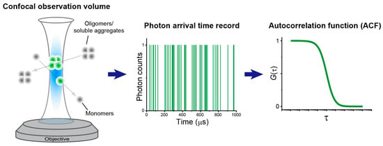
At the core of FCS lies the analysis of fluorescence intensity fluctuations generated as molecules diffuse through a defined observation volume (typically in confocal optics). These fluctuations are statistically evaluated using the temporal autocorrelation function (ACF), which was originally introduced as a general tool to extract temporal or spatial similarities from arbitrary signals [15] (Figure 1). In the context of conventional confocal FCS, the ACF for diffusion measurements is typically parameterized by the average number of particles within the observation volume, the characteristic diffusion time, and the so-called confocal structure parameter representing the observation volume size [16,17]. Importantly, FCS enables the noninvasive tracking of size and brightness changes that accompany molecular assembly, making it uniquely valuable for research on biological supramolecular complexes and aggregates.

Figure 1.
Confocal FCS. (Left) Confocal observation volume formed at the focal point of the objective lens and the passage of monomers or oligomers (or soluble aggregates) through it. Fluorescence molecules are excited and emit fluorescence only within the confocal observation volume. (Middle) Example of photon arrival time records measured when fluorescent molecules traverse the confocal observation volume, where each detected photon is counted as a digital signal (0 or 1). (Right) Example of an autocorrelation function, G(τ), calculated from photon arrival times. τ denotes lag time.
In biological supramolecular complexes and aggregate studies, monomers and various oligomers often coexist. Nonlinear regression with multi-component diffusion models allows estimation of the relative fractions of these species [18,19]. Two-component models are most commonly successful, though some studies have reported three-component analyses in live cells [20]. However, such cases remain rare, and further advances in nonlinear regression methods are required. Improvements under investigation include refined least-squares fitting, better handling of correlation noise, Bayesian estimation, and machine learning approaches to improve parameter estimation accuracy [21,22,23,24].
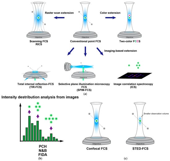
When multi-component analysis fails to resolve monomers and oligomers, fluorescence cross-correlation spectroscopy (FCCS or two-color FCS) using two spectrally distinct fluorophores can be employed (Figure 2a) [25]. FCCS directly detects homo- and hetero-interactions between molecules and is also applicable to the evaluation of drug-mediated inhibition of complex formation [26]. Although FCCS is typically performed with two colors, extensions to three or more colors have been demonstrated (multi-color FCCS) [27]. The main challenge lies in spectral crosstalk between detection channels, which can cause false positives. Methods such as pulsed interleaved excitation and spectral separation detection have been developed to overcome this limitation [28,29].

Figure 2.
Overview of various extensions originating from conventional FCS. (a) (Top center) Illustration of the observation volume (blue area) formed in the microscope system of conventional FCS and fluorescent molecules passing through it. (Top right) Extension to two-color FCS, which enables direct detection of molecular interactions. (Top left) Extension to raster-scan–based methods. (Bottom left) TIR-FCS, a type of imaging FCS, allows selective excitation and observation of fluorescent molecules located near the coverslip surface. Unable to image focal planes that are axially displaced from the coverslip. (Bottom center) SPIM-FCS, another type of imaging FCS. In the SPIM illumination system, excitation light is delivered as a single plane at an arbitrary focal depth perpendicular to the detection objective lens, offering greater flexibility in focal positioning compared with TIR-illumination; however, an additional objective lens is required to generate this specialized observation volume. (Bottom right) Extension to imaging-based methods such as ICS, which can detect changes in particle shape and molecular number. These analyses are independent of the confocal optical setup or raster scan speed and are performed directly from the acquired images. (b) A method for analyzing the distribution of fluorescence intensity. As an advanced form of FCS, it requires a clearly defined observation volume and relies on fluorescence fluctuations generated by molecular motion for analysis. Because it statistically analyzes fluorescence intensity distributions, this approach is well suited for studying heterogeneous molecular populations. (c) Visualization of changes in the observation volume achieved by optical super-resolution techniques such as STED.
Brightness analysis methods such as photon counting histogram (PCH) and number and brightness (N&B) analysis estimate oligomerization numbers from photon statistics, and are useful even when slow diffusion makes correlation analysis difficult (Figure 2b) [30,31]. Image-based spatial PCH analysis is a useful approach for identifying biomolecular oligomerization events that occur relatively statically within living cells [32]. While these methods offer significant advantages, they are also sensitive to inaccuracies in observation volume modeling, necessitating careful optical calibration. Hybrid approaches such as correlated PCH and fluorescence intensity and lifetime distribution analysis (FILDA) are being explored to improve reliability [33,34], and these concepts would be transferable to N&B analysis as well.
Image-based FCS (Imaging FCS; ImFCS) and raster image correlation spectroscopy (RICS) are powerful approaches that extend traditional FCS to image series, enabling spatial mapping of molecular diffusion and interactions [35,36]. ImFCS analyzes pixel fluctuations in image series acquired with advanced optical systems that provide spatially confined fluorescence detection, such as total internal reflection fluorescence (TIRF), or selective plane illumination microscopy (SPIM), generating spatial maps of diffusion and concentration [35,37,38] (Figure 2a). These techniques are beneficial for visualizing the environment surrounding biological supramolecular complexes and aggregates within cells. Scanning FCS is conceptually similar but reduces excitation frequency, making it more suitable for slow molecular motions in biological supramolecular complexes and aggregates [39,40]. However, image- and scanning-based methods sacrifice temporal resolution compared to single-point FCS, limiting their ability to simultaneously capture both ranging from the submicrosecond to tens of microsecond-scale fluorescence blinking by photophysics (e.g., intersystem crossing, photoisomerization, and other dynamic quenching processes) and molecular movement [41,42]. Pair correlation function (pCF) analysis serves as a bridge in temporal resolution between single-point FCS and ImFCS, enabling the detection of hetero-oligomerization of the transcription factor STAT3 [43]. Multipoint FCS, in contrast, has the potential to overcome these limitations of ImFCS and scanning FCS by enabling simultaneous acquisition of fluorescence fluctuations from multiple observation spots without compromising temporal resolution [44].
The hallmark of FCS and its derivative techniques lies in utilizing fluorescence fluctuations arising from molecular motion. In contrast, image correlation spectroscopy (ICS) analyzes spatial or temporal correlations of fluorescence intensity variations within static images or time-series image data (Figure 2a). This approach enables the quantification of particle size, spatial arrangement, dynamic regularity, and particle number [17]. Using ICS, the degree of protein aggregation within living cells has been quantitatively evaluated [45].
Finally, spatially super-resolved FCS approaches such as stimulated emission depletion (STED)-FCS combine FCS with stimulated emission depletion microscopy to achieve nanoscale resolution of molecular diffusion and phase behaviors [46]. These approaches are gaining prominence as conceptual advances that enable nanoscale resolution in the study of molecular assemblies and their dynamics [47]. Furthermore, although minimizing the observation volume remains a key challenge, this approach allows measurements at higher sample concentrations and thus may enable the detection of weak molecular interactions that are undetectable by conventional FCS (Figure 2c).
3. Definition and Properties of Biological Clusters
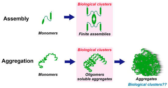
The term “biological cluster” can be defined as a functional unit that extends beyond the level of conventional supramolecular complexes, encompassing assemblies of proteins, nucleic acids, lipids, and other molecules; however, definitions vary and interpretations of their scale and properties remain inconsistent across the literature [5,48]. Although the distinction is self-evident from a physicochemical standpoint, recent biological studies have often conflated diverse molecular assemblies with phase-separated condensates or droplets. It is thus crucial to stress that biological clusters represent a distinct concept. The finiteness and convergence of the number of molecules represent one of the critical factors that help define biological clusters. Functional complexes such as kinetochores or receptor nanoclusters on cell membranes are defined by their functional requirement to assemble at specific and limited scales from a few molecules to hundreds or thousands [49,50]. Assemblies that grow indefinitely might more appropriately be considered aggregates rather than biological clusters [51]. Although such aggregates can also exhibit growth-arrest mechanisms or saturation phenomena that limit their expansion [52], their characteristic molecular scales remain fundamentally distinct from those of clusters. Therefore, “biological clusters” may be regarded as biomolecular assemblies that stabilize at finite molecular numbers that are not excessively large and serve critical biological functions (Figure 3).

Figure 3.
Concepts of biological clusters. In both assembly (top) and aggregation (bottom), monomers associate, leading to an increase in the number of molecules. However, assemblies stabilize at finite molecular numbers to form complexes, whereas aggregates involve vastly larger numbers of molecules and may not necessarily cease growing. Before progressing to large aggregates, intermediate states such as oligomers or soluble aggregates can exist as metastable species.
Initial small oligomers—often referred to as pre-aggregates or soluble aggregates—may also be regarded as biological clusters because they can exist as quasi-stable assemblies comprising a limited number of molecules. Small oligomers of Aβ, α-synuclein, and TDP-43, typically consisting of only a few to several molecules, have been identified as major neurotoxic species that trigger neuronal dysfunction and cell death [53]. However, these small oligomers differ fundamentally from phase-separated droplets. The droplets represent large-scale demixing events observable at the microscopic level, whereas oligomers remain nanoscale assemblies with finite molecular numbers. In this context, FCS provides an indispensable tool for distinguishing and characterizing such nanoscale biological clusters, as it can directly measure molecular numbers and diffusion coefficients. Indeed, FCS has been successfully applied to quantify concentration gradients of cNap1 across centrosomal regions, demonstrating its potential for broader applications to diverse biological clusters [54].
4. Applications of FCS to Biological Clusters and Aggregates
4.1. Protein Clusters
FCS has been employed as a powerful technique to directly measure the dynamics of such biological clusters. The diffusion properties of G protein-coupled receptors were analyzed using FCS, demonstrating their clustering and localization within membrane microdomains [55]. However, in single-color FCS experiments that measure diffusion to study the oligomerization of protein clusters, there is a limitation in that the diffusion coefficient is relatively insensitive to changes in molecular weight. To overcome this limitation, the use of two-color FCS (FCCS) is extremely effective [56]. ImFCS was applied to reveal heterogeneity in plasma membranes, suggesting that cluster dynamics are directly linked to additional complexity in steady state membrane organization [57]. In this way, in addition to utilizing diffusion-based measurements such as FCS and ImFCS, selecting methods like FCCS that can directly quantify the number of interacting molecules will enable deeper investigation into various protein clusters.
4.2. Protein Aggregates
FCS has been used to track Aβ oligomer formation and determine its critical aggregation concentration [58]. Förster resonance energy transfer (FRET) is a spectroscopic technique that reports on nanometer-scale proximity between fluorophores, and when combined with FCS (FRET-FCS), it provides a powerful tool for probing intermolecular interactions [59]. FRET-FCS has enabled the detection of rare assemblies of Aβ oligomers at high sensitivity [60]. In α-synuclein studies, FCS has clarified oligomerization dynamics, while FCCS has been applied to detect complex assemblies on oligomer formation [61]. FCS tracks in vivo dynamics and oligomerization of expanded polyQ aggregation in Caenorhabditis elegans [62]. Soluble aggregates of TDP-43 and its C-terminal fragments, key proteins in ALS pathology, have also been quantified using FCS [63]. Similar studies have extended to SOD1, further linking FCS analysis to disease mechanisms [64]. In the detection of protein aggregates, the strategies and methodologies used are similar to those for detecting protein clusters. However, while FCS, which specializes in diffusion dynamics, is well suited for detecting transient states during the formation of protein clusters or aggregates, ICS is considered more appropriate for detecting clusters or aggregates that have already formed and exist in a quasi-stable state [45,65]. Since temporal analysis of ICS is also possible [66], it is important to select the method best suited to the characteristics of the target under observation.
4.3. Nucleic Acid Clusters
RNA acts as a nucleating factor in ribonucleoprotein condensation. Since FCS has been applied to quantify RNA copy numbers [67], quantitative determination of RNA numbers and their aggregation states within biological clusters and condensates will likely be required. Local DNA compaction at a locus in zebrafish embryos promotes the formation of transcription factor–DNA clusters, particularly involving Nanog, a homeobox transcription factor essential for maintaining pluripotency [68]; thus, these DNA-including biological clusters may serve as valuable targets for quantitative analysis by FCS. Nucleic acids are among the biomolecules for which fluorescent imaging and spectroscopic analysis can be performed relatively easily compared to proteins, since they can be chemically synthesized, fluorescently labeled, and introduced into cells. By taking advantage of this property in FCS and its advanced methodologies, structural analysis of nucleic acids within living cells can be conducted more broadly [69].
4.4. Lipid Clusters
STED-FCS has enabled direct observation of protein and lipid clustering in membranes beyond the diffraction limit, and Eggeling and colleagues measured the nanoscale diffusion of lipid molecules and their restricted mobility in living cell membranes [70]. On the other hand, conventional FCS has also been applied to analyze lipid membrane environments through the use of mixed membranes incorporating fluorescently labeled lipids or environment-sensitive fluorescent probes [71,72]. The application of spatio-temporal ICS has contributed to elucidating how membrane protein clustering and the laws governing lateral diffusion are manifested in living cell membranes [65,73]. These studies have advanced our understanding of lipid membrane organization at the molecular level, providing indirect implications for how clustering phenomena may modulate physiological events such as signal transduction. Fluorescent visualization of lipids has long been pursued in conjunction with the broader theme of cellular imaging; however, the synthesis of fluorescently labeled lipids is not as straightforward as that of proteins or nucleic acids [74]. In particular, the requirement for synthetic expertise in organic chemistry poses a substantial barrier to entry for researchers in molecular cell biology and biophysics. Therefore, to advance the study of lipid clusters, progress should not be sought solely from the perspective of developing advanced FCS methodologies, but also through technological innovations in probe design from diverse fields such as chemical biology, organic chemistry, and lipid biology.
5. Experimental Considerations and Artifacts
Several experimental factors require attention in FCS. Labeling fractions may be better kept low to avoid self-quenching, artificially induced aggregation, and/or excessive photon emission from the fluorophore. Triplet states and photobleaching can distort correlation functions, requiring correction terms in fitting models [42,75,76]. When fluorescence bleaching or drift of the observed specimen unrelated to diffusion occurs, applying detrending procedures to the time-resolved photon data is recognized as an effective approach to facilitate the extraction of the intended molecular behavior [36,76,77]. Afterpulsing in avalanche photodiodes (APDs) can impact correlations at the sub-microsecond scale, necessitating correction algorithms [22,78]. High concentrations or large aggregates may overwhelm the observation volume, complicating data interpretation [79]. Overall, various artifacts in FCS measurements should be properly anticipated, requiring the use of more appropriate model equations, calibration, and correction.
6. Future Perspectives
Future developments for FCS can be anticipated along several lines. One important direction is the improvement of measurements for quite slow molecular dynamics. This includes strategies to suppress photobleaching during prolonged acquisitions, as well as the application of detrending algorithms to correct bleaching-related components in time-resolved photon data [76]. Another critical avenue is the refinement of parameter estimation. Statistical, Bayesian, and machine-learning-based fitting approaches hold promise for overcoming the limitations inherent in conventional nonlinear regression [21,47,80]. The integration of such advanced methods into commercial software platforms will significantly improve accessibility and accelerate the broader adoption of FCS.
The development of multi-color FCCS represents a further frontier, enabling the simultaneous tracking of multiple proteins, nucleic acids, and other biomolecules in the study of biological clusters and aggregates. Achieving widespread implementation of three- or higher-order multi-color FCCS would considerably expand the utility of this methodology for dissecting complex molecular networks. In parallel, integration with super-resolution microscopy—such as STED and MINFLUX—will provide the means to quantify nanoscale clusters and phase boundaries [81]. Although localization-based super-resolution methods are inherently limited in temporal resolution, correlation-based analyses remain feasible. Combining FCS with diverse super-resolution modalities, therefore, promises substantial advances in resolving dynamic molecular assemblies. Furthermore, ImFCS is an attractive approach for capturing slow molecular dynamics in living cells, but improvements in frame rates and quantum efficiency of image sensors will enable the successful observation of a wider variety of dynamic phenomena, such as various dynamics on different timescales. For example, photon-counting cameras based on Single-Photon APD (SPAD) arrays are expected to offer significant advantages [82].
Transient state (TRAST) monitoring is an imaging approach that modulates the excitation pulse width to probe transitions into dynamic non-emissive states, such as intersystem crossing—a radiationless transition between electronic states of different spin multiplicity (e.g., singlet–triplet transitions)—as well as photoisomerization into dark conformers [83]. Unlike FCS, TRAST monitoring does not depend on diffusion dynamics, enabling noninvasive sensing of local physicochemical environments even for immobilized or slowly moving biological clusters, as well as aggregates, droplets, or membrane microdomains. While direct applications to protein aggregates have not yet been reported, TRAST monitoring has been applied to flow-based barcoding of unilamellar liposomes and to sensing organelle environments [84,85]. A report showing that quenching population measured by TRAST monitoring can be corrected using immobile fractions estimated by FRAP—thereby enabling the in-cell estimation of diffusive RNA structures [69]—TRAST monitoring, when coupled with FRAP, could provide a more comprehensive picture of molecular states and microenvironments in complex systems. The integration of TRAST and FCS holds promise for enabling detailed and quantitative readouts of biological clusters and a wide range of intracellular structures, encompassing both static features and dynamic processes. This synergistic approach is expected to open new and attractive perspectives for future investigations.
Finally, integration with artificial intelligence (AI) is expected to reshape the usability of FCS [24,47,86]. Calibration and data analysis remain technically demanding tasks that often present a barrier for non-specialists. In this context, conversational large language models such as ChatGPT could provide real-time calibration guidance, troubleshooting support, and interpretation assistance. Looking forward, AI-driven analysis pipelines integrated directly into FCS platforms may dramatically increase throughput, reduce the technical entry barrier, and facilitate wider dissemination of the methodology across the life sciences.
FCS is already established as a fundamental technology for studying biological clusters and aggregates. Nevertheless, most published studies have focused on systems that were technically feasible to measure, leaving more challenging or complex targets insufficiently explored. Extending FCS to previously inaccessible systems will not only enhance quantitative accuracy but also uncover novel forms of molecular assembly.
7. Conclusions
This review summarizes the development and application of FCS in the analysis of biological clusters. By enabling sensitive quantification of particle numbers and diffusion, FCS has illuminated assemblies that were previously difficult to detect, including protein oligomers, lipid nanodomains, and RNA-containing clusters. Emerging technologies such as TRAST monitoring, super-resolution FCS, multi-color FCCS, and AI-assisted analysis will further extend the scope of this methodology.
The term “biological clusters” remains ambiguous, and its precise definition will ultimately depend on future experimental and theoretical developments. However, biological clusters can be distinguished from phase-separated condensates and indefinite aggregates by their functional convergence at finite molecular numbers. FCS provides one of the few experimental approaches capable of directly probing this finiteness and convergence. Continued technical and analytical innovations in FCS are expected to clarify the definition of biological clusters, and once established, studies of these clusters will provide direct insights into disease mechanisms and inform future drug discovery efforts.
Funding
This research was funded by grants from Japan Society for Promotion of Science (JSPS) Grant-in-Aid for Transformative Research Areas (A) (24H02286) and Forming Japan’s Peak Research Universities (J-PEAKS) (JPJS00420230001); the Japan Agency for Medical Research and Development (JP22gm6410028); Suhara-kinenn Zaidann Co., Ltd.; and Hagiwara Foundation of Japan.
Institutional Review Board Statement
Not applicable.
Informed Consent Statement
Not applicable.
Data Availability Statement
No new data were created or analyzed in this study. Data sharing is not applicable to this article.
Conflicts of Interest
The author declares no conflicts of interest. The funders had no role in the design of the study; in the collection, analyses, or interpretation of data; in the writing of the manuscript; or in the decision to publish the results.
Abbreviations
The following abbreviations are used in this manuscript:
| AI | Artificial intelligence |
| DNA | Deoxyribonucleic acid |
| FRAP | Fluorescence recovery after photobleaching |
| RNA | Ribonucleic acid |
References
- Toledo, P.L.; Gianotti, A.R.; Vazquez, D.S.; Ermacora, M.R. Protein nanocondensates: The next frontier. Biophys. Rev. 2023, 15, 515–530. [Google Scholar] [CrossRef]
- Alberti, S.; Hyman, A.A. Biomolecular condensates at the nexus of cellular stress, protein aggregation disease and ageing. Nat. Rev. Mol. Cell Biol. 2021, 22, 196–213. [Google Scholar] [CrossRef]
- Ripin, N.; Parker, R. Formation, function, and pathology of RNP granules. Cell 2023, 186, 4737–4756. [Google Scholar] [CrossRef]
- Levental, I.; Levental, K.R.; Heberle, F.A. Lipid Rafts: Controversies Resolved, Mysteries Remain. Trends Cell Biol. 2020, 30, 341–353. [Google Scholar] [CrossRef]
- Morris, R.; Black, K.A.; Stollar, E.J. Uncovering protein function: From classification to complexes. Essays Biochem. 2022, 66, 255–285. [Google Scholar] [CrossRef]
- Haustein, E.; Schwille, P. Fluorescence correlation spectroscopy: Novel variations of an established technique. Annu. Rev. Biophys. Biomol. Struct. 2007, 36, 151–169. [Google Scholar] [CrossRef] [PubMed]
- Magde, D.; Elson, E.; Webb, W.W. Thermodynamic Fluctuations in a Reacting System—Measurement by Fluorescence Correlation Spectroscopy. Phys. Rev. Lett. 1972, 29, 705–708. [Google Scholar] [CrossRef]
- Elson, E.L.; Magde, D. Fluorescence correlation spectroscopy. I. Conceptual basis and theory. Biopolymers 1974, 13, 1–27. [Google Scholar] [CrossRef]
- Magde, D.; Elson, E.L.; Webb, W.W. Fluorescence correlation spectroscopy. II. An experimental realization. Biopolymers 1974, 13, 29–61. [Google Scholar] [CrossRef] [PubMed]
- Ehrenberg, M.; Rigler, R. Rotational brownian motion and fluorescence intensify fluctuations. Chem. Phys. 1974, 4, 390–401. [Google Scholar] [CrossRef]
- Borejdo, J. Motion of myosin fragments during actin-activated ATPase: Fluorescence correlation spectroscopy study. Biopolymers 1979, 18, 2807–2820. [Google Scholar] [CrossRef]
- Thompson, N.L.; Axelrod, D. Immunoglobulin surface-binding kinetics studied by total internal reflection with fluorescence correlation spectroscopy. Biophys. J. 1983, 43, 103–114. [Google Scholar] [CrossRef] [PubMed]
- Qian, H.; Elson, E.L. Analysis of confocal laser-microscope optics for 3-D fluorescence correlation spectroscopy. Appl. Opt. 1991, 30, 1185–1195. [Google Scholar] [CrossRef]
- Rigler, R.; Mets, U.; Widengren, J.; Kask, P. Fluorescence Correlation Spectroscopy with High Count Rate and Low-Background—Analysis of Translational Diffusion. Eur. Biophys. J. Biophy 1993, 22, 169–175. [Google Scholar] [CrossRef]
- Elson, E.L. Fluorescence correlation spectroscopy: Past, present, future. Biophys. J. 2011, 101, 2855–2870. [Google Scholar] [CrossRef]
- Kim, S.A.; Heinze, K.G.; Schwille, P. Fluorescence correlation spectroscopy in living cells. Nat. Methods 2007, 4, 963–973. [Google Scholar] [CrossRef] [PubMed]
- Kitamura, A.; Kinjo, M. State-of-the-Art Fluorescence Fluctuation-Based Spectroscopic Techniques for the Study of Protein Aggregation. Int. J. Mol. Sci. 2018, 19, 964. [Google Scholar] [CrossRef] [PubMed]
- Nath, S.; Meuvis, J.; Hendrix, J.; Carl, S.A.; Engelborghs, Y. Early aggregation steps in alpha-synuclein as measured by FCS and FRET: Evidence for a contagious conformational change. Biophys. J. 2010, 98, 1302–1311. [Google Scholar] [CrossRef]
- Chakraborty, M.; Kuriata, A.M.; Nathan Henderson, J.; Salvucci, M.E.; Wachter, R.M.; Levitus, M. Protein oligomerization monitored by fluorescence fluctuation spectroscopy: Self-assembly of rubisco activase. Biophys. J. 2012, 103, 949–958. [Google Scholar] [CrossRef]
- Tsutsumi, M.; Muto, H.; Myoba, S.; Kimoto, M.; Kitamura, A.; Kamiya, M.; Kikukawa, T.; Takiya, S.; Demura, M.; Kawano, K.; et al. In vivo fluorescence correlation spectroscopy analyses of FMBP-1, a silkworm transcription factor. Febs Open Bio 2016, 6, 106–125. [Google Scholar] [CrossRef]
- Kohler, J.; Hur, K.H.; Mueller, J.D. Statistical analysis of the autocorrelation function in fluorescence correlation spectroscopy. Biophys. J. 2024, 123, 667–680. [Google Scholar] [CrossRef]
- Zhao, M.; Jin, L.; Chen, B.; Ding, Y.; Ma, H.; Chen, D. Afterpulsing and its correction in fluorescence correlation spectroscopy experiments. Appl. Opt. 2003, 42, 4031–4036. [Google Scholar] [CrossRef]
- Sun, G.; Guo, S.M.; Teh, C.; Korzh, V.; Bathe, M.; Wohland, T. Bayesian model selection applied to the analysis of fluorescence correlation spectroscopy data of fluorescent proteins in vitro and in vivo. Anal. Chem. 2015, 87, 4326–4333. [Google Scholar] [CrossRef]
- Enderlein, J. Machine learning and advanced statistical analysis for fluorescence correlation spectroscopy. Biophys. J. 2024, 123, 651–652. [Google Scholar] [CrossRef]
- Bacia, K.; Kim, S.A.; Schwille, P. Fluorescence cross-correlation spectroscopy in living cells. Nat. Methods 2006, 3, 83–89. [Google Scholar] [CrossRef]
- Mikuni, S.; Kodama, K.; Sasaki, A.; Kohira, N.; Maki, H.; Munetomo, M.; Maenaka, K.; Kinjo, M. Screening for FtsZ Dimerization Inhibitors Using Fluorescence Cross-Correlation Spectroscopy and Surface Resonance Plasmon Analysis. PLoS ONE 2015, 10, e0130933. [Google Scholar] [CrossRef][Green Version]
- Hwang, L.C.; Gosch, M.; Lasser, T.; Wohland, T. Simultaneous multicolor fluorescence cross-correlation spectroscopy to detect higher order molecular interactions using single wavelength laser excitation. Biophys. J. 2006, 91, 715–727. [Google Scholar] [CrossRef] [PubMed]
- Previte, M.J.; Pelet, S.; Kim, K.H.; Buehler, C.; So, P.T. Spectrally resolved fluorescence correlation spectroscopy based on global analysis. Anal. Chem. 2008, 80, 3277–3284. [Google Scholar] [CrossRef] [PubMed][Green Version]
- Dunsing, V.; Petrich, A.; Chiantia, S. Multicolor fluorescence fluctuation spectroscopy in living cells via spectral detection. eLife 2021, 10, e69687. [Google Scholar] [CrossRef] [PubMed]
- Chen, Y.; Muller, J.D.; So, P.T.; Gratton, E. The photon counting histogram in fluorescence fluctuation spectroscopy. Biophys. J. 1999, 77, 553–567. [Google Scholar] [CrossRef] [PubMed]
- Digman, M.A.; Dalal, R.; Horwitz, A.F.; Gratton, E. Mapping the number of molecules and brightness in the laser scanning microscope. Biophys. J. 2008, 94, 2320–2332. [Google Scholar] [CrossRef]
- Godin, A.G.; Rappaz, B.; Potvin-Trottier, L.; Kennedy, T.E.; De Koninck, Y.; Wiseman, P.W. Spatial Intensity Distribution Analysis Reveals Abnormal Oligomerization of Proteins in Single Cells. Biophys. J. 2015, 109, 710–721. [Google Scholar] [CrossRef]
- Vetri, V.; Ossato, G.; Militello, V.; Digman, M.A.; Leone, M.; Gratton, E. Fluctuation methods to study protein aggregation in live cells: Concanavalin A oligomers formation. Biophys. J. 2011, 100, 774–783. [Google Scholar] [CrossRef]
- Scales, N.; Swain, P.S. Resolving fluorescent species by their brightness and diffusion using correlated photon-counting histograms. PLoS ONE 2019, 14, e0226063. [Google Scholar] [CrossRef]
- Yu, L.; Lei, Y.; Ma, Y.; Liu, M.; Zheng, J.; Dan, D.; Gao, P. A Comprehensive Review of Fluorescence Correlation Spectroscopy. Front. Phys. 2021, 9, 644450. [Google Scholar] [CrossRef]
- Digman, M.A.; Wiseman, P.W.; Horwitz, A.R.; Gratton, E. Detecting protein complexes in living cells from laser scanning confocal image sequences by the cross correlation raster image spectroscopy method. Biophys. J. 2009, 96, 707–716. [Google Scholar] [CrossRef]
- Digman, M.A.; Brown, C.M.; Sengupta, P.; Wiseman, P.W.; Horwitz, A.R.; Gratton, E. Measuring fast dynamics in solutions and cells with a laser scanning microscope. Biophys. J. 2005, 89, 1317–1327. [Google Scholar] [CrossRef] [PubMed]
- Singh, A.P.; Wohland, T. Applications of imaging fluorescence correlation spectroscopy. Curr. Opin. Chem. Biol. 2014, 20, 29–35. [Google Scholar] [CrossRef]
- Petrasek, Z.; Schwille, P. Precise measurement of diffusion coefficients using scanning fluorescence correlation spectroscopy. Biophys. J. 2008, 94, 1437–1448. [Google Scholar] [CrossRef]
- Fujioka, Y.; Alam, J.M.; Noshiro, D.; Mouri, K.; Ando, T.; Okada, Y.; May, A.I.; Knorr, R.L.; Suzuki, K.; Ohsumi, Y.; et al. Phase separation organizes the site of autophagosome formation. Nature 2020, 578, 301–305. [Google Scholar] [CrossRef]
- Widengren, J.; Rigler, R.; Mets, Ü. Triplet-state monitoring by fluorescence correlation spectroscopy. J. Fluoresc. 1994, 4, 255–258. [Google Scholar] [CrossRef]
- Widengren, J.; Mets, U.; Rigler, R. Fluorescence Correlation Spectroscopy of Triplet-States in Solution—A Theoretical and Experimental-Study. J. Phys. Chem. 1995, 99, 13368–13379. [Google Scholar] [CrossRef]
- Hinde, E.; Pandzic, E.; Yang, Z.; Ng, I.H.; Jans, D.A.; Bogoyevitch, M.A.; Gratton, E.; Gaus, K. Quantifying the dynamics of the oligomeric transcription factor STAT3 by pair correlation of molecular brightness. Nat. Commun. 2016, 7, 11047. [Google Scholar] [CrossRef] [PubMed]
- Yamamoto, J.; Mikuni, S.; Kinjo, M. Multipoint fluorescence correlation spectroscopy using spatial light modulator. Biomed. Opt. Express 2018, 9, 5881–5890. [Google Scholar] [CrossRef]
- Ma, Y.; Pandzic, E.; Nicovich, P.R.; Yamamoto, Y.; Kwiatek, J.; Pageon, S.V.; Benda, A.; Rossy, J.; Gaus, K. An intermolecular FRET sensor detects the dynamics of T cell receptor clustering. Nat. Commun. 2017, 8, 15100. [Google Scholar] [CrossRef] [PubMed]
- Kastrup, L.; Blom, H.; Eggeling, C.; Hell, S.W. Fluorescence Fluctuation Spectroscopy in Subdiffraction Focal Volumes. Phys. Rev. Lett. 2005, 94, 178104. [Google Scholar] [CrossRef] [PubMed]
- Sankaran, J.; Wohland, T. Current capabilities and future perspectives of FCS: Super-resolution microscopy, machine learning, and in vivo applications. Commun. Biol. 2023, 6, 699. [Google Scholar] [CrossRef]
- Koukos, P.I.; Bonvin, A.M.J.J. Integrative Modelling of Biomolecular Complexes. J. Mol. Biol. 2020, 432, 2861–2881. [Google Scholar] [CrossRef]
- Ariyoshi, M.; Fukagawa, T. An updated view of the kinetochore architecture. Trends Genet. 2023, 39, 941–953. [Google Scholar] [CrossRef]
- Liu, W.; Wang, H.; Xu, C. Antigen Receptor Nanoclusters: Small Units with Big Functions. Trends Immunol. 2016, 37, 680–689. [Google Scholar] [CrossRef]
- Housmans, J.A.J.; Wu, G.; Schymkowitz, J.; Rousseau, F. A guide to studying protein aggregation. FEBS J. 2023, 290, 554–583. [Google Scholar] [CrossRef]
- Banani, S.F.; Lee, H.O.; Hyman, A.A.; Rosen, M.K. Biomolecular condensates: Organizers of cellular biochemistry. Nat. Rev. Mol. Cell Biol. 2017, 18, 285–298. [Google Scholar] [CrossRef]
- Rinauro, D.J.; Chiti, F.; Vendruscolo, M.; Limbocker, R. Misfolded protein oligomers: Mechanisms of formation, cytotoxic effects, and pharmacological approaches against protein misfolding diseases. Mol. Neurodegener. 2024, 19, 20. [Google Scholar] [CrossRef]
- Mahen, R. cNap1 bridges centriole contact sites to maintain centrosome cohesion. PLoS Biol. 2022, 20, e3001854. [Google Scholar] [CrossRef]
- Kilpatrick, L.E.; Hill, S.J. The use of fluorescence correlation spectroscopy to characterise the molecular mobility of G protein-coupled receptors in membrane microdomains: An update. Biochem. Soc. Trans. 2021, 49, 1547–1554. [Google Scholar] [CrossRef]
- Kaliszewski, M.J.; Shi, X.; Hou, Y.; Lingerak, R.; Kim, S.; Mallory, P.; Smith, A.W. Quantifying membrane protein oligomerization with fluorescence cross-correlation spectroscopy. Methods 2018, 140–141, 40–51. [Google Scholar] [CrossRef] [PubMed]
- Bag, N.; Holowka, D.A.; Baird, B.A. Imaging FCS delineates subtle heterogeneity in plasma membranes of resting mast cells. Mol. Biol. Cell 2020, 31, 709–723. [Google Scholar] [CrossRef] [PubMed]
- Garai, K.; Sureka, R.; Maiti, S. Detecting amyloid-beta aggregation with fiber-based fluorescence correlation spectroscopy. Biophys. J. 2007, 92, L55–L57. [Google Scholar] [CrossRef]
- Lazaro-Alfaro, A.; Nicholas, S.L.N.; Sanabria, H. FRET-FCS: Advancing comprehensive insights into complex biological systems. Biophys. J. 2025, 124, 3329–3341. [Google Scholar] [CrossRef]
- Wennmalm, S.; Chmyrov, V.; Widengren, J.; Tjernberg, L. Highly Sensitive FRET-FCS Detects Amyloid beta-Peptide Oligomers in Solution at Physiological Concentrations. Anal. Chem. 2015, 87, 11700–11705. [Google Scholar] [CrossRef] [PubMed]
- Santos, J.; Gracia, P.; Navarro, S.; Pena-Diaz, S.; Pujols, J.; Cremades, N.; Pallares, I.; Ventura, S. alpha-Helical peptidic scaffolds to target alpha-synuclein toxic species with nanomolar affinity. Nat. Commun. 2021, 12, 3752. [Google Scholar] [CrossRef]
- Beam, M.; Silva, M.C.; Morimoto, R.I. Dynamic imaging by fluorescence correlation spectroscopy identifies diverse populations of polyglutamine oligomers formed in vivo. J. Biol. Chem. 2012, 287, 26136–26145. [Google Scholar] [CrossRef]
- Kitamura, A.; Fujimoto, A.; Kawashima, R.; Lyu, Y.; Sasaki, K.; Hamada, Y.; Moriya, K.; Kurata, A.; Takahashi, K.; Brielmann, R.; et al. Hetero-oligomerization of TDP-43 carboxy-terminal fragments with cellular proteins contributes to proteotoxicity. Commun. Biol. 2024, 7, 743. [Google Scholar] [CrossRef]
- Das, B.; Roychowdhury, S.; Mohanty, P.; Rizuan, A.; Chakraborty, J.; Mittal, J.; Chattopadhyay, K. A Zn-dependent structural transition of SOD1 modulates its ability to undergo phase separation. EMBO J. 2023, 42, e111185. [Google Scholar] [CrossRef] [PubMed]
- Abu-Arish, A.; Pandzic, E.; Luo, Y.; Sato, Y.; Turner, M.J.; Wiseman, P.W.; Hanrahan, J.W. Lipid-driven CFTR clustering is impaired in cystic fibrosis and restored by corrector drugs. J. Cell Sci. 2022, 135, jcs259002. [Google Scholar] [CrossRef] [PubMed]
- Kolin, D.L.; Wiseman, P.W. Advances in image correlation spectroscopy: Measuring number densities, aggregation states, and dynamics of fluorescently labeled macromolecules in cells. Cell Biochem. Biophys. 2007, 49, 141–164. [Google Scholar] [CrossRef]
- Sasaki, A.; Yamamoto, J.; Kinjo, M.; Noda, N. Absolute Quantification of RNA Molecules Using Fluorescence Correlation Spectroscopy with Certified Reference Materials. Anal. Chem. 2018, 90, 10865–10871. [Google Scholar] [CrossRef]
- Chabot, N.M.; Purkanti, R.; Del Panta Ridolfi, A.; Nogare, D.D.; Oda, H.; Kimura, H.; Jug, F.; Dal Co, A.; Vastenhouw, N.L. Local DNA compaction creates TF-DNA clusters that enable transcription. bioRxiv 2024. [Google Scholar] [CrossRef]
- Kitamura, A.; Tornmalm, J.; Demirbay, B.; Piguet, J.; Kinjo, M.; Widengren, J. Trans-cis isomerization kinetics of cyanine dyes reports on the folding states of exogeneous RNA G-quadruplexes in live cells. Nucleic Acids Res. 2023, 51, e27. [Google Scholar] [CrossRef]
- Eggeling, C.; Ringemann, C.; Medda, R.; Schwarzmann, G.; Sandhoff, K.; Polyakova, S.; Belov, V.N.; Hein, B.; von Middendorff, C.; Schonle, A.; et al. Direct observation of the nanoscale dynamics of membrane lipids in a living cell. Nature 2009, 457, 1159–1162. [Google Scholar] [CrossRef] [PubMed]
- Schwille, P.; Korlach, J.; Webb, W.W. Fluorescence correlation spectroscopy with single-molecule sensitivity on cell and model membranes. Cytometry 1999, 36, 176–182. [Google Scholar] [CrossRef]
- Rose, M.; Hirmiz, N.; Moran-Mirabal, J.M.; Fradin, C. Lipid Diffusion in Supported Lipid Bilayers: A Comparison between Line-Scanning Fluorescence Correlation Spectroscopy and Single-Particle Tracking. Membranes 2015, 5, 702–721. [Google Scholar] [CrossRef]
- Di Rienzo, C.; Gratton, E.; Beltram, F.; Cardarelli, F. Fast spatiotemporal correlation spectroscopy to determine protein lateral diffusion laws in live cell membranes. Proc. Natl. Acad. Sci. USA 2013, 110, 12307–12312. [Google Scholar] [CrossRef]
- Rubio, V.; McInchak, N.; Fernandez, G.; Benavides, D.; Herrera, D.; Jimenez, C.; Mesa, H.; Meade, J.; Zhang, Q.; Stawikowski, M.J. Development and characterization of fluorescent cholesteryl probes with enhanced solvatochromic and pH-sensitive properties for live-cell imaging. Sci. Rep. 2024, 14, 30777. [Google Scholar] [CrossRef]
- Davis, L.M.; Shen, G. Accounting for triplet and saturation effects in FCS measurements. Curr. Pharm. Biotechnol. 2006, 7, 287–301. [Google Scholar] [CrossRef]
- Hodges, C.; Kafle, R.P.; Hoff, J.D.; Meiners, J.-C. Fluorescence Correlation Spectroscopy with Photobleaching Correction in Slowly Diffusing Systems. J. Fluoresc. 2018, 28, 505–511. [Google Scholar] [CrossRef]
- Pinto-Cámara, R.; Linares, A.; Moreno-Gutiérrez, D.S.; Hernández, H.O.; Martínez-Reyes, J.D.; Rendón-Mancha, J.M.; Wood, C.D.; Guerrero, A. FCSlib: An open-source tool for fluorescence fluctuation spectroscopy analysis for mobility, number and molecular brightness in R. Bioinformatics 2020, 37, 1930–1931. [Google Scholar] [CrossRef] [PubMed]
- Ziarkash, A.W.; Joshi, S.K.; Stipcevic, M.; Ursin, R. Comparative study of afterpulsing behavior and models in single photon counting avalanche photo diode detectors. Sci. Rep. 2018, 8, 5076. [Google Scholar] [CrossRef] [PubMed]
- Nagy, A.; Wu, J.; Berland, K.M. Characterizing observation volumes and the role of excitation saturation in one-photon fluorescence fluctuation spectroscopy. J. Biomed. Opt. 2005, 10, 44015. [Google Scholar] [CrossRef] [PubMed]
- Seltmann, A.; Carravilla, P.; Reglinski, K.; Eggeling, C.; Waithe, D. Neural network informed photon filtering reduces fluorescence correlation spectroscopy artifacts. Biophys. J. 2024, 123, 745–755. [Google Scholar] [CrossRef]
- Eilers, Y.; Ta, H.; Gwosch, K.C.; Balzarotti, F.; Hell, S.W. MINFLUX monitors rapid molecular jumps with superior spatiotemporal resolution. Proc. Natl. Acad. Sci. USA 2018, 115, 6117–6122. [Google Scholar] [CrossRef]
- Slenders, E.; Castello, M.; Buttafava, M.; Villa, F.; Tosi, A.; Lanzano, L.; Koho, S.V.; Vicidomini, G. Confocal-based fluorescence fluctuation spectroscopy with a SPAD array detector. Light. Sci. Appl. 2021, 10, 31. [Google Scholar] [CrossRef] [PubMed]
- Sanden, T.; Persson, G.; Thyberg, P.; Blom, H.; Widengren, J. Monitoring kinetics of highly environment sensitive states of fluorescent molecules by modulated excitation and time-averaged fluorescence intensity recording. Anal. Chem. 2007, 79, 3330–3341. [Google Scholar] [CrossRef] [PubMed]
- Du, Z.; Piguet, J.; Baryshnikov, G.; Tornmalm, J.; Demirbay, B.; Ågren, H.; Widengren, J. Imaging Fluorescence Blinking of a Mitochondrial Localization Probe: Cellular Localization Probes Turned into Multifunctional Sensors. J. Phys. Chem. B 2022, 126, 3048–3058. [Google Scholar] [CrossRef] [PubMed]
- Sandberg, E.; Demirbay, B.; Kulkarni, A.; Liu, H.; Piguet, J.; Widengren, J. Fluorescence Bar-Coding and Flowmetry Based on Dark State Transitions in Fluorescence Emitters. J. Phys. Chem. B 2024, 128, 125–136. [Google Scholar] [CrossRef] [PubMed]
- Tang, W.H.; Sim, S.R.; Aik, D.Y.K.; Nelanuthala, A.V.S.; Athilingam, T.; Rollin, A.; Wohland, T. Deep learning reduces data requirements and allows real-time measurements in imaging FCS. Biophys. J. 2024, 123, 655–666. [Google Scholar] [CrossRef]
Disclaimer/Publisher’s Note: The statements, opinions and data contained in all publications are solely those of the individual author(s) and contributor(s) and not of MDPI and/or the editor(s). MDPI and/or the editor(s) disclaim responsibility for any injury to people or property resulting from any ideas, methods, instructions or products referred to in the content. |
© 2025 by the author. Licensee MDPI, Basel, Switzerland. This article is an open access article distributed under the terms and conditions of the Creative Commons Attribution (CC BY) license (https://creativecommons.org/licenses/by/4.0/).