Article

Gestational Age Is Positively Associated with Retinol and α-Tocopherol in Preterm Infants: The Mediating Role of Birth Weight

by
Panos Papandreou
1,
Paraskevi Detopoulou
2 and
Maria Skouroliakou
3,*
1
Department of Nutrition, IASO Hospital, 15123 Athens, Greece
2
Department of Clinical Nutrition, General Hospital Korgialenio Benakio, 11526 Athens, Greece
3
Department of Dietetics and Nutritional Science, School of Health Science and Education, Harokopio University, 17671 Athens, Greece
*
Author to whom correspondence should be addressed.
Dietetics 2023, 2(4), 366-376; https://doi.org/10.3390/dietetics2040027
Submission received: 23 October 2023 / Revised: 11 December 2023 / Accepted: 14 December 2023 / Published: 18 December 2023

Abstract

:
Oxidative stress is thought to be one of the common mechanisms for several neonatal diseases in premature infants. Moreover, fat-soluble antioxidant vitamins, i.e., retinol and α-tocopherol, have been found to be low in preterm neonates; however, data are limited. The aim of this was to assess the circulating α-tocopherol and retinol concentrations in preterm infants at birth and investigate if they are related to gestational age. Retinol and α-tocopherol were measured on the first day after birth in 30 preterm neonates with HPLC. Means ± SD of serum retinol and α-tocopherol were 392.0 ± 162.9 μg/L and 6.83 ± 3.02 mg/L, respectively. In total, 73% of infants had a very low birth weight (<1500 g) and 23.3% were small for gestational age (SGA). Moreover, 10% of neonates had a retinol deficiency and 20% had an α-tocopherol deficiency. The retinol concentration was lower in SGA infants compared to appropriate for gestational age ones (340.85 ± 75.89 vs. 407.60 ± 179.83 μg/L, correspondingly p = 0.030). Retinol was linearly related to gestational age (Pearson’s rho = 0.84, p < 0.001) but the association did not remain significant after an adjustment for birth weight (partial rho = 0.193, p = 0.316). α-tocopherol was nonlinearly associated with gestational age (Spearman’s rho = 0.470, p = 0.044). The assessment of the vitamin status and potential deficiency in neonates is crucial in order to appropriately support the nutritional needs of newborns.

1. Introduction

The interest surrounding the effects of vitamins A and E in human health has significantly increased in the context of their role as antioxidants [1,2]. Vitamin A is essential for cellular growth and differentiation [3], lung development and alveoli formation [4], as well as immune function [5]. Retinoic acid, can modulate gene expression through its binding to responsive elements in the genome, including genes involved in antioxidant systems [6].
Vitamin E occurs in nature in several chemical forms (four tocopherols, α-,β-,γ- and δ- and four tocotrienols, α-,β-,γ- and δ-). However, α-tocopherol represents ~90% of this vitamin in humans [7]. Vitamin E has an antioxidant action by reducing lipid peroxidation [7]. Moreover, other properties have been attributed to vitamin E, such as neuroprotection, reduction of inflammation, and anti-cancer activity [8]. In pregnancy, vitamin E is involved in embryo development, placenta maturation, and defense against oxidative stress [9].
The role of vitamins A and E in the development of neonates is being studied, but the data are relatively limited [9]. Low concentrations of vitamin A and E have been documented in preterm infants [10,11], possibly showing that the accumulation of these vitamins in the fetus mostly occurs in the last trimester of pregnancy [12]. However, not all studies agree with this [13,14]. In parallel, preterm infants are at a deficiency risk for these vitamins, since fat storage is low, intake may be inadequate, malabsorption may be present, and nutritional needs are high [15]. It is also noted that fat-soluble vitamins require carrier systems (lipoproteins) [16] or specific carrier proteins [17] for solubility in the blood. For example, α-tocopherol is mostly carried in low-density lipoprotein and high-density lipoproteins [16], while retinoids are carried in chylomicrons, very low-density lipoprotein particles and low-density lipoprotein particles [18]. In parallel, retinol binds to retinol-binding protein, albumin, and other proteins, which may be low in malnourished pregnant women [19]. The intestinal absorption of vitamins depends on the co-presence of fat, as well as fat absorption [18,20].
Vitamins A and E cross the placenta by simple or facilitated diffusion [21]. The fetus is unable to synthesize vitamins and relies on the placental delivery of retinol and α-tocopherol. The accumulation of vitamins in the fetus takes place throughout pregnancy, and especially during the third trimester, and depends on the maternal status. Therefore, blood concentrations and body stores at birth may be lower in preterm infants of poorly nourished mothers [22]. In fact, an improvement in neonatal vitamin A and E concentrations can be achieved with a good nutritional status of the mother, while [23] the maternal nutritional status can have an impact on birth weight [24].
Vitamin A deficiency has been connected to retinopathy of prematurity, bronchopulmonary dysplasia, and chronic lung disease [5]. Indeed, the pathogenesis of retinopathy of prematurity includes oxidative stress-related damage, which could be improved with the antioxidant actions of vitamin A [25]. It is also noteworthy that, even in adults with lung inflammatory disease, liver stores of vitamin A are depleted [26]. Moreover, the incidence of an intraventricular hemorrhage is higher in infants a with low vitamin A liver status [27]. Vitamin A deficiency has also been connected to infant mortality [5]. In the same context, animal studies have shown that severe vitamin A deficiency of the mother leads to early fetal death [28].
Vitamin E deficiency has been connected to hemolytic anemia, bronchopulmonary dysplasia, myopathy, edema, thrombocytosis, hemolytic anemia, and intraventricular hemorrhage in infants [29,30]. In addition, cardiomyopathy may occur due to muscle degeneration [22]. Additional effects of perinatal vitamin E deficiency have also been proposed. For example, vitamin E deficiency has been shown to be implicated in neural development and childhood cognition [1].
Oxidative stress is thought to be one of the common mechanisms for several pathological conditions in preterm infants. Indeed, the process of childbirth per se is related to an increase in the oxidative environment [31,32]. The fetus is transferred from a hypoxic intrauterine environment (pO2 of 20–25 mm Hg) to an environment of high oxygen content (pO2 of 100 mm Hg) and it is exposed to inhaled air [31,32]. This change results in greater oxidative stress simply due to the existence of normal oxygen levels in the new extrauterine environment [31]. In addition, the cell membranes of infants are more vulnerable to oxidative stress since they have a higher content of polyunsaturated fatty acids. This fact actually increases infantile vitamin E requirements [33].
The oxidative aggression suffered by the neonate is counteracted by the maturation of effective antioxidant mechanisms. There are complex antioxidant defense systems against the effects of oxygen free radicals on biological macromolecules, including both enzymatic (superoxide dismutase, catalase, glutathione peroxidase, etc.) and nonenzymatic (vitamins A, E, C, etc.) components [34]. In the case of a preterm neonate, both the adaptations to physiological oxidative stress and the antioxidant defenses are reduced [31,35,36]. In this scenario, the maternal–fetal transfer of antioxidant nutrients has not been completed in the premature neonate, while some of the antioxidant enzymatic systems have not had time to mature sufficiently [35,36]. For all these reasons, the preterm neonate is more vulnerable to dysregulation of the antioxidant/prooxidant balance, and is more likely to suffer from the “neonatal diseases from oxygen free radicals” [31]. In addition, it is also probable that the prenatal and neonatal oxidative stress connect to other diseases in later life, such as obesity [37].
Given the deleterious effects of oxidative stress and the consequences of retinol and α-tocopherol deficiency in preterm neonates, it is crucial to assess the status of these vitamins as early as possible. In fact, early postnatal supplementation of vitamin A administered to preterm infants improves their respiratory functions, but probably only in those with low baseline levels of this vitamin [38]. This means that the determination of the vitamin status at birth is important. Moreover, the relation of vitamins A and E and gestational age is not clear [39,40]. Thus, the aim of this study was to assess the circulating α-tocopherol and retinol in preterm infants at birth and investigate if they are related to gestational age. To our knowledge, there are scarce data in Greece regarding the status of these vitamins in preterm infants, and these data are also relatively old [41].

2. Materials and Methods

2.1. Study Design

This is a cross-sectional study including 30 preterm infants from the intensive care unit at Iaso Hospital. The study protocol was approved by the Scientific Committee of Iaso Hospital (number: A21112018). Mothers of infants signed an informed consent form in order to participate in this study.

2.2. Participants

The study sample consisted of preterm neonates admitted to the neonatal intensive care unit (NICU) of the “IASO” Maternity Hospital (May 2020 to September 2020). The inclusion criteria were (a) gestational age 26–35 weeks and (b) parental consent for participation in this study. The exclusion criteria were (a) gestational diabetes or pre-eclampsia of the mother, (b) congenital infections, (c) perinatal asphyxia, (d) major congenital anomalies, and (e) no parental consent. The neonates had not received corticosteroids postnatally at the time of measurements. It is noted that all mothers were of Greek nationality and lived in Athens and near suburbs (Attiki area).

2.3. Anthropometric Measurements

Weight was measured on a digital scale with an accuracy of 0.005 Kg (Model AND SK-WP 1–10 kg). Low birth weight was defined as weight <2500 g and very low birth weight was defined as weight <1500 g, as suggested by the World Health Organization [42]. Small for gestational age (SGA) infants were determined as infants with weight at birth of less than the 10th percentile for gestational age [43]. For this determination, gender-specific reference charts of birth weight for gestational age of the World Health Organization were used [43].

2.4. Biochemical Measurements

In this study, 3 mL samples of blood were collected in glass tubes one day after birth of each infant. Blood samples were centrifuged (3000 rpm, 10 min), and sera were removed and stored at −20 °C until analysis. The determination of retinol and α-tocopherol was carried out simultaneously with high-performance liquid chromatography (HPLC) (AGILENT, series number 1100 with constant flow pump) using a UV detector and a reagent kit of Chromsystems. More particularly, an isocratic elution program was used; the solvent flow was 1.5 mL/min, the column temperature was 25 °C, and the injection volume was 50 μL. The UV detector was set at 325 and 295 nm. The retention time of retinol was about 2.5 min and that of α-tocopherol was about 10 min. Thus, vitamin A was first eluted and detected with the UV detector at 325 nm. At 3.5 min, the wavelength of the detector was changed to 295 nm for the determination of α-tocopherol (at 10 min). The internal standard had a seizure time of about 5 min.
The deficiency in retinol and α-tocopherol was defines according to the World Health Organization criteria (retinol < 200 μg/L and α-tocopherol < 5.0 mg/L) [15].

2.5. Statistical Analysis

The Kolmogorov–Smirnoff test was applied to test normality. Normally distributed variables are shown as means ± standard deviation (SD), while non-normally distributed variables are shown as medians and 25th–75th percentiles. Birth weight was logarithmized to achieve normality. Absolute numbers and frequencies (%) are shown for categorical variables. The t-test was applied for comparisons between normally distributed or log-transformed continuous variables between two groups. The Chi-squared test was used for group comparisons of categorical variables (i.e., males vs. females). Pearson’s rho correlations and partial correlations adjusted for birth weight were applied between normal or transformed variables. Moreover, Spearman’s correlations were applied to identify potential nonlinear relationships between variables, as in the case of the associations of α-tocopherol to birth weight and gestational age. p-values were based on two-sided tests. The significance level was set at 5%. The statistical program used for analysis was SPSS Statistics for Windows (version 22.0, IBM Corp: Armonk, NY, USA).

3. Results

In total, 30 preterm infants were studied (16 girls and 14 boys). The gestational age ranged from 26 to 35 weeks. The birth weight and vitamin status of preterm infants are shown in Table 1. As it is shown, the median birth weight was 1185 g, with 73% of infants having a very low birth weight (<1500 g) and 100% of infants having low birth weight (<2500 g). SGA infants represented 23.3% of the total sample. More particularly, 42.8% of boys were SGA and only 6% of girls were SGA (p = 0.018). Means s± SD of serum retinol and α-tocopherol were 392.0 ± 162.9 μg/L and 6.83 ± 3.02 mg/L, respectively. Ten percent of neonates had a retinol deficiency and 20% of neonates had an α-tocopherol deficiency. No sex differences were documented in the vitamin status.
Furthermore, the vitamin status was compared between SGA infants and appropriate for gestational age infants. Regarding retinol, its concentration was 340.85 ± 75.89 μg/L and 407.60 ± 179.83 μg/L in SGA infants and appropriate for gestational age infants, correspondingly (p = 0.030). α-tocopherol was not significantly different between SGA infants (8.04 ± 2.64 mg/L) and appropriate for gestational age (6.46 ± 3.08 mg/L) infants (p = 0.792).
In Table 2, Pearson’s correlations and weight-adjusted partial correlations are presented. It is noted that gestational age was highly correlated with birth weight (Pearson’s rho = 0.914, p < 0.0001). Therefore, birth weight-adjusted correlations were also performed. As can be seen, retinol was linearly correlated with gestational age, but the correlation was not significant when birth weight was taken into account (Table 2). A nonlinear association was detected between α-tocopherol and gestational age, as revealed through the use of Spearman’s correlation (rho = 0.470, p = 0.044). Similarly, a nonlinear association was detected between α-tocopherol and birth weight (rho = 0.368, p = 0.046). These associations were validated through a graphical representation, as demonstrated in Figure 1.
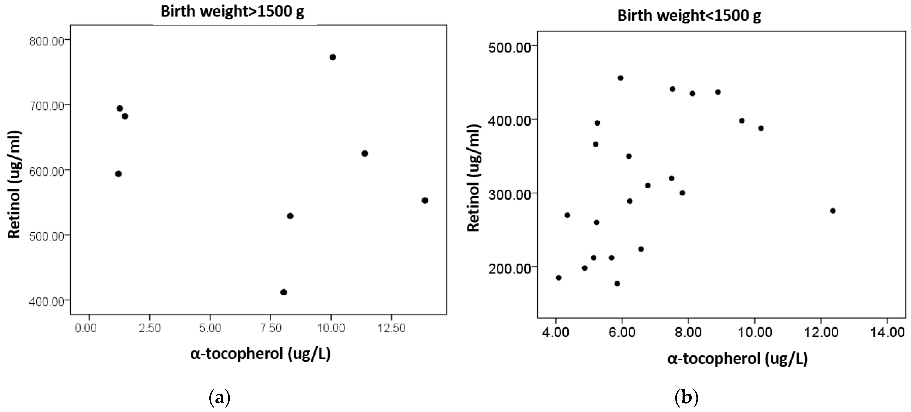
In Figure 2, a scatter plot between α-tocopherol and retinol is presented in neonates with a birth weight of >1500 g and neonates with a birth weight of <1500 g. As can be seen, a positive association of the two vitamins is present in very low birth weight infants (Spearman’s rho = 0.554, p = 0.007).

4. Discussion

In this study, about 10% of preterm infants had a retinol deficiency and about 20% of preterm infants had an α-tocopherol deficiency. SGA infants represented 23.3% of the sample and had lower retinol levels. Moreover, this study documented a positive relationship between gestational age and lipid-soluble antioxidant vitamins, which was dependent on birth weight regarding retinol.
Although retinol cutoffs for defining a deficiency differ across studies, a retinol concentration of less than 200 μg/mL was considered as a deficiency [15] in this and other studies [11,40,44]. The retinol deficiency of infants in this study (10%) was lower than that reported by others (42–82%) [11,45,46,47]. Moreover, SGA infants have been reported to have low levels of retinol-binding protein [48], which is well correlated to serum retinol [49]. Both retinol and its binding protein in cord blood have been shown to increase after the 36th week [50]. It is also noted that birth weight is related to the mother’s status of vitamin A [51]. In this study, neither the data on mothers’ nutrition nor the data on their vitamin statuses were collected. It is most probable that the mothers in this study were not malnourished because this study was conducted in a private hospital, which primarily hosts mothers with higher incomes due to its nature. In other words, it can be hypothesized that mothers had good nutritional statuses, which may also explain the relatively low presence of retinol deficiency in this study.
Several studies have assessed vitamin E deficiency by using different cutoffs, such as from <3.5 mmol/L to <11.6 mmol/L, which corresponds to <1.50 mg/L to 4.99 mg/L [9]. In this study, a cutoff point of <5.0 mg/L was used to determine α-tocopherol deficiency [11], and 20% of neonates were deemed deficient. Studies using a similar cutoff point have found higher levels of deficiency, reaching 62% in the first 24 h of life without supplementation [52]. The study of Chan et al. documented that 38% of preterm infants were deficient, but their study used a lower cutoff point [53]. As recently reviewed, vitamin E deficiency in neonates ranges from 19% to 100% in newborns, reflecting a very high variability [9]. In addition, there is no overall consensus on optimal α-tocopherol levels in preterm infants. The ESPGHAN/ESPEN/ESPR/CSPEN propose that if α-tocopherol is 1–2 mg/L, treatment should be initiated [54]. However, “the more the better” rule seems not to be applicable, since the supplementation of preterm infants to achieve levels of >35 mg/dL (81 μmol/L) is associated with an increased risk of sepsis [55]. Moreover, it has been shown that the α-tocopherol in plasma and red blood cells gradually increases after the first month of birth in very low birth weight infants [56].
Given that breast milk is the main nutritional source for infants, it is important to define its content in retinol and α-tocopherol. The concentration of both vitamins was lower in the breast milk of women having preterm birth versus those having a full-term birth [57]. In absolute numbers, the concentration of colostrum in α-tocopherol was about 250 µg/dL [58], 260 µg/dL [59], and 470 µg/dL [57] in Canadian, Russian, and Mexican women with preterm birth, respectively, although higher values have also been reported [60]. The concentration of colostrum in retinol has been reported to be 36 to 57.5 μg/dL, with a high standard deviation [57,61]. In the cases where donor milk was given, the pasteurization destroyed the pathogenic bacteria, as well as the microbiota of human milk [62]. Fat content [63] and fat-soluble vitamins are not changed after milk pasteurization [64], although some data point to a reduction in retinol content after milk processing [65] and a possible photosensitivity-related retinol reduction if transparent vials are used [66]. In parallel, the freezing of human milk has no effect on its vitamin E content [67,68].
The relatively low content of breast milk in the retinol and α-tocopherol of preterm infants suggests that the supplementation of mothers and/or preterm infants with these vitamins may be useful. However, fat-soluble vitamins are not easily excreted from the body and tend to accumulate. Therefore, they can produce toxicity. This implies that vitamins should be measured, and the supplementation of neonates should be performed on a case-by-case basis. For example, the incidental supplementation of vitamin A in preterm neonates (by using multivitamins) was related to higher intakes of vitamin A than recommended (for infants) [69]. Moreover, as already mentioned, high circulating α-tocopherol is related to a risk of sepsis [55].
No sex differences were observed regarding the vitamin status in our study. This is in line with the findings of other studies for retinol [40,45] and α-tocopherol [53]. It is noted that male infants had a lower retinol concentration than females in another study [70]; however, after adjusting for birth weight and length, the difference was not significant [70]. This finding also underlines the importance of anthropometric variables in interpreting serum retinol concentrations in infants.
SGA infants constituted only 23.3% of the neonates in the present sample. SGA infants aim to maximize the chance of survival and preserve the function of central organs [71]. This often leads to a reduction in the supply of nutrients, a reduction in body fat stores and nutrient depots, along with an alteration of physical and neurological characteristics [72]. In this study, SGA infants presented lower retinol levels. In the literature, SGA infants have been shown to have low circulating retinol-binding protein [48], which is correlated to serum retinol [49]. Moreover, the maternal retinol concentration during mid-pregnancy has been related to the risk of giving birth to SGA infants, while maternal α-tocopherol levels were not related to the birth of SGA infants [73]. Similarly, in our study, the levels of α-tocopherol were not differentiated in SGA infants.
Gestational age and weight, in particular, were correlated to retinol and α-tocopherol levels in our study. This observation may be explained through the following mechanisms: (i) fat-soluble vitamins are deposited in larger quantities as adipose tissue increases and (ii) vitamin A has an effect on growth and possibly on weight gain [3]. Indeed, infants with normal vitamin A levels have been found gain weight more easily [11]. It is also possible that higher oxidative stress present in preterm neonates may “consume” serum antioxidants, such as retinol and α-tocopherol. Indeed, serum antioxidant capacity was positively associated with birth weight in neonates with a wide range of weights (800–3700 g) [74]. Taterno et al. reported a correlation between vitamin E levels and birth weight, but they did not include preterm infants [39]. Chan et al. found a weak but significant association between vitamin E and gestational age [53], while the levels of vitamin E between preterm and term infants were similar in other studies, but no correlation coefficient was reported [13,14]. Liver content in retinol was correlated to gestational age in the fetuses of Indian mothers [75]. In another study, no association was found between gestational age and serum retinol, but blood was taken at 48 h after birth [40]. Similarly, the study of Tao et al. found no association between retinol vs. birthweight and gestational age, but cord blood was measured [45].
The positive association of vitamins E and A should be considered regarding lipid solubility and shared metabolic pathways through lipoproteins [76]. For this reason, some researchers “normalize” their results by dividing lipid-soluble vitamins with total lipids [32]. In addition, weight may influence this association, which is in line with the reports showing that serum antioxidant capacity is positively associated with birth weight [74]. However, an inverse correlation has been observed between serum retinol and α-tocopherol in the colostrum of lactating women [77], suggesting that these vitamins may antagonize each other and affect their bioavailability [78].
The strengths of this study is that it adds much knowledge to this field at a regional level. The levels of α-tocopherol in our country have been previously reported to be around 2.0 to 2.5 mg/L in a smaller sample of preterm infants [41], which is much lower than that the ones documented in this work. The comparability of our results with those from other studies is limited since most studies have been conducted earlier. This means that the previous detection methods may not be as sensitive as the current ones. For example, in the study of Chan et al., the detection limit was 0.60 mg/L, and 14% of the samples could not be detected [53]. Moreover, maternal diet and medical monitoring throughout pregnancy are now more advanced. In addition, few studies have been conducted in Europe [79]. Indeed, most studies have been conducted in participants from Asia, Africa, Brazil, and the US [9,47,75]. In this study, neonates had not received corticosteroids after birth. This is particularly important for the interpretation of our findings, since dexamethasone administration in preterm neonates for facilitating extubation has been connected with a decrease in retinol levels for the first 7 days after birth and a subsequent two-fold increase in retinol levels at ~1 month [80].
Several limitations should be considered along with the interpretation of our results. Firstly, no data on delivery status were available and it is possible that a caesarian delivery affects the status of fat-soluble vitamins [81]. However, not all studies agree on this, since vitamin E levels in fetuses born with a caesarean section and vaginal delivery have also been found to be similar [82]. Supplement use was not recorded in this work and this is also the case for other studies on this topic, with some exceptions [79]. Moreover, no data on mothers’ vitamin statuses were recorded. Indeed, a positive correlation has been documented between maternal and cord levels of vitamins A and E [83]. This study also lacked a control group of full-term infants, but the initial aim was to investigate the vitamin statuses of preterm infants. There are no data on the nutritional support of neonates, but blood samples were collected on their first day of life, so it is mostly improbable that exogenous factors, such as nutrient delivery or breast milk delivery, have affected their vitamin levels. Moreover, no measures of functionality are available, such as the Apgar score. lastly, the serum concentrations of fat-soluble vitamins do not always reflect tissue concentrations [84]. For example, the “gold standard” method for the assessment of vitamin A status is the measurement of liver vitamin A depots [12]. Although retinol levels may reflect the availability of the blood-carrying retinol-binding protein, blood concentrations of retinol are the most frequently used biomarkers [85].

5. Conclusions

This study revealed a positive relationship between gestational age and lipid-soluble antioxidant vitamins, i.e., retinol and α-tocopherol in preterm infants, which can be partially explained by their weight differences. Further studies are needed to establish related functional long-term outcomes in relation to the vitamin status of infants at birth.

Author Contributions

Conceptualization, M.S. and P.D.; methodology, M.S. and P.P.; formal analysis, P.D.; investigation, M.S. and P.P.; resources, M.S.; data curation, P.D.; writing—original draft preparation, P.D.; writing—review and editing, M.S. and P.P.; supervision, M.S.; project administration, M.S. and P.P.; funding acquisition, M.S. All authors have read and agreed to the published version of the manuscript.

Funding

This research received no external funding.

Institutional Review Board Statement

This study was conducted in accordance with the Declaration of Helsinki and was approved by the Scientific Committee of Iaso Hospital (number A21112018).

Informed Consent Statement

Informed consent was obtained from all subjects involved in this study.

Data Availability Statement

Data are available upon request.

Conflicts of Interest

The authors declare no conflict of interest.

References

  1. Traber, M.G.; Head, B. Vitamin E: How much is enough, too much and why! Free Radic. Biol. Med. 2021, 177, 212–225. [Google Scholar] [CrossRef] [PubMed]
  2. Didier, A.J.; Stiene, J.; Fang, L.; Watkins, D.; Dworkin, L.D.; Creeden, J.F. Antioxidant and Anti-Tumor Effects of Dietary Vitamins A, C, and E. Antioxidants 2023, 12, 632. [Google Scholar] [CrossRef] [PubMed]
  3. Ross, A.C.; Gardner, E.M. The Function of Vitamin A in Cellular Growth and Differentiation, and Its Roles during Pregnancy and Lactation. In Nutrient Regulation during Pregnancy, Lactation, and Infant Growth; Allen, L., King, J., Lönnerdal, B., Eds.; Advances in Experimental Medicine and Biology; Springer: Boston, MA, USA, 1994; Volume 352, pp. 187–200. ISBN 978-1-4899-2577-0. [Google Scholar]
  4. Massaro, D.; Massaro, G.D. Lung Development, Lung Function, and Retinoids. N. Engl. J. Med. 2010, 362, 1829–1831. [Google Scholar] [CrossRef] [PubMed]
  5. Dowling, J.E. Vitamin A: Its many roles—From vision and synaptic plasticity to infant mortality. J. Comp. Physiol. A 2020, 206, 389–399. [Google Scholar] [CrossRef] [PubMed]
  6. Blaner, W.S.; Shmarakov, I.O.; Traber, M.G. Vitamin A and Vitamin E: Will the Real Antioxidant Please Stand Up? Annu. Rev. Nutr. 2021, 41, 105–131. [Google Scholar] [CrossRef] [PubMed]
  7. Kamal-Eldin, A.; Appelqvist, L.-Å. The chemistry and antioxidant properties of tocopherols and tocotrienols. Lipids 1996, 31, 671–701. [Google Scholar] [CrossRef]
  8. Ungurianu, A.; Zanfirescu, A.; Nițulescu, G.; Margină, D. Vitamin E beyond Its Antioxidant Label. Antioxidants 2021, 10, 634. [Google Scholar] [CrossRef]
  9. Assunção, D.G.F.; da Silva, L.T.P.; Camargo, J.D.d.A.S.; Cobucci, R.N.; Ribeiro, K.D.d.S. Vitamin E Levels in Preterm and Full-Term Infants: A Systematic Review. Nutrients 2022, 14, 2257. [Google Scholar] [CrossRef]
  10. Henriksen, C.; Helland, I.B.; Rønnestad, A.; Grønn, M.; Iversen, P.O.; Drevon, C.A. Fat-soluble vitamins in breast-fed preterm and term infants. Eur. J. Clin. Nutr. 2006, 60, 756–762. [Google Scholar] [CrossRef]
  11. Kositamongkol, S.; Suthutvoravut, U.; Chongviriyaphan, N.; Feungpean, B.; Nuntnarumit, P. Vitamin A and E status in very low birth weight infants. J. Perinatol. 2011, 31, 471–476. [Google Scholar] [CrossRef]
  12. Shenai, J.P.; Chytil, F.; Jhaveri, A.; Stahlman, M.T. Plasma vitamin A and retinol-binding protein in premature and term neonates. J. Pediatr. 1981, 99, 302–305. [Google Scholar] [CrossRef] [PubMed]
  13. Hågå, P.; Lunde, G. SELENIUM AND VITAMIN E IN CORD BLOOD FROM PRETERM AND FULL TERM INFANTS. Acta Paediatr. 1978, 67, 735–739. [Google Scholar] [CrossRef] [PubMed]
  14. Leonard, P.J.; Doyle, E.; Harrington, W. Levels of vitamin E in the plasma of newborn infants and of the mothers. Am. J. Clin. Nutr. 1972, 25, 480–484. [Google Scholar] [CrossRef] [PubMed]
  15. Tsang, R.C.; Uauy, R.; Koletzko, B.; Zlotkin, S.H. (Eds.) Nutrition of the Preterm Infant: Scientific Basis and Practical Guidelines; Digital Educational Publishing: Cincinnati, OH, USA, 2005; pp. 141–173. [Google Scholar]
  16. Traber, M.; Cohn, W.; Muller, D. Absorption, Transport and Delivery to Tissues. In Vitamin E in Health and Disease; Packer, L., Fuchs, J., Eds.; Marcel Dekker: New York, NY, USA, 1992; pp. 35–53. [Google Scholar]
  17. Kono, N.; Arai, H. Platelet-activating factor acetylhydrolases: An overview and update. Biochim. Biophys. Acta (BBA)-Mol. Cell Biol. Lipids 2018, 1864, 922–931. [Google Scholar] [CrossRef] [PubMed]
  18. O’Byrne, S.M.; Blaner, W.S. Retinol and retinyl esters: Biochemistry and physiology. J. Lipid Res. 2013, 54, 1731–1743. [Google Scholar] [CrossRef] [PubMed]
  19. Giacoia, G.P. Concentration of Serum Prealbumin and Retinol-Binding Proteins During Pregnancy. South. Med. J. 1984, 77, 1261–1263. [Google Scholar] [CrossRef] [PubMed]
  20. Jeanes, Y.M.; Hall, W.L.; Ellard, S.; Lee, E.; Lodge, J.K. The absorption of vitamin E is influenced by the amount of fat in a meal and the food matrix. Br. J. Nutr. 2004, 92, 575–579. [Google Scholar] [CrossRef]
  21. Marceau, G.; Gallot, D.; Lemery, D.; Sapin, V. Metabolism of Retinol During Mammalian Placental and Embryonic Development. In Vitamins & Hormones; Elsevier: Amsterdam, The Netherlands, 2007; Volume 75, pp. 97–115. ISBN 978-0-12-709875-3. [Google Scholar]
  22. Debier, C.; Larondelle, Y. Vitamins A and E: Metabolism, roles and transfer to offspring. Br. J. Nutr. 2005, 93, 153–174. [Google Scholar] [CrossRef]
  23. Gannon, B.M.; Rogers, L.M.; Tanumihardjo, S.A. Metabolism of Neonatal Vitamin A Supplementation: A Systematic Review. Adv. Nutr. Int. Rev. J. 2020, 12, 942–958. [Google Scholar] [CrossRef]
  24. Yang, W.; Jiao, M.; Xi, L.; Han, N.; Luo, S.; Xu, X.; Zhou, Q.; Wang, H. The association between maternal fat-soluble vitamin concentrations during pregnancy and infant birth weight in China. Br. J. Nutr. 2020, 125, 1058–1066. [Google Scholar] [CrossRef]
  25. Rao, N.A.; Wu, G.S. Oxygen free radicals and retinopathy of prematurity. Br. J. Ophthalmol. 1996, 80, 387. [Google Scholar] [CrossRef] [PubMed]
  26. Paiva, S.; Godoy, I.; Vannucchi, H.; Fávaro, R.M.; Geraldo, R.R.; Campana, A. Assessment of vitamin A status in chronic obstructive pulmonary disease patients and healthy smokers. Am. J. Clin. Nutr. 1996, 64, 928–934. [Google Scholar] [CrossRef] [PubMed]
  27. Zachman, R.D.; Samuels, D.P.; Brand, J.M.; Winston, J.F.; Pi, J.T. Use of the intramuscular relative-dose-response test to predict bronchopulmonary dysplasia in premature infants. Am. J. Clin. Nutr. 1996, 63, 123–129. [Google Scholar] [CrossRef] [PubMed]
  28. Clagett-Dame, M.; DeLuca, H.F. THEROLE OFVITAMINAINMAMMALIANREPRODUCTION ANDEMBRYONICDEVELOPMENT. Annu. Rev. Nutr. 2002, 22, 347–381. [Google Scholar] [CrossRef]
  29. Barekatain, B.; Saraeian, S.; Farghadani, M.; Armanian, A.M.; Shahsanaee, A.; Rouhani, E.; Safaei, A. Effect of vitamin E in prevention of intraventricular hemorrhage in preterm neonates. Int. J. Prev. Med. 2018, 9, 97. [Google Scholar] [CrossRef] [PubMed]
  30. Carey, A.N.; Duggan, C. 50 Years Ago in The Journal of Pediatrics. J. Pediatr. 2017, 181, 162. [Google Scholar] [CrossRef]
  31. Cannavò, L.; Perrone, S.; Viola, V.; Marseglia, L.; Di Rosa, G.; Gitto, E. Oxidative Stress and Respiratory Diseases in Preterm Newborns. Int. J. Mol. Sci. 2021, 22, 12504. [Google Scholar] [CrossRef]
  32. Ogihara, T.; Mino, M. Vitamin E and preterm infants. Free Radic. Biol. Med. 2021, 180, 13–32. [Google Scholar] [CrossRef]
  33. Miyake, M.; Miki, M.; Yasuda, H.; Ogihara, T.; Mino, M. Vitamin E and the Peroxidizability of Erythrocyte Membranes in Neonates. Free Radic. Res. Commun. 1991, 15, 41–50. [Google Scholar] [CrossRef]
  34. Demirci-Çekiç, S.; Özkan, G.; Avan, A.N.; Uzunboy, S.; Çapanoğlu, E.; Apak, R. Biomarkers of Oxidative Stress and Antioxidant Defense. J. Pharm. Biomed. Anal. 2021, 209, 114477. [Google Scholar] [CrossRef]
  35. Matyas, M.; Hasmasanu, M.G.; Zaharie, G. Antioxidant Capacity of Preterm Neonates Assessed by Hydrogen Donor Value. Medicina 2019, 55, 720. [Google Scholar] [CrossRef] [PubMed]
  36. Dani, C.; Poggi, C.; Auten, R.L.; Farrow, K.N.; Collins, J.J.; Thébaud, B.; Wedgwood, S.; Steinhorn, R.H.; Berkelhamer, S.K.; Sdrimas, K.; et al. The Role of Genetic Polymorphisms in Antioxidant Enzymes and Potential Antioxidant Therapies in Neonatal Lung Disease. Antioxid. Redox Signal. 2014, 21, 1863–1880. [Google Scholar] [CrossRef] [PubMed]
  37. Duh-Leong, C.; Ghassabian, A.; Kannan, K.; Gross, R.S.; Ortiz, R.; Gaylord, A.; Afanasyeva, Y.; Lakuleswaran, M.; Spadacini, L.; Trasande, L. Prenatal oxidative stress and rapid infant weight gain. Int. J. Obes. 2023, 47, 583–589. [Google Scholar] [CrossRef] [PubMed]
  38. Rakshasbhuvankar, A.A.; Pillow, J.J.; Simmer, K.N.; Patole, S.K. Vitamin A supplementation in very-preterm or very-low-birth-weight infants to prevent morbidity and mortality: A systematic review and meta-analysis of randomized trials. Am. J. Clin. Nutr. 2021, 114, 2084–2096. [Google Scholar] [CrossRef] [PubMed]
  39. Tateno, M.; Oshima, A. The relation between serum-vitamin E levels in the perinatal period and the birth weight of the neonate. Acta Obstet. Gynaecol. Jpn. 1973, 20, 177–181. [Google Scholar] [PubMed]
  40. Chen, H.-J.; Hsu, C.-H.; Chiang, B.-L. Serum retinol levels and neonatal outcomes in preterm infants. J. Formos. Med Assoc. 2017, 116, 626–633. [Google Scholar] [CrossRef]
  41. Skouroliakou, M.; Konstantinou, D.; Koutri, K.; Kakavelaki, C.; Stathopoulou, M.; Antoniadi, M.; Xemelidis, N.; Kona, V.; Markantonis, S. A double-blind, randomized clinical trial of the effect of ω-3 fatty acids on the oxidative stress of preterm neonates fed through parenteral nutrition. Eur. J. Clin. Nutr. 2010, 64, 940–947. [Google Scholar] [CrossRef]
  42. World Health Organization. International Statistical Classification of Diseases and Related Health Problems, 10th Revision, 2nd ed.; World Health Organization: Geneva, Switzerland, 2004. [Google Scholar]
  43. Battaglia, F.C.; Lubchenco, L.O. A practical classification of newborn infants by weight and gestational age. J. Pediatr. 1967, 71, 159–163. [Google Scholar] [CrossRef]
  44. Deger, I.; Ertuğrul, S.; Yılmaz, S.T.; Özbey, Z.K.; Yolbaş, I.; Kaplan, I. The relationship between Vitamin A and Vitamin E levels and neonatal morbidities. Eur. Rev. Med. Pharmacol. Sci. 2022, 26, 1963–1969. [Google Scholar] [CrossRef]
  45. Tao, E.; Chen, C.; Chen, Y.; Cai, L.; Yuan, T. The relationship between umbilical cord blood vitamin A levels and late preterm infant morbidities: A prospective cohort study. Eur. J. Pediatr. 2020, 180, 791–797. [Google Scholar] [CrossRef]
  46. Zhang, X.; Chen, K.; Wei, X.-P.; Qu, P.; Liu, Y.-X.; Chen, J.; Li, T.-Y. Perinatal Vitamin A Status in Relation to Neurodevelopmental Outcome at two Years of Age. Int. J. Vitam. Nutr. Res. 2009, 79, 238–249. [Google Scholar] [CrossRef] [PubMed]
  47. Kiatchoosakun, P.; Jirapradittha, J.; Panthongviriyakul, M.C.; Khampitak, T.; Yongvanit, P.; Boonsiri, P. Vitamin A supplementation for prevention of bronchopulmonary dysplasia in very-low-birth-weight premature Thai infants: A randomized trial. J. Med. Assoc. Thai. 2014, 97 (Suppl. 10), S82–S88. [Google Scholar] [PubMed]
  48. Giacomozzi, C.; Ghirri, P.; Lapolla, R.; Bartoli, A.; Scirè, G.; Serino, L.; Germani, D.; Boldrini, A.; Cianfarani, S. Retinol-binding protein 4 in neonates born small for gestational age. J. Endocrinol. Investig. 2010, 33, 217–221. [Google Scholar] [CrossRef]
  49. Smith, F.R.; Goodman, D.S.; Arroyave, G.; Viteri, F. Serum vitamin A, retinol-binding protein, and prealbumin concentrations in protein-calorie malnutrition. Am. J. Clin. Nutr. 1973, 26, 982–987. [Google Scholar] [CrossRef]
  50. Galinier, A.; Périquet, B.; Lambert, W.; Garcia, J.; Assouline, C.; Rolland, M.; Thouvenot, J.-P. Reference range for micronutrients and nutritional marker proteins in cord blood of neonates appropriated for gestational ages. Early Hum. Dev. 2005, 81, 583–593. [Google Scholar] [CrossRef] [PubMed]
  51. Wang, Y.-Z.; Ren, W.-H.; Liao, W.-Q.; Zhang, G.-Y. Concentrations of Antioxidant Vitamins in Maternal and Cord Serum and Their Effect on Birth Outcomes. J. Nutr. Sci. Vitaminol. 2009, 55, 1–8. [Google Scholar] [CrossRef] [PubMed]
  52. Bell, E.F.; Hansen, N.I.; Brion, L.P.; Ehrenkranz, R.A.; Kennedy, K.A.; Walsh, M.C.; Shankaran, S.; Acarregui, M.J.; Johnson, K.J.; Hale, E.C.; et al. Serum Tocopherol Levels in Very Preterm Infants After a Single Dose of Vitamin E at Birth. Pediatrics 2013, 132, e1626–e1633. [Google Scholar] [CrossRef] [PubMed]
  53. Chan, D.K.-L.; Lim, M.S.-F.; Choo, S.H.-T.; Tan, I.-K. Vitamin E status of infants at birth. J. Perinat. Med. 1999, 27, 395–398. [Google Scholar] [CrossRef] [PubMed]
  54. Braegger, C.; Bronsky, J.; Cai, W.; Campoy, C.; Carnielli, V.; Darmaun, D.; Decsi, T.; Domellöf, M.; Embleton, N.; Fewtrell, M.; et al. ESPGHAN/ESPEN/ESPR/CSPEN guidelines on pediatric parenteral nutrition: Vitamins. Clin. Nutr. 2018, 37, 2366–2378. [Google Scholar] [CrossRef]
  55. Brion, L.P.; Bell, E.F.; Raghuveer, T.S. Vitamin E supplementation for prevention of morbidity and mortality in preterm infants. Cochrane Database Syst. Rev. 2003, 2010, CD003665. [Google Scholar] [CrossRef]
  56. Tanaka, H.; Mino, M.; Takeuchi, T. A nutritional evaluation of vitamin E status in very low birth weight infants with respect to changes in plasma and red blood cell tocopherol levels. J. Nutr. Sci. Vitaminol. 1988, 34, 293–307. [Google Scholar] [CrossRef] [PubMed]
  57. Sámano, R.; Martínez-Rojano, H.; Hernández, R.M.; Ramírez, C.; Quijano, M.E.F.; Espíndola-Polis, J.M.; Veruete, D. Retinol and α-Tocopherol in the Breast Milk of Women after a High-Risk Pregnancy. Nutrients 2017, 9, 14. [Google Scholar] [CrossRef] [PubMed]
  58. Bishara, R.; Dunn, M.S.; Merko, S.E.; Darling, P. Nutrient Composition of Hindmilk Produced by Mothers of Very Low Birth Weight Infants Born at Less Than 28 Weeks’ Gestation. J. Hum. Lact. 2008, 24, 159–167. [Google Scholar] [CrossRef] [PubMed]
  59. Martysiak-Żurowska, D.; Szlagatys-Sidorkiewicz, A.; Zagierski, M. Concentrations of alpha- and gamma-tocopherols in human breast milk during the first months of lactation and in infant formulas. Matern. Child Nutr. 2012, 9, 473–482. [Google Scholar] [CrossRef] [PubMed]
  60. Haug, M.; Laubach, C.; Burke, M.; Harzer, G. Vitamin E in Human Milk from Mothers of Preterm and Term Infants. J. Pediatr. Gastroenterol. Nutr. 1987, 6, 605–609. [Google Scholar] [CrossRef] [PubMed]
  61. Fares, S.; Sethom, M.M.; Khouaja-Mokrani, C.; Jabnoun, S.; Feki, M.; Kaabachi, N. Vitamin A, E, and D Deficiencies in Tunisian Very Low Birth Weight Neonates: Prevalence and Risk Factors. Pediatr. Neonatol. 2014, 55, 196–201. [Google Scholar] [CrossRef] [PubMed]
  62. de Segura, A.G.; Escuder, D.; Montilla, A.; Bustos, G.; Pallás, C.; Fernández, L.; Corzo, N.; Rodríguez, J.M. Heating-induced Bacteriological and Biochemical Modifications in Human Donor Milk After Holder Pasteurisation. J. Pediatr. Gastroenterol. Nutr. 2012, 54, 197–203. [Google Scholar] [CrossRef]
  63. Paulaviciene, I.J.; Liubsys, A.; Eidukaite, A.; Molyte, A.; Tamuliene, L.; Usonis, V. The Effect of Prolonged Freezing and Holder Pasteurization on the Macronutrient and Bioactive Protein Compositions of Human Milk. Breastfeed. Med. 2020, 15, 583–588. [Google Scholar] [CrossRef]
  64. Van Zoeren-Grobben, D.; Schrijver, J.; Berg, H.V.D.; Berger, H.M. Human milk vitamin content after pasteurisation, storage, or tube feeding. Arch. Dis. Child. 1987, 62, 161–165. [Google Scholar] [CrossRef]
  65. Ribeiro, K.D.S.; Melo, I.L.P.; Pristo, A.Z.O.; Dimenstein, R. The effect of processing on the Vitamin A content of human milk. J. Pediatr. 2005, 81, 61–64. [Google Scholar] [CrossRef]
  66. Chaves, J.O.; Fernandes, A.M.d.F.; Parreiras, P.M.; Breguez, G.S.; Passos, M.C.; da Cunha, L.R.; Menezes, C.C. Effect of storage on retinol content and total antioxidant capacity of human milk. Br. Food J. 2019, 122, 606–616. [Google Scholar] [CrossRef]
  67. Romeu-Nadal, M.; Castellote, A.; López-Sabater, M. Effect of cold storage on vitamins C and E and fatty acids in human milk. Food Chem. 2008, 106, 65–70. [Google Scholar] [CrossRef]
  68. Kaya, Ö.; Çınar, N. The effects of freezing and thawing on mature human milk’s contains: A systematic review. Midwifery 2023, 118, 103519. [Google Scholar] [CrossRef] [PubMed]
  69. Oliver, C.; Watson, C.; Crowley, E.; Gilroy, M.; Page, D.; Weber, K.; Messina, D.; Cormack, B. Vitamin and Mineral Supplementation Practices in Preterm Infants: A Survey of Australian and New Zealand Neonatal Intensive and Special Care Units. Nutrients 2019, 12, 51. [Google Scholar] [CrossRef] [PubMed]
  70. Fernandes, T.F.D.S.; Andreto, L.M.; Vieira, C.S.D.S.; De Arruda, I.K.G.; Diniz, A.D.S. Serum Retinol Concentrations in Mothers and Newborns at Delivery in a Public Maternity Hospital in Recife, Northeast Brazil. J. Health Popul. Nutr. 2014, 32, 28–35. [Google Scholar] [PubMed]
  71. MacDonald, M.; Seshia, M. Avery’s Neonatology, Pathophysiology and Management of Newborn, 6th ed.; Wolters Kluwer: Gurgaon, India, 2005. [Google Scholar]
  72. Lapillonne, A.; Braillon, P.; Claris, O.; Chatelain, P.; Delmas, P.; Salle, B. Body composition in appropriate and in small for gestational age infants. Acta Paediatr. 1997, 86, 196–200. [Google Scholar] [CrossRef] [PubMed]
  73. Cohen, J.; Kahn, S.; Platt, R.; Basso, O.; Evans, R.; Kramer, M.S. Small-for-gestational-age birth and maternal plasma antioxidant levels in mid-gestation: A nested case-control study. BJOG Int. J. Obstet. Gynaecol. 2015, 122, 1313–1321. [Google Scholar] [CrossRef] [PubMed]
  74. Sullivan, J.L.; Newton, R.B. Serum antioxidant activity in neonates. Arch. Dis. Child. 1988, 63, 748–750. [Google Scholar] [CrossRef]
  75. Shah, R.S.; Rajalakshmi, R.; Bhatt, R.V.; Hazra, M.N.; Patel, B.C.; Swamy, N.B.; Patel, T.V. Liver stores of vitamin A in human fetuses in relation to gestational age, fetal size and maternal nutritional status. Br. J. Nutr. 1987, 58, 181–189. [Google Scholar] [CrossRef]
  76. Kramer, M.S.; Kahn, S.R.; Platt, R.W.; Genest, J.; Rozen, R.; Chen, M.F.; Goulet, L.; Séguin, L.; Dassa, C.; Lydon, J.; et al. Antioxidant Vitamins, Long-Chain Fatty Acids, and Spontaneous Preterm Birth. Epidemiology 2009, 20, 707–713. [Google Scholar] [CrossRef]
  77. de Lira, L.Q.; Lima, M.S.R.; de Medeiros, J.M.S.; da Silva, I.F.; Dimenstein, R. Correlation of vitamin A nutritional status on alpha-tocopherol in the colostrum of lactating women. Matern. Child Nutr. 2011, 9, 31–40. [Google Scholar] [CrossRef] [PubMed]
  78. Ametaj, B.N.; Nonnecke, B.J.; Franklin, S.T.; Horst, R.L.; Bidlack, W.R.; Stuart, R.L.; Beitz, D.C. Dietary Vitamin A Modulates the Concentrations of RRR-α-tocopherol in Plasma Lipoproteins from Calves Fed Milk Replacer. J. Nutr. 2000, 130, 629–636. [Google Scholar] [CrossRef] [PubMed]
  79. Kiely, M.; Cogan, P.; Kearney, P.; Morrissey, P. Concentrations of tocopherols and carotenoids in maternal and cord blood plasma. Eur. J. Clin. Nutr. 1999, 53, 711–715. [Google Scholar] [CrossRef] [PubMed]
  80. Rossholt, M.E.; Wendel, K.; Bratlie, M.; Aas, M.F.; Gunnarsdottir, G.; Fugelseth, D.; Pripp, A.H.; Domellöf, M.; Størdal, K.; Stiris, T.; et al. Vitamin A Status in Preterm Infants Is Associated with Inflammation and Dexamethasone Exposure. Nutrients 2023, 15, 441. [Google Scholar] [CrossRef] [PubMed]
  81. González-Corbella, M.; López-Sabater, M.; Castellote-Bargalló, A.; Campoy-Folgoso, C.; Rivero-Urgell, M. Influence of caesarean delivery and maternal factors on fat-soluble vitamins in blood from cord and neonates. Early Hum. Dev. 1998, 53, S121–S134. [Google Scholar] [CrossRef] [PubMed]
  82. Woods, J.R.; Cavanaugh, J.L.; Norkus, E.P.; Plessinger, M.A.; Miller, R.K. The effect of labor on maternal and fetal vitamins C and E. Am. J. Obstet. Gynecol. 2002, 187, 1179–1183. [Google Scholar] [CrossRef] [PubMed]
  83. Baydas, G.; Karatas, F.; Gursu, M.; Bozkurt, H.; Ilhan, N.; Yasar, A.; Canatan, H. Antioxidant Vitamin Levels in Term and Preterm Infants and Their Relation to Maternal Vitamin Status. Arch. Med Res. 2002, 33, 276–280. [Google Scholar] [CrossRef]
  84. Embleton, N.D.M.; Moltu, S.J.; Lapillonne, A.; Akker, C.H.v.D.; Carnielli, V.; Fusch, C.; Gerasimidis, K.; van Goudoever, J.B.; Haiden, N.M.; Iacobelli, S.; et al. Enteral Nutrition in Preterm Infants (2022): A Position Paper From the ESPGHAN Committee on Nutrition and Invited Experts. J. Pediatr. Gastroenterol. Nutr. 2022, 76, 248–268. [Google Scholar] [CrossRef]
  85. Tanumihardjo, S.A.; Russell, R.M.; Stephensen, C.B.; Gannon, B.M.; Craft, N.E.; Haskell, M.J.; Lietz, G.; Schulze, K.; Raiten, D.J. Biomarkers of Nutrition for Development (BOND)—Vitamin A Review. J. Nutr. 2016, 146, 1816S–1848S. [Google Scholar] [CrossRef]
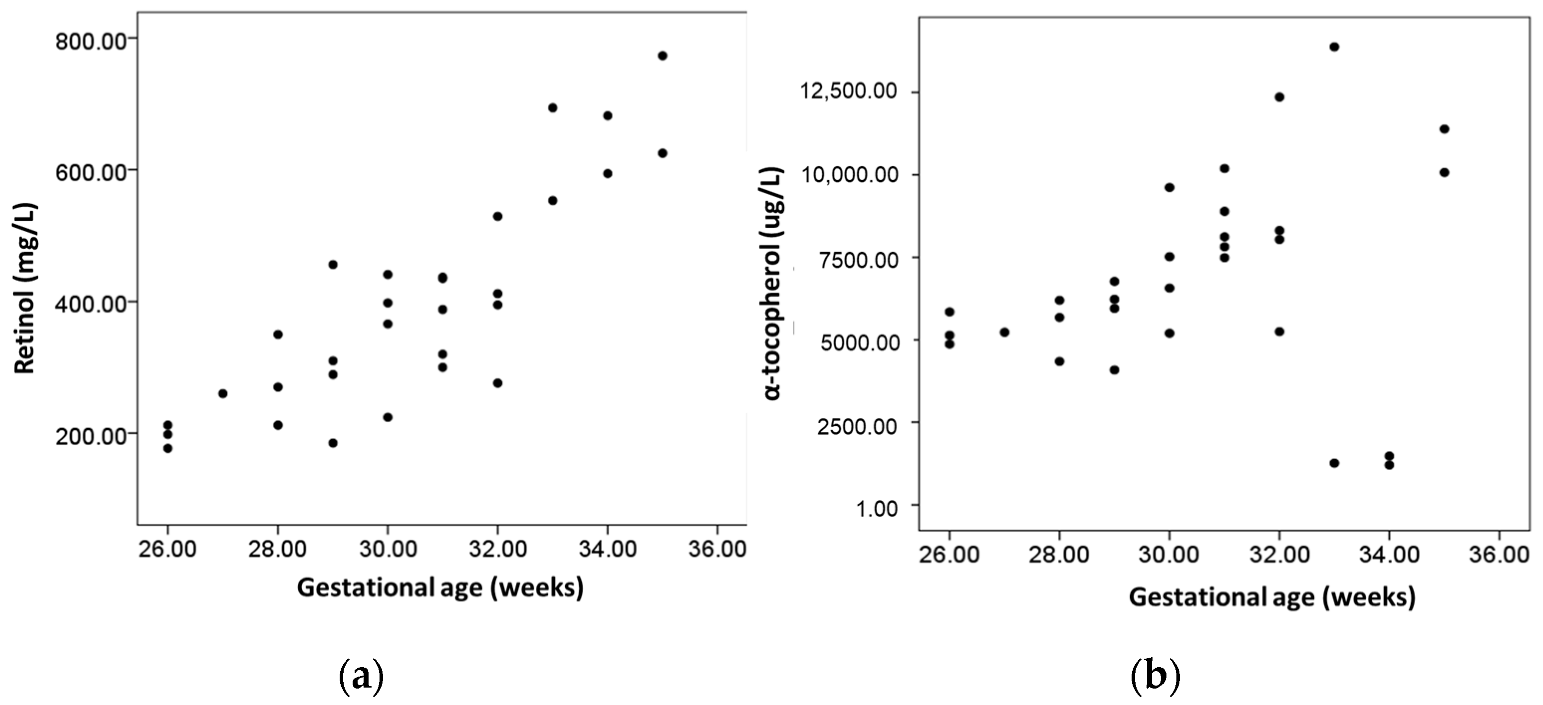
Figure 1. Scatter plot depicting the relation of (a) serum retinol and (b) α-tocopherol vs. gestational age.
Dietetics 02 00027 g001
Figure 2. Scatter plot depicting the relation of serum retinol and α-tocopherol in infants with (a) weight > 1500 and (b) weight < 1500 g.
Dietetics 02 00027 g002
Table 1. Weight and vitamin status of preterm neonates.
Total Sample
(n = 30)
Males
(n = 14)
Females
(n = 16)
p-Value
Gestational age (weeks)30.4 ± 2.5530.6 ± 2.230.1 ± 2.80.628
Birth weight (g) 1185 (1017–1535)1185 (1017–1637)1210 (1010–1502)0.881
Low birth weight (%) *100100100ΝA
Very low birth weight (%) **73.371.475.00.574
Small for gestational age (%)23.342.860.018
Retinol (μg/L)392.0 ± 162.9405.7 ± 148.7380.0 ± 178.30.669
Retinol deficiency (n, %) §3 (10%)1 (7.1%)2 (12.5%)0.552
α-tocopherol (mg/L)6.83 ± 3.026.63 ± 3.307.01 ± 2.850.742
α-tocopherol deficiency (n, %) 6 (20%)3 (21.4%)3 (18.8%)0.605
Values were logarithmized prior to statistical comparisons. ΝA: not applicable; * low birth weight was defined as weight < 2500 g; ** very low birth weight was defined as weight < 1500 g; § retinol deficiency was defined as retinol < 200 μg/L [15]; α-tocopherol deficiency was defined as α-tocopherol < 5.0 mg/L [15].
Table 2. Pearson’s correlations and partial correlations between serum retinol, α-tocopherol, weight and gestational age.
Pearson’s CorrelationWeight-Adjusted Correlation
Retinol (ng/L)α-Tocopherol (ng/L)Retinol (ng/L)α-Tocopherol (ng/L)
Gestational age (weeks)0.845 (p < 0.001)0.255 (p = 0.173)0.193 (p= 0.316)0.180 (p = 0.351)
Birth weight (g) 0.884 (p < 0.001)0.201 (p = 0.286) NANA
Retinol (μg/L)ΝA0.273 (p = 0.144)ΝA−0.238 (p = 0.215)
α-tocopherol (mg/L)0.070 (p = 0.715)ΝA−0.238 (p= 0.215)ΝA
Values were logarithmized prior to statistical comparisons. Spearman’s correlation coefficient was 0.470 between α-tocopherol and gestational age (p = 0.044) and 0.368 between α-tocopherol and birthweight (p = 0.046); ΝA: not applicable.
Disclaimer/Publisher’s Note: The statements, opinions and data contained in all publications are solely those of the individual author(s) and contributor(s) and not of MDPI and/or the editor(s). MDPI and/or the editor(s) disclaim responsibility for any injury to people or property resulting from any ideas, methods, instructions or products referred to in the content.