Review

The Vaginal Microbiome during Pregnancy in Health and Disease

by
George L. Mendz
School of Medicine, Sydney Campus, University of Notre Dame Australia, Darlinghurst, NSW 2010, Australia
Appl. Microbiol. 2023, 3(4), 1302-1338; https://doi.org/10.3390/applmicrobiol3040089
Submission received: 2 November 2023 / Revised: 27 November 2023 / Accepted: 28 November 2023 / Published: 30 November 2023
(This article belongs to the Special Issue Microbiome in Ecosystem 2.0)

Abstract

:
This study appraises the progress in the understanding of the composition of the vaginal microflora with a focus on the microbiome during pregnancy. This knowledge is presented with the background of the global health contribution, along with the importance of these microbial communities to pregnancy. A brief review of current methods employed to investigate the structure of these microbial populations is included. Two types of studies, cross-sectional and longitudinal, have been used to characterise the vaginal microbiota; both types are reviewed since they provide information that serves to piece together a more complete picture of the vaginal microflora and its changes during pregnancy. The identity of microbes present in the vagina are examined in the context of health and disease, and, more specifically, in the setting of pregnancy outcomes. The protective role of lactobacilli in maintaining a healthy vaginal environment is evaluated, with analyses of the different roles of various Lactobacillus spp. Classifications of the vaginal microbiota into vagitypes in non-pregnant and pregnant women are discussed. The associations of specific taxa with three adverse pregnancy results, namely, miscarriage, stillbirth, and preterm birth, are examined in some detail. Longitudinal studies investigating changes in the bacterial community composition and taxa abundance demonstrate that this microbiota decreases in richness and diversity relative to those present in non-pregnant microbiomes. Notwithstanding the significant effort made to characterise the vagina bacterial microbiota, a large number of issues remain to be fully understood.

1. Introduction

1.1. Scope of the Study

This work reviews the bacterial microbiota present in the vagina in health and disease to provide a background for understanding the vaginal bacterial populations and their roles during pregnancy, employing up-to-date literature. It presents the bacterial populations in the context of their contributions to maternal and child health and disease. This microbiome has been studied extensively to determine the bacterial communities that live in the vagina during healthy pregnancies, as well as during those with adverse outcomes; this review focuses on miscarriage, stillbirth, and PTB. The presentation is structured according to two types of studies, cross-sectional and longitudinal, that can be identified in investigations of the vaginal microbiome of gravid women. Both types are reviewed since they provide information that serves to piece together a more complete picture of the vaginal microbiota and its changes during pregnancy. Notwithstanding the significant effort made to characterise the vagina bacterial microbiota, a large number of issues remain to be fully understood.

1.2. United Nations Goals on Maternal and Child Health

In the year 2000, the Millenium Summit of the United Nations established eight international development goals called the Millennium Development Goals to be achieved by 2015. The Fifth Goal was “To improve maternal health”: it included reducing the rate of maternal mortality and achieving universal access to antenatal care coverage [1]. In September 2015, the United Nations General Assembly adopted 17 Sustainable Development Goals for the future of human development to be achieved by 2030. Maternal and child health are included in Goal 3: Good Health and Wellbeing [2].

1.3. Adverse Pregnancy Outcomes

In the 2021 Report of the UN Inter-agency Group for Child Mortality Estimation, the rates of miscarriage, defined as the loss of pregnancy before viability, was estimated at 11–22% of pregnancies [3]; an estimated 23 million miscarriages occur globally every year [4]. The rates of neonatal deaths and stillbirths, defined as a child born dead after 28 weeks of gestation, are estimated at 16.7/1000 and 13.9/1000 births, respectively [5]. The global burden of stillbirth is enormous: about 2 million infants die every year. Stillbirth rates vary significantly between countries from 1.4 to 32.2 per 1000 births; 34% of global stillbirths occur in Central and Southern Asia and 42% in Sub-Saharan Africa. In contrast, an average of about 1% of global stillbirths occur in North America, Europe, Australia, and Oceania [5].
Focussing on preterm birth (PTB, before 37 completed weeks of gestation), every year, over 15 million babies are born preterm, and this number is rising [3]. Across 184 countries, PTB rates range from 5% to 18% of babies born [3], and the average global PTB rate is about 11% [6]. It is the leading cause of death amongst children; one million children die due to PTB before the age of five years, and it accounts for 35% of all deaths amongst newborns (age < 28 days) and for 18% of all deaths amongst children aged under five years [6]. The rates of PTB and mortality vary significantly between countries and within countries, and, besides perinatal mortality, it is a major cause of serious neonatal morbidity and moderate to severe childhood disability in developed and underdeveloped countries. The burden of PTB is particularly high in low- and middle-income countries. For example, in the Pacific Islands, the prevalence for low birth weight and for PTB is 12% and 13%, respectively [7]. Notwithstanding the global efforts to achieve the Fifth Millennium Goal and the Third Sustainable Development Goal, substantial problems in maternal and child health remain. In particular, to reach target number 3.2 of Goal 3 that aims to end all preventable deaths of newborns and children aged under five years requires, amongst other interventions, a determined effort to reduce the rates of adverse pregnancy outcomes.

1.4. Health Risks in Pregnancy

Ordinarily, pregnancy is a normal state of health, but changes in hormone levels and immune system function can make gravid women more susceptible to diseases that pose health risks to the mother, the child, or both. Complications during pregnancy have complex causes including alcohol or drug use, smoking, inadequate prenatal care, genetic background, maternal age, hypertension, nutritional status, diabetes, genitourinary tract infections, and several other risk factors [8].
The aetiology of pregnancy disorders is multifactorial, but the importance of the microbiota of the female genital tract (vagina and uterus) for maternal and infant health is shown by links between the dysbiosis of this tract and negative pregnancy outcomes. Some diseases that create problems primarily for the mother are urinary tract infections, vaginitis, postpartum infections, and pneumonia. For the child, adverse pregnancy outcomes related to maternal infections are miscarriage, stillbirth, PTB, low birth weight, and neonatal and perinatal mortality.
A little over a decade ago in 2011, volume 118 of the British Journal of Obstetrics and Gynaecology devoted a special issue to ‘Infections in Pregnancy’. The editorial stated “Infections of mothers and their babies (both in utero and ex utero) are a major global health challenge” [9]. The threat remains today.

2. Types of Infections during Pregnancy

Many aetiological agents pose a risk to pregnancy; in particular, infections of the genital tract by various pathogens that can cause serious health issues for both mothers and their fetuses have become a major public health issue all over the world for their high and growing prevalence. Genital infections can have a long duration, and women with these infections before pregnancy might also have increased risks of adverse outcomes including macrosomia, PTB, and spontaneous abortion [10].
Multiple types of viral, bacterial, fungal, and parasitic infections have been identified that can affect the health of the pregnant woman, the pregnancy, and the infant after delivery [11].

2.1. Viral Infections

Different types of viruses have been identified as risk factors for maternal and infant heath (Table 1). Arboviruses can cross the placenta and have been linked with fetal morbidity and mortality. Venezuelan equine encephalitis, West Nile, and Zika viruses have been associated with fetal malformations and demise, and neonatal death. Moreover, dengue and chikungunya viruses have been connected with adverse infant and mother outcomes [12].
The Hepatitis A virus is an important cause of materno-fetal infections in endemic regions owing to the spread of the disease. Most women infected with Hepatitis A during pregnancy and their infants do not experience serious complications. Nonetheless, this infection poses a higher risk of preterm labour, especially if it occurs during the second or third trimester. Other increased risks associated with Hepatitis A infection may include: premature uterine contractions. placental abruption, and the preterm prelabour rupture of membranes (PPROM). An increased chance for birth defects following exposure to Hepatitis A in pregnancy has not been shown [13]. Chronic Hepatitis B virus infection in pregnant women increases the risk of intrahepatic cholestasis, gestational diabetes mellitus, pre-eclampsia, and PTB rates [14]. In Hepatitis-E-endemic countries, there is a high burden of infection amongst pregnant women; this virus can cause fulminant hepatic failure, and, in third trimester infections, it has been associated with up to 30% mortality. Hepatitis E virus can be transmitted from infected mothers to their infants with significant perinatal morbidity and mortality [15].
The influenza virus has been related to various negative pregnancy outcomes; however, the results of a recent meta-analysis suggested a correlation with an increased risk of stillbirth, but no clear effects on small-for-gestational-age development, low birth weight, PTB, and fetal death [16]. Pregnant women with measles infection are at risk of severe pregnancy complications. The virus is not teratogenic but causes a dysregulation of the entire immune system that may interfere with the normal course of pregnancy. Measles virus infection alters the physiological mechanisms of immunotolerance present during pregnancy such that it can lead to a reaction similar to rejection, manifested by the spontaneous abortion or premature expulsion of the fetus. Measles in pregnancy can also lead to perinatal infections in the newborn, associated with neurological complications and a high mortality [17].
Pregnant women who contract rubella (German measles) are at risk of miscarriage or stillbirth, and their developing infants are at risk of severe birth defects. The timing of the infection is crucial: contracted by the fetus during the first 12 weeks of gestation, it is likely to result in many infant problems; in infections acquired between 12 and 20 weeks of pregnancy, usually, the problems will be milder; in infections acquired at later gestational ages, commonly, there will not be problems with the infant [18]. Congenital rubella syndrome (CRS) can affect almost everything in the developing infant’s body. The most common birth defects from CRS are: cataracts, deafness, heart defects, liver and spleen injury, intellectual disabilities, low birth weight, and birth skin rash [19].
Herpes simplex (HSV) infection during pregnancy increases the risk of stillbirth, spontaneous abortion, and premature birth. These infections are well-known causes of childhood sequelae [20]. Human cytomegalovirus is a major cause of adverse nonhereditary birth outcomes, including hearing and visual loss, neurologic deficits, and intrauterine growth retardation; the virus may contribute to outcomes such as stillbirth and preterm delivery [21]. Human-papilloma-virus-positive women have an increased risk of pregnancy problems, such as miscarriage, pregnancy-induced hypertensive disorders, intrauterine growth restriction, low birth weight, the premature rupture of membranes, and PTB [22].
Untreated maternal human immunodeficiency (HIV) virus infection increases the risk of stillbirth, preterm delivery, low birth weight, and small-for-gestational-age infants. In 2013, the World Health Organization recommended the use of antiretroviral therapy for all pregnant and breastfeeding women for the prevention of mother-to-child HIV transmission [23].
Viruses and their disease risks and/or adverse outcomes in pregnancy are shown in Table 1. The sources of information are in Section 2.1 Viral Infections. The availability of universal anti-retroviral therapy has not diminished the adverse pregnancy outcomes that are two to three times higher amongst HIV-positive women compared to HIV-negative [24].
Acute syndrome coronavirus 2 (SARS-CoV-2) infections during pregnancy have been related to adverse maternal outcomes such as pneumonia, prematurity, venous thrombotic events, severe pre-eclampsia, and death [12]. In a study of 489,471 delivery hospitalisations, 6550 of women diagnosed with COVID-19 had an increased risk of a 1.2 adjusted relative risk for preterm labour with preterm delivery [25].

2.2. Bacterial Infections

Bacterial colonisation of the female genital tract has been studied for a long time and extensively. These investigations have resulted in a considerable body of knowledge about the bacteria that have their habitats in the genital tract of healthy women, and those associated with disease; the latter represent a risk to the outcome of pregnancy.
Prominent amongst beneficial bacteria are four well-studied species of lactobacilli, L. crispatus, L. iners, L. gasseri, and L. jensenii [26]. A characteristic of female genital infections is that, usually, they are polymicrobial, of mixed aerobes and anaerobes that frequently outnumber aerobes. Common bacteria in genital tract infections are Chlamydia trachomatis, Clostridium spp., Gardnerella vaginalis, Mobiluncus mulieris, Nesseria gonorrhoeae, Peptostreptococcus spp., Porphyromonas spp., Prevotella bivia, Streptococcus agalactiae (Group B Streptococus, GBS), Ureaplasma urealyticum, and others [26]. Many of these infectious taxa pose a considerable risk of disease such as bacterial vaginosis (BV), endometritis, salpingitis, tubo-ovarian abscesses, pelvic inflammatory disease, amnionitis, septic pelvic thrombophlebitis, intrauterine contraceptive device-associated infection, septic abortion, etc.
Commonly, the presence of pathogens is tested by cultivation to identify the bacteria involved in genital infections and apply appropriate therapeutic measures, but, in many instances, no diagnosis had been possible owing to the absence of growth in cultures from genital tract swabs. Knowledge of this microflora has improved remarkably by employing culture-independent new sequencing methods that have yielded a vast increase in the number of taxa identified in the female genital tract. The work that led to this new understanding of the genital bacterial microbiome and the main components of this microbiota are discussed in the following sections.

2.3. Fungal Infections

The female genital mycobiota cincludes fungi residing in the reproductive tract: common taxa are Aspergillus, Candida, Sacharomyces, Sporobolomyces, and others. During pregnancy, increased levels of progesterone and oestrogen affect the vaginal microenvironment; these physiological changes are a risk factor for candidiasis (commonly known as thrush). Candida is the leading genus of fungal cervicovaginal infections present in approximately 20% of non-pregnant women; it increases to an average of 30% during pregnancy, with a risk that can be as high as 50% in the third trimester [27], and it can lead to intrauterine infection, endometritis, and choroamnionitis [28]. Owing to the widespread presence of candidiasis, studies are conducted to establish the effectiveness of treating women with asymptomatic vaginal thrush in reducing spontaneous PTB and late miscarriage. Symptomatic vaginal thrush is not harmful to the developing baby, but the infection is debilitating, and there is also a risk that the Candida fungus can be passed to the infant during childbirth [29]. Table 2 summarises adverse outcomes of fungal infections on pregnancy.
Fungi and parasites and their disease risks and/or adverse outcomes in pregnancy are shown in Table 2. The sources of information are in Section 2.3 and Section 2.4.

2.4. Parasitic Infections

Parasitic infections are common among pregnant women, owing to reduced body immunity, and they can affect the physiology of the mother’s body (Table 2). Helminth infection may be particularly detrimental during pregnancy by increasing the risk of PTB, low birth weight, and maternal anaemia. Prenatal exposures to these infections are linked to infant impaired behavioral development [30]. In contrast, urogenital schistosomiasis is not significantly associated with adverse pregnancy outcomes [31].
Scabies mite infection accounts for 2% to 6% of skin conditions during pregnancy, but the infection is not known to cause directly negative pregnancy and fetal outcomes. Scratching the scabies rash may lead to secondary infections and even sepsis. Scabies-related infections may also increase the risk of having kidney diseases and rheumatic heart disease [32].
Pregnant women infected with malaria have adverse consequences on birth outcomes, including having small-for-gestational-age and preterm infants [33], as well as low birth weights, that predisposes them to infant mortality and lifelong morbidities [34]. An investigation involving 361 pregnant women with simple malaria and 85 women with severe malaria showed significant differences in the perinatal poor outcomes between simple and severe malaria and by the trimester of infection [35].
The global annual prevalence of congenital toxoplasmosis is estimated to be over 190,000 cases. A primary Toxoplasma gondii infection in pregnant women can cause the vertical transmission of the parasite and result in miscarriage, stillbirth, premature birth, abortions, malformations, neonatal mortality, and a variety of neurological sequelae [36]. Infants with congenital toxoplasmosis may exhibit clinical signs of hydrocephalus, mental retardation, eye disease, and other severe sequelae. The poor health condition of infants with congenital toxoplasmosis contributes to their heavy global health burden [37].
Trichomonas vaginalis infection is the most common non-viral sexually transmitted disease in the world. Often, it is asymptomatic and can be eliminated with proper treatment. Untreated trichomoniasis in pregnant women is associated with preterm delivery, the prelabour rupture of membranes, and low birth weight [38].

3. The Human Microbiome

3.1. The Human Microbiome Project

The microbial populations in an ecosystem make up its microbiome, and all the micro-organisms that live in or on the human body constitute the human microbiome, distributed across body sites. With the realisation of the fundamental role of the human microflora in health and disease, in 2007, the National Institutes of Health launched the Human Microbiome Project initiative. Between 2007 and 2014, the Human Microbiome Project Phase I investigated microbial communities from 300 healthy human subjects at five different body sites: skin, nasal passages, the oral cavity, the gastrointestinal tract, and the female urogenital tract [39].
In 2014, Phase II started, known as the Integrative Human Microbiome Project, with the aim of producing resources to characterise this microbiome, focussing on understanding the presence of the microbiota in health and disease states. Its study methods “included 16S rRNA gene profiling, whole metagenome shotgun sequencing, whole-genome sequencing, metatranscriptomics, metabolomics/lipidomics, and immunoproteomics”. [40]. Phase II encompasses three sub-projects. One of them is the Multi-Omic Microbiome Study Pregnancy Initiative (MOMS-PI) conducted by the Vaginal Microbiome Consortium at Virginia Commonwealth University in collaboration with the Global Alliance to Prevent Prematurity and Stillbirth (GAPPS) at Seattle Children’s Hospital; it aims to understand the changes throughout pregnancy of the vaginal microbiome and their influence in the microbiome of neonates. The Initiative investigates the roles of the mother’s genetic background, urogenital conditions, physiological states, and environmental factors in the structure and composition of the vaginal microbiome [41].
These projects and other investigations made important advances in the knowledge of the microbial flora that inhabit humans. The composition of the diverse, complex, and abundant microbiota of the human body is largely determined by body site, host genetics, environmental exposures, and time [39]. The picture that emerged from the sites that were investigated more comprehensively, such as the gut, the mouth, and the skin, is that: (i) the human body is host to a much greater number of microbial species, and, specifically, bacteria, than previously thought, and (ii) a significant number of the taxa of the microflora in various sites of the human body remain incompletely characterised [39]. Importantly, it confirmed that the microbiome plays a significant role in human physiology in health and disease beyond what was suspected, and our knowledge of it continues to unfold.
The female genital tract during pregnancy has been less comprehensively studied than other body sites owing to the difficulty in accessing the intrauterine space. Nonetheless, microbiotas of various loci within the female genital tract form a continuum of bacterial communities that change from the vagina to the ovaries, challenging the traditional view of human fetal development as occurring in a sterile environment. Indeed, the vertical transmission of the mothers’ microbial populations before birth could be the norm throughout the animal kingdom [42].

3.2. Methods of Investigation

A brief discussion of the techniques and tools employed to elucidate the genital microbiome will serve to introduce the overview of the vaginal microbiota. Advances in molecular techniques to identify micro-organisms and bioinformatics tools to analyse very large datasets provided the foundation to characterise and investigate the microbial communities that inhabit the human body. In terms of the diversity of taxa, the majority of microbes on and in the human body are bacteria; thus, the review focuses on the vaginal bacteriome, which, following common nomenclature, is referred to generically as the microbiome.
The advent of high-throughput new-generation (HTNG) sequencing had a significant impact on disease diagnosis, particularly, in human genetic diseases and cancers, and, to a lesser extent, in microbial-related illnesses. The effectiveness of HTNG techniques was demonstrated by identifying aetiological agents in samples where traditional bacterial culture techniques failed, and in cases where multiple agents were involved. Important limitations to a wider use of these techniques are the lack of ability of diagnostic centres to perform fast sample HTNG analyses, and the capacity to analyse the large datasets generated by these methods. Two important techniques for bacterial identification and characterisation that use HTNG are 16S rRNA sequencing and metagenomics sequencing.

3.2.1. 16S rRNA Sequencing

For a long time, it was thought that, in healthy pregnancies, the intra-amniotic space was sterile; thus, studies of the genital microbiota concentrated on the vagina. For more than a hundred years, there have been investigations in the composition of the vaginal microflora, and, up to about 25 years ago, most conclusions about the vaginal microbial populations of post-pubertal women were based on techniques that employed microbial cultivation [43], and, more recently, culture-independent targeted polymerase chain reaction (PCR) methods [44]. The composition of the bacterial communities identified by these approaches was biased and incomplete because many micro-organisms in these communities are refractory to culture or not culturable in vitro, and targeted PCR was unable to identify the diverse array of bacteria present in the vagina. A result is that, in current clinical practice, microbiological analyses of the female genital microbiota focus on a limited number of species of bacteria.
The discovery that bacterial 16S ribosomal RNA (16S rRNA) genes have both highly conserved and variable regions allowed them to be used to determine microbial phylogeny with accuracy [45]. Cultivation-independent broad-range PCR analyses of 16S rRNA gene sequences have the advantages of the universality and high copy number of 16S genes and their limited horizontal transfer, and provide a detailed description of the microbial DNA in a sample. Their disadvantages are: (i) the inability to differentiate between some strains in a taxon or closely related taxa, (ii) the variability of the copy number per genome between taxa distorting their relative abundances, (iii) the inability to assess the bacterial load, (iv) the lack of information on the viability, metabolic function, antibiotic resistance, and interaction with the maternal host of the bacteria present, and (v) PCR DNA amplification biases that result in the inaccurate determination of the relative abundances, because different primers focus on different variable regions V1–V2, V3–V4, or V4, and, depending upon the primer set, some genera can be under- or over-represented; thus, results from studies employing different primers cannot be compared. Nonetheless, this is the method most commonly used in microbiome research; its application suggests that only a small percentage of bacteria in nature have been identified, even in well-studied environments.
Work on the genital microflora employing this approach continues, although it has revealed a richer microbiota with a wider range of taxa, and a larger proportion of polymicrobial infections than those identified employing culturing methods [46,47]. In particular, the full identity and diversity of the genital bacterial populations during pregnancy remain unknown for various racial backgrounds, health status, and lifestyles. Moreover, infections can be tackled with only limited efficiency without the knowledge of the complex interactions of the various taxa of the genital microflora.
The potential of HTNG sequencing is evidenced by its capacity to gather data and employ statistical analyses to identify microbial taxa involved in healthy pregnancies and those associated with adverse pregnancy outcomes. Such work is serving to refine more targeted clinical screening approaches.

3.2.2. Metagenomics Sequencing

More complete analyses, both taxonomical and functional, are achieved by sequencing entire genomes. In shotgun sequencing, genomes are sheared randomly into DNA fragments that are sequenced individually and reassembled into whole genomes employing bioinformatic tools. Through capturing the entire genome of micro-organisms, the method allows analyses of viral, bacterial, fungal, and parasitic genomes. In addition to taxonomic information, other advantages of the method are the ability to provide functional profiles of the microbes identified, and how they communicate, form symbioses, or compete. The disadvantages of metagenomics are: (i) higher costs owing to needing many more sequencing reads to reach the depth required to characterise unusual micro-organisms, and (ii) the large bioinformatics processing demands to analyse the data generated. The method is limited in characterising previously unknown microbes, owing to the need to rely on reference databases to assemble their genomes [48].
Advances in molecular techniques have promoted the widespread use of next-generation sequencing platforms for both amplicon and whole-genome sequencing, and further enhanced the knowledge of these organisms, particularly those that represent very small proportions of a given microbial community. Studies on the female bacterial microbiota during pregnancy have contributed a wealth of information to the knowledge of which micro-organisms colonise the vagina and uterus.

3.2.3. Omics Techniques

The integration of data from various omics techniques and advances in computational bioinformatics methods have made significant contributions to the understanding of bacterial–human interactions in health and disease [49]. Omics techniques such as genomics, epigenomics, transcriptomics, proteomics, metabolomics, etc. have served to develop a multi-omics approach to genotyping and phenotyping that, besides having an impact on infectious diseases in pregnancy, is applied to many other areas such as aging mechanisms, cancer aetiology, cardio-metabolic diseases, organ transplantation, probiotics studies, etc. [50,51].

4. The Human Vaginal Microbiome

Human vaginal tissue is made up of stratified squamous epithelium covered by cervicovaginal secretions [52]. The vaginal mucosa is a relatively anaerobic habitat owing to a limited blood supply that requires obtaining oxygen and nutrients from underlying submucosal tissues [53]. An indigenous complex microbial community lives in a symbiotic relationship with the host and constitutes the normal vaginal microbiome. Billions of microbes inhabit this microbial ecosystem; a systematic analysis of the bacterial biomass of females of reproductive age employing 16S rRNA gene sequencing indicated that the vagina contains 1010–1011 bacteria. At the interface between the host and the environment are apical layers with a key role in resisting colonisation by other microbes [42]. The vaginal bacterial community is an ecosystem strongly influenced by characteristics of the host such as age, menstruation, race/ethnic group, genetics, diet, sexual behaviour, smoking, etc. [54].
The health status of the vagina is associated with its resident microflora. A healthy vaginal microbial population modulates immune responses to invading pathogens and contributes to host immunity through the production of factors such as bacteriocins, hydrogen peroxide, and biosurfactants [55]. Many pathogenic microbes causing vaginal infections have long been identified, but new methods of analysis of this human flora revealed associations of other micro-organisms with disease, as well as the presence of presumed pathogens in healthy women.
Multiple studies employing culture-independent, high-throughput new-generation sequencing methods, and state-of-the-art biostatical analyses have been applied to address the scientific challenge of characterising the vaginal microbiome, and basic questions have been answered such as what bacterial taxa inhabit this tract, the role of racial background in determining the composition of this microbiota, the presence of pathogens hitherto unknown and the changes they induce in this microbiome, etc. Nonetheless, many characteristics of this ecosystem remain to be elucidated fully such as the type and rate of changes in the vaginal microflora during pregnancy, and the differences in the composition of bacterial communities of women giving birth at term, preterm, or with other adverse pregnancy outcomes

Lactobacillus ssp. Protective Role

The vagina contains various types of cells and receptors associated with the immune system that recognise and respond to the presence of micro-organisms [56]. Both commensal and pathogenic bacteria are recognised by pattern recognition receptors such as toll-like receptors, dectin-1 receptor, and nucleotide-binding oligomerisation domain. These receptors are present on both the squamous epithelial cells lining the vagina and the columnar cells lining the upper section of the female genitalia [57].
Lactobacillus species are present in the vaginal microbiota of almost every woman; the relative abundances and proportions of various species depend on the racial background of the woman. Over 250 bacterial species have been identified in the vagina, and, commonly, the genus Lactobacillus is found as the predominant commensal bacteria in the healthy human vagina. The most abundant species include L. crispatus, L. iners, L. jensenii, and L. gasseri [58]. These lactobacilli are regarded as beneficial to the vaginal econiche with a homeostatic role that contributes to exclude competitively the invasion and establishment of genitourinary pathogens partly through forming large biofilm tightly attached to the vaginal epithelial surface, thus, creating a first line of defence mechanism against potential pathogens [59]. Lactobacilli have been used as probiotics to clear the polymicrobial biofilms formed by pathogenic bacteria, functionally to impede bacterial virulence and suppress infection in the vagina [60]. Exclusion, displacement, and competition experiments showed that L. crispatus and L. gasseri inhibited significantly the adhesion to primary vaginal cells of the pathogens G. vaginalis, M. mulieris, Candida albicans, S. agalactiae, Staphilococcus aureus, Escherichia coli, and Enterococcus [61]. In co-cultures, various Lactobacillus species were able to inhibit C. albicans biofilm formation and biofilm-related gene expression [62].
Lactobacilli also produce organic acids, hydrogen peroxide, bacteriocin, and other antimicrobial compounds that protect the vaginal tissues from invasion by pathogens [63]. In most instances, the depletion of Lactobacillus spp. and the overgrowth of non-Lactobacillus microbes is a characteristic of the disruption of the vaginal microbiota equilibrium. Typically, the overgrowth of anaerobic bacteria can result in aberrant conditions, such as bacterial vaginosis- sexually transmitted infections, as well as pregnancy-related complications [64].
The protective role of Lactobacillus spp. in the vagina has been well-described, e.g., regarding their antimicrobial properties [60,65] supporting the establishment of pregnancy [66], and preventing necrotising enterocolitis in infants [67]. Conversely, the paucity of Lactobacillus spp. in this microbiome is a risk factor for several disease states such bacterial vaginosis [51], and adverse pregnancy outcomes such as miscarriage [64] and PTB [68,69]. Studies have showed that L. crispatus-dominated vaginal microbiomes are associated with a lower prevalence of bacterial vaginosis, vulvovaginal candidiasis, and sexually transmitted infections (STI) [70] such as infection with Chlamydia trachomatis [71], HIV [55], Neisseria gonorrhoeae [72], and HSV-2 [73].
L. crispatus is the second most common Lactobacillus species in the vagina; its dominance is associated with a more stable microbiome and a reduced probability of bacterial vaginosis. Preterm delivery is correlated with lower levels of L. crispatus and higher levels of other taxa [74]. Vitamin D levels correlate with vaginal L. crispatus abundance and could, thus, prevent pregnancy complications [75].
L. iners is one of the most prevalent lactobacilli in the vagina of healthy women [76], or individuals with vaginal dysbiosis [77]. L. crispatus usually dominates the healthy vagina of reproductive-age women, but it decreases during menstruation and is partly replaced by L. iners [78]. Current knowledge indicates that L. iners is the predominant species in the vaginal microbiota amongst subpopulations of older women, pregnant women [79], and women of Afro-American [80] and sub-Saharan African descent [81]. In addition, it is found in women diagnosed with vaginal diseased states, e.g., BV [82], and sexually transmitted infections [83], and shortly after BV treatment [84]. L. iners appears to be capable of adapting to the changing conditions prevailing in the vaginal niche and it is not easily displaced by pathogenic microbes. Evidence suggests that it is a transitional species that colonises after the vaginal environment is disturbed and offers overall less protection against vaginal dysbiosis. There is no sufficient evidence to conclude that L. iners causes BV by itself, but it is present in BV.
Function analysis of proteins encoded by the L. iners genome revealed that it could show both commensal and pathogenic properties. Two characteristics of this bacterium that enable it to adapt to the fluctuating environment of the vaginal niche are complex nutrient requirements [85] and a strong adhesive ability to vaginal epithelial cells [86]. The size of the L. iners genome is unique in the Lactobacillus genus; the AB-1 strain has the smallest genome amongst Lactobacillus spp., consisting of a single chromosome of approximately 1.3 Mbp, in contrast to other lactobacilii whose genome size is approximately 3–4 Mbp [87]. Nonetheless, the L. iners genome encodes proteins predicted to be involved in the optimal adaptation to the variable conditions of the vagina, such as iron-sulfur proteins and the σ factor, suggesting that its metabolism may change with rapid shifts in the environment of the vagina. Moreover, given its ability to scavenge a variety of nutrients, it may survive during times of transition in conditions where other microbes do not, e.g., after the host takes an antibiotic.
Several genes identified in the L. iners genome suggest that it may be an opportunistic pathogen [82]. The genomes of some L. iners strains encode the toxin inerolysin [88], related to the vaginolysin of G. vaginalis [89]. It appears that there may be some clonal variants within the L. iners genus that show commensal properties in some cases and pathogenic properties in others [82]. Thus, depending on the conditions in the vagina, L. iners is a genuine vaginal symbiont, but it can also be an opportunistic pathogen [90].
Associations with viral infections have been observed in vaginal microbiomes dominated by L. iners. The titers of torquetenovirus in 494 second trimester pregnant women identified by gene amplification and analysis were decreased in the presence of L. crispatus and increased in the presence of G. vaginalis and L. iners, suggesting that the presence of specific bacterial species influenced local changes in immune status [91]. Human papillomavirus (HPV) is an essential but not sufficient prerequisite of cervical cancer, owing to the fact that the infection in the majority of infected women is controlled by the immune response [92]. In instances when L. crispatus predominates, there is generally a lower risk of HPV infection, cervical intraepithelial neoplasia, and cervical carcinoma, in contrast those risks are increased when L. iners predominates [93]. Thus, the vaginal microbiota may be an important co-factor in the development of cervical cancer, especially when lactobacilli are involved [92]. In premenopausal women with cervical cancer, L. iners predominated in 4 of 5 patients, whereas, in the cervical microbiome of 20 healthy women, L. crispatus was dominant in 10 and L. iners in 3 [94]. In a cohort of 135 Chinese women of reproductive age in Xinjiang comprising 43 healthy HPV-negative, 58 HPV-positive with no lesions, and 34 HPV-positive participants with low-grade squamous intraepithelial lesions, the structure of the vaginal microbiota was found to change in persistent HPV infections in asymptomatic women. In women with low-grade squamous intraepithelial lesions, the dominance of L. iners and the presence of Shuttleworthia may be markers for HPV infection [95].
Co-cultures of C. albicans with L. iners resulted in an upregulation of the expression of biofilm-related genes ALS3 and ECE1, suggesting that the presence of the bacterium may be indicative of a shift to vaginal dysbiosis. Cell-free supernatants from L. iners cultures induced a significant increase in biofilm formation by moderate or weak biofilm producers of C. albicans clinical isolates. The effect was correlated with the upregulation of hyphae-associated genes and an enhancement of hyphal/pseudohyphal growth. The results of this study highlighted the complexity of the interactions between C. albicans and vaginal lactobacilli and suggested that L. iners contributes to the pathogenesis of vulvovaginal candidiasis [96]. Thus, L. iners should not be used as a probiotic intervention for C. albicans infection [97]. Understanding fungal–bacterial interactions could prove essential for the development of new strategies for treating vulvovaginal candidiasis.
Evidence assessing the relationships between genital female microbiome and fertility confirmed that Lactobacillus-dominated flora in the vagina played a pivotal role in determining fertility, with L. crispatus occupying a central role. Fertility was adversely affected by the presence of pathogens in the genital tract, such as C. trachomatis, G. vaginalis, Ureaplasma spp., and various Gram-negative bacteria; asymptomatic bacterial vaginosis also negatively affected fertility [98].
The probiotic effect of Lactobacillus on the vaginal microenvironment has led to the view that these bacteria may play a positive role on fertility. However, the adherence of genital lactobacilli to sperm at high bacterial loads may result in reduced motility and/or inactivation and produce an impairment of fertility. Nonetheless, since Lactobacillus species generally do not appear to influence fertility significantly, the impairment effects of vaginal lactobacilli under normal physiological conditions would be relatively mild [99]. A comparison of the vaginal microbiota of a group of 84 healthy women and 116 healthy women with infertility problems found in the latter group a relative decrease in lactobacilli, and higher frequencies of Candida infection and of bacterial vaginosis including asymptomatic vaginosis [100].
Analyses of the bacterial populations in the vaginal lavages of 25 infertile couples with idiopathic infertility and 22 with explained infertility showed that lactobacilli were dominant in the vaginal lavages of both patient groups, with L. iners as the most abundant species, suggesting a link to a decreased fertility rate [76]. The comparison of the microbiota in vaginal lavages of C. trachomatis-negative healthy women and C. trachomatis-positive infertile women showed significantly lower biodiversity in women with tubal infertility. The latter group of women presented a unique L. iners-dominated vaginal microbiota rather than one dominated by L. crispatus, and displayed a decrease in the taxon relative abundances of Lactobacillus, Bifidobacterium, Enterobacter, Atopobium, and Streptococcus, accompanied by decreased levels of cytokines such as interferon IFN-γ and interleukin IL-10 [101].

5. Healthy and Abnormal Vaginal Microbiomes in Non-Pregnant Women

Information on the human vaginal bacteriome refers mostly to the populations inhabiting non-pregnant women of reproductive age. The results of these studies suggested that a healthy vaginal microbiome after menarche and during reproductive age: (i) is maintained by the levels of oestrogens and progestin [102]; (ii) is made up by five different community state types called vagitypes [103]; (iii) is related to the host’s racial background [103]; and (iv) has an increased microbial diversity during infections, commonly of anaerobes but also of aerobes [104]. A brief review of the vaginal microbiome in non-pregnant women will help to understand the vaginal microbiota during pregnancy.

5.1. Normal Flora

The normal vaginal microflora comprises diverse micro-organisms coexisting in a dynamic balance, with complex interconnections with each other and the host. A gene sequencing investigation employing 16S rRNA analyses performed on a cohort of 396 asymptomatic and sexually active women from four different racial backgrounds found a large number of resident bacterial taxa, and classified them into five main vaginal microbial community state types, namely, vagitypes (Table 3).
The presence of L. crispatus and L. iners was identified in the majority of vagitypes IV, but not as the predominant taxa. Lactobacillus-dominated communities were found in 90% of Caucasian, 80% of Asian, 65% of Hispanic, and 61% of African-American women. Interestingly, Prevotella spp. were detected in 68% of all samples with an abundance of up to 45% [103].
An investigation of the vaginal microbiome of non-pregnant American women employing HTNG methods confirmed Lactobacillus spp. to be most commonly present taxa. Other taxa frequently found in the vagina were Actinobacteria, Prevotella, Veillonellaceae, Streptococcus, Proteobacteria, Bifidobacteriaceae, Bacteroides, and Burkholderiales [105].
The variations in the vaginal microbiome observed in women of different racial backgrounds may be driven by host genetic factors. Other factors considered important are the immune system, ligands on the surface of the epithelial cells, and the quantity and components of vaginal discharge; behavioural and cultural differences also make a contribution but are regarded as less important. Studies with cohorts of American women support the view that the microbiota of the healthy vagina shows differences between racial backgrounds. Vagitypes I, II, III, and V are present in 89.7% of Caucasian women, 80.2% of Asian-American women, 61.9% of African-American women and 59.6% of Hispanic women, respectively [54]. An analysis of the vaginal microbiome of 90 asymptomatic Italian Caucasian women without any vaginal complaints aged 18–40 years old reported that vagitypes I, II, and III were present in 72.5% of participants and vagitype IV in 25% of women. The study suggested a new vagitype present in 2.5% of the women and consisting predominantly of Bifidobacteria, which, as lactic acid producers, could have a similar protective role to that of Lactobacillus [106].
The microbiota in the female genital tract in 95 Chinese women of reproductive age who underwent surgery for conditions not involving infections was systematically sampled along the tract, and taxa were identified at various sites. In the lower third of the vagina, posterior fornix, and cervical mucus, the microbiota was dominated by Firmicutes at the phylum level. The most dominant taxa belonged to the Lactobacillus genus with relative abundances greater than 99% for the two vaginal sites, and 98% for the cervix site. At the species level, the vaginal samples contained mostly L. crispatus and L. iners, whereas the mucus contained a greater variety of of Lactobacillus spp., also including L. gasseri and L. jensenii [107].
Studies with African-American women found the L. jensenii vagitype to be absent, very small relative abundances of L. crispatus, and a high prevalence of Prevotella, Sneathia, and the novel bacteria of the order Clostridiales, referred to as bacterial-vaginosis-associated bacteria, BVAB 1-3 [47]. In contrast, earlier research with Nigerian women reported the presence of L. gasseri, L. crispatus, and a high relative abundance of L. iners [108]. A cohort investigation of the vaginal microbiota of South African, Rwandan, and Kenyan women found L. iners (75%) and L. crispatus (35%) dominating a stable microbiota [109]. More differences were observed in the microbiome of black South African populations, and three vagitypes were identified with high abundances of L. crispatus (8.5%), or L. iners (28.5%), or Gardnerella (28%), and a fourth type with a heterogenous mix of vaginotype IV (35%) that included Prevotella amnii, M. mulieris, Sneathia amnii, and S. sanguinegens [110]. At variance with these findings, another South African study found the vaginal microbiota dominated by G. vaginalis, Prevotella, and Lactobacillus spp. [111], and an investigation with young females showed vagitypes I and III only in a few women, with the vagina of 58% of the cohort dominated by Gardnerella, Prevotella, Megasphaera, Sneathia, and Shuttleworthia [112]. P. bivia was found in significant proportions in Tanzanian women [113]. The inconsistency of the results for African-American, Nigerian, South African, and Tanzanian women led to the hypothesis that, besides the racial background and known common factors, the vaginal microbiome may be influenced by geographical location, diet, immunity, and other factors [114].

5.2. Abnormal Flora

The healthy vaginal microbiota provides host protection against invading pathogenic viruses, bacteria, fungi, and parasites. This microbiome has a role in mucosal integrity that can be affected by changes in its composition and function, and lead to increased susceptibility to infection. The risk of damage to the vaginal epithelium is increased by depletion of normal commensal microbiota. Approximately twenty lower-genital-tract-related infections caused by bacteria, fungi, protozoa, mycoplasma, and viruses have been recognised. Pathogenic vaginal microbiota disrupt the mucosal epithelial barrier through the secretion of metabolites and enzymes that mediate inflammation. Specific bacterial species have been associated with vaginal disease; for instance, the overgrowth of G. vaginalis and P. bivia is associated with the release of inflammatory cytokines, and infection with A. vaginae increased the production of inflammatory cytokines that interfere with the innate mucosal barrier function. An abnormal composition of the vaginal microbiota can also suppress local host antiviral defence mechanisms and increase the susceptibility to invading viral pathogens [55]. Moreover, different studies show that women with a natural low abundance of lactobacilli are more frequently colonised with vaginal E. coli than those with lactobacilli-dominated microbiomes, which, consequently, modulates the risk of the development of urinary tract infections [115,116].
The abnormal microbiota in various vaginal dysbioses such as BV, vulvovaginitis, and aerobic vaginosis (AV) appear to be less connected with racial background and more related to a decrease in Lactobacillus spp. or other taxa that could perform similar protective functions, and an increase in the abundance of taxa associated with the specific type of disease. Furthermore, vaginal infections could contribute to numerous health disorders, including HIV and HPV viral infections, pelvic inflammatory disease, and adverse pregnancy outcomes [26].
Bacterial vaginosis is a very common lower genital tract disorder affecting women of reproductive age around the world [117]. It is characterised by the loss or a marked reduction in the total number of Lactobacillus species, as well as a commensurate 100–1000-fold rise in the abundance of facultative or obligate anaerobic bacteria, including Gardnerella, Atopobium, Prevotella, Bifidobacterium, Mobiluncus, Leptotrichia, Sneathia, and BVAB 1–3 [26,102].
A common component of BV is the development of dense polymicrobial biofilms formed on vaginal epithelial cells in which G. vaginalis is the dominant bacterial species [118]. It has been hypothesised that this bacterium initiates biofilm formation that forms a scaffold for the attachment of second colonisers such as A. vaginae [119] and BVAB, that further enhance the biofilm thickness [120]. Moreover, Gardnerella biofilms serve as barriers preventing the penetration of antibiotics and protecting other bacteria in the film [121]. It is generally accepted that the high rate of BV recurrence is partly due to the formation of these biofilms that, besides protecting bacteria from antibiotic treatment, serve as a reservoir for pathogen regrowth [90].
Women with BV are at a higher risk of HSV infection, chlamydiasis, and trichomoniasis [26]. Sexually transmitted HIV infections have been associated with several bacterial taxa, specifically, with the abundance of Prevotella spp., Parvimonas spp. type 1 and type 2, Veillonella montpellierensis, Eggerthella spp. Type 1, Mycoplasma spp., Gemella asaccharolytica, S. sanguinegens, and vaginal Megasphaera spp. [122]. Key pathophysiological mechanisms are the ability of these bacteria to instigate a vigorous inflammatory response in the cervicovaginal milieu with the subsequent recruitment of CCR5+ CD4+ T cells (the primary cells targeted by HIV), and increased concentrations of interleukins IL-1β, IL-17, and IL-23 [112,123].
Using inflammation as a criterion for disease status, the vaginal microbiome in 74 young South Africans was characterised employing sequencing and proteomics. Both techniques gave similar profiles for the taxa relative abundances, but it was estimated that the proteomics results were more accurate. At low inflammation, L. iners was the most abundant taxon, followed by L. crispatus; other taxa were in lower abundances. At high inflammation, Gardnerella, Prevotella, Megasphaera, Sneathia, and Atopobium were in relative greater abundance than L. crispatus and L. iners [124].
Fungi of the Candida spp., commonly, C. albicans, are found in the vaginal mucosa as symbiotes forming a complex ecosystem with bacteria [125]. Candida colonisation is frequently asymptomatic but an imbalance in the microbiota and overgrowth of the yeast may lead to vulvovaginal candidiasis (VVC) with vulvar itching, burning, pain while urinating, vaginal discharge, and aggressive host response. The pathogenesis of VVC follows a sequence of three steps: the adherence of the fungus to epithelial cells, followed by invasion; the formation of biofilm; and the secretion of virulence factors [126]. It is one of the most common forms of vaginitis and is estimated to affect about 75% of women at least once in their lifetime [127], and recurrent VVC affects about 8% of women in the world [128].
The relationship between Candida colonisation and the vaginal microbiome was studied in 255 Caucasian and African-American non-pregnant women. The bacterial microbiomes were characterised by 16S rRNA profiling and the Candida status by reverse quantitative PCR (qPCR). The microbiomes were grouped as L. crispatus-dominant, L. iners-dominant, and Diverse. Forty-two women were Candida-positive, of which 62% were African-American and 32% Caucasian. There was co-variance between the microbiome composition, race, and Candida colonisation. Candida prevalence was higher in African-American women and L. iners-dominant communities than in Caucasian women and L. crispatus-dominant communities. The results of the study suggested different relationships between Candida and Lactobacillus spp. The growth inhibition of C. albicans in vitro was significantly greater in cell-free supernatants of L. crispatus cultures than of L. iners supernatants; the former cultures had a lower pH. The growth difference was eliminated by buffering the cultures to neutrality, suggesting that L. crispatus expresses pH-dependent factors that inhibit C. albicans growth more effectively [129].
An investigation of the vaginal microbiota of a cohort of 79 Italian women including healthy controls and patients who had VVC, Chlamydia infection, or BV showed that the microbiome of the healthy controls dominated by L. crispatus shifted in the diseased patients to L. iners, accompanied by a profound overall reduction in the relative abundance of lactobacilli, and an increase in the anaerobes Gardnerella, Prevotella, Megasphaera, Roseburia, and Atopobium [130].
Aerobic vaginitis is a condition in which women experience vaginal complaints such as abnormal discharge, inflammation with redness and swelling, and small erosions or ulcerations; there is thinning of the vaginal epithelium and a disturbed bacterial community. Commonly found are facultative anaerobic, enteric, or aerobic bacteria, such as S. aureus, S. agalactiae, E. coli, Enterococcus faecalis, and Klebsiella spp. [131]. These are pro-inflammatory micro-organisms; thus, AV should also be considered as a potential contributing factor to materno-fetal infections. An investigation with 20 premenopausal women in Tienen, Belgium yielded reduced absolute abundances of bacteria in general, and, specifically, of lactobacilli measured by qPCR; the genera most relatively abundant were Gardnerella, Streptococcus, and, surprisingly, anaerobic Prevotella, which were also associated with AV [132]. A similar decrease in the abundance of Lactobacillus was found in the vaginal microbial profiles of Italian women with AV, accompanied by an increase in aerobic taxa including S. agalactiae, E. faecalis, E. coli, and S. aureus [106]. An investigation with 115 Tunisian women included 65 who had unexplained repeated pregnancy loss (RPL) and 50 healthy controls; the prevalence of AV in women with RPL was 64.6% and 12% in women with healthy pregnancies, suggesting a possible role of AV in the aetiology of RPL [131]. Employing standard microbiological culture conditions, vaginal discharge samples were cultivated and bacteria grown were identified using 16S rRNA gene sequence analysis. The most abundant isolates were Enterococcus spp. (52.0%), Staphylococcus spp. (26.0%), and Streptococcus spp. (6.0%), and at lesser frequencies were E. coli, Klebsiella spp., and three Lactobacillus spp.
In a highly dense bioenvironment such as the vagina, the function of any single microbe is regulated by other microbes [107]. The transition of the vaginal microflora from a normal symbiotic state to an abnormal dysbiotic state is characterised both by the change of the abundance of individual key microbes, as well as by the interaction networks of all microbes as a cohesive whole. Employing a network analysis tool to establish microbe interactions that determine aerobic AV, a study in Tianjin with 240 participants of which 160 were healthy women and 80 had gynaecological AV showed that the role of Lactobacillus in maintaining vaginal microbial symbiosis is enabled by upregulation from other microbes, rather than only through any intrinsic capacity of these bacteria. A set of microbes was identified at the genus level that differentiated the vaginal microbiome of women in the healthy and AV groups denoted by a significant decrease in the latter of Lactobacillus and an increase in the abundance of aerobes of the genera Aerococcus, Atopobium, Escherichia, Gardnerella, Staphyloccocus, and others [133].
Mixed vaginitis is the vaginal dysbiosis resulting from the simultaneous presence of two or more types of vaginitis that cause symptoms of vaginal disease; generally, it involves the formation of biofilms including more than one microbial species. Mixed vaginitis is different from vaginal coinfections in which the clinical manifestations and signs are caused by a single pathogen with concomitant asymptomatic colonisation by other pathogens. Understanding mixed vaginitis is at an early stage that requires further elucidation of the pathogenic mechanisms and correlations of symptoms with the various presentations of these infections. The prevalence of mixed vaginitis is significant; it was detected in 20.3% of the patients in studies from the last five years with 14,290 women. The most common forms of vaginitis contributing to mixed vaginitis are AV, BV, cytolytic vaginosis, VVC, and Trichomonas vaginalis infection. In these investigations, 59% of the women diagnosed with mixed vaginitis had the VVC with BV presentation [134].
In samples collected at a Beijing clinic from 48 symptomatic patients diagnosed with VVC complicated with BV, the vaginal microflora was determined before and after treatment with metronidazole combined with local clotrimazole. Treatment outcomes clustered into four groupings: patients underwent a cure for VVC, BV, both infections, or did not experience any cure. Before antibiotic treatment, over 75% of the relative abundances of the microbiomes comprised the genera Lactobacillus, Gardnerella, Prevotella, and Atopobium in various proportions, with a predominance of L. iners and Prevotella. After treatment, Atopobium and Prevotella were eradicated in the groups where VVC or BV were cured, and their abundance was drastically reduced in the fourth group. Although several other bacterial genera were present in the microbiota, the data suggested an effectiveness of metronidazole against Atopobium. All Candida infections were sensitive to clotrimazole [135].

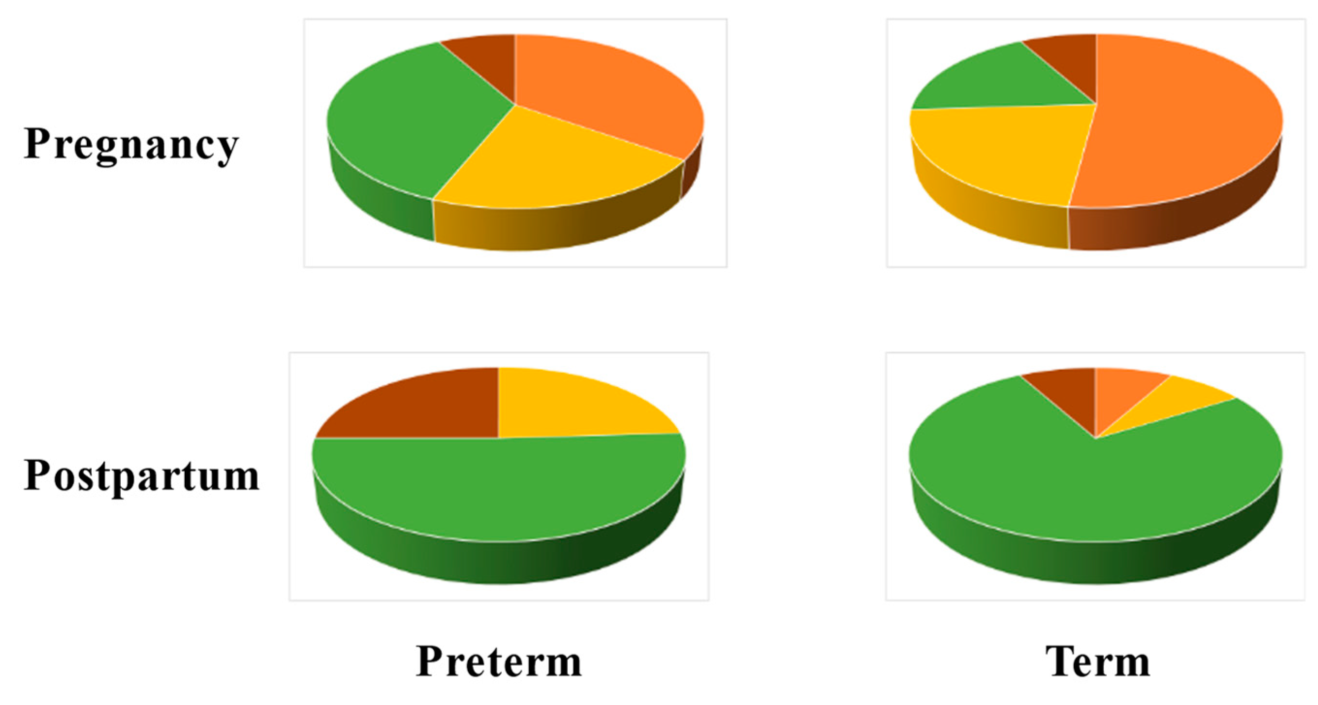
6. The Vaginal Microbiome during Pregnancy

During pregnancy, various physiological changes take place to adapt the mother’s body to the fetus and vice versa. This adaptation is governed by hormonal changes that lead to immune modulation, behavioural changes, physicochemical changes in the genital mucosa, and other adjustments in the genital tract. All these factors drive a modulation in the structure and function of the pregnant woman microbiome, making it different from that of non-pregnant females [104]. In addition to many other hormonal changes, pregnancy is characterised by high circulating oestrogen concentrations, mainly produced by the placenta, and increased glycogen deposition on the vaginal epithelium [136]. Such conditions favour the proliferation and dominance of Lactobacillus, which metabolises glycogen’s breakdown products to lactic acid [137]. As a result, during a healthy pregnancy, often, the vaginal microbiome has low bacterial species diversity.
Commonly, during the course of gestation, the vaginal microbiome of individual women with a healthy pregnancy undergoes a reduction in the diversity of microbial communities (alpha-diversity), but there are increases in microbial diversity between subjects (beta-diversity) and in the overall number of different taxa that result in a larger number of vagitypes being identified in pregnancies than in non-pregnancies. Also characteristic of pregnancy are shifts in vagitypes, discussed in more detail in the description of longitudinal studies. These changes are unrelated to those that arise from the pre-pregnancy body mass index, the development of gestational diabetes, or the number of previous births.
The prevalence of a specific vagitype stems from the stability of the microbial community, the lack of stability of other vagitypes, and the probabilities of switching among different dominant communities. Vagitypes dominated by lactobacilli, in particular, L. crispatus, are quite stable during pregnancy; others, for example, ones dominated by G. vaginalis, exhibit less stability [138]. Racial background plays an important role in the vaginal microbiota composition. For instance, the vaginal microbiota in non-pregnant or pregnant women of African ancestry includes commonly anaerobic bacteria such as G. vaginallis, A. vaginae, and S. amnii, and fewer Lactobacillus species. This occurrence increases the risk of bacterial vaginosis and PTB in women of African ancestry relative to women in other racial groups [137].
In addition to the known vaginal bacterial communities, there is a vaginal virome abundant in double-stranded DNA viruses, a lesser number of single-stranded DNA viruses, and a few unidentified viruses. Pathogenic viruses found in the vagina are taxa belonging to the order Herpesvirales and the family Papillomaviridae. Little information exists about the vaginal mycobiome [104].
Investigations of the vaginal microbiota in pregnant women can be classified as cross-sectional studies that provide snapshots at specific moments during pregnancy and longitudinal studies that address the temporal changes in this microbiome as pregnancy progresses. These studies have characterised normal pregnancies that finish at term, and/or abnormal pregnancies that end before a complete gestational period.

6.1. Cross-Sectional Studies

The vaginal microbiomes of pregnant women show similarities to those of non-pregnant women but with the following differences: (i) significant changes after delivery, and the inference that significant changes would have taken place too at the beginning of the pregnancy; (ii) stable populations throughout healthy pregnancies; and (iii) a varying number of vagitypes. Cross-sectional investigations highlighted the complexity and diversity of vaginal bacterial communities, indicating that there are racial differences in the vaginal microbiomes of women with healthy pregnancies. The findings of these studies also clarified that there are differences in the taxa abundance and diversity between healthy term pregnancies and those with adverse outcomes such as miscarriage, stillbirth, and preterm. However, many of these studies comprised a limited number of subjects, and yielded only an initial understanding of the vaginal microbiome during pregnancy.

6.1.1. Healthy Pregnancies

In pregnant women, the taxa more frequently observed and in greater abundance are Lactobacillus spp.; also present in the vagina with high frequency are the Actinomycetales, Clostridiales, and Bacteroidales taxa [139]. Lactobacillus spp. are abundant in the vaginal anaerobic habitat and create an environment hostile to many other bacteria. In particular, lactic acid keeps the vaginal pH below 4.5, thus establishing a defence against invading pathogens and contributing to a healthy vaginal environment. The most important role, as an antibacterial of the two L- and D-lactic acid forms, corresponds to the D-isomer from which hydrogen peroxide can be generated. Notably, the dominant Lactobacillus species play an important role in the protection of the vaginal ecosystem. The health and high stability of the vaginal community are enhanced by L. crispatus that produces D- and L-lactic acids [140]. L. iners does not generate D-lactic acid [141], and dysbiosis and lower stability are more commonly present in some racial backgrounds where the vaginal microbiota is dominated by this Lactobacillus.
The vaginal flora of 175 Korean pregnant women was characterised as normal, employing Nugent scores and several clinical symptoms. It showed that most of the women had vagitype I bacterial populations (n = 102), numerically in second place were women with vagitype III (n = 62), and present in lower numbers were vagitypes IV (n = 29), II (n = 5), and V (n = 4) [142].
To investigate the vaginal microbiome diversity that arises from racial background, the vaginal microflora of 300 Caucasian-, African-, or Hispanic-ancestry pregnant women were compared to those of 300 non-pregnant women case-matched for race, gestational age, and household income. The study found that pregnant women overall have a significantly higher prevalence of the four most common Lactobacillus vagitypes and a lower prevalence of vagitypes dominated by other taxa. Central to these differences was a higher prevalence of the L. iners vagitype during pregnancy at the expense of the G. vaginalis and other more complex vagitypes. Relative to non-pregnant women, a lower prevalence of G. vaginalis, A. vaginae, Prevotella cluster 2, P. bivia, and S. amnii was observed amongst African-American pregnant women, and a higher abundance of A. vaginae and P. bivia in Hispanic pregnant women. A lower prevalence of L. crispatus, accompanied by a higher prevalence of L. iners, was found in Caucasian pregnant women relative to non-pregnant women. In this group, the microbiome shifted towards a more stable, generally Lactobacillus-dominated profile; in women of African or Hispanic lineage, a major shift was observed in the early stages of pregnancy [138].
A study with 454 Chinese women who delivered at term or preterm identified several other factors that affected the composition of the vaginal microbiome, namely, hypertensive disorders, a history of abortion, the mode of delivery, and age. The relative abundance of more than 30 bacterial taxa varied between women with high blood pressure and controls with normal blood pressure resulting in a significantly higher alpha-diversity in the vaginal microbiome of the hypertensive group. High blood pressure did not reduce the relative abundances of Lactobacillus spp., but was accompanied by an increase in the proportion of Gardnerella, Atopobium, and Sneathia, and had an impact on maternal physiology that caused considerable changes in the diversity of bacterial species [143]. A difference in community structure was observed between pregnant women with and without a history of abortion. The vaginal microbiota of women in the latter group had significantly more similarity, suggesting that abortion might have increased the heterogeneity between individuals. Furthermore, the microbiomes of women who delivered vaginally had fewer microbial species postpartum and more clustered community structures than those who delivered by Caesarean section, indicating greater intragroup similarity. Lactobacillus was more abundant in the postpartum vagina of the pregnant women who delivered vaginally [143]. Maternal age was associated with variation in the postpartum vaginal microbiome. Samples collected from elder or younger women clustered into two distinct groups, with elder women showing a more compact community structure and the absence of L. iners and L. reuteri after delivery [143]. In addition, bacterial species during pregnancy differed significantly between women with PROM-related PTB and non-PROM-related PTB. The vagitype IVA was more prevalent during pregnancy in the PTB cases with PROM, suggesting that specific bacterial species could serve to distinguish between different types of PTB [143].
Many studies with varied populations have revealed Lactobacillus to be the dominant genus of the vaginal flora in most of the healthy, pregnant females [104]. Nonetheless, several factors affect, in various degrees, the vaginal microbiome in healthy pregnancies; of central importance are racial background and maternal age. The diverse vagitypes of this microbiota are governed by changes in the environment of the genital tract.

6.1.2. Pregnancies with Adverse Outcomes

Remarkable progress has been made in understanding the role of the vaginal microbiome in maintaining health and in preventing obstetric and gynaecological diseases, but further work is required to elucidate fully the contribution that a balanced vaginal microbiota makes to pregnancy. Cross-sectional investigations of the vaginal microbiome during pregnancy have served to discover relationships between taxa and the adverse pregnancy outcomes of miscarriage (a child born dead before 22 weeks of pregnancy), stillbirth (several criteria are employed—commonly, a child born dead at 22 weeks of pregnancy or later), and PTB (a live birth that occurs before 37 completed weeks of pregnancy).

Miscarriage

Miscarriage is an adverse obstetric outcome associated with a large number of pregnancy losses [144]. Statistics on the rate of miscarriage are difficult to obtain, but it is estimated that up to 10% of clinically recognised pregnancies end in miscarriage. A retrospective analysis was conducted on data from 24,835 pregnant women divided into two groups according to whether they had a first trimester threatened miscarriage or not. The women with the risk of miscarriage were older, and with higher rates of assisted reproduction and nulliparity. In pregnancies of this group of women, hyperemesis gravidarum, gestational diabetes mellitus, and placenta previa were more frequent than in the control group, and these women had lower gestational age and child birth weight [145]. Miscarriage is associated with a variety of conditions including embryo genetic and epigenetic disorders, immunological and endocrine factors, uterine malformations, maternal age, and lifestyle.
The abnormal microbiota composition of the female reproductive system was investigated as a potential cause of miscarriage in euploid and aneuploid pregnancies [146]. A study that included 10 women who suffered spontaneous first trimester miscarriage found that it was associated with an abnormal vaginal microbiome. Higher levels than normal of inosine, fumarate, xanthine, benzoate, and ascorbate were measured in the vaginal environment. The increased concentration of specific metabolites could be linked to changes in the microbial composition: for example, fumarate and ethanolamine are recognised as BV-associated metabolites. In euploid miscarriages, Lactobacillus spp.-depleted vaginal microbiota was associated with higher concentrations of pro-inflammatory cytokine IL-1beta, IL-6, and IL-18 compared to a viable term pregnancy [147].
To understand the relationship between miscarriage and vaginal bacterial populations, the effects of changes in this microbiota during the first trimester of pregnancy on the risk of miscarriage in the second trimester were analysed in a project including 418 women of African-American (65%), Hispanic (27%), and Caucasian (4%) racial backgrounds, of whom 74 experienced an early pregnancy loss. This group had a significantly higher relative abundance of the Clostridiales BVAB 3 taxon. The study also found a significantly decreased risk of miscarriage among women with higher abundances of Leptotrichia/Sneathia species or Megasphaera phylotype 1-like species early in the pregnancy [148].
The vaginal microbiota of women who miscarried correlated with a reduction in the number of Lactobacillus spp. in the first or second trimester of pregnancy. The risk of pregnancy loss in the second trimester among women with confirmed BV diagnosed in the first trimester increased but was not statistically significant. However, women with the most severe BV presentations in the vaginal microbiota had a twofold increase in the risk of pregnancy loss in the second trimester compared to women with normal vaginal microbiota [144]. Similarly, a comparison of the vaginal microbiomes of 25 women with normal pregnancies and 25 women with embryonic miscarriages showed that 80% of the latter group had a significantly lower abundance of Lactobacillus that was predominant in the healthy vaginas. The presence of Gardnerella and Prevotella, as well as Megatrobila and Cyclospora, increased the risk of miscarriage [149].
Recurrent miscarriage (RM) is defined as three or more consecutive pregnancy losses [150]. Vaginal infections have been associated with RM, but the pathophysiology is poorly understood. The innate immune system has various mechanisms for interacting with pathogens to protect vaginal tissues and to allow the survival of the commensal microbiota. In pregnancy, an organised immune response is essential for implantation, placentation, and blood vessel transformation, which an active infection may disturb, resulting in miscarriage. The mucosal epithelium and neutrophils in the vagina provide a first line of defence against micro-organisms, and, by secreting cytokines and chemokines, they attract innate immune cells such as macrophages, dendritic cells, and natural killer (NK) cells [151].
In particular, the presence of G. vaginalis in the vagina of women with RM correlated with higher levels of peripheral natural killer (pNK) cells. In 248 RM patients, the prevalence of G. vaginalis was 19.0%, Gram-negative anaerobes 20.5%, Candida species 7.9%, S. agalactiae 11.0%, and Enterobacteriaceae 14.8%,; commensal lactobacilli were absent in 14.5% of these women. In this investigation too, women with elevated pNK cells had a significantly higher prevalence of G. vaginalis and Gram-negative anaerobes than patients with normal pNK levels [152]. Another study comparing the microbial communities of 10 Chinese women who experienced unexplained RM and 10 women with healthy pregnancies found a lower microbial alpha-diversity and richness in the samples of women with miscarriage. Further, principal component and beta-diversity analyses indicated that the bacterial community structures in both groups were different. Although lactobacilli were present in both groups, Atopobium, Prevotella, and Streptococcus were more abundant in women with RM, and Lactobacillus and Gardnerella were more abundant in the healthy control group. These results showed that associated with miscarriage was a quantitative and qualitative shift from commonly occurring lactobacilli in a balanced vaginal microflora to a mixed microbiota dominated by anaerobic bacteria [153].
A prospective observational study characterised the vaginal microbiomes of 24 women in the first trimester of pregnancy. There were significant differences between the microbiomes of women who suffered miscarriage and those who continued to term delivery, both in the overall microbiome populations and in the abundances of individual taxa (Figure 1) [154].
L. crispatus, L. gasseri, and Bifidobacerium were more abundant in women who delivered at term, whereas L. iners, L. jansenii, Gardnerella, Prevotella, and Escherichia/Shigella were found at higher relative abundances in women who suffered miscarriage [154]. Regarding pregnancy history, L. crispatus was the dominant bacterium in 50% of nulliparous women and L. iners was the dominant bacterium in 50% in women with a history of prior miscarriage and who had a miscarriage in the study, compared to 15% in women with no history of miscarriage, supporting the conclusion that the composition of the vaginal microbiome varies with pregnancy history. Lactobacillus spp. in the vaginal microbiome had a positive correlation with fertilisation success, implantation, and early embryonic development [155]. Notwithstanding the overall positive effects of Lactobacillus spp. on pregnancy and the known interactions between these bacteria and the components of the immune system located in the vagina, more detail remains to be known [146]. Lactic acid produced by lactobacilli shows immunomodulatory properties by inducing an anti-inflammatory response in vaginal and cervical epithelial cells [156] and, together with hydrogen peroxide, can lower the levels of pro-inflammatory cytokines, thus positively affecting fertility and pregnancy maintenance. In contrast, lipopolyssacharides from Gram-negative bacteria are potent immunostimulators that can activate peripheral natural killer cells with negative effects on reproduction [152].
In response to pathogens, neutrophil and macrophages produce extracellular traps composed of DNA strands, histones, elastase, and various peptides and enzymes that act as mechanisms to fight invading bacteria, but, if produced in excess, they can induce autoimmune and coagulation disorders and metastasis [157], and cause damage to the placenta and fetal membranes [158]. In women who had a miscarriage, elements of extracellular networks were found in the placenta, suggesting a relationship with this adverse outcome [159]. Whether Lactobacillus spp. can inhibit the formation of extracellular traps remains to be established, but, if demonstrated experimentally, it will provide evidence of new immunomodulatory properties of these bacteria in pregnancy.
The way in which a balanced vaginal microbiome helps prevent gynaecological diseases in women and maintain health remains to be fully elucidated, but an abnormal vaginal microbial composition appears to be one of the factors that increase the risk of miscarriage. Lactobacillus spp. are the most common bacteria within the healthy reproductive tract. Microbiological tests before conception and in early pregnancy to determine the vaginal microbial composition may serve to understand the mechanisms that promote successful embryo implantation and placenta formation, and reduce the incidence of miscarriage [146]. Future research should focus on determining whether the presence of Lactobacilli significantly prevents pregnancy loss in all racial backgrounds and the protection mechanism that the presence of these bacteria affords pregnancies.

Stillbirth

In 2021, an estimated 2 million babies were stillborn, with a global rate of 13.9 stillbirths per 1000 total births [5], with large regional variations from 22.8 stillbirths per 1000 total births in West and Central Africa to 2.9 per 1000 in Western Europe [160]. Common causes of stillbirth include infections, birth defects, and pregnancy complications like pre-eclampsia. In more than 1 of every 10 stillbirths, the death was likely caused either by an infection in the fetus or in the placenta, or by a serious microbial disease in the mother. Pathogens were a more common cause of death in stillbirths before week 24 than later in the pregnancy. In the United States, stillbirths are more than twice as likely among African-American women than among Caucasian women, and it is an important factor in the former group [161].
The rate of pregnancy loss varies with the gestational age at the moment of infection and by the specific invading pathogen. For example, infection with Treponema pallidum causes pregnancy loss or fetal death in up to 50% of cases, whereas parvovirus B19 (PVB 19) infection causes pregnancy loss or stillbirth in less than 3% of cases. The mechanisms of pregnancy loss owing to pathogens that traverse the maternal–fetal barrier and cause congenital disease in the fetus are known. Infections by Toxoplasma gondii, rubella virus, cytomegalovirus, or HSV (TORCH) can be pathogen-mediated, placenta-mediated, and/or through inflammation-induced previable delivery [162]. Other bacteria linked to stillbirth include S. agalactiae, E. coli, Haemophilus influenza, and species of the genera Klebsiella, Enterococcus, Chlamydia, Mycoplasma, and Ureaplasma [160,163].
Group B Streptococcus infection is an important, potentially preventable, cause of worldwide burden of stillbirths, accounting for around 1% of cases in developed countries and 4% in Sub-Saharan Africa. Infection of the infant starting before delivery likely proceeds from bacteria ascending in utero from the maternal genitourinary tract. Evidence has been obtained from whole-genome sequencing studies that demonstrated GBS isolated at birth from the skin of newborns delivered by Caesarean section to be identical to those colonising the mother. Moreover, GBS isolated from postmortem stillbirth blood cultures were genetically identical to maternal GBS isolates [164].
A secondary analysis of 512 stillbirths in a prospective, multisite, geographically, racially, and ethnically diverse population in the United States identified infection as a possible or probable cause of death in 66 cases. In the study, infection-related stillbirths occurred earlier than non-infection-related stillbirth. The most common bacterial pathogens identified were E. coli, S. agalactiae, and Enterococcus spp., and the most common viral pathogen was cytomegalovirus (CMV) [165]. A study from a tertiary care centre in Mexico considered placental and/or fetal infection as probable cause of death in 15.1% of stillbirths. The pathogens identified were E. coli, Staphylococcus warnerii, Enterobacter spp., Klebsiella pneumoniae, Listeria monocytogenes, M. tuberculosis, C. albicans, Candida krusei, CMV, and Zika virus [166].
Viruses known to cause fetus damage during infection, and, thus, potentially associated with stillbirth, are rubella, measles, CMV, varicella zoster, HSV, PVB19, and the human HIV [167,168]. Term placentas from congenital CMV infection showed pathologic findings that suggested the viral infection contributed to intrauterine growth restriction; in addition, CMV has been identified in several tissues of fetus with intrauterine death [169].
Another virus known to impact perinatal outcomes is SARS-CoV-2, and fetal death was associated with its intrauterine transmission. Placenta and umbilical cord blood tested positive for the virus, confirming transplacental transmission [170]. An international study covering 52 million births in 26 countries investigated the impact of pandemic-related lockdowns on stillbirth. It found evidence for: (i) an increased risk of stillbirth in the first month of lockdown in high-income countries and in Brazil, and (ii) an association between a lockdown and stillbirth in the second, third, and fourth months of lockdown. Although SARS-CoV-2 infection increases the risk of stillbirth, the increase in stillbirth with a lockdown in some countries might reflect reduced access to timely quality antenatal and intrapartum care rather than other factors, since only a small fraction of pregnant women experienced SARS-CoV-2 infection at the early stage of the pandemic [171].

Preterm Birth

The length of gestation is considered a key indicator of child health, and PTB is associated with poorer health outcomes in infants who have increased risks of short-term complications, mainly owing to the immaturity of multiple organ systems and neurodevelopmental disorders. Yearly, approximately 15 million infants are born preterm worldwide at a rate of 10.6%, with significant regional variations from 8.7% to 13.4% [172], with a higher prevalence in low- and middle-income countries [173]. Prematurity is the cause of about 35% of newborn deaths, and of 18% of children younger than 5 years old. Reducing PTB is part of the United Nations Sustainable Development Goal 3, target 3.2, which endeavours to end all preventable deaths of newborns and children aged under 5 years by 2030 [173].
Spontaneous PTB is associated with multiple, complex, and not completely understood causes of disease, but a leading cause of PTB is infection resulting from the microbial invasion of the amniotic cavity. A characteristic of acute chorioamnionitis is neutrophilic infiltration at the maternal–fetal interface, with ensuing inflammation. Commonly, acute chorioamnionitis is a result of ascending infection and recent studies suggest a link between vaginal dysbiosis, vaginal inflammation, and ascending infection. Less commonly, microbes can invade the maternal–fetal interface via the haematogenous route, e.g., Zika virus, CMV, and L. monocytogenes, where they can cause placental villitis and severe fetal inflammation and injury [174]. Although some studies found preterm labour to be associated with sterile intra-amniotic inflammation or infection [175], this does not preclude the involvement of the microbiota in the vagina modulating systemic host responses that could trigger an inflammatory response in the amniotic cavity leading to PTB. Studies linking the vaginal microbiota to PTB have yielded mixed results, exposing the need for further investigations [143].
A high proportion of PTB are preceded by PPROM that provide microbes an entry to the amniotic space and may contribute to maternal and neonate morbidities, including sepsis [176]. A multicentre cohort study that included 78 women who suffered PPROM found in the vaginal microbiota a depletion of the Lactobacillus spp. relative abundance and a relative enrichment of Ureaplasma parvum. These changes were accompanied by increases in bacterial diversity, evenness, and richness. It was determined that the presence of the Escherichia/Shigella and Facklamia taxa in the vaginal microbiota at birth were risk factors for early onset neonatal sepsis (EONS), and Anaerococcus obesiensis and Campylobacter ureolyticus taxa have protective effects against sepsis. The study concluded that analyses of the vaginal and neonatal microbiota after PPROM could be employed for a risk assessment of EONS [177].
A prospective study with 464 Caucasian-American women and 360 African-American women analysed the association between the vaginal microbiome at midpregnancy, race, and spontaneous PTB [178]. In the Caucasian-American cohort, 375 women delivered at term and 89 preterm, and, in the African-American cohort, 276 women delivered at term and 84 preterm. The vaginal microbiomes of both cohorts were significantly different; the African-American women had higher microbial diversity, a greater abundance of L. iners, and a lower abundance of L. crispatus. The vaginal microbiomes of both groups of women were significantly associated with race, PTB, and maternal factors such as poverty, education, marital status, age, and douching. A higher L. crispatus abundance in term controls was the main difference with the microbiota of women who delivered PTB. In L. crispatus-dominated microbiomes, diversity was significantly lower than in L. iners-dominated ones, suggesting that the former species is better at suppressing bacterial vaginosis-like microbiomes [178]. This result is consistent with the observation that L. iners-dominated vagitypes shift more often toward a diverse bacterial community structure than those where L. crispatus is predominant [82]. Moreover, it was found that the risk of PTB in vagitypes dominated by L. gasseri and L. jensenii was higher than those of L. crispatus-dominated vagitypes, indicating that the former species are not as protective as the latter [178].
The vagitypes and taxa abundance were characterised in 8–14-week-gestation vaginal samples from African-American women whose pregnancy resulted in full-term birth (n = 231), early-term birth (n = 84), or PTB (n = 44). Close to half of the women had vagitype IV, one-third had vagitype III, and 16% had vagitypes I, II, or V. Vagitypes III and IV were associated with PTB with an adjusted odds ratio of 4.1 and 7.7, respectively, compared to vagitypes I, II, or V. In contrast, no vagitype was associated with early-term birth [179].
Samples were obtained at each pregnancy trimester from a cohort of women of African descent comprising 45 women who delivered through spontaneous preterm, and 90 case-matched women who gave birth at term. The dominant bacterial taxon in samples was used to stratify the community states of the vaginal microbiomes into vagitypes, and, according to this criterion, 13 vagitypes were identified. The ten most frequent vagitypes in each group of women are given in Table 4 [74].
The diversity of taxa measured by the Shannon index or the inverse Simpson index was greater in samples from women who experienced premature delivery, as reported in other studies with women of various racial backgrounds [74]. L. iners was predominant in women who delivered at term or preterm, with increasing abundance in successive trimesters. Next in relative abundance were L. crispatus in the term group and L. jensenii in the preterm group. Other taxa more abundant in the preterm group than in the term group were Lachnospiraceae BVAB1, Prevotella cluster 2, S. amnii, Dialister cluster 51; P. amnii, Clostridiales BVAB2; Coriobacteriaceae, Dialister micraerophilus, and Parvimonas. A model constructed to predict the risk of PTB showed high associations with S. amnii and Lachnospiraceae BVAB1 infections [74].
Bacterial taxa commonly associated with infection and preterm birth are G. vaginalis, A. vaginae, P. bivia, U. urealyticum, M. hominis, and S. agalactiae. Polymicrobial biofilms that form on the vaginal epithelium have a central role in bacterial vaginosis. G. vaginalis is a primary coloniser that can establish a scaffold for the attachment of other microbes associated with infection such as A. vaginae and P. bivia, thus enabling the development of polymicrobial biofilms [119]. Gardnerellla biofilms are found also in the endometrium and fallopian tubes, indicating that it is able to migrate to the upper genital tract and lead to adverse pregnancy outcomes [180]. G. vaginalis taxa with various potentials to form biofilms were identified in 155 samples collected from Iraqi women who experienced various forms of PTB [181]. The strict anaerobe A. vaginae is a second important coloniser of polymicrobial biofilms often found in bacterial vaginosis [182]. Supporting these results was a study with Korean pregnant women describing the abnormal microbiota of 37 women; the most abundant taxa in this cohort were L. iners, Gardnerella, Prevotella, and Atopobium [142].
A study examined whether vaginal U. urealyticum/M. hominis colonisation was predictive of PTB in patients exhibiting signs of threatened PTB or in those with an asymptomatic short cervix. The median gestational age of the 94 enrolled patients was 29 weeks and 6 days, and 54 (57%) of the patients were vaginal U. urealyticum/M. hominis-positive. The PTB rate in the positive group was higher than in the negative group. Vaginal Ureaplasma/Mycoplasma positivity was found an independent risk factor for PTB in patients with symptomatic threatened preterm labour and/or a short cervix [183].
The effect of Ureaplasma or Mycoplasma infection in the vagina of pregnant women on the duration of pregnancy was investigated in a cohort 200 Northern Iranian women. Half of the women delivered at term and the other half delivered preterm. Genital Mycoplasma was detected in 78 cases (39%) and colonisation rates were 60% and 18% in the samples from preterm and term pregnancies, respectively. The findings suggested a significant association between the presence of genital mycoplasmas and the risk of preterm labour [184].

Other Adverse Outcomes

The most frequent disease outcomes of S. agalactiae colonisation during pregnancy are infant meningitis or sepsis, which are accompanied by high mortality risk. Other infrequent outcomes include stillbirths, maternal infections, and prematurity; however, given the wide prevalence of this genital infection, the potential annual burden of GBS-associated PTB has been estimated for the first time at 50,000 births, with a wide uncertainty range [185].
A pre-pregnancy health examination program included 89 women whose pregnancy outcomes were followed up for 1 year. Vaginal swabs were collected, 16S rRNA genes were sequenced, and M. hominis colonisation was confirmed by qPCR. Cox models were used to estimate the fecundability odds ratio (FOR) for women with M. hominis [186]. The prevalence of M. hominis was 22.47% (20/89) with a relatively low abundance. The Simpson index of the Mycoplasma-positive group was significantly lower than that of the negative group (p  =  0.003), suggesting that microbiome diversity appeared to increase with M. hominis positivity. The relative abundance of M. hominis was negatively correlated with L. crispatus (p  =  0.024), but positively correlated with G. vaginalis, A. vaginae, and P. bivia (p <  0.05 for all). The cumulative one-year pregnancy rate for the Mycoplamas-positive group was lower than that in the negative group (58.96% vs. 66.76%, p  =  0.029). After controlling for potential confounders, the risk of pregnancy in the positive group was reduced by 38% when compared with the negative group [186].

6.2. Longitudinal Studies

Longitudinal studies of the vaginal microbiome examine the abundance of the microbial taxa and their change throughout pregnancy in the context of a dynamic environment. Childbearing may be considered a pro-inflammatory condition in which the vaginal microbiota might possess immunomodulation properties based on the modification of bacterial species; in particular, lactobacilli seem to play a key role in this process [137]. The vaginal bacterial composition of pregnant women exhibits considerable convergence across different populations, becoming less rich and diverse compared to the vaginal bacterial populations of non-pregnant women. During pregnancy, Lactobacillus taxa become the predominant bacteria in the vagina in most women of various racial backgrounds, leading to a decrease in alpha-diversity [104].
The vaginal ecosystem of 64 Caucasian women with normal pregnancies was characterised in the first, second, and third trimester. Advancing in the pregnancy, there was a significant decrease in cases of bacterial vaginosis and an increase in cases with normal microbiota. The relative abundance of lactobacilli increased from 50% in the first trimester, to 73.4% in the second trimester, and to 79.7% in the third trimester. This shift was associated with marked changes in the vaginal metabolome: several metabolites, such as lactate, glycine, phenylalanine, leucine, and isoleucine, ordinarily found in healthy vaginas, reached their highest concentrations at the end of pregnancy. Concomitantly, the abundance of microbiota present in bacterial vaginosis decreased throughout the pregnancy, and there was a progressive reduction in the levels of metabolites such as biogenic amines, alcohols, propionate, and acetate associated with dysbiosis. The levels of cytokines IL-6 and IL-8 were positively correlated, and their lowest concentrations were measured in the second trimester. A total of 19 women had a Candida infection, with 10 cases throughout the pregnancy; associated with this infection were higher levels of IL-8, 4-hydroxyphenyllactate, choline, and O- acetylcholine, as well as a higher concentration of leukocytes [147].
The vaginal microbiome throughout pregnancy was assessed in a longitudinal study of 12 healthy Chinese women with uncomplicated singleton pregnancies and no medical problems or adverse outcomes during any previous pregnancy who delivered at term (38–42 weeks). The vaginal microbial community was studied at pre-pregnancy, 8–12, 24–28, and 37–38 weeks of gestation, and puerperium. L. crispatus, L. gasseri, L. jensenii, and L. johnsonii were abundant before and during pregnancy, but, post-delivery, the vaginal microflora increased in diversity. L. crispatus comprised over 40% and L. iners about 30% of the total lactobacilli population in the vaginal microflora before or during pregnancy. The pre-pregnancy abundance of L. delbrueckii was 0.8% and had an average abundance of 4.5% during the complete pregnancy period. Post-pregnancy, L. delbrueckii and L. jensenii with abundances of 41.8% and 24.0%, respectively, dominated over L. crispatus and L iners, whose abundances decreased to 18.1% and 2%, respectively. Post-delivery, Lactobacillus was largely replaced by Bacillus, Phyllobacterium, Prevotella, Pseudomonas, and Streptococcus. These changes may be the result of fluctuations in oestrogen levels, stress of delivery, involution, lochia, etc. [187].
A prospective study with weekly sampling of the vaginal microbiome was conducted with 40 pregnant women, of which 11 delivered preterm. The vagitypes identified corresponded well with the five described for non-pregnant women. The taxonomic composition and diversity of the microbiota was generally stable in each individual with some vagitype transitions during the pregnancy; the Lactobacillus-dominated vagitypes I, II, III, and V were more stable than those of vagitype IV. Pregnancies with vagitype IV exhibited a stronger association with PTB, at every time window during gestation. G. vaginalis was strongly associated with PTB; a high Ureaplasma abundance combined with a low abundance of Lactobacillus was associated with PTB as well. Twenty-five women provided a postpartum sample; their microbiota indicated that delivery generally was accompanied by a significant, sudden, and durable increase in bacterial community diversity, albeit not in all cases [188].
In 90 pregnant women of African or non-African ancestry who delivered at term, there was a transition of the vaginal microbiome beginning early in the pregnancy towards Lactobacillus-dominated profiles at the expense of taxa often associated with vaginal dysbiosis. The changes were minimal in women of European ancestry, and larger in women of African and Hispanic ancestry. In the latter group, the main change was to a higher prevalence of the L. iners-dominated vagitype at the expense of G. vaginalis and other more complex vagitypes. Hispanic women experienced a lesser increase in the four Lactobacillus vagitypes. These observations suggested that, in pregnancy, physiological or environmental changes in the vagina favour Lactobacillus taxa and population shifts to a less complex microbiota. The changes occurred early in pregnancy, followed predictable patterns, were associated with a simplification of the metabolic capacity of the microbiome, and were significant only in women of African or Hispanic ancestry [74].
The vaginal samples from 48 pregnant women (Caucasian 56.35%, Asian 12.5%, Hispanic 4.2%, and African-American 2.1%) collected prospectively during the first trimester, third trimester, and postpartum were assessed by measuring the vaginal fluids and epithelial cells [189]. Linear mixed-effects models were employed to investigate alterations in secretions involved in host physiology and immunity that would accompany changes in the vaginal microbiome. Elevated levels of hyaluronate and heatshock protein 70 and decreased concentrations of the D- and L-lactic acid isomers were measured, indicating an increase in the concentrations of metabolites associated with physiological stress or degradation of the extracellular matrix, and a decrease in the levels of metabolites produced chiefly by lactobacilli. The bacterial communities in the first and third trimesters tended to be very similar, whereas the vaginal microbiome was more diverse during the postpartum period, consistent with previous findings. Amongst the ten more abundant taxa during the first trimester were L. crispatus (49%), L. jensenii (16.2%), L. gasseri (10.2%), and L. iners (2.7%). Postpartum, there was a lower relative abundance of L. crispatus (6.7%), a higher abundance of L. iners (11%), and negligible abundances of L. jensenii or L. gasseri. Moreover, the relative abundances of Streptococcus anginosus (a facultative anaerobe involved in aerobic vaginitis), P. bivia, and G. vaginalis were significantly higher postpartum. Most of the vagitype transitions that occurred between the third trimester and postpartum were from communities dominated by Lactobacillus spp. to vagitypes that were not. During the postpartum transition, an increase in alpha-diversity accompanied the overall decrease in Lactobacillus spp. with a concomitant increase in the diversity of taxa including Peptoniphilus, Anaerococcus, Bacteroidia, Prevotella, Veillonella, Porphyromonas, and Megasphaera. Notably, there were no transitions in the opposite direction. The metabolite changes post-delivery profoundly altered the host habitat and created an environment that contributed to the survival and proliferation of diverse bacterial communities in the vagina [189].
A longitudinal study of 474 women primarily of African-American background examined the associations between maternal age and parity, and the composition of the vaginal microbiota throughout gestation that led to term delivery [190]. The contributions to the variance in the composition and structure of the vaginal microbiota by age and parity were small: 0.2–1.4% and 0.3–1.9%, respectively. The changes in the overall structure of the vaginal microbiota likely reflected physiological alterations in the vaginal microenvironment throughout gestation that favoured the growth of some bacterial taxa, e.g., Lactobacillus spp. Seven vagitypes were identified, four corresponding to Lactobacillus-dominated I, II, III, and V, and three different ones corresponding to vagitype IV. The latter all included L. iners, G. vaginalis, and Megasphaera spp., with candidatus Lachnocurva vaginae, A. vaginae, and Bifidobacterium spp. being relatively more abundant in vagitypes IVA, IVB, and IVC, respectively. The abundance of vagitypes I, III, and V generally increased with advancing gestational age and the abundance of vagitypes IVA and IVB declined steadily as term approached. In agreement with other reports, the richness and evenness of the vaginal microbiota decreased with gestational age from the first to the third trimester, at variance with observations with Caucasian cohorts where measures of alpha-diversity are generally low and consistent throughout gestation [190].
Parity was positively correlated with the richness and evenness of the bacterial populations, suggesting that the reduction in these microbiota characteristics during pregnancy was mitigated by parity. The explanation offered for this observation is that the increase in diversity that takes place after birth accumulates across pregnancies, similar to the imprinted regulatory memory of human NK-cells in the immunity of the uterine mucosae [191]. Parity was also associated with a decrease in vagitype III, and high maternal parity with an increase in vagitype IV bacteria such as Gardnerella, Megasphaera, Prevotella, and Sneathia, and lower maternal parity with an increase in Lactobacillus [191].
The potential association of socioeconomic status with the longitudinal stability of the vaginal microbiota from early (8–14 weeks) to later (24–30 weeks) gestational age was investigated in 110 African-American women. It was found that a low level of education (high school or less) was associated with an increase in vaginal microbiome diversity during pregnancy, suggesting that variables linked to socioeconomic status should be considered when assessing associations between the microbiota composition and health outcomes [175].
An exploratory longitudinal investigation of the vaginal microbiota composition of eight Mexican women with healthy pregnancies collected samples in the third trimester of pregnancy and, subsequently, at childbirth at term. The vaginal microbiota was dominated by the Lactobacillus genus at both time points. There were no statistically significant differences in relative abundances, absolute read count, bacterial richness, community diversity, evenness, and beta-diversity between the third trimester of pregnancy and the time of childbirth. Nonetheless, compared to the third trimester of pregnancy, a trend was observed of higher absolute read counts for the genera Gardnerella, Faecalibaculum, Ileibacterium, and Lactococcus, and lower absolute read counts of Lactobacillus spp. at childbirth, but these changes in absolute read counts did not result in significant statistical differences between the microbial populations at both times. The results suggest that the vaginal bacterial composition is stable, and Lactobacillus genus is the dominant taxa in Mexican women’s vagina at the third trimester of pregnancy and at childbirth [137].
Analyses of vaginal samples from a cohort of Malawi women collected between 5 and 583 days post-delivery demonstrated that Lactobacillus spp. were present in less than a third of the women. Postpartum vaginal communities resembled those of vagitypes III and IV. Also the abundances of L. iners increased with time throughout the postpartum stage [192]. In comparison to other Lactobacillus-dominated communities, those dominated by L. iners tend to be more diverse and less stable as they often transition to communities resembling vagitype IV [82]. The observed shift in the bacterial community composition during the postpartum period can occur as early as the onset of labour and may persist for up to 1 year [193].
Vagitypes I, II, IVA, and IVB were identified in a study that characterised the vaginal microbiota of 356 Chinese women sampled during the first and third trimesters of pregnancy and of 98 Chinese women sampled in a six-week postpartum period. No significant changes in overall community structures or alpha- and beta-diversity were observed during pregnancy. The relative abundances of vagitypes I, II, IVA, and IVB in the women who delivered at term were 52%, 22%, 18%, and 8%, respectively. In women who delivered preterm, the relative abundances for the same vagitypes were 35%, 21%, 36%, and 8%, respectively. Postpartum, the relative abundances of vagitypes I, II, IVA, and IVB for women who delivered at term were 8%, 8%, 76%, and 8%, respectively, and, for women who delivered preterm, they were 0%, 24%, 51% and 25%, respectively (Figure 2). Of note was the postpartum loss of Lactobacillus spp. and the increase in the abundance of anaerobic bacteria. During pregnancy, the vaginal samples were more closely clustered than the postpartum samples. Both the stability of the vagitypes during pregnancy and their shifts postpartum that indicated significant increases in diversity were similar to those found for other racial groups. The alpha-diversity of the vaginal microbiome was higher in pregnant women with high blood pressure. Hypertensive disorder was associated with relative abundance increases in the genera Gardnerella, Atopobium, and Sneathia, but not with a reduction in Lactobacillus abundance [143].
A prospective case–control study described alterations in the composition of vaginal microbiota in pregnant women with a SARS-CoV-2 (COVID-19) active infection or who were infected within a month. Vaginal swabs were collected from uninfected pregnant women (n  =  28) and pregnant women with COVID-19 (n  =  19). Aside from the viral infection, both groups had similar maternal demographic and clinical characteristics. There were no significant differences in the alpha-diversity of the vaginal microbiota between the second and third trimesters in uninfected women. The relative abundances of L. crispatus, L. iners, L. gasseri, and L. jensenii were lower in the COVID-19 group than in uninfected pregnant women. In women infected with COVID-19 disease, L. iners decreased the most among Lactobacillus spp., and the relative abundances of G. vaginalis, M. hominis, and Ureaplasma spp. increased. The amount of Ureaplasma spp. was higher in women with moderate/severe disease than those of asymptomatic/mild disease. These findings indicated that the composition of vaginal microbiota was unfavourably affected by COVID-19 disease and there is a prominent dysbiosis during active COVID-19 infection [194].

7. Conclusions

In healthy pregnancies, the bacterial communities in the vagina have a relatively lower number of taxa than in non-pregnant women, and are dominated by a few species, namely, those of the genus Lactobacillus, although other taxa such as A. vaginae, G. vaginalis, and Prevotella spp. are also found in normal pregnancies. Interestingly, although the smaller number of taxa in individuals commonly results in a lower alpha-diversity, marked differences in vagitypes between individuals effect a higher beta-diversity during pregnancy. A relationship has been found between the vaginal microbiome and racial background; women of Hispanic or African ancestry harbour more anaerobic flora in their microbiota than women of Asian or Caucasian backgrounds who commonly have Lactobacillus spp. as the dominant taxa. Besides racial background, other factors that modulate the vaginal microbiota include maternal age, previous pregnancies, blood pressure, behavioural habits, and various environmental factors.
Epidemiological studies provide evidence that the urogenital microbiota is linked to obstetric diseases. A marker of pregnancy complications is the proliferation in the genital microbiota to the significant abundance or dominance of aerobes such as Acinetobacter baumannii, E. coli, E. faecalis, S. aureus, and S. agalactiae [195,196], or anaerobes like A. vaginae, Clostridiales BVAB 1-3, D. microaerophilus, G. vaginalis, M. hominis, P. timonensis, and U. urealyticum. The viral infections discussed in Section 2.1 are also an important contributor to adverse pregnancy outcomes [197]. These infections are risk factors for miscarriage [154], stillbirth [198], prematurity [178], and other maternal diseases including cervicovaginal cancers [199], as well as infant diseases [177].
Investigations on the changes induced in the vaginal bacterial populations by pregnancy in health and disease indicated that the vaginal microbiota is stable in the absence of infections. These studies confirmed that considerable changes in the vaginal community composition occur immediately following pregnancy, as well as postpartum.
The stability of the vaginal microbiome during a healthy pregnancy is such that alterations in the bacterial flora dominated by Lactobacillus spp. reflect the status of various obstetric conditions and are predictive biomarkers for certain pregnancy-adverse outcomes. Thus, it has been proposed that the composition of the vaginal microbiome may be a useful prognostic indicator of preterm labour and serve as a monitoring tool for tocolytic treatment to prevent PTB [200].
Nonetheless, much more needs to be learned about the pathogenicity and mechanisms of host defence to these micro-organisms. Future investigations will serve to elucidate the functional effect of the microbial communities and/or specific bacterial species on homeostasis and disease during pregnancy. A better understanding of the host–micro-organism interactions might reveal new opportunities for disease prevention, therapy, and improving women’s quality of life and overall health.
In addition, the complex gut microbiomes are involved in host immunity, metabolism, digestion, and the functioning of the nervous system, and are important for the health of the mother and child. During pregnancy, changes may occur naturally in the microbiomes of the oral cavity, intestine, and breast milk. Changes in the structure and composition of the gut microbiomes with an increase in the abundance of various genera of micro-organisms, e.g., Acinetobacter, Actinobacter, Klebsiella, Rothia, etc., and a decrease in others, e.g., Bacteroides, Bifidobacterium, Eubacterium, etc., can manifest in pregnancy complications such as gestational diabetes, gestational obesity, pre-eclampsia, diseases of the digestive tract, and autoimmune disease. The relationships between imbalances of the maternal gut microbiomes and their physiological effects during pregnancy are starting to be elucidated, but many more investigations are required to provide a comprehensive picture that would serve to foster maternal and offspring health [201].

Funding

A grant from the Cerebral Palsy Alliance (R00020) was employed to support, in part, this investigation.

Data Availability Statement

The data presented in this study are available in this article.

Conflicts of Interest

The author declares no conflict of interest.

References

  1. UN Millennium Development Goals. Available online: https://www.un.org/millenniumgoals/ (accessed on 15 July 2022).
  2. UN Department of Economic and Social Affairs. Transforming Our World: The 2030 Agenda for Sustainable Development UN General Assembly A/RES/70/1; UN: New York, NY, USA, 2015; Available online: https://sdgs.un.org/2030agenda (accessed on 15 July 2022).
  3. UN Inter-agency Group for Child Mortality Estimation. Levels & Trends in Child Mortality, Report 2021; UNICEF: New York, NY, USA, 2021; Available online: https://www.who.int/news-room/fact-sheets/detail/preterm-birth (accessed on 15 July 2022).
  4. Editorial: Miscarriage-Worldwide reform of care is needed. Lancet 2021, 397, 1507.
  5. UN Inter-Agency Group for Child Mortality Estimation. A Neglected Tragedy: The Global Burden of Stillbirth Report 2020; UNICEF: New York, NY, USA, 2020; Available online: https://www.unicef.org/media/84851/file/UN-IGME-the-global-burden-of-stillbirths-2020.pdf (accessed on 30 July 2022).
  6. Walani, S.R. Global burden of preterm birth. Int. J. Gynecol. Obstet. 2020, 150, 31–33. [Google Scholar] [CrossRef] [PubMed]
  7. Kaforau, L.S.K.; Tessema, G.A.; Jancey, J.; Dhamrait, G.; Bugoro, H.; Pereira, G. Prevalence and risk factors of adverse birth outcomes in the Pacific Island Region: A scoping review. Lancet Reg. Health-West. Pac. 2022, 21, 100402. [Google Scholar] [CrossRef] [PubMed]
  8. Baskaradoss, J.K.; Geevarghese, A.; Al Dosari, A.A.F. Causes of adverse pregnancy outcomes and the role of maternal periodontal status. Open Dent. J. 2012, 6, 79–84. [Google Scholar] [CrossRef] [PubMed]
  9. Hussein, J.; Ugwumadu, A.; Witkin, S.S. Editor’s Choice. Br. J. Obst. Gynaecol. 2011, 118, i–ii. [Google Scholar] [CrossRef]
  10. Zeng, M.; Yang, L.; Mao, Y.; Yang He, Y.; Li, M.; Liu, J.; Zhu, Q.; Chen, L.; Zhou, W. Preconception reproductive tract infections status and adverse pregnancy outcomes: A population-based retrospective cohort study. BMC Pregnancy Childbirth 2022, 22, 501. [Google Scholar] [CrossRef] [PubMed]
  11. National Institute of Child Health and Human Development. What Infections Can Affect Pregnancy? Available online: https://www.nichd.nih.gov/health/topics/pregnancy/conditioninfo/infections# (accessed on 15 July 2022).
  12. Pomar, L.; Baud, D. Emerging virus infections in adverse pregnancy outcomes. Viruses 2022, 14, 285. [Google Scholar] [CrossRef]
  13. Chaudhry, S.A.; Koren, G. Hepatitis A infection during pregnancy. Can. Fam. Physician 2015, 61, 963–964. [Google Scholar]
  14. Sirilert, S.; Tongsong, T. Hepatitis B virus infection in pregnancy: Immunological response, natural course and pregnancy outcomes. J. Clin. Med. 2021, 10, 2926. [Google Scholar] [CrossRef]
  15. Wu, C.; Wu, X.; Xia, J. Hepatitis E virus infection during pregnancy. Virol. J. 2020, 17, 73. [Google Scholar] [CrossRef]
  16. Wang, R.; Yan, W.; Du, M.; Tao, L.; Liu, J. The effect of influenza virus infection on pregnancy outcomes: A systematic review and meta-analysis of cohort studies. Int. J. Infect. Dis. 2021, 105, 567–578. [Google Scholar] [CrossRef] [PubMed]
  17. Ragusa, R.; Platania, A.; Cuccia, M.; Zappalà, G.; Giorgianni, G.; D’Agati, P.; Bellia, M.A.; Marranzano, M. Measles and pregnancy: Immunity and immunization-What can be learned from observing complications during an epidemic year. J. Pregnancy 2020, 2020, 6532868. [Google Scholar] [CrossRef] [PubMed]
  18. Canadian Paediatric Society. Rubella (German measles) in Pregnancy. Paediatr. Child Health 2007, 12, 798. [Google Scholar] [CrossRef] [PubMed]
  19. Centers for Disease Control and Prevention. Pregnancy and Rubella; National Center for Immunixation and Respiratory Disease (NCIRD), Division of Viral Diseases: Atlanta, GA, USA, 2020. Available online: https://www.cdc.gov/rubella/pregnancy.html#:~:text=Pregnant%20women%20who%20contract%20rubella (accessed on 30 July 2022).
  20. Shi, T.-L.; Huang, L.-J.; Xiong, Y.-Q.; Zhong, Y.-Y.; Yang, J.-J.; Fu, T.; Lei, X.-F.; Chen, Q. The risk of herpes simplex virus and human cytomegalovirus infection during pregnancy upon adverse pregnancy outcomes: A meta-analysis. J. Clin. Virol. 2018, 104, 48–55. [Google Scholar] [CrossRef] [PubMed]
  21. Njue, A.; Coyne, C.; Margulis, A.V.; Wang, D.; Marks, M.A.; Russell, K.; Das, R.; Sinha, A. The role of congenital cytomegalovirus infection in adverse birth outcomes: A review of the potential mechanisms. Viruses 2021, 13, 20. [Google Scholar] [CrossRef] [PubMed]
  22. Condrat, C.E.; Filip, L.; Gherghe, M.; Cretoiu, D.; Suciu, N. Maternal HPV infection: Effects on pregnancy outcome. Viruses 2021, 13, 2455. [Google Scholar] [CrossRef]
  23. World Health Organisation. Consolidated Guidelines on the Use of Antiretroviral Drugs for Treating and Preventing HIV Infection: Recommendations for a Public Health Approach. 2013. Available online: https://www.who.int/hiv/pub/guidelines/arv2013/download/en/ (accessed on 30 July 2022).
  24. Tukei, V.J.; Hoffman, H.J.; Greenberg, L.; Thabelo, R.; Nchephe, M.; Mots’oane, T.; Masitha, M.; Chabela, M.; Mokone, M.; Mofenson, L.; et al. Adverse pregnancy outcomes among HIV-positive women in the era of universal antiretroviral therapy remain elevated compared with HIV-negative women. Pediatr. Infect. Dis. J. 2021, 40, 821–826. [Google Scholar] [CrossRef]
  25. Ko, J.Y.; DeSisto, C.L.; Simeone, R.M.; Ellington, S.; Galang, R.R.; Oduyebo, T.; Gilboa, S.M.; Lavery, A.M.; Gundlapalli, A.V.; Shapiro-Mendoza, C.K. Adverse pregnancy outcomes, maternal complications, and severe illness among US delivery hospitalizations with and without a coronavirus disease 2019 (COVID-19) Diagnosis. Clin. Infect. Dis. 2021, 73 (Suppl. S1), S24–S31. [Google Scholar] [CrossRef]
  26. Yeruva, T.; Rajkumar, H.; Donugama, V. Vaginal lactobacilli profile in pregnant women with normal & abnormal vaginal flora. Indian J. Med. Res. 2017, 146, 534–540. [Google Scholar]
  27. Godoy-Vitorino, F.; Romaguera, J.; Zhao, C.; Vargas-Robles, D.; Ortiz-Morales, G.; Vázquez-Sánchez, F.; Sanchez-Vázquez, M.; de la Garza-Casillas, M.; Martinez-Ferrer, M.; White, J.R.; et al. Cervicovaginal fungi and bacteria associated with cervical intraepithelial neoplasia and high-risk human papillomavirus infections in a Hispanic population. Front. Microbiol. 2018, 9, 2533. [Google Scholar] [CrossRef]
  28. Maki, Y.; Fujisaki, M.; Sato, Y.; Sameshima, H. Candida chorioamnionitis leads to preterm birth and adverse fetal-neonatal outcome. Infect. Dis. Obstet. Gynecol. 2017, 2017, 9060138. [Google Scholar] [CrossRef] [PubMed]
  29. MotherSafe Royal Hospital for Women. Thrush and Pregnancy. NSW Health 2021. Available online: https://www.seslhd.health.nsw.gov.au/sites/default/files/groups/Royal_Hospital_for_Women/Mothersafe/documents/thrushpreg2021 (accessed on 30 July 2022).
  30. Garrison, A.; Boivin, M.; Khoshnood, B.; Courtin, D.; Alao, J.; Mireku, M.; Ibikounle, M.; Massougbodji, A.; Cot, M.; Bodeau-Livinec, F. Soil-transmitted helminth infection in pregnancy and long-term child neurocognitive and behavioral development: A prospective mother-child cohort in Benin. PLoS Negl. Trop. Dis. 2021, 15, e0009260. [Google Scholar] [CrossRef] [PubMed]
  31. Murenjekwa, W.; Makasi, R.; Ntozini, R.; Chasekwa, B.; Mutasa, K.; Moulton, L.H.; Tielsch, J.M.; Humphrey, J.H.; Smith, L.E.; Prendergast, A.J.; et al. Determinants of urogenital schistosomiasis among pregnant women and its association with pregnancy outcomes, neonatal deaths, and child growth. J. Infect. Dis. 2021, 223, 1433–1444. [Google Scholar] [CrossRef]
  32. Weill, A.; Bernigaud, C.; Mokni, M.; Gil, S.; Elefant, E.; Chosidow, O. Scabies-infested pregnant women: A critical therapeutic challenge. PLoS Negl. Trop. Dis. 2021, 15, e0008929. [Google Scholar] [CrossRef] [PubMed]
  33. Chua, C.L.L.; Hasang, W.; Rogerson, S.J.; Teo, A. Poor birth outcomes in malaria in pregnancy: Recent insights into mechanisms and prevention approaches. Front. Immunol. 2021, 12, 621382. [Google Scholar] [CrossRef] [PubMed]
  34. Thompson, J.M.; Eick, S.M.; Dailey, C.; Dale, A.P.; Mehta, M.; Nair, A.; Cordero, J.F.; Welton, M. Relationship between pregnancy-associated malaria and adverse pregnancy outcomes: A systematic review and meta-analysis. J. Trop. Pediatr. 2020, 66, 327–338. [Google Scholar] [CrossRef]
  35. Kwizera, A.; Ntasumumuyange, D.; Small, M.; Rulisa, S.; Moscovitz, A.N.; Magriples, U. Assessment of perinatal outcomes of pregnant women with severe versus simple malaria. PLoS ONE 2021, 16, e0247053. [Google Scholar] [CrossRef] [PubMed]
  36. Arranz-Solis, D.; Mukhopadhyay, D. Toxoplasma effectors that affect pregnancy outcomes. Trends Parasitol. 2021, 27, 283–295. [Google Scholar] [CrossRef]
  37. Li, X.L.; Wei, H.X.; Zhang, H.; Peng, H.J.; Lindsay, D.S. A meta-analysis on risks of adverse pregnancy outcomes in Toxoplasma gondii infection. PLoS ONE 2014, 9, e97775. [Google Scholar] [CrossRef]
  38. Van Gerwen, O.T.; Craig-Kuhn, M.C.; Jones, A.T.; Schroeder, J.A.; Deaver, J.; Buekens, P.; Kissinger, P.J.; Muzny, C.A. Trichomoniasis and adverse birth outcomes: A systematic review and meta-analysis. Br. J. Obst. Gynaecol. 2021, 128, 1907–1915. [Google Scholar] [CrossRef]
  39. Human Microbiome Project Phase I. 2007. Available online: https://hmpdacc.org/hmp/ (accessed on 30 July 2022).
  40. NIH Human Microbiome Portfolio Analysis Team. A review of 10 years of human microbiome research activities at the US National Institutes of Health, Fiscal Years 2007–2016. Microbiome 2019, 7, 31. [Google Scholar] [CrossRef] [PubMed]
  41. MOMS-PI. 2014. Available online: https://www.vmc.vcu.edu/projects.html (accessed on 30 July 2022).
  42. Nakama, C.; Thompson, B.; Szybala, C.; McBeth, A.; Dobner, P.; Zwickey, H. The continuum of microbial ecosystems along the female reproductive tract: Implications for health and fertility. Pathogens 2022, 11, 1244. [Google Scholar] [CrossRef] [PubMed]
  43. Choi, S.J.; Park, S.D.; Jang, I.H.; Uh, Y.; Lee, A. The prevalence of vaginal microorganisms in pregnant women with preterm labor and preterm birth. Ann. Lab. Med. 2012, 32, 194–200. [Google Scholar] [CrossRef] [PubMed]
  44. Wen, A.; Srinivasan, U.; Goldberg, D.; Owen, J.; Marrs, C.F.; Misra, D.; Wing, A.; Ponnaluri, S.; Miles-Jay, A.; Bucholz, B.; et al. Selected vaginal bacteria and risk of preterm birth: An ecological perspective. J. Infect. Dis. 2014, 209, 1087–1094. [Google Scholar] [CrossRef] [PubMed]
  45. Woese, C.R.; Fox, G.E. Phylogenetic structure of the prokaryotic domain: The primary kingdoms. Proc. Natl. Acad. Sci. USA 1977, 74, 5088–5090. [Google Scholar] [CrossRef] [PubMed]
  46. Srinivasan, S.; Hoffman, N.G.; Morgan, M.T.; Matsen, F.A.; Fiedler, T.L.; Hall, R.W.; Ross, F.J.; McCoy, C.O.; Bumgarner, R.; Marrazzo, J.M.; et al. Bacterial communities in women with bacterial vaginosis: High resolution phylogenetic analyses reveal relationships of microbiota to clinical criteria. PLoS ONE 2012, 7, e37818. [Google Scholar] [CrossRef] [PubMed]
  47. Fettweis, J.M.; Serrano, M.G.; Sheth, N.U.; Mayer, C.M.; Glascock, A.L.; Brooks, J.P.; Jefferson, K.K. Species-level classification of the vaginal microbiome. BMC Genom. 2012, 13 (Suppl. S8), S17. [Google Scholar] [CrossRef]
  48. Gotschlich, E.C.; Colbert, R.A.; Gill, T. Methods in microbiome research: Past, present, and future. Best Pract. Res. Clin. Rheumatol. 2019, 3, 101498. [Google Scholar] [CrossRef]
  49. Babu, M.; Snyder, M. Multi-Omics profiling for health. Mol. Cell. Proteomics 2023, 22, 100561. [Google Scholar] [CrossRef]
  50. Wang, C.; Segal, L.N.; Hu, J.; Zhou, B.; Hayes, R.B.; Ahn, J.; Li, H. Microbial risk score for capturing microbial characteristics, integrating multi-omics data, and predicting disease risk. Microbiome 2022, 10, 121. [Google Scholar] [CrossRef]
  51. Kwoji, I.D.; Aiyegoro, O.A.; Okpeku, M.; Adeleke, M.A. Multi-omics’ data integration: Applications in probiotics studies. NPJ Sci. Food 2023, 7, 25. [Google Scholar] [CrossRef] [PubMed]
  52. Pekmezovic, M.; Mogavero, S.; Naglik, J.R.; Hube, B. Host-pathogen interactions during female genital tract infections. Trends Microbiol. 2019, 27, 982–996. [Google Scholar] [CrossRef] [PubMed]
  53. Linhares, I.M.; Summers, P.R.; Larsen, B.; Giraldo, P.C.; Witkin, S.S. Contemporary perspectives on vaginal pH and lactobacilli. Am. J. Obstet. Gynecol. 2011, 204, 120.e1–120.e5. [Google Scholar] [CrossRef] [PubMed]
  54. Lehtoranta, L.; Ala-Jaakkola, R.; Laitila, A.; Maukonen, J. Healthy vaginal microbiota and influence of probiotics across the female life span. Front. Microbiol. 2022, 13, 819958. [Google Scholar] [CrossRef] [PubMed]
  55. Bayigga, L.; Kateete, D.P.; Anderson, D.J.; Sekikubo, M.; Nakanjako, D. Diversity of vaginal microbiota in sub-Saharan Africa and its effects on HIV transmission and prevention. Am. J. Obstet. Gynecol. 2019, 220, 155–166. [Google Scholar] [CrossRef] [PubMed]
  56. Monin, L.; Whettlock, E.M.; Male, V. Immune responses in the human female reproductive tract. Immunology 2020, 160, 106–115. [Google Scholar] [CrossRef] [PubMed]
  57. Villa, P.; Cipolla, C.; D’Ippolito, S.; Amar, I.D.; Shachor, M.; Ingravalle, F.; Scaldaferri, F.; Puca, P.; Di Simone, N.; Scambia, G. The interplay between immune system and microbiota in gynecological diseases: A narrative review. Eur. Rev. Med. Pharmacol. Sci. 2020, 24, 5676–5690. [Google Scholar] [PubMed]
  58. Alonzo Martinez, M.C.; Cazorla, E.; Canovas, E.; Martinez-Blanch, J.F.; Chenoll, E.; Climent, E.; Navarro-Lopez, V. Study of the vaginal microbiota in healthy women of reproductive age. Microorganisms 2021, 9, 1069. [Google Scholar] [CrossRef]
  59. Subramanyam, D. Recent findings of Lactobacillus diversity and their functional role in vaginal ecosystems. In Recent Developments in Applied Microbiology and Biochemistry; Buddolla, V., Ed.; Academic Press: London, UK, 2019; Chapter 1; pp. 3–12. [Google Scholar]
  60. Chee, W.J.Y.; Chew, S.Y.; Than, L.T.L. Vaginal microbiota and the potential of Lactobacillus derivatives in maintaining vaginal health. Microb. Cell Factories 2020, 19, 203. [Google Scholar] [CrossRef]
  61. He, Y.; Niu, X.; Wang, B.; Na, R.; Xiao, B.; Yang, H. Evalution of the inhibitory effects of Lactobacillus gasseri and Lactobacillus crispatus on the adhesion fo seven common lower genital tract infection-causing pathogens to vaginal epithelial cells. Front. Med. 2020, 7, 284. [Google Scholar] [CrossRef]
  62. McKloud, E.; Delaney, C.; Sherry, L.; Kean, R.; Williams, S.; Metcalfe, R.; Thomas, R.; Richardson, R.; Gerasimidis, K.; Nile, C.J.; et al. Recurrent vulvovaginal candidiasis: A dynamic interkingdom biofilm disease of Candida and Lactobacillus. mSystems 2021, 6, e0062221. [Google Scholar] [CrossRef] [PubMed]
  63. Petrova, M.I.; Lievens, E.; Malik, S.; Imholz, N.; Lebeer, S. Lactobacillus species as biomarkers and agents that can promote various aspects of vaginal health. Front. Physiol. 2015, 6, 81. [Google Scholar] [CrossRef] [PubMed]
  64. Chang, D.-H.; Shin, J.; Rhee, M.-S.; Park, K.-R.; Cho, B.-K.; Lee, S.-K.; Kim, B.-C. Vaginal microbiota profiles of native Korean women and associations with high-risk pregnancy. J. Microbiol. Biotechnol. 2020, 30, 248–258. [Google Scholar] [CrossRef] [PubMed]
  65. Scillato, M.; Spitale, A.; Mongelli, G.; Privitera, G.F.; Mangano, K.; Cianci, A.; Stefani, S.; Santagati, M. Antimicrobial properties of Lactobacillus cell-free supernatants against multidrug-resistant urogenital pathogens. Microbiol. Open 2021, 10, e1173. [Google Scholar] [CrossRef] [PubMed]
  66. Bernabeu, A.; Lledo, B.; Díaz, M.C.; Lozano, F.M.; Ruiz, V.; Fuentes, A.; Lopez-Pineda, A.; Moliner, B.; Castillo, J.C.; Ortiz, J.A.; et al. Effect of the vaginal microbiome on the pregnancy rate in women receiving assisted reproductive treatment. J. Assist. Reprod. Genet. 2019, 6, 2111–2119. [Google Scholar] [CrossRef]
  67. Liu, D.; Shao, L.; Zhang, Y.; Kang, W. Safety and efficacy of Lactobacillus for preventing necrotizing enterocolitis in preterm infants. Int. J. Surg. 2020, 76, 79–87. [Google Scholar] [CrossRef]
  68. Gerson, K.D.; McCarthy, C.; Elovitz, M.A.; Ravel, J.; Sammel, M.D.; Burris, H.H. Cervicovaginal microbial communities deficient in Lactobacillus species are associated with second trimester short cervix. Am. J. Obs. Gyn. 2020, 222, 491.e1–491.e8. [Google Scholar] [CrossRef]
  69. Kosti, I.; Lyalina, S.; Pollard, K.S.; Butte, A.J.; Sirota, M. Meta-analysis of vaginal microbiome data provides new insights into preterm birth. Front. Microbiol. 2020, 8, 11476. [Google Scholar] [CrossRef]
  70. Redelinghuys, M.; Geldenhuys, J.; Jung, H.; Kock, M.M. Bacterial vaginosis: Current diagnostic avenues and future opportunities. Front. Cell. Infect. Microbiol. 2020, 10, 354. [Google Scholar] [CrossRef]
  71. Chen, H.; Min, S.; Wang, L.; Zhao, L.; Luo, F.; Lei, W.; Wen, Y.; Luo, L.; Zhou, Q.; Peng, L.; et al. Lactobacillus modulates Chlamydia infectivity and genital tract pathology in vitro and in vivo. Front. Microbiol. 2022, 13, 877223. [Google Scholar] [CrossRef]
  72. Foschi, C.; Salvo, M.; Cevenini, R.; Parolin, C.; Vitali, B.; Marangoni, A. Vaginal lactobacilli reduce Neisseria gonorrhoeae viability through multiple strategies: An in vitro study. Front. Cell. Infect. Microbiol. 2017, 7, 502. [Google Scholar] [CrossRef] [PubMed]
  73. Mousavi, E.; Makvandi, M.; Teimoori, A.; Ataei, A.; Ghafari, S.; Samarbaf-Zadeh, A. Antiviral effects of Lactobacillus crispatus against HSV-2 in mammalian cell lines. J. Chin. Med. Assoc. 2018, 81, 262–267. [Google Scholar] [CrossRef] [PubMed]
  74. Fettweis, J.M.; Serrano, M.G.; Brooks, J.P.; Edwards, D.J.; Girerd, P.H.; Parikh, H.I.; Huang, B.; Arodz, T.J.; Edupuganti, L.; Glascock, A.L.; et al. The vaginal microbiome and preterm birth. Nat. Med. 2019, 25, 1012–1021. [Google Scholar] [CrossRef] [PubMed]
  75. Jefferson, K.K.; Parikh, H.I.; Garcia, E.M.; Edwards, D.J.; Serrano, M.G.; Hewison, M.; Shary, J.R.; Powell, A.M.; Hollis, B.W.; Fettweis, J.M.; et al. Relationship between vitamin D status and the vaginal microbiome during pregnancy. J. Perinatol. 2019, 39, 824–836. [Google Scholar] [CrossRef] [PubMed]
  76. Campisciano, G.; Iebba, V.; Zito, G.; Luppi, S.; Martinelli, M.; Fischer, L.; De Seta, F.; Basile, G.; Ricci, G.; Comar, M. Lactobacillus iners and gasseri, Prevotella bivia and HPV belong to the microbiological signature negatively affecting human reproduction. Microorganisms 2021, 9, 39. [Google Scholar] [CrossRef]
  77. Han, Y.; Liu, Z.; Chen, T. Role of vaginal microbiota dysbiosis in gynecological diseases and the potential interventions. Front. Microbiol. 2021, 12, 643422. [Google Scholar] [CrossRef]
  78. Santiago, G.L.D.S.; Cools, P.; Verstraelen, H.; Trog, M.; Missine, G.; El Aila, N.; Verhelst, R.; Tency, I.; Claeys, G.; Temmerman, M.; et al. Longitudinal study of the dynamics of vaginal microflora during two consecutive menstrual cycles. PLoS ONE 2011, 6, e28180. [Google Scholar]
  79. Mls, J.; Stranik, J.; Kacerovsky, M. Lactobacillus iners-dominated vaginal microbiota in pregnancy. Ceska Gynekol. 2019, 84, 463–467. [Google Scholar]
  80. Wells, J.; Chandler, R.; Dunn, A.; Brewster, G. The vaginal microbiome in U.S. black women: A systematic review. J. Womens Health 2020, 29, 362–375. [Google Scholar] [CrossRef]
  81. Borgdorfff, H.; van der Veer, C.; van Houdt, R.; Alberts, C.J.; de Vries, H.J.; Bruisten, S.M.; Snijder, M.B.; Prins, M.; Geerlings, S.E.; van der Loeff, M.F.S.; et al. The association between ethnicity and vaginal microbiota composition in Amsterdam, the Netherlands. PLoS ONE 2017, 12, e0181135. [Google Scholar] [CrossRef]
  82. Petrova, M.I.; Reid, G.; Vaneechoutte, M.; Lebeer, S. Lactobacillus iners: Friend or foe? Trends Microbiol. 2017, 25, 182–191. [Google Scholar] [CrossRef] [PubMed]
  83. Vaneechoutte, M. Lactobacillus iners, the unusual suspect. Res. Microbiol. 2017, 168, 826–836. [Google Scholar] [CrossRef] [PubMed]
  84. Carter, K.A.; Fischer, M.D.; Petrova, M.I.; Balkus, J.E. Epidemiologic evidence on the role of Lactobacillus iners in sexually transmitted infections and bacterial vaginosis: A series of systematic reviews and meta-analyses. Sex. Transm. Dis. 2022, 50, 224–235. [Google Scholar] [CrossRef] [PubMed]
  85. Kwak, W.; Han, Y.H.; Seol, D.; Kim, H.; Ahn, H.; Jeong, M.; Kang, J.; Kim, H.; Kim, T.H. Complete genome of Lactobacillus iners KY using Flongle provides insight into the genetic background of optimal adaption to vaginal econiche. Front. Microbiol. 2020, 11, 1048. [Google Scholar] [CrossRef] [PubMed]
  86. McMillan, A.; Macklaim, J.M.; Burton, J.P.; Reid, G. Adhesion of Lactobacillus iners AB-1 to human fibronectin: A key mediator for persistence in the vagina? Reprod. Sci. 2013, 20, 791–796. [Google Scholar] [CrossRef]
  87. Macklaim, J.M.; Fernandes, A.D.; Di Bella, J.M.; Hammond, J.-A.; Reid, G.; Gloor, G.B. Comparative meta-RNA-seq of the vaginal microbiota and differential expression by Lactobacillus iners in health and dysbiosis. Microbiome 2013, 1, 12. [Google Scholar] [CrossRef]
  88. Rampersaud, R.; Lewis, E.L.; LaRocca, T.J.; Ratner, A.J. Environmental pH modulates inerolysin activity via post-binding blockade. Sci. Rep. 2018, 8, 1542. [Google Scholar] [CrossRef]
  89. Ragaliauskas, T.; Plečkaitytė, M.; Jankunec, M.; Labanauskas, L.; Baranauskiene, L.; Valincius, G. Inerolysin and vaginolysin, the cytolysins implicated in vaginal dysbiosis, differently impair molecular integrity of phospholipid membranes. Sci. Rep. 2019, 9, 10606. [Google Scholar] [CrossRef]
  90. Zheng, N.; Guo, R.; Wang, J.; Zhou, W.; Ling, Z. Contribution of Lactobacillus iners to vaginal health and diseases: A systematic review. Front. Cell. Infect. Microbiol. 2021, 11, 792787. [Google Scholar] [CrossRef]
  91. Tozetto-Mendoza, T.R.; Mendes-Correa, M.C.; Moron, A.F.; Forney, L.J.; Linhares, I.M.; Ribeiro da Silva, A., Jr.; Layla Honorato, L.; Steven, S.; Witkin, S.S. The vaginal Torquetenovirus titer varies with vaginal microbiota composition in pregnant women. PLoS ONE 2022, 17, e0262672. [Google Scholar] [CrossRef]
  92. Castanheira, C.P.; Sallas, M.L.; Nunes, R.A.L.; Lorenzi, N.P.C.; Termini, L. Microbiome and cervical cancer. Pathobiology 2021, 88, 187–197. [Google Scholar] [CrossRef] [PubMed]
  93. Norenhag, J.; Du, J.; Olovsson, M.; Verstraelen, H.; Engstrand, L.; Brusselaers, N. The vaginal microbiota, human papillomavirus and cervical dysplasia: A systematic review and network meta-analysis. Br. J. Obst. Gyn. 2020, 27, 171–180. [Google Scholar] [CrossRef] [PubMed]
  94. Zeber-Lubecka, N.; Kulecka, M.; Lindner, B.; Krynicki, R.; Paziewska, A.; Nowakowski, A.; Bidzinski, M.; Ostrowski, J. Increased diversity of a cervical microbiome associates with cervical cancer. Front. Oncol. 2022, 12, 1005537. [Google Scholar] [CrossRef] [PubMed]
  95. Xia, Y.; Feng, Y.; Qin, T.; Zhao, X.; Lu, J.; Ma, C. Characteristics of vaginal microbiome in reproductive-age females with HPV infection in Xinjiang, China. J. Evid. Based Complement. Altern. Med. 2022, 2022, 7332628. [Google Scholar] [CrossRef] [PubMed]
  96. Sabbatini, S.; Visconti, S.; Gentili, M.; Lusenti, E.; Nunzi, E.; Ronchetti, S.; Perito, S.; Gaziano, R.; Monari, C. Lactobacillus iners cell-free supernatant enhances biofilm formation and hyphal/pseudohyphal growth by Candida albicans vaginal isolates. Microorganisms 2021, 9, 2577. [Google Scholar] [CrossRef]
  97. Ponomarova, O.; Gabrielli, N.; Sévin, D.C.; Mülleder, M.; Zirngibl, K.; Bulyha, K.; Andrejev, S.; Kafkia, E.; Typas, A.; Uwe Sauer, U.; et al. Yeast creates a niche for symbiotic lactic acid bacteria through nitrogen overflow. Cell Syst. 2017, 5, 345–357. [Google Scholar] [CrossRef]
  98. Vitale, S.G.; Ferrari, F.; Ciebiera, M.; Zgliczyńska, M.; Rapisarda, A.M.C.; Vecchio, G.M.; Pino, A.; Angelico, G.; Knafel, A.; Riemma, G.; et al. The role of genital tract microbiome in fertility: A systematic review. Int. J. Mol. Sci. 2022, 23, 180. [Google Scholar] [CrossRef]
  99. Zhang, F.; Dai, J.; Chen, T. Role of Lactobacillus in female infertility via modulating sperm agglutination and immobilization. Front. Cell. Infect. Microbiol. 2020, 10, 620529. [Google Scholar] [CrossRef]
  100. Babu, G.; Singaravelu, B.G.; Srikumar, R.; Reddy, S.V.; Kokan, A. Comparative study on the vaginal flora and incidence of asymptomatic vaginosis among healthy women and in women with Infertility Problems of Reproductive Age. J. Clin. Diagn. Res. 2017, 11, DC18–DC22. [Google Scholar] [CrossRef]
  101. Chen, H.; Wang, L.; Zhao, L.; Luo, L.; Min, S.; Wen, Y.; Lei, W.; Shu, M.; Li, Z. Alterations of vaginal microbiota in women with infertility and Chlamydia trachomatis infection. Front. Cell. Infect. Microbiol. 2021, 11, 698840. [Google Scholar] [CrossRef]
  102. Auriemma, R.S.; Scairati, R.; Del Vecchio, G.; Liccardi, A.; Verde, N.; Pirchio, R.; Pivonello, R.; Ercolini, D.; Colao, A. The vaginal microbiome: A long urogenital colonization throughout woman life. Front. Cell. Infect. Microbiol. 2021, 11, 686167. [Google Scholar] [CrossRef]
  103. Ravel, J.; Gajer, P.; Abdo, Z.; Schneider, G.M.; Koenig, S.S.; McCulle, S.L.; Karlebach, S.; Gorle, R.; Russell, J.; Tacket, C.O.; et al. Vaginal microbiome of reproductive-age women. Proc. Natl. Acad. Sci. USA 2011, 108 (Suppl. S1), 4680–4687. [Google Scholar] [CrossRef] [PubMed]
  104. Gupta, P.; Singh, M.P.; Goyal, K. Diversity of vaginal microbiome in pregnancy: Deciphering the obscurity. Front. Public Health 2020, 8, 326. [Google Scholar] [CrossRef] [PubMed]
  105. Aagaard, K.; Riehle, K.; Ma, J.; Segata, N.; Mistretta, T.-A.; Coarfa, C.; Raza, S.; Rosenbaum, S.; Van den Veyver, I.; Aleksandar Milosavljevic, A.; et al. A Metagenomic Approach to Characterization of the Vaginal Microbiome Signature in Pregnancy. PLoS ONE 2012, 7, e36466. [Google Scholar] [CrossRef] [PubMed]
  106. De Seta, F.; Campisciano, G.; Zanotta, N.; Ricci, G.; Comar, M. The Vaginal Community State Types Microbiome-Immune Network as Key Factor for Bacterial Vaginosis and Aerobic Vaginitis. Front. Microbiol. 2019, 10, 2451. [Google Scholar] [CrossRef] [PubMed]
  107. Chen, C.; Song, X.; Wei, W.; Zhong, H.; Dai, J.; Lan, Z.; Li, F.; Yu, X.; Feng, Q.; Wang, Z.; et al. The microbiota continuum along the female reproductive tract and its relation to uterine related diseases. Nat. Commun. 2017, 8, 875. [Google Scholar] [CrossRef] [PubMed]
  108. Anukam, K.C.; Osazuwa, E.O.; Ahonkhai, I.; Reid, G. Lactobacillus vaginal microbiota of women attending a reproductive health care service in Benin City, Nigeria. Sex. Transm. Dis. 2006, 33, 59–62. [Google Scholar] [CrossRef] [PubMed]
  109. Jespers, V.; Kyongo, J.; Joseph, S.; Hardy, L.; Cools, P.; Crucitti, T.; Mwaura, M.; Ndayisaba, G.; Delany-Moretlwe, S.; Buyze, J.; et al. A longitudinal analysis of the vaginal microbiota and vaginal immune mediators in women from sub-Saharan Africa. Sci. Rep. 2017, 7, 11974. [Google Scholar] [CrossRef]
  110. Anahtar, M.N.; Byrne, E.H.; Doherty, K.E.; Bowman, B.A.; Yamamoto, H.S.; Soumillon, M.; Padavattan, N.; Ismail, N.; Moodley, A.; Sabatini, M.E.; et al. Cervicovaginal bacteria are a major modulator of host inflammatory responses in the female genital tract. Immunity 2015, 42, 965–976. [Google Scholar] [CrossRef]
  111. Lennard, K.; Dabee, S.; Barnabas, S.L.; Havyarimana, E.; Blakney, A.; Jaumdally, S.Z.; Botha, G.; Mkhize, N.N.; Bekker, L.G.; Lewis, D.A.; et al. Microbial composition predicts genital tract inflammation and persistent bacterial vaginosis in South African adolescent females. Infect. Immun. 2017, 86, e00410-17. [Google Scholar] [CrossRef]
  112. Gosmann, C.; Anahtar, M.N.; Handley, S.A.; Farcasanu, M.; Abu-Ali, G.; Bowman, B.A.; Padavattan, N.; Desai, C.; Droit, L.; Moodley, A.; et al. Lactobacillus-deficient cervicovaginal bacterial communities are associated with increased HIV acquisition in Young South African Women. Immunity 2017, 46, 29–37. [Google Scholar] [CrossRef] [PubMed]
  113. Hummelen, R.; Fernandes, A.D.; Macklaim, J.M.; Dickson, R.J.; Changalucha, J.; Gloor, G.B.; Reid, G. Deep sequencing of the vaginal microbiota of women with HIV. PLoS ONE. 2010, 5, e12078. [Google Scholar] [CrossRef] [PubMed]
  114. Odogwu, N.M.; Olayemi, O.O.; Omigbodun, A.O. The vaginal microbiome of sub-Saharan African women: Revealing important gaps in the era of next-generation sequencing. PeerJ 2020, 8, e9684. [Google Scholar] [CrossRef] [PubMed]
  115. Lewis, A.L.; Gilbert, N.M. Roles of the vagina and the vaginal microbiota in urinary tract infection: Evidence from clinical correlations and experimental models. GMS Infect. Dis. 2020, 8, Doc02. [Google Scholar] [PubMed]
  116. Meštrović, T.; Matijašić, M.; Perić, M.; Paljetak, H.C.; Barešić, A.; Verbanac, D. The Role of Gut, Vaginal, and Urinary Microbiome in Urinary Tract Infections: From Bench to Bedside. Diagnostics 2020, 11, 7. [Google Scholar] [CrossRef] [PubMed]
  117. Javed, A.; Parvaiz, F.; Manzoor, S. Bacterial vaginosis: An insight into the prevalence, alternative treatments regimen and it’s associated resistance patterns. Microb Pathog. 2019, 127, 21–30. [Google Scholar] [CrossRef] [PubMed]
  118. Rosca, A.S.; Castro, J.; Sousa, L.G.V.; França, A.; Vaneechoutte, M.; Cerca, N. In vitro interactions within a biofilm containing three species found in bacterial vaginosis (BV) support the higher antimicrobial tolerance associated with BV recurrence. J. Antimicrob. Chemother. 2022, 77, 2183–2190. [Google Scholar] [CrossRef] [PubMed]
  119. Castro, J.; Rosca, A.S.; Cools, P.; Vaneechoutte, M.; Cerca, N. Gardnerella vaginalis enhances Atopobium vaginae viability in an in vitro model. Front. Cell. Infect. Microbiol. 2020, 10, 83. [Google Scholar] [CrossRef]
  120. Muzny, C.A.; Łaniewski, P.; Schwebke, J.R.; Herbst-Kralovetz, M.M. Host-vaginal microbiota interactions in the pathogenesis of bacterial vaginosis. Curr. Opin. Infect. Dis. 2020, 33, 59–65. [Google Scholar] [CrossRef]
  121. Gustin, A.T.; Thurman, A.R.; Chandra, N.; Schifanella, L.; Alcaide, M.; Fichirova, R.; Doncel, G.F.; Gale, M., Jr. Recurrent bacterial vaginosis following metronidazole treatment is associated with microbiota richness at diagnosis. Am. J. Obs. Gynecol. 2022, 226, 225.E1–225.E15. [Google Scholar] [CrossRef]
  122. McClelland, R.; Lingappa, J.R.; Srinivasan, S.; Kinuthia, J.; John-Stewart, G.; C Jaoko, W.; Richardson, B.A.; Yuhas, K.; Fiedler, T.L.; Mandaliya, K.N.; et al. Evaluation of the association between the concentrations of key vaginal bacteria and the increased risk of HIV acquisition in African women from five cohorts: A nested case-control study. Lancet Infect. Dis. 2018, 18, 554–564. [Google Scholar] [CrossRef] [PubMed]
  123. Torcia, M.G. Interplay among vaginal microbiome, immune response and sexually transmitted viral infections. Int. J. Mol. Sci. 2019, 20, 266. [Google Scholar] [CrossRef] [PubMed]
  124. Alisoltani, A.; Manhanzva, M.T.; Potgieter, M.; Balle, C.; Bell, L.; Ross, E.; Iranzadeh, A.; du Plessis, M.; Radzey, N.; McDonald, Z.; et al. Microbial function and genital inflammation in young South African women at high risk of HIV infection. Microbiome 2020, 8, 165. [Google Scholar] [CrossRef] [PubMed]
  125. Papon, N.; Van Dijck, P. A complex microbial interplay underlies recurrent vulvovaginal candidiasis pathobiology. mSystems 2021, 6, e0106621. [Google Scholar] [CrossRef] [PubMed]
  126. Kalia, N.; Singh, J.; Kaur, M. Microbiota in vaginal health and pathogenesis of recurrent vulvovaginal infections: A critical review. Ann. Clin. Microbiol. Antimicrob. 2020, 19, 5. [Google Scholar] [CrossRef] [PubMed]
  127. de Cássia Orlandi Sardi, J.; Silva, D.R.; Anibal, P.C.; Carvalho Moraes de Campos Baldin, J.J.; Rodrigues Ramalho, S.R.; Rosalen, P.L.; Rodrigues Macedo, M.L.; Hofling, J.F. Vulvovaginal candidiasis: Epidemiology and risk factors, pathogenesis, resistance, and new therapeutic options. Curr. Fungal Infect. Rep. 2021, 15, 32–40. [Google Scholar] [CrossRef]
  128. Denning, D.W.; Kneale, M.; Sobel, J.D.; Rautemaa-Richardson, R. Global burden of recurrent vulvovaginal candidiasis: A systematic review. Lancet Infect. Dis. 2018, 18, e339–e347. [Google Scholar] [CrossRef]
  129. Tortelli, B.A.; Lewis, W.G.; Allsworth, J.E.; Member-Meneh, N.; Foster, L.R.; Reno, H.E.; Peipert, J.F.; Fay, J.C.; Lewis, A.L. Associations between the vaginal microbiome and Candida colonization in women of reproductive age. Am. J. Obstet. Gynecol. 2020, 222, 471.e1–471.e9. [Google Scholar] [CrossRef]
  130. Ceccarani, C.; Foschi, C.; Parolin, C.; D’Antuono, A.; Gaspari, V.; Consolandi, C.; Laghi, L.; Camboni, T.; Vitali, B.; Severgnini, M.; et al. Diversity of vaginal microbiome and metabolome during genital infections. Sci. Rep. 2019, 9, 14095. [Google Scholar] [CrossRef]
  131. Ncib, K.; Bahia, W.; Leban, N.; Mahdhi, A.; Trifa, F.; Mzoughi, R.; Haddad, A.; Jabeur, C.; Donders, G. Microbial diversity and pathogenic properties of microbiota associated with aerobic vaginitis in women with recurrent pregnancy loss. Diagnostics 2022, 12, 2444. [Google Scholar] [CrossRef]
  132. Oerlemans, E.F.M.; Wuyts, S.; Bellen, G.; Wittouck, S.; De Boeck, I.; Ruban, K.; Allonsius, C.N.; van den Broek, M.F.L.; Donders, G.G.G.; Lebeer, S. The dwindling microbiota of aerobic vaginitis, an inflammatory state enriched in pathobionts with limited TLR stimulation. Diagnostics 2020, 10, 879. [Google Scholar] [CrossRef] [PubMed]
  133. Wang, Q.; Dong, A.; Zhao, J.; Wang, C.; Griffin, C.; Gragnoli, C.; Xue, F.; Wu, R. Vaginal microbiota networks as a mechanistic predictor of aerobic vaginitis. Front. Microbiol. 2022, 13, 998813. [Google Scholar] [CrossRef]
  134. Qi, W.; Li, H.; Wang, C.; Li, H.; Zhang, B.; Dong, M.; Fan, A.; Han, C.; Xue, F. Recent advances in presentation, diagnosis and treatment for mixed vaginitis. Front. Cell. Infect. Microbiol. 2021, 11, 759795. [Google Scholar] [CrossRef] [PubMed]
  135. Xiao, B.; A, D.; Qin, H.; Mi, L.; Zhang, D. Correlation analysis of vaginal microbiome changes and bacterial vaginosis plus vulvovaginal candidiasis mixed vaginitis prognosis. Front. Cell. Infect. Microbiol. 2022, 12, 860589. [Google Scholar] [CrossRef] [PubMed]
  136. Amabebe, E.; Anumba, D.O.C. The vaginal microenvironment: The physiologic role of Lactobacilli. Front. Med. 2018, 5, 181. [Google Scholar] [CrossRef] [PubMed]
  137. González-Sánchez, A.; Reyes-Lagos, J.J.; Peña-Castillo, M.A.; Nirmalkar, N.; García-Mena, J.; Pacheco-López, G. Vaginal microbiota is stable and mainly dominated by Lactobacillus at third trimester of pregnancy and active childbirth: A longitudinal study of ten Mexican women. Curr. Microbiol. 2022, 79, 230. [Google Scholar] [CrossRef]
  138. Serrano, M.G.; Parikh, H.I.; Brooks, J.P.; Edwards, D.J.; Arodz, T.J.; Edupuganti, L.; Huang, B.; Girerd, P.H.; Bpkhari, Y.A.; Bradley, S.P.; et al. Racioethnic diversity in the dynamics of the vaginal microbiome during pregnancy. Nat. Med. 2019, 25, 1001–1011. [Google Scholar] [CrossRef] [PubMed]
  139. Chen, X.; Lu, Y.; Chen, T.; Li, R. The female vaginal microbiome in health and bacterial vaginosis. Front. Cell. Infect. Microbiol. 2021, 11, 631972. [Google Scholar] [CrossRef]
  140. van der Veer, C.; Hertzberger, R.Y.; Bruisten, S.M.; Tytgat, H.L.P.; Swanenburg, J.; de Kat Angelino-Bart, A.; Schuren, F.; Molenaar, D.; Reid, G.; de Vries, H.; et al. Comparative genomics of human Lactobacillus crispatus isolates reveals genes for glycosylation and glycogen degradation: Implications for in vivo dominance of the vaginal microbiota. Microbiome 2019, 7, 49. [Google Scholar] [CrossRef]
  141. Edwards, V.L.; Smith, S.B.; McComb, E.J.; Tamarelle, J.; Ma, B.; Humphrys, M.S.; Gajer, P.; Gwilliam, K.; Schaefer, A.M.; Lai, S.K.; et al. The cervicovaginal microbiota-host interaction modulates Chlamydia trachomatis infection. mBio 2019, 10, e01548-19. [Google Scholar] [CrossRef]
  142. Lee, S.; Oh, K.Y.; Hong, H.; Jin, C.H.; Shim, E.; Kim, S.H.; Kim, B.-Y. Community state types of vaginal microbiota and four types of abnormal vaginal microbiota in pregnant Korean women. Front. Public Health 2020, 8, 507024. [Google Scholar] [CrossRef] [PubMed]
  143. Zhang, X.; Zhai, Q.; Wang, J.; Ma, X.; Xing, B.; Fan, H.; Gao, Z.; Zhao, F.; Liu, W. Variation of the vaginal microbiome during and after pregnancy in Chinese women. Genom. Proteom. Bioinform. 2022, 20, 7. [Google Scholar] [CrossRef] [PubMed]
  144. Al-Memar, M.; Bobdiwala, S.; Fourie, H.; Mannino, R.; Lee, Y.S.; Smith, A.; Marchesi, J.R.; Timmerman, D.; Bourne, T.; Bennett, P.R.; et al. The association between vaginal bacterial composition and miscarriage: A nested case–control study. Br. J. Obst. Gynaecol. 2020, 127, 264–274. [Google Scholar] [CrossRef] [PubMed]
  145. Kanmaz, A.G.; İnan, A.H.; Beyan, E.; Budak, A. The effects of threatened abortions on pregnancy outcomes. Ginekol. Pol. 2019, 90, 195–200. [Google Scholar] [CrossRef] [PubMed]
  146. Grewal, K.; Lee, Y.S.; Smith, A.; Brosens, J.J.; Bourne, T.; Al-Memar, M.; Kundu, S.; MacIntyre, D.A.; Bennett, P. Chromosomally normal miscarriage is associated with vaginal dysbiosis and local inflammation. BMC Med. 2022, 20, 38. [Google Scholar] [CrossRef] [PubMed]
  147. Marangoni, A.; Laghi, L.; Zagonari, S.; Patuelli, G.; Zhu, C.; Foschi, C.; Morselli, S.; Pedna, M.F.; Sambri, V. New insights into vaginal environment during pregnancy. Front. Mol. Biosci. 2021, 8, 656844. [Google Scholar] [CrossRef] [PubMed]
  148. Nelson, D.B.; Hanlon, A.L.; Wu, G.; Liu, C.; Fredricks, D.N. First trimester levels of BV-associated bacteria and risk of miscarriage among women early in pregnancy. Matern. Child Health J. 2015, 19, 2682–2687. [Google Scholar] [CrossRef]
  149. Xu, L.; Huang, L.; Lian, C.; Xue, H.; Lu, Y.; Chen, X.; Xia, Y. Vaginal microbiota diversity of patients with embryonic miscarriage by using 16S rDNA High-Throughput Sequencing. Int. J. Genom. 2020, 2020, 1764959. [Google Scholar] [CrossRef]
  150. Christiansen, O.B.; Steffensen, R.; Nielsen, H.S.; Varming, K. Multifactorial etiology of recurrent miscarriage and its scientific and clinical implications. Gynecol. Obstet. Investig. 2008, 66, 257–267. [Google Scholar] [CrossRef]
  151. Giakoumelou, S.; Wheelhouse, N.; Cuschieri, K.; Entrican, G.; Howie, S.E.; Horne, A.W. The role of infection in miscarriage. Hum. Reprod. Update 2016, 22, 116–133. [Google Scholar] [CrossRef]
  152. Kuon, R.J.; Togawa, R.; Vomstein, K.; Weber, M.; Goeggl, T.; Strowitzki, T.; Markert, U.R.; Zimmermann, S.; Daniel, V.; Dalpke, A.H.; et al. Higher prevalence of colonization with Gardnerella vaginalis and Gram-negative anaerobes in patients with recurrent miscarriage and elevated peripheral natural killer cells. J. Reprod. Immunol. 2017, 120, 15–19. [Google Scholar] [CrossRef] [PubMed]
  153. Zhang, F.; Zhang, T.; Ma, Y.; Huang, Z.; He, Y.; Pan, H.; Fang, M.; Ding, H. Alteration of vaginal microbiota in patients with unexplained recurrent miscarriage. Exp. Ther. Med. 2019, 17, 3307–3316. [Google Scholar] [CrossRef] [PubMed]
  154. Shahid, M.; Quinlivan, J.A.; Peek, M.; Castaño-Rodríguez, N.; Mendz, G.L. Is there an association between the vaginal microbiome and first trimester miscarriage? A prospective observational study. J. Obs. Gyn. Res. 2022, 48, 119–128. [Google Scholar] [CrossRef] [PubMed]
  155. Fu, M.; Zhang, X.; Liang, Y.; Lin, S.; Qian, W.; Fan, S. Alterations in vaginal microbiota and associated metabolome in women with recurrent implantation failure. mBio 2020, 1, e03242-19. [Google Scholar] [CrossRef] [PubMed]
  156. Valenti, P.; Rosa, L.; Capobianco, D.; Lepanto, M.S.; Schiavi, E.; Cutone, A.; Paesano, R.; Mastromarino, P. Role of lactobacilli and lactoferrin in the mucosal cervicovaginal defense. Front. Immunol. 2018, 9, 376. [Google Scholar] [CrossRef] [PubMed]
  157. Nija, R.J.; Sanju, S.; Sidharthan, N.; Mony, U. Extracellular trap by blood cells: Clinical implications. Tissue Eng. Regen. Med. 2020, 17, 141–153. [Google Scholar] [CrossRef]
  158. Tong, M.; Abrahams, V.M. Neutrophils in preterm birth: Friend or foe? Placenta 2020, 102, 17–20. [Google Scholar] [CrossRef]
  159. Omeljaniuk, W.J.; Jabłońska, E.; Garley, M.; Pryczynicz, A.; Ratajczak-Wrona, W.; Socha, K.; Borawska, M.H.; Charkiewicz, A.E. Biomarkers of neutrophil extracellular traps (NETs) and nitric oxide-(NO)-dependent oxidative stress in women who miscarried. Sci. Rep. 2020, 10, 13088. [Google Scholar] [CrossRef]
  160. Hug, L.; You, D.; Blencowe, H.; Mishra, A.; Wang, Z.; Fix, M.J.; Wakefield, J.; Moran, A.C.; Gaigbe-Togbe, V.; Suzuki, E.; et al. Global, regional, and national estimates and trends in stillbirths from 2000 to 2019: A systematic assessment. Lancet 2021, 398, 772–785. [Google Scholar] [CrossRef]
  161. Eunice Kennedy Shriver National Institute of Child Health and Human Development. What Are Possible Causes of Stillbirth? Available online: https://www.nichd.nih.gov/health/topics/stillbirth/topicinfo/causes# (accessed on 22 December 2022).
  162. Megli, C.J.; Coyne, C.B. Infections at the maternal–fetal interface: An overview of pathogenesis and defence. Nat. Rev. Microbiol. 2022, 20, 67–82. [Google Scholar] [CrossRef]
  163. Stillbirth, Cleveland Clinic. 2023. Available online: https://my.clevelandclinic.org/health/diseases/9685-stillbirth (accessed on 15 February 2023).
  164. Seale, A.C.; Blencowe, H.; Bianchi-Jassir, F.; Embleton, N.; Bassat, Q.; Ordi, J.; Menéndez, C.; Cutland, C.; Briner, C.; Berkley, J.A.; et al. Stillbirth with Group B Streptococcus disease worldwide: Systematic review and meta-analyses. Clin. Infect. Dis. 2017, 65 (Suppl. S2), S125–S132. [Google Scholar] [CrossRef] [PubMed]
  165. Page, J.M.; Bardsley, T.; Thorsten, V.; Allshouse, A.A.; Varner, M.W.; Debbink, M.P.; Dudley, D.J.; Saade, G.R.; Goldenberg, R.L.; Stoll, B.; et al. Stillbirth associated with infection in a diverse U.S. cohort. Obstet. Gynecol. 2019, 134, 1187–1196. [Google Scholar] [CrossRef] [PubMed]
  166. Aguinaga, M.; Valdespino, Y.; Medina, D.; Espino y Sosa, S.; Sevilla, R.; Miranda, O.; Acevedo, S.; Monroy, I.E.; Helguera, A.C.; Pérez, J.; et al. Causal analysis of fetal death in high-risk pregnancies. J. Perinat. Med. 2021, 49, 740–747. [Google Scholar] [CrossRef] [PubMed]
  167. Williams, E.J.; Embleton, N.D.; Clark, J.E.; Bythell, M.; Ward Platt, M.P.; Berrington, J.E. Viral infections: Contributions to late fetal death, stillbirth, and infant death. J. Pediatr. 2013, 163, 424–428. [Google Scholar] [CrossRef]
  168. Herrera-Salazar, A.; Flores-Hernández, L.A.; Valdespino-Vázquez, M.Y.; Fonseca-Coronado, S.; Moreno-Verduzco, E.R. Viral infections in stillbirth: A contribution underestimated in Mexico? J. Perinat. Med. 2022, 50, 786–795. [Google Scholar] [CrossRef]
  169. Joseph, A.; Mahida, N.; Clark, G.; Irving, W.; Soo, S. Congenital citomegalovirus infection. Paediatr. Child Health. 2018, 28, 6. [Google Scholar] [CrossRef]
  170. Stonoga, E.T.S.; de Almeida, L.A.; Zadorosnei, R.P.; Permegiani de Oliveira, A.L.; Chiste, J.A.; Arias Fugaça, C.; Prá, D.M.M.; Percicote, A.P.; Rossoni, A.; Nogueira, M.B.; et al. Intrauterine transmission of SARS-CoV-2. Emerg. Infect. Dis. 2021, 27, 638–641. [Google Scholar] [CrossRef]
  171. Calvert, C.; Brockway, M.; Zoega, H.; Miller, J.E.; Been, J.V.; Amegah, A.K.; Racine-Poon, A.; Oskoui, S.E.; Abok, I.I.; Aghaeepour, N.; et al. Changes in preterm birth and stillbirth during COVID-19 lockdowns in 26 countries. Nat. Hum. Behav. 2023, 7, 529–544. [Google Scholar] [CrossRef]
  172. Cao, G.; Liu, J.; Liu, M. Global, regional, and national incidence and mortality of neonatal preterm birth, 1990–2019. JAMA Pediatr. 2022, 176, 787–796. [Google Scholar] [CrossRef]
  173. Ohuma, E.O.; Moller, A.-B.; Bradley, E.; Chakwera, S.; Hussain-Alkhateeb, L.; Lewin, A.; Okwaraji, Y.B.; Mahanani, W.R.; Johansson, E.W.; Lavin, T.; et al. National, regional, and global estimates of preterm birth in 2020, with trends from 2010: A systematic analysis. Lancet 2023, 402, 1261–1271. [Google Scholar] [CrossRef]
  174. Cappelletti, M.; Presicce, P.; Kallapur, S.G. Immunobiology of acute chorioamnionitis. Front. Immunol. 2020, 11, 649. [Google Scholar] [CrossRef] [PubMed]
  175. Gomez-Lopez, N.; Romero, R.; Tarca, A.L.; Miller, D.; Panaitescu, B.; Schwenkel, G.; Gudicha, D.W.; Hassan, S.S.; Pacora, P.; Jung, E.; et al. Gasdermin D: Evidence of pyroptosis in spontaneous preterm labor with sterile intra-amniotic inflammation or intra-amniotic infection. Am. J. Reprod. Immunol. 2019, 82, e13184. [Google Scholar] [CrossRef] [PubMed]
  176. Bennett, P.R.; Brown, R.G.; MacIntyre, D.A. Vaginal microbiome in preterm rupture of membranes. Obstet. Gynecol. Clin. N. Am. 2020, 47, 503–521. [Google Scholar] [CrossRef]
  177. dos Anjos, L.G.; Pastuschek, J.; Heimann, Y.; Dawczynski, K.; PEONS study group; Schleußner, E.; Pieper, D.H.; Zöllkau, J. Vaginal and neonatal microbiota in pregnant women with preterm premature rupture of membranes and consecutive early onset neonatal sepsis. BMC Med. 2023, 21, 92. [Google Scholar] [CrossRef]
  178. Sun, S.; Serrano, M.G.; Fettweis, J.M.; Basta, P.; Rosen, E.; Ludwig, K.; Sorgen, A.A.; Blakley, I.C.; Wu, M.C.; Nancy Dole, N. Race, the vaginal microbiome, and spontaneous preterm birth. mSystems 2022, 7, 3. [Google Scholar] [CrossRef] [PubMed]
  179. Dunlop, A.L.; Satten, G.A.; Hu, Y.-J.; Knight, A.K.; Hill, C.C.; Wright, M.L.; Smith, A.K.; Read, T.D.; Pearce, B.D.; Corwin, E.J. Vaginal microbiome composition in early pregnancy and risk of spontaneous preterm and early term birth among African-American women. Front. Cell. Infect. Microbiol. 2021, 11, 641005. [Google Scholar] [CrossRef] [PubMed]
  180. Freitas, A.C.; Bocking, A.; Hill, J.E.; Money, D.M. Increased richness and diversity of the vaginal microbiota and spontaneous preterm birth. Microbiome 2018, 6, 117. [Google Scholar] [CrossRef] [PubMed]
  181. Bunyan, I.A.; Lamees, A.; Abdul-Lateef, L.A.; Al-Rubaeaee, A.A. Molecular Detection of Gardnerella vaginalis isolated from preterm labor patients and study of some factors on biofilm formation in Al-Hilla City, Iraq. J. Glob. Pharm. Technol. 2017, 9, 265–270. [Google Scholar]
  182. Chen, S.; Xue, X.; Zhang, Y.; Zhang, H.; Huang, X.; Chen, X.; Deng, G.; Luo, S.; Gao, J. Vaginal Atopobium is Associated with Spontaneous Abortion in the First Trimester: A Prospective Cohort Study in China. Microbiol. Spectr. 2022, 10, e0203921. [Google Scholar] [CrossRef]
  183. Miyoshi, Y.; Suga, S.; Sugimi, S.; Kurata, N.; Yamashita, H.; Yasuhi, I. Vaginal Ureaplasma urealyticum or Mycoplasma hominis and preterm delivery in women with threatened preterm labor. J. Matern. Fetal Neonatal Med. 2022, 35, 878–883. [Google Scholar] [CrossRef]
  184. Alinezhad, S.; Bakhshandehnosrat, S.; Baniaghil, A.S.; Livani, S.; Bazouri, M.; Shafipour, M.; Behnampour, N.; Ghaemi, E.A. The role of genital mycoplasmas in preterm labor. J. Reprod. Infertil. 2022, 23, 114–119. [Google Scholar] [CrossRef] [PubMed]
  185. Gonçalves, B.P.; Procter, S.R.; Paul, P.; Chandna, J.; Lewin, A.; Seedat, F.; Koukounari, A.; Dangor, Z.; Leahy, S.; Santhanam, S.; et al. Group B streptococcus infection during pregnancy and infancy: Estimates of regional and global burden. Lancet Glob. Health 2022, 10, e807–e819. [Google Scholar] [CrossRef] [PubMed]
  186. Hong, X.; Yin, J.; Wang, W.; Zhao, F.; Ding, X.; Yu, H.; Zhang, X.; Huang, B. The associations between low abundance of Mycoplasma hominis and female fecundability: A pregnancy-planning cohort study. BMC Microbiol. 2022, 22, 121. [Google Scholar] [CrossRef] [PubMed]
  187. Li, D.; Chi, X.-Z.; Zhang, L.; Chen, R.; Cao, J.-R.; Sun, X.-Y.; Yang, H.-Q.; Liao, Q.-P. Vaginal microbiome analysis of healthy women during different periods of gestation. Biosci. Rep. 2020, 40, BSR20201766. [Google Scholar] [CrossRef] [PubMed]
  188. Di Giulio, D.B.; Callahan, B.J.; McMurdie, P.J.; Costello, E.K.; Lyella, D.J.; Robaczewska, A.; Suna, C.L.; Goltsman, D.S.A.; Wong, R.J.; Shawa, G.; et al. Temporal and spatial variation of the human microbiota during pregnancy. Proc. Natl. Acad. Sci. USA 2015, 112, 11060–11065. [Google Scholar] [CrossRef]
  189. Nunn, K.L.; Witkin, S.S.; Schneider, G.M.; Boester, A.; Nasioudis, D.; Minis, E.; Gliniewicz, K.; Forney, L.J. Changes in the vaginal microbiome during the pregnancy to postpartum transition. Reprod. Sci. 2021, 28, 1996–2005. [Google Scholar] [CrossRef] [PubMed]
  190. Romero, R.; Theis, K.R.; Gomez-Lopez, N.; Winters, A.D.; Panzer, J.; Lin, H.; Galaz, J.; Greenberg, J.M.; Shaffer, Z.; Kracht, D.J.; et al. The vaginal microbiota of pregnant women varies with gestational age, maternal age, and parity. bioRxiv 2023, 11, e03429-22. [Google Scholar] [CrossRef] [PubMed]
  191. Feyaerts, D.; Joosten, I.; van der Molen, R.G. A pregnancy to remember: Trained immunity of the uterine mucosae. Mucosal Immunol. 2021, 14, 539–541. [Google Scholar] [CrossRef]
  192. Doyle, R.; Gondwe, A.; Fan, Y.-M.; Maleta, K.; Ashorn, P.; Klein, N.; Harris, K.A. A Lactobacillus-deficient vaginal microbiota dominates postpartum women in rural Malawi. Appl. Environ. Microbiol. 2018, 84, e02150-17. [Google Scholar] [CrossRef]
  193. Avershina, E.; Slangsvold, S.; Simpson, M.R.; Storrø, O.; Johnsen, R.; Øien, T.; Rudi, K. Diversity of vaginal microbiota increases by the time of labor onset. Sci. Rep. 2017, 7, 17558. [Google Scholar] [CrossRef]
  194. Celik, E.; Ozcan, G.; Vatansever, C.; Paerhati, E.; Kuşkucu, M.A.; Dogan, O.; Cekic, S.G.; Ergonul, O.; Gürsoy, A.; Keskin, Ö.; et al. Alterations in vaginal microbiota among pregnant women with COVID-19. J. Med. Virol. 2022, 95, e28132. [Google Scholar] [CrossRef]
  195. Nguyen, A.T.C.; Le Nguyen, N.T.; Hoang, T.T.A.; Nguyen, T.T.; Tran, T.T.Q.; Tran, D.N.T.; Nguyen, A.T.K.; Tran, L.M.; Nguyen, D.H.C.; Le, T.M.; et al. Aerobic vaginitis in the third trimester and its impact on pregnancy outcomes. BMC Pregnancy Childbirth 2022, 22, 432. [Google Scholar] [CrossRef] [PubMed]
  196. Ma, X.; Wu, M.; Wang, C.; Li, H.; Fan, A.; Wang, Y.; Cha, H.; Xue, F. The pathogenesis of prevalent aerobic bacteria in aerobic vaginitis and adverse pregnancy outcomes: A narrative review. Reprod. Health 2022, 19, 21. [Google Scholar] [CrossRef] [PubMed]
  197. Price, J.T.; Vwalika, B.; Hobbs, M.; Nelson, J.A.E.; Stringer, E.M.; Zou, F.; Rittenhouse, K.J.; Azcarate-Peril, A.; Kasaro, M.P.; Stringer, J.S.A. Highly diverse anaerobe-predominant vaginal microbiota among HIV-infected pregnant women in Zambia. PLoS ONE 2021, 14, e0223128. [Google Scholar] [CrossRef] [PubMed]
  198. Holliday, M.; Uddipto, K.; Castillo, G.; Vera, L.E.; Quinlivan, J.A.; Mendz, G.L. Insights into the Genital Microbiota of Women Who Experienced Fetal Death in Utero. Microorganisms 2023, 11, 1877. [Google Scholar] [CrossRef]
  199. Łaniewski, P.; Ilhan, Z.E.; Herbst-Kralovetz, M.M. The microbiome and gynaecological cancer development, prevention and therapy. Nat. Rev. Urol. 2020, 17, 232–250. [Google Scholar] [CrossRef]
  200. Sakabe, Y.; Nishizawa, H.; Kato, A.; Noda, Y.; Ohwaki, A.; Yoshizawa, H.; Kato, T.; Sekiya, T.; Fujii, T.; Kurahashi, H. Longitudinal study of the vaginal microbiome in pregnancies involving preterm labor. Fujita Med. J. 2022, 8, 96–101. [Google Scholar]
  201. Yao, Y.; Cai, X.; Chen, C.; Fang, H.; Zhao, Y.; Fei, W.; Chen, F.; Zheng, C. The Role of microbiomes in pregnant women and offspring: Research progress of recent years. Front. Pharmacol. 2020, 11, 643. [Google Scholar] [CrossRef]
Figure 1. Comparison of the composition of microbiomes of women who miscarried (inverted triangles) and controls (upright triangles) in 3 dimensions. The outlines refer to the miscarriage (red oval) and control (blue oval) microbiomes [154].
Applmicrobiol 03 00089 g001
Figure 2. Relative abundance of vagitypes I (tan), II (yellow), IVA (green), and IVB (brown) in the vaginal microbiome during pregnancy and postpartum in women who delivered preterm or term. Data from [143].
Applmicrobiol 03 00089 g002
Table 1. Viral infections during pregnancy and their adverse effects.
VirusDisease Risk/Manifestation
ArbovirusesFD, FM, PTB, TE, NI
ChikungunyaMI, NI, IM
CytomegalovirusSI, PTB, BD
Coronavirus disease-19PE, PTB
DengueMI, SB, PTB, LBW, NI
Hepatitis APPROM, PA, PTB
Hepatitis BGDM, PE, PTB
Hepatitis EFD, FHF
Herpes simplexSB, AB, PTB
Human immunodeficiencySI, PTB, LBW
InfluenzaSB, FD, LBW
MeaslesMI, SB, PTB, LBW
RubellaMI, SB, LBW, BD, ID
Venezuelan equine encephalitisMI, SB, PTB
West NileFM, FD
ZikaFM, FD, TE
Abbreviation keys: AB, abortion; BD, birth defects; FD, fetal demise; FHF, fulminant hepatic failure; GDM, gestational diabetes mellitus; FM, fetal malformations; ID, intellectual disability; IM, infant morbidity; LBW, low birth weight; MI, miscarriage; NI, neonatal infections; PA, placental abruptio; PE, pre-eclampsia; PPROM, premature rupture of membranes; PTB, preterm birth; SB, stillbirth; SI, small-for-gestational-age infant; TE, teratogenic effects.
Table 2. Fungal and parasitic infections during pregnancy and their adverse effects.
PathogenDisease Risk/Manifestation
Fungi
AspergillusMI, SB, SI, PTB, LBW, NJ
CandidaSB, AB, PTB, NI
SaccharomycesGDM
SporobolomycesGDM
Parasites
HelminthsPTB, LBW, NI, CI, MS, AM, ST
ScabiesBI, NI
MalariaFD, PI, SI, PTB, LBW, CI
ToxoplasmosisSB, SI, FD, BD, ID, MS
TrichomoniasisPPROM, PTB, LBW, ID
Abbreviation keys: AB, abortion; AM, association with malaria, CI, cognitive impairment; MS, motor skills impairment; ST, sensitivity to tuberculosis; BI, bacterial infections; FD, fetal demise; ID, intellectual disability; LBW, low birth weight; MI, miscarriage; NI, neonatal infections; NJ, neonatal jaundice; PI, placental insufficiency PPROM, premature rupture of membranes; PTB, preterm birth; SB, stillbirth; SI, small-for-gestational-age infant; TE, teratogenic effects.
Table 3. Healthy vaginal community state types (vagitypes), with the corresponding predominant taxon [103].
VagitypePredominant Taxon
IL. crispatus
IIL. gasseri
IIIL. iners
IVVarious obligate anaerobes
VL. jensenii
Table 4. Order of the 10 most frequent vagitypes in women who delivered at term or preterm in a cohort of predominantly African-American descent.
Term BirthPreterm Birth
1. L. iners1. L. iners
2. L. crispatus2. No specific type
3. Lachnospiraceae BVAB13. Lachnospiraceae BVAB1
4. G. vaginalis4. G. vaginalis
5. No specific type5. L. crispatus
6. A. vaginae6. A. vaginae
7. L. gasseri7. Prevotella cluster 2
8. L. delbrueckii8. S. amnii
9. Streptococcus cluster 299. S. agalactiae
10. S. amnii10. L. gasseri
Disclaimer/Publisher’s Note: The statements, opinions and data contained in all publications are solely those of the individual author(s) and contributor(s) and not of MDPI and/or the editor(s). MDPI and/or the editor(s) disclaim responsibility for any injury to people or property resulting from any ideas, methods, instructions or products referred to in the content.

