Article

Reduced Infestation Levels of Lepeophtheirus salmonis in Atlantic Salmon (Salmo salar) following Immersion Exposure to Probiotic Aliivibrio spp.

by
Marius Steen Dobloug
1,2,*,
Camilla Skagen-Sandvik
1,
Øystein Evensen
2,
Koestan Gadan
2,
Marit Jørgensen Bakke
2,
Henning Sørum
2 and
Kira Salonius
1
1
Previwo AS, Ullevålsveien 68, 0454 Oslo, Norway
2
Department of Paraclinical Sciences, Faculty of Veterinary Medicine, Norwegian University of Life Sciences, Elizabeth Stephansens vei 15, 1430 Ås, Norway
*
Author to whom correspondence should be addressed.
Appl. Microbiol. 2023, 3(4), 1339-1354; https://doi.org/10.3390/applmicrobiol3040090
Submission received: 29 October 2023 / Revised: 26 November 2023 / Accepted: 28 November 2023 / Published: 30 November 2023

Abstract

:
Salmon lice (Lepeophtheirus salmonis) constitute a major challenge during the production of farmed Atlantic salmon in Norway. Preventive measures are considered to have a higher impact on sustainable control than lice treatment. Therefore, the studies presented here aimed to document the preventive effects of probiotic Aliivibrio spp. on lice infestation in experimental challenges. A reduction in salmon lice attachment success (58–65%) was observed in two separate aquarium trials, where Atlantic salmon were exposed to different compositions of Aliivibrio species 91 and 155 days prior to lice challenge. In a third trial, no difference in attachment was observed in groups exposed to probiotics 58 days prior to lice challenge compared to controls. However, a relative reduction in lice counts was seen on movable stages later in the trial. High levels of probiotic bacteria had no impact on lice viability in an in vitro bioassay on the preadult life stage; thus, the mechanism behind the preventative effect remains unknown. In conclusion, probiotic Aliivibrio bacteria can likely be used as a preventive tool to reduce salmon louse infestations in the salmon industry. The mechanism is still unknown, and this novel approach to lice control warrants further investigation to understand its optimal use and potential.

1. Introduction

The salmon louse Lepeophtheirus salmonis belongs to the class of Copepoda, family Caligidae. L. salmonis is found in the northern hemisphere in areas where salmonids enter marine waters such as in the Northern Atlantic and in the Pacific Ocean [1]. L. salmonis is a host-specific parasite, and its hosts in Norwegian waters are Atlantic salmon (Salmo salar) and sea trout (Salmo trutta) [2,3,4]. Sexually mature and fertilized female lice have been shown to produce 11 pairs of egg strings during their lifetime, and each pair contains several hundred eggs released by hatching [5]. The salmon louse life cycle post-hatching begins with a free-living phase of two nauplii stages and the infective copepodid stage with a finite energy reserve that can be used to settle on a salmonid host [6]. After successful settlement, the louse moults into the first chalimus stage, attaching itself via a frontal filament. Together with the second chalimus stage, this is considered to be the stationary phase. The final mobile phase, distinguishable from the stationary phase by the ability to temporarily detach from the host, is composed of two pre-adult stages and the final adult stage [7].
All stages, except free-living, can feed on tissue from the fish, causing disturbed skin function due to erosion, fibrosis, hyperplasia, and necrosis [8,9]. Severe mechanical epidermal damage occurs mainly after lice reach the preadult stage [10] when the epidermal tissue is broken and the pre-adult I start to consume blood [11]. This mechanical damage yields pathophysiological changes such as osmoregulatory problems [3] and stress [12,13]. Stress is an energy-demanding process, and chronic stress will demand resources that otherwise would be available for important life processes [14]. Stress has been described as causing three levels of responses, where the tertiary level related to chronic stress is known to cause immunosuppression [15]. While some salmonid species can reject the ectoparasite, the Atlantic salmon is more susceptible to pathogenic effects and long-term infestations [12,16,17] that may also result in secondary infections [18,19,20]. Sea lice have also been suggested to be a potential reservoir for fish pathogens [21]. Data on the importance of the interaction between sea lice and the skin microbiota are scarce, and more research is needed to detail the interactions [22]. Lice have evolved advanced mechanisms for locating their host, possibly via salmonid-specific odour and chemical cues [23,24,25]. The exact nature and the multitude of compounds produced by salmon that attract copepodids and induce attachment are largely unknown [26]. However, ionotropic receptors in the salmon louse have been associated with host-seeking behaviour [27], and molecules found in salmon mucus have been associated with lice activation [28] and lice resistance [29]. The reported cost to the industry from lice treatment is high—USD 0.42 (NOK 4.25) per kilo salmon produced [30]—in addition to biological costs associated with growth reduction and biomass losses estimated to USD 0.44 (NOK 4.4) per kilo [31] in Norway based on figures from 2016. The economic impacts described in 2016 are still highly relevant in 2023, as the Norwegian lice situation remains an industry challenge, with the same reported average amount of adult female lice per farmed salmon [32]. However, due to lice developing resistance to medicinal treatments, the treatment strategies have changed. In 2016, the primary lice control approach was by medicinal treatments, but since then, there has been a shift towards non-medicinal treatments, including mechanical removal and thermal baths, which have been associated with increased mortality and welfare challenges [33]. A recent study describes the total profit in avoiding four thermal treatments to be EUR 535 313 per cage in a 1-yearling production, highlighting the economic potential of preventive measures [34].
Probiotics are defined as “live microorganisms which when administered in adequate amounts confer a health benefit on the host” [35,36]. Traditionally, probiotic research has focused on the gastrointestinal tract. This organ contains a mucosal surface hosting large and complex microbial communities within the host lumen. In contrast to mammals, fish skin has a mucosal surface, and the fish skin hosts its own microbial communities. It has been shown that Atlantic salmon infested with salmon lice develop a reduction in microbial diversity and richness in addition to a destabilization of the composition of the microbial community [20]. A good composite microbiota has the potential to outcompete and inhibit pathogens, known as “colonization resistance” [37]. Furthermore, the ability of fish to maintain a healthy balance between commensal and opportunistic bacteria is suggested to represent a key factor in sustaining fish health. This potential relies on a delicate balance between the microbiota and the immune system. In mice, a commensal microbial strain to seed the skin was an important driver of innate immunity to aid in protection from a protozoan parasite [38]. In fish, probiotic interventions have traditionally been delivered by inclusion in feed and shown to inhibit and out-compete pathogens directly and by immunomodulatory effect in addition to other positive effects on the fish [39,40,41,42,43]. Two such probiotics have been shown to increase resistance to ectoparasites in fish [44,45]. No prophylactic measures of bacterial origin with sufficient efficacy against sea lice attachment have been documented for wide-scale use as an aid in lice prevention [46].
The Aliivibrio spp. strains employed in this study are used in a microbial enhancement product administered by immersion in a saltwater bath as a means of maintaining skin health against bacterial ulcerative conditions [47,48]. Given the demonstrated positive impact of Aliivibrio spp. on the skin health of Atlantic salmon, we postulate that these probiotic bacteria may have an impact on other ulcerative causes, such as lice infestation. The aim of the current study was therefore to investigate if salmon lice numbers are reduced in Atlantic salmon exposed to Aliivibrio spp. in controlled infection trials.

2. Materials and Methods

2.1. Isolation and Cultivation of Probiotic Bacteria for Infection Trials

In trials 1–3, various combinations of three Aliivibrio spp. strains were employed. Aliivibrio sp. Vl1, NCIMB 42593; Aliivibrio sp. Vl2, NCIMB 42592; and Aliivibrio sp. Vl4, NCIMB 43939 isolated from the mandibulae and head kidney of farmed Atlantic salmon were used. The use of Aliivibrio sp. Vl1, Vl2, Vl3, and Vl4 as probiotic bacteria for fish are patented (NO342578, NO346318, NO346319, DK181056, CL67310, US11266168, and EP3481182). The bacteria were primarily cultivated on blood agar plates (OXOID, CM0271, blood agar base No. 2, with 5% bovine blood and 2.5% (w/v) NaCl) at 10 °C for 2–4 days. Single cultures were then plated secondary in monoculture with the same type of blood agar plates and incubated at 10 °C for 2–4 days. The incubation span of 2–4 days was determined by convenience as the cultures remained viable throughout this duration. Later, the cultivated monocultures were suspended in freeze broth (Luria–Bertani broth—1% Bacto-tryptone; 0.5% Bacto-yeast extract with 2.5% NaCl) with 20% glycerol and stored at −80 °C.
When needed, frozen bacterial seeds were thawed and cultivated in the laboratory by inoculation into 10 mL Luria–Bertani broth with 2.5% NaCl, diluted 1:10 every three days. The cultivation temperature was 12 °C with 120 RPM. Harvested cultures were sampled for viable cell count and purity by serial dilution plating and OD600 measurements.
Composition 1 was made by co-culturing active cultures of VI1 and VI2, as previously described by Klakegg et al. [47,48], resulting in ≈30% VI1 and 70% VI2. This was controlled by colony morphology on serially diluted cultures on blood agar plates. Composition 2 was produced by mixing equal amounts of monocultured Vl1, Vl2, and Vl4. All cultures were diluted to the same OD600 before mixing. Composition 3 was made by producing composition 1 as previously described and substituting 33% of the volume with monocultured Vl4, resulting in a ratio of ≈20% VI1, 47% VI2, and 33% Vl4. Samples were collected from every probiotic bath, and effective doses were determined by serial diluted blood agar plates (Table 1).

2.2. Fish, Identification and Facilities

Atlantic salmon were maintained during the trials at the Marine Research Station of the Norwegian Institute of Water Research (NIVA), Drøbak, Norway. Fish were weighed at the start (50–220 g) and end (161–507 g), with exact weights specified under each trial below. The Animal Welfare Approval (FOTS) number was 19688 for Trial 1, 29259 for Trial 2, and 24725 for trial 3. Fish welfare and water quality were surveyed, and care provided by the responsible veterinarian or fish health biologist throughout all trials. The ARRIVE guidelines for the use of research animals have been followed.
Fish (male and female) were held in tanks with a flow-through water supply, with tank size and water parameters specified under each trial below. All tanks receive water from the same source and empty in a common collecting pipe with no water being shared between tanks. In all trials, seawater was supplied from a 50 m depth in the Oslofjord and treated with UV irradiation (2 BetaLine-Eco BLE3.250L2/NW100/Us1/Mc/230V50Hz/PS, 100 m3/h maximum flow, UV T10 mm 95–98%, 45 mJ/cm2 UV dose) before entry to the research facility. Overviews of trials 1, 2, and 3 are presented in Figure 1.
Trial 1. The pre-smolt salmon were hatched and grown at the NMBU freshwater hatchery (Ås, Norway) and transferred to the NIVA (Drøbak, Norway). Atlantic salmon were smoltified by photoperiod smoltification of four weeks of 12 h:12 h light:darkness per day and two weeks of 24 h light per day. The fish were gradually acclimatized to full seawater (≈33‰) at the time of trial onset, and no mortality due to poor smoltification was observed. In total, 160 fish were randomly distributed into eight 280 L tanks, 20 fish/tank. One triplicate included fish exposed to probiotic composition 1, one triplicate control fish, and one duplicate with a mix of 50% control fish and 50% exposed to composition 1. Each fish in the cohabitant tanks was PIT-tagged with 12 mm RFID, itag 162 under anaesthesia (as explained in Section 2.3). PIT tags were ISO 11784 and 11785 approved and IEC 8-2-6/29 tested. PIT-tags were used as recommended by BTS-ID (Helsingborg, Sweden) and injected intraperitoneally with an N125 needle. Fish held separately were not PIT tagged. All fish were housed at ≈33‰ salinity with an average water temperature of 10.3 (8.6–12.6) °C throughout the lice infestation. Due to a water supply error in the research facility, one tank of fish exposed to probiotic composition 1 was lost. All fish were weighed at the start of the trial. At day 0, the average fish weight was 150 g. At day 155, the average fish weight was 507 g.
Trial 2. A total of 540 Atlantic salmon from Sørsmolt AS (Sannidal, Norway) were transferred to NIVA’s research station. Fish were randomly distributed into eighteen 180 L tanks containing freshwater and 30 fish each. These 18 tanks were randomized into six groups, where triplicate tanks were subjected to either (i) single probiotic bacteria (Vl1, Vl2 or Vl4), (ii) a probiotic composition 1 or 2 (iii) or a control, respectively. All groups were later exposed to photoperiod-induced smoltification (as described above). All fish were housed at ≈33‰ salinity with an average water temperature of 9.9 (8.1–11.9) °C throughout the lice infestation. Five fish from each group (n = 35) were weighed at the start of the trial (day 0); the average fish weight was 70 g. All fish were weighed and measured 3 weeks after the second louse count (day 136); the average fish weight was 206.8 g and 25.9 cm in length.
Trial 3. A total of 300 Atlantic salmon from Sørsmolt AS (Sannidal, Norway) were transferred to NIVA’s research station. The fish were randomly distributed into ten 180 L tanks with 30 fish in each. A triplicate was exposed to (i) probiotic composition 3 by bath, (ii) the same composition delivered by spray onto skin, and (iii) control (4 tanks). The fish were then acclimatized to saltwater via photoperiod smoltification completed 12 days prior to the lice challenge as described in trial 1. All fish were housed at ≈33.3 ‰ salinity with an average water temperature of 8.6 (5.4–10.1) °C throughout the lice infestation. Ten fish from each tank (n = 100) were weighed at the start of the trial (day 0); the average fish weight was 50 g. All fish were weighed and measured one week after the louse count (day 130); the average fish weight was 161 g and 23.8 cm in length.

2.3. Administration of Probiotic Bacteria

In all trials, fish were immersed for 2 min in a bath of probiotic bacteria (specified below and in Table 1) with the anaesthetic chemical Benzoak Vet 200 mg/mL (ACD Pharmaceuticals AS, Leknes, Norway) as per the manufacturer’s instructions. The bathing was timed according to the time to reach stage II anaesthesia, as defined by the inability to stay in a normal vertical position in the water and was acquired in 1–2 min.
Trial 1. Live, pure cultures of VI1 and VI2 (composition 1) were added to the anaesthetic bath at a 1/20 dilution in seawater. The fish were then netted out and PIT tagged as previously described or placed directly in the respective holding tank.
Trial 2. Active cultures of Vl1, Vl2, Vl4, Composition 1 (Vl1 and Vl2 co-culture), or composition 2 (Vl1, Vl2, and Vl4 mixed) were added to individual anaesthetic baths in 10‰ saltwater. The rest of the administration process was as described in trial 1, but the pre-smolt fish were not PIT tagged and subsequently smoltified as previously described.
Trial 3. Active cultures of composition 3 (VI1 and VI2 cocultured mixed with Vl4 monoculture) were added to the anaesthetic bath at a 1/20 dilution of pure cultures in 10‰ saltwater. The same composition was added to a plastic spray bottle, delivering 1 mL of fluid per pump. The administration of pre-smolt fish was conducted as in trials 1 and 2 for group 1. Group 2 was submerged in anaesthetic baths and, when reaching stage II anaesthesia, picked up and sprayed once on a single flank. They were then placed back into a bucket containing only freshwater for 5 min before going back to their respective holding tanks and subsequently smoltified as previously described.

2.4. Salmon Lice

Copepodids for challenge were purchased from ILAB (Industrial and Aquatic Laboratory, Bergen, Norway).
The lice used for testing of direct effect of bacteria on pre-adult lice were hatched locally at NIVA from the source population of lice (1st generation) from ILAB. Two hours before application of the various bacteria, the pre-adult I stage salmon lice were picked from infested Atlantic salmon by forceps and transferred to a clear glass jar with marine water from the tank inlet.

2.5. Lice Challenges and Sampling

Prior to all challenges, copepodid morphology and activity were observed in a microscope to ensure that the lice were of good quality.
Trial 1. The lice challenge was initiated 155 days after the application of probiotic bacteria to the fish. The fish were challenged with 30 copepodids per fish for 30 min. Water flow was stopped, and oxygen was added to the tanks (>80% saturation throughout). All fish were assessed 34 days after the challenge via stage II anaesthesia and blinded lice counting (Tables S1 and S2).
Trial 2. Fish were challenged as in trial 1, 58 days after probiotic application. Lice infestation levels were counted as in trial 1, at 30 days and 57 days post-challenge (Table S3).
Trial 3. Fish were challenged as described in trial 1, 91 days from probiotic application. Lice infestation levels were counted as in trial 1, 32 days post challenge (Table S4).
All counts in all trials were blinded, and in trial 1, lice count in the cohabitation tanks was attributed to PIT tags rather than tanks. Trial 2 was also randomized and double-blinded. All control fish were given an identical mock bath without probiotic bacteria.

2.6. Cultivation and Application of Bacteria to Pre-Adult Salmon Lice

Transparent glass jars were filled with 250 mL marine inlet water, and 10 or 5 pre-adult lice were introduced to each glass jar. Within a few minutes, the pre-adult salmon lice attached to the side walls of all glass jars.
Thirty minutes after attachment, 3 mL water was removed from the 250 mL jar, and 3 mL Luria-Bertani broth with 2.5% NaCl with or without bacteria was added. The jars containing more than one bacterial strain consisted of equal monocultured amounts of each strain.
Cultures of the Aliivibrio sp. Vl1 NCIMB 42593, Aliivibrio sp. Vl2 NCIMB 42592, Aliivibrio sp. Vl3 NCIMB 42594, Aliivibrio salmonicida NCIMB 2262, Aliivibrio wodanis NVI 06/09/139 Ft 5426, Moritella viscosa NVI 06/09/139 Ft 5427, Aliivibrio sp. NCIMB 42181, Aliivibrio sp. NCIMB 42953, Aliivibrio sp. NCIMB 42952, and Aliivibrio sp. NCIMB 42954 were made from frozen stock cultures. The stock was kept at −80 °C in Luria broth with 2.5% NaCl and 20% glycerol and grown on 5% bovine blood agar plates with 2.5% NaCl. The plates were incubated for 3 days at 10 °C before being introduced to Luria broth with 2.5% NaCl and incubated under constant shaking at 250 RPM at 10 °C for 3 days before use. The OD600 was ≈1.5 when the cultures were used in the louse experiment. The colony-forming units per mL marine water in the lice-containing glass jars were ≈107.
The glass jars with attached pre-adult salmon lice and added bacteria with controls, were incubated at 12 °C for 24 h before assessing lice vitality and survival. This was carried out by assessing the number of attached lice after gentle application of 10 circular movements of the jar. Immobilized and dead lice were carried around with the water flow at the bottom of the jar, whereas unaffected lice stayed attached to the side wall. After the first count, the glass jars were incubated for another 17 h and examined with the same method.

2.7. Statistics

The effects of the group on lice attachment were analyzed in the statistical software Jamovi 2.2.5 and R 4.1.2 using RStudio with the Tidyverse package. The significance of the efficacy on lice count data was determined using Generalized Mixed Models with Poisson Mixed Model (PMM) or Negative Binomial Mixed Model (NBMM) when overdispersed. H0 is no difference between groups. When comparing groups in different tanks, the group was treated as a fixed effect and the tank as a random factor. Sample sizes were chosen based on power analyses with a 90% target. In trial 1 cohabitation tanks, all fish are experimental units due to the common garden setup (n = 40). Tank is the experimental unit in trial 1 separate tanks (n = 5), trial 2 (n = 18) and trial 3 (n = 10). Post hoc tests were carried out using Bonferroni correction. Relative differences in lice counts were compared using group means. All data except for one extreme outlier (fish) are included. All results are reported as untransformed values. All statistical assumptions are fulfilled.

3. Results

3.1. Lice Counts

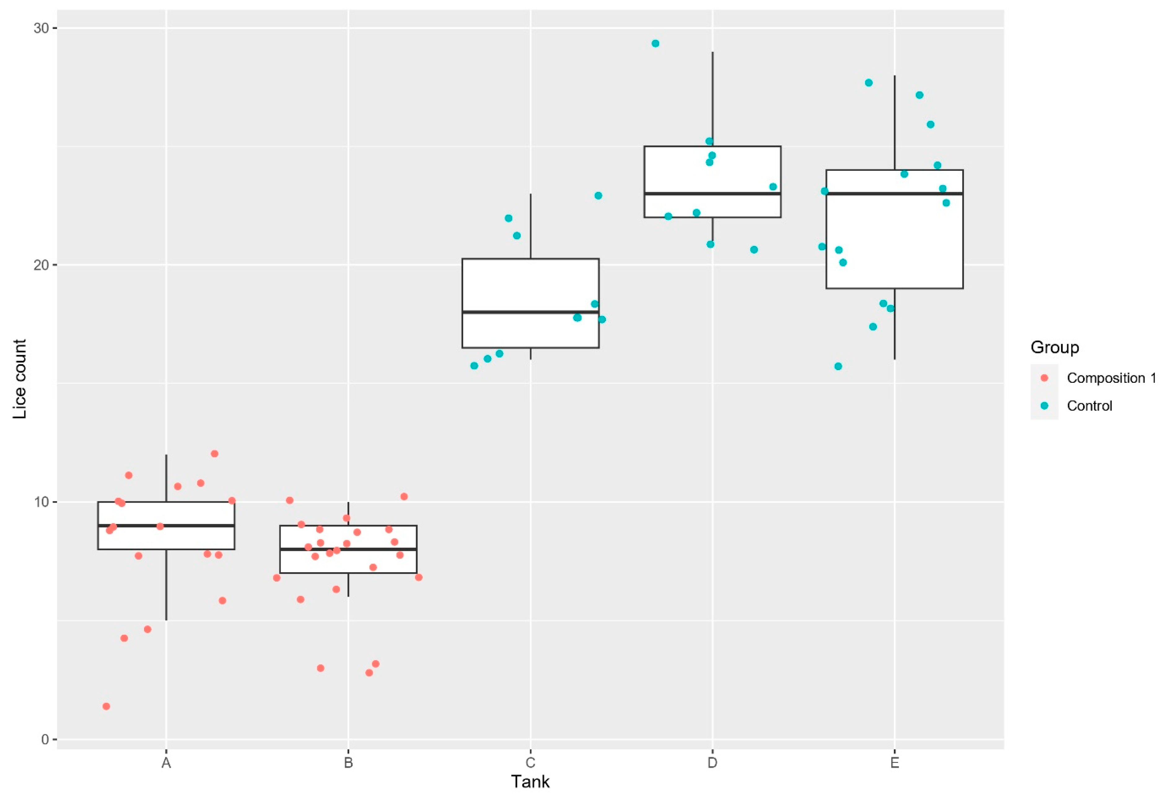
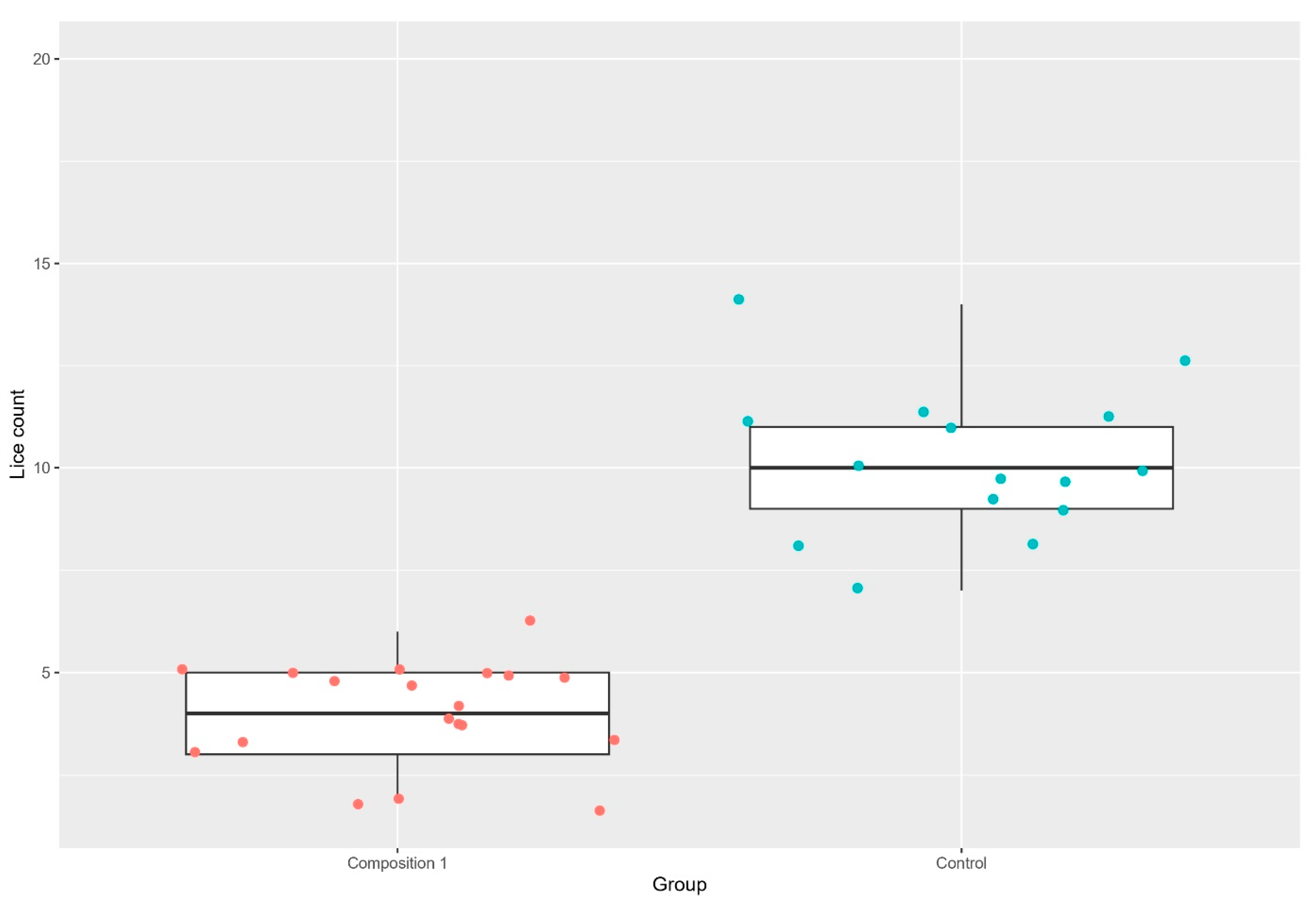
Trial 1. As presented in Table 2 and Figure 2 and Figure 3, the exposure to composition 1 (VI1 and VI2 co-culture) was associated with a significant reduction in the number of attached lice (p < 0.001 and p < 0.001 by NBMM and PMM) following copepodid challenge assessed 34 days post-challenge. The total number of attached lice was counted for each fish.
Fish immersed in composition 1 in separate tanks had an average lice reduction of 63.38%. When fish exposed to composition 1 and non-exposed fish were cohabited from challenge, there was a 60.06% average reduction in attached lice in the exposed fish. Most lice had reached chalimus or preadult stages.
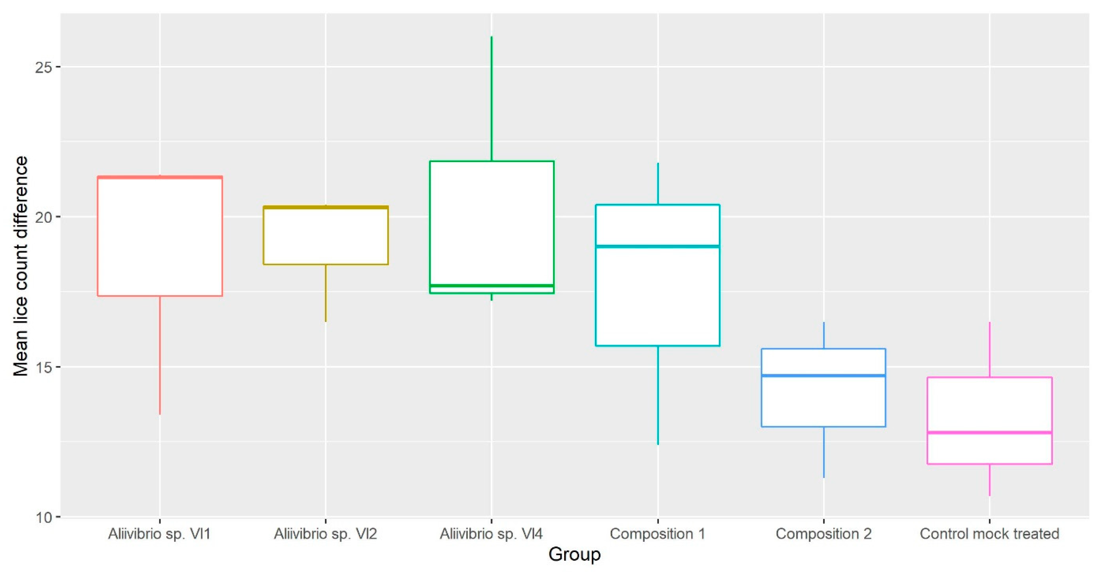
Trial 2. As shown in Table 2 and Figure 1, this trial compared five different probiotic formulations. At first lice count, there was no reduction in lice numbers for any groups compared with the control. Only 5% of the lice developed beyond the chalimus stage. A second count was conducted when most lice (≈90%) had reached pre-adult/adult stages 27 days later, and groups immersed in Composition 1, Composition 2, and Vl4 had lower lice counts than the control, while Vl1 and Vl2 did not. As illustrated in Figure 4, the lice counts were overall reduced when compared to the first count, with the control group having the lowest relative reduction, although not significantly different (p > 0.05 by NBMM).
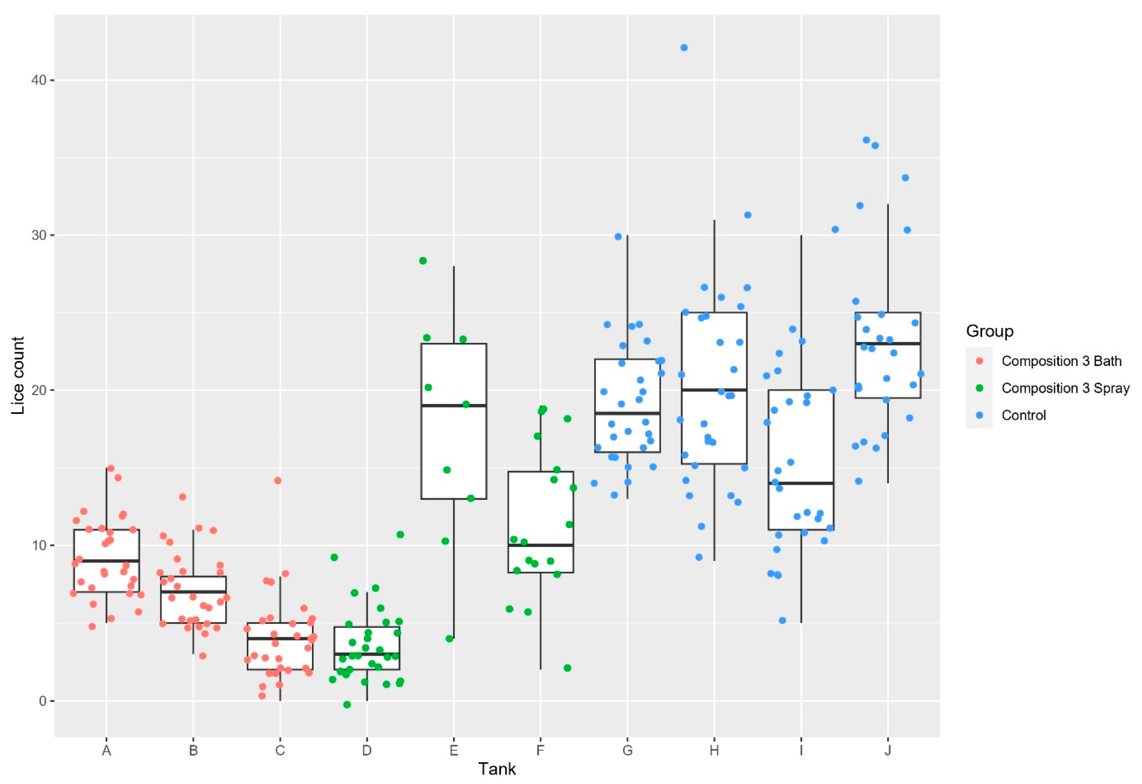
Trial 3. In this trial, we compared administration by bath or spray using composition 3 (Figure 5). The lice count was conducted 32 days post-challenge, with all lice having reached chalimus or pre-adult stages. The reduction for the bath group compared to control was 65% and for spray, 57.8%.

3.2. Application of Bacteria to Pre-Adult Salmon Lice

Next, we wanted to document if probiotics applied directly to water baths containing salmon lice impacted the viability of the lice. The probiotic Aliivibrio bacteria had no observed negative impact on the pre-adult salmon lice by 24 h of incubation, with the exception of 2 of 10 pre-adult salmon lice exposed to Vl3 (Table 3). All lice exposed to a combination of Vl1, Vl2, and Vl3 survived.
For the fish pathogenic Aliivibrio salmonicida, Moritella viscosa, Aliivibrio sp. NCIMB 42181, and Aliivibrio sp. NCIMB 42953, Aliivibrio sp. NCIMB 42952, and Aliivibrio sp. NCIMB 42954, all pre-adult salmon lice were detached and non-motile by 24 h incubation, while no salmon lice detached or died when exposed to A. wodanis.
After an additional 17 h (41 h exposure in total), all pre-adult salmon lice were detached and dead on the bottom of all glass jars, including the negative control.

4. Discussion

In these trials we have studied the effects of immersing Atlantic salmon in baths containing probiotic Aliivibrio spp. prior to the experimental lice challenge and attempted to identify the most effective preventive bacterial composition and administration strategy. We have also studied the mortality of pre-adult salmon lice after exposure to various probiotic and pathogenic strains of bacteria in an attempt to understand the mechanisms involved.

4.1. Lice Attachment

In trial 1, Composition 1 gave significantly fewer lice compared to the control (p < 0.001 by NBMM and PMM). Separate tanks within the same group were not statistically different from each other (p > 0.5 by NBMM). Keeping treated and controls in separate tanks gave a higher percentage reduction compared to when treated and controls were kept in a common garden setup during challenge. An observation here is that the fish in the common garden approach had overall lower infestation levels (4 and 10 versus 8 and 21 attached lice). The attachment process of copepodids involves a complex sequence of initial attraction, landing, docking, and attaching, a process that needs 4–5 days to complete [7,26,49]. As the salmon copepod only has a finite energy level available to find a host, the selection process is likely not random. If half the fish in a tank is less attractive to the parasite, it may have to spend twice the energy to find the optimal host. By this reasoning, one can speculate if fish in common garden setups can receive passive protection from their cohabitants.
In trial 2, there were no differences between the groups at the first count (p > 0.05 by NBMM), and the lice were all less developed than expected. Water temperatures are one of the key factors in lice development [50], and the salmon lice in trial 2 developed much slower than expected at an average temperature of 9.9 °C. At the second count, we observed unexpected significant differences in the relative mean count. The lice batch in this trial could be the reason, even though the preliminary visual evaluation of the copepodids did not reveal any abnormalities. Another concern would be the entry of water carrying Moritella viscosa, shown to kill salmon lice when exposed directly to the lice in the water in vitro. Controlled co-infections of salmon lice with Moritella viscosa have been shown to decrease the lice count over time [51]. Although no winter ulcers were visible during the trial, Moritella viscosa was isolated from several fish two weeks after the trial ended. Another possibility is that the fish sourced in this trial had been exposed to these bacteria previously, resulting in less effect, as these are naturally occurring bacteria, and there are not yet developed screening methods for the presence of these probiotic bacteria in fish. Either way, the lice reduction in all groups between counts was reduced in the control group compared to those administered probiotics (Figure 4). Although this trial was overall non-significant (p > 0.05 by NBMM), the monoculture of Vl4 appeared to be a good candidate for further studies.
In trial 3, we selected composition 1 and monoculture Vl4, which was cultured separately and mixed at the time of administration. The group administered probiotic composition 3 by bath had significantly fewer lice than the control group (p < 0.001 by NBMM). The spray group was not significantly different from the bath group or the control, due to large tank variations, although there were 57.7% fewer lice in this group overall.
When we compare these trials, the overall reduction in salmon lice attachment success in trial 1 and 3 are quite different from trial 2. In addition to previously discussed aspects, there are several factors in the design used in the present study that may explain the variation in attachment success between the three trials included. The most probable explanation for the differences between trials is the time from probiotic bath to exposure of salmon lice. In trial 2, fish were infected only 58 days after probiotic immersion, while we waited 155 and 91 days in trials 1 and 3, respectively. Some probiotics have been shown to modulate the microbiota [52], and time is a factor for in vivo adaptions in different species [53]. Hence, 58 days after probiotic administration may be too brief for the anti-lice effects to take place.
High fish density is a known stressor, and stress is regarded as an important factor that may increase host susceptibility to infection [17]. Still, fish density at the end of trial 2 was 34.5 kg/m3 three weeks after the second lice count, lower than what is shown to cause negative effects when other parameters are within recommended levels [54,55]. In addition, trial 1 density was 36.2 kg/m3 at the lice count and trial 3 density was 26.8 kg/m3 one week after the lice count, indicating that all trials likely had similar densities throughout the lice challenge period, far below density limits for post-smolt suggested by Calabrese et al. Growth is one of the main parameters affected by chronic stress [14]. The fish in trials 2 and 3 were of similar size to the crowding experiments of Hosfeld et al., and growth observed in trials 2 and 3 surpassed even the most crowded fish in these experiments, which indicates that the fish in these trials were likely not chronically stressed. Two studies have also shown that chronic stress from long-term crowding and cortisol implants did not increase salmon lice attachment in Atlantic salmon [56,57]. Because of this, we believe that the trial differences in lice attachment are not caused by the fish densities. Different bacterial doses administered are another possible cause, but in trial 2, lower doses were administered than in trial 1 and higher doses than in trial 3. The same composition in trial 1 was used in trial 2, which indicates that the bacterial doses administered cannot explain the differences in lice attachment.

4.2. Bacterial Composition

The Aliivibrio spp. used in these trials are originally cultivated from farmed Atlantic salmon and Vibrionaceae are naturally highly abundant in the aquatic environment [58]. Composition 1 is the first formulation where we saw significant differences and has therefore remained a constant across all trials. As monocultured Vl4 stood out in trial 2, we mixed it with composition 1, resulting in composition 3, which showed the largest observed reductions in attached lice during these three trials. The differences between monocultures compared to the same bacteria cocultured in trial 2 are not surprising, as Aliivibrio species has previously been shown to highly affect other bacteria in its presence [59,60]. As the lice attachment success was not statistically different between groups in trial 2, one can hypothesize that the effect on reduced attachment success is not related to bacterial strain. If so, any immersion in non-pathogenic species within this genus may lead to similar effects. This can also be supported by trials 1 and 3 as the results from immersions are very similar despite different bacterial compositions and doses and should be investigated further.

4.3. Administration Method

In trial 3, we also compared the administration methods of bath versus spray. The group of three tanks immersed in the probiotic composition showed a low overall attachment in unison, while the spray group had a higher degree of variation. The spray tank with the lowest lice numbers was the lowest tank overall in trial 3 but given the high lice numbers in the two other spray-administered tanks, it appears that this method can be more difficult in delivering an effective dose, although possible. As lice attachment success was reduced in fish exposed to the probiotic bacteria on skin only and even further reduced in fish where the whole fish surface was submersed, the bacterial relationship with fish skin may be relevant to the probiotic success. Fish skin is a mucosal surface, hosting a wide variety of bacterial species that the salmon louse will encounter when parasitizing the salmon. Fish skin has also been reported to be a potential entry port for bacteria [61] and has been demonstrated for Aliivibrio salmonicida [62], a bacterium phylogenetically close to the Aliivibrio species used as probiotics in this study. Salmon skin as the domain or entry port of these bacteria seems likely given the literature and results after bath and spray administration but is yet to be shown convincingly and in-depth.

4.4. Mechanism

Numerous studies have demonstrated that probiotics cause health benefits in aquatic organisms, and probiotic modes of action on disease resistance vary [40]. Some studies have also shown that probiotics can have an effect on the control of parasites in mammals, avians and cell cultures [63]. The probiotic Aliivibrio species studied here did not increase the mortality of pre-adult lice when they were exposed to approximately the same concentrations of bacteria that are used in the probiotic bath of the Atlantic salmon. This indicates that the reduced lice attachment is likely not due to lice being rapidly killed by direct pathogenic effects from the probiotics but via a secondary mechanism. Colonization of external surfaces is one of the possible fates of bacteria coming in contact with fish [64], and the microbial composition contributes directly to the mucosal environment. We speculate that this, in turn, can affect the level of lice attachment, as salmon skin and mucus have been shown to activate the salmon louse host-finding behaviour [24]. Antimicrobial peptides released from the skin of salmon will also trigger this behaviour by activation of chemosensory neural activity [28]. Recently, specific volatile organic compounds in the fish mucus were shown to be associated with salmon lice resistance [29]. As the exact sequence of events that ends with successful attachment is still not understood, host mucus composition undoubtedly plays a part in the host selection process. We suggest that submersion or spray of probiotics to fish skin and mucus can interfere with the salmon louse decision process for host selection.
To our knowledge, this is the first time that fish pathogenic bacteria have been shown to directly increase mortality in pre-adult salmon lice. The underlying mechanism for the pathogenic effect on salmon louse is unknown, but the same virulence features as those causing disease in fish could be responsible. The only fish pathogenic bacteria tested in this study that did not impact the salmon lice copepodid mortality was A. wodanis. Although surprising, A. wodanis interplay and pathogenicity are not yet fully understood [60].
Despite slow lice development and a large lice number decrease in trial 2, the relative reduction between counts is an interesting observation. This attachment reduction appeared during development from chalimus to adult salmon louse and was lowest in the control group. One of the main changes in the salmon lice during this period is the start of blood consumption [11]. This is also where many key immunological components are located, which mobile salmon lice will be exposed to for the first time since settling on the host [65]. This could indicate that the content of the host blood is relevant to the probiotic mechanism, and the possibility of skin as an entry port should be investigated further.
No matter the mechanism, this is a unique example of interaction between three kingdoms of life to keep a natural balance of three biological agents: the probiotic bacteria, the salmon host, and the parasitic salmon louse. Our results may open an intriguing research path towards understanding more from the natural relations that can help human society farm Atlantic salmon in a more sustainable way in the future. Further development of this approach could have a significant impact as a non-medicinal approach to salmon louse control and appears to be the first report of significant reductions in lice load on Atlantic salmon after probiotic exposure.

Supplementary Materials

The following supporting information can be downloaded at: https://www.mdpi.com/article/10.3390/applmicrobiol3040090/s1, Table S1: Lice count trial 1, fish immersed in Composition 1 (tanks A and B) or unexposed control fish. Lice count conducted 189 days after probiotic exposure and 34 days post infestation with 30 copepodids per fish. A third tank containing fish administered Aliivibrio sp. Vl1 + Vl2 was lost due to a water stoppage in the research facility.; Table S2: Lice count trial 1, fish cohabitant in the two tanks (G and F, respectively) with approximately 50% exposed to Composition 1 and 50% non-treated controls. Lice count conducted 189 days after probiotic exposure and 34 days post infestation with 30 copepodids per fish.; Table S3: Lice counts trial 2 per tank and per group;Table S4: Lice count trial 3 per tank and per group. Lice count conducted day 32 post lice exposure and 123 days post probiotic application.

Author Contributions

M.S.D.: Investigation, Data Curation, Writing—original draft, review and editing, Formal analysis, Visualization. C.S.-S.: Investigation, Data Curation, Writing—review and editing. Ø.E.: Conceptualization, Methodology, Writing—review and editing. K.G.: Investigation, Methodology, Writing—review and editing. M.J.B.: Investigation, Data Curation, Writing—review and editing. H.S.: Resources, Conceptualization, Investigation, Supervision, Writing—review and editing. K.S.: Conceptualization, Methodology, Investigation, Writing—original draft, review and editing. All authors have read and agreed to the published version of the manuscript.

Funding

The research was funded by Previwo AS with support from the Research Council of Norway (NFR), project no. 322983. Ø Evensen and K Gadan received funding from the Norwegian Seafood Research Fund, project no. 901566 “Host immunity and skin microbiome interplay—importance for protection against sea lice infection in Atlantic salmon”.

Institutional Review Board Statement

The Animal Welfare Approval (FOTS) number was 19688 for Trial 1, 29259 for Trial 2, and 24725 for trial 3.

Informed Consent Statement

Not applicable.

Data Availability Statement

The data that supports the findings of this study are available from the corresponding author upon request.

Acknowledgments

Thanks to Aud Kari Fauske, Cristopher Nilsson, and Nils Sandqvist for the preparation of the probiotic cultures for the trials. Thanks to Øystein Klakegg, Stanislav Iakhno, and Anne Bakke Fylling for review and editing. Thanks to NMBU and especially Stein Helge Skjelde (Sørsmolt) for the salmon used in these studies.

Conflicts of Interest

Aside from the employee relationships as written in the author affiliations, Henning Sørum and Kira Salonius are shareholders in Previwo AS, a company that produces a probiotic product, StembiontTM, including two of the Aliivibrio strains under investigation in this report.

References

  1. Skern-Mauritzen, R.; Torrissen, O.; Glover, K.A. Pacific and Atlantic Lepeophtheirus salmonis (Krøyer, 1838) are allopatric subspecies: Lepeophtheirus salmonis salmonis and L. salmonis oncorhynchi subspecies novo. BMC Genet 2014, 15, 32. [Google Scholar] [CrossRef] [PubMed]
  2. Dawson, L.H.J.; Mcvicar, A.H. Comparison of the susceptibility of sea trout and Atlantic salmon for sea lice. ICES J. Mar. Sci. 1997, 54, 1129–1139. [Google Scholar] [CrossRef]
  3. Finstad, B.; Bjørn, P.A.; Grimnes, A.; Hvidsten, N.A. Laboratory and field investigations of salmon lice [Lepeophtheirus salmonis (Krøyer)] infestation on Atlantic salmon (Salmo salar L.) post-smolts. Aquac. Res. 2000, 31, 795–803. [Google Scholar] [CrossRef]
  4. Murray, A.G. Using observed load distributions with a simple model to analyse the epidemiology of sea lice (Lepeophtheirus salmonis) on sea trout (Salmo trutta). Pest Manag. Sci. 2002, 58, 585–594. [Google Scholar] [CrossRef] [PubMed]
  5. Heuch, P.A.; Nordhagen, J.R.; Schram, T.A. Egg production in the salmon louse [Lepeophtheirus salmonis (Krøyer)] in relation to origin and water temperature. Aquac. Res. 2000, 31, 805–814. [Google Scholar] [CrossRef]
  6. Tucker, C.S.; Sommerville, C.; Wootten, R. An Investigation into the Larval Energetics and Settlement of the Sea Louse, Lepeophtheirus salmonis, an Ectoparasitic Copepod of Atlantic salmon, Salmo salar. Fish Pathol. 2000, 35, 137–143. [Google Scholar] [CrossRef]
  7. Hamre, L.A.; Eichner, C.; Caipang, C.M.A.; Dalvin, S.T.; Bron, J.E.; Nilsen, F.; Boxshall, G.; Skern-Mauritzen, R. The Salmon Louse Lepeophtheirus salmonis (Copepoda: Caligidae) Life Cycle Has Only Two Chalimus Stages. PLoS ONE 2013, 8, e73539. [Google Scholar] [CrossRef]
  8. Jones, M.W.; Sommerville, C.; Bron, J. The histopathology associated with the juvenile stages of Lepeophtheirus salmonis on the Atlantic salmon, Salmo salar L. J. Fish Dis. 1990, 13, 303–310. [Google Scholar] [CrossRef]
  9. Johnson, S.C. The Biology of Lepeophtheirus salmonis. Ph.D. Thesis, Simon Fraser University, Burnaby, BC, Canada, 1991. [Google Scholar]
  10. Bjørn, P.A.; Finstad, B. The development of salmon lice (Lepeophtheirus salmonis) on artificially infected post smolts of sea trout (Salmo trutta). Can. J. Zool. 1998, 76, 970–977. [Google Scholar] [CrossRef]
  11. Heggland, E.I.; Dondrup, M.; Nilsen, F.; Eichner, C. Host gill attachment causes blood-feeding by the salmon louse (Lepeophtheirus salmonis) chalimus larvae and alters parasite development and transcriptome. Parasites Vectors 2020, 13, 225. [Google Scholar] [CrossRef]
  12. Fast, M.D.; Muise, D.M.; Easy, R.E.; Ross, N.W.; Johnson, S.C. The effects of Lepeophtheirus salmonis infections on the stress response and immunological status of Atlantic salmon (Salmo salar). Fish Shellfish. Immunol. 2006, 21, 228–241. [Google Scholar] [CrossRef] [PubMed]
  13. Ugelvik, M.S.; Mæhle, S.; Dalvin, S. Temperature affects settlement success of ectoparasitic salmon lice (Lepeophtheirus salmonis) and impacts the immune and stress response of Atlantic salmon (Salmo salar). J. Fish Dis. 2022, 45, 975–990. [Google Scholar] [CrossRef] [PubMed]
  14. Schreck, C.B.; Tort, L. The concept of stress in fish. In Fish Physiology, 1st ed.; Academic Press: Cambridge, MA, USA, 2016; Volume 35, pp. 1–34. [Google Scholar]
  15. Eissa, N.; Wang, H.P. Transcriptional stress responses to environmental and husbandry stressors in aquaculture species. Rev. Aquac. 2016, 8, 61–88. [Google Scholar] [CrossRef]
  16. Braden, L.M.; Monaghan, S.J.; Fast, M.D. Salmon immunological defence and interplay with the modulatory capabilities of its ectoparasite Lepeophtheirus salmonis. Parasite Immunol. 2020, 42, e12731. [Google Scholar] [CrossRef]
  17. Johnson, S.C.; Albright, L.J. Comparative susceptibility and histopathology of the response of naive Atlantic. Chinook and coho salmon to experimental infection with Lepeophtheirus salmonis (Copepoda: Caligidae). Dis. Aquat. Org. 1992, 14, 179–193. [Google Scholar] [CrossRef]
  18. Barker, S.E.; Bricknell, I.R.; Covello, J.; Purcell, S.; Fast, M.D.; Wolters, W.; Bouchard, D.A. Sea lice, Lepeophtheirus salmonis (Krøyer 1837), infected Atlantic salmon (Salmo salar L.) are more susceptible to infectious salmon anemia virus. PLoS ONE 2019, 14, e0209178. [Google Scholar] [CrossRef] [PubMed]
  19. Lhorente, J.P.; Gallardo, J.A.; Villanueva, B.; Carabaño, M.J.; Neira, R. Disease Resistance in Atlantic Salmon (Salmo salar): Coinfection of the Intracellular Bacterial Pathogen Piscirickettsia salmonis and the Sea Louse Caligus rogercresseyi. PLoS ONE 2014, 9, e95397. [Google Scholar] [CrossRef] [PubMed]
  20. Llewellyn, M.S.; Leadbeater, S.; Garcia, C.; Sylvain, F.-É.; Custodio, M.; Ang, K.P.; Powell, F.; Carvalho, G.R.; Creer, S.; Elliot, J.; et al. Parasitism perturbs the mucosal microbiome of Atlantic Salmon. Sci. Rep. 2017, 7, srep43465. [Google Scholar] [CrossRef]
  21. Gonçalves, A.T.; Collipal-Matamal, R.; Valenzuela-Muñoz, V.; Nuñez-Acuña, G.; Valenzuela-Miranda, D.; Gallardo-Escárate, C. Nanopore sequencing of microbial communities reveals the potential role of sea lice as a reservoir for fish pathogens. Sci. Rep. 2020, 10, 2895. [Google Scholar] [CrossRef]
  22. Gallardo-Escárate, C.; Valenzuela-Muñoz, V.; Núñez-Acuña, G.; Carrera, C.; Gonçalves, A.T.; Valenzuela-Miranda, D.; Benavente, B.P.; Roberts, S. Catching the complexity of salmon-louse interactions. Fish Shellfish. Immunol. 2019, 90, 199–209. [Google Scholar] [CrossRef]
  23. Bailey, R.J.E.; Birkett, M.A.; Ingvarsdóttir, A.; Mordue, A.J.; Mordue, W.; O’Shea, B.; Pickett, J.A.; Wadhams, L.J. The role of semiochemicals in host location and non-host avoidance by salmon louse (Lepeophtheirus salmonis) copepodids. Can. J. Fish. Aquat. Sci. 2006, 63, 448–456. [Google Scholar] [CrossRef]
  24. Devine, J.G.; Ingvarsdóttir, A.; Mordue, W.; Pike, W.A.; Pickett, J.; Duce, I.; Mordue, A.J. Salmon Lice, Lepeophtheirus salmonis, Exhibit Specific Chemotactic Responses to Semiochemicals Originating from the Salmonid, Salmo salar. J. Chem. Ecol. 2000, 26, 1833–1847. [Google Scholar] [CrossRef]
  25. Ingvarsdóttir, A.; Birkett, M.A.; Duce, I.; Genna, R.L.; Mordue, W.; Pickett, J.A.; Wadhams, L.J.; Mordue, A.J. Semiochemical strategies for sea louse control: Host location cues. Pest Manag. Sci. 2002, 58, 537–545. [Google Scholar] [CrossRef] [PubMed]
  26. Mordue, A.J.; Birkett, M.A. A review of host finding behaviour in the parasitic sea louse, Lepeophtheirus salmonis(Caligidae: Copepoda). J. Fish Dis. 2009, 32, 3–13. [Google Scholar] [CrossRef]
  27. Komisarczuk, A.Z.; Grotmol, S.; Nilsen, F. Ionotropic receptors signal host recognition in the salmon louse (Lepeophtheirus salmonis, Copepoda). PLoS ONE 2017, 12, e0178812. [Google Scholar] [CrossRef]
  28. Núñez-Acuña, G.; Gallardo-Escárate, C.; Fields, D.M.; Shema, S.; Skiftesvik, A.B.; Ormazábal, I.; Browman, H.I. The Atlantic salmon (Salmo salar) antimicrobial peptide cathelicidin-2 is a molecular host-associated cue for the salmon louse (Lepeophtheirus salmonis). Sci. Rep. 2018, 8, 13738. [Google Scholar] [CrossRef] [PubMed]
  29. Difford, G.F.; Haugen, J.-E.; Aslam, M.L.; Johansen, L.H.; Breiland, M.W.; Hillestad, B.; Baranski, M.; Boison, S.; Moghadam, H.; Jacq, C. Variation in volatile organic compounds in Atlantic salmon mucus is associated with resistance to salmon lice infection. Sci. Rep. 2022, 12, 4839. [Google Scholar] [CrossRef]
  30. Iversen, A.; Hermansen, Ø.; Nystøyl, R.; Hess, E.J. Kostnadsutvikling i lakseoppdrett, Nofima, Tromsø, Norway, Report 24/2017, pp. 1–47. Available online: https://nofima.no/publikasjon/1523319/ (accessed on 3 November 2023).
  31. Berle, H.; Rim, S.Y.; Thesis, M. The Cost of Sea Lice and Its Implications for the Future of the Norwegian Aquaculture Industry—A Study on Sea Lice and Recommendations. Master’s Thesis, Norwegian School of Economics, Bergen, Norway, 2018. [Google Scholar]
  32. Fiskehelserapporten 2022. Norwegian Veterinary Institute’s Report Series, 5a/2023, Ås, Norway. Available online: https://www.vetinst.no/rapporter-og-publikasjoner/rapporter/2023/norwegian-fish-health-report-2022 (accessed on 3 November 2023).
  33. Overton, K.; Dempster, T.; Oppedal, F.; Kristiansen, T.S.; Gismervik, K.; Stien, L.H. Salmon lice treatments and salmon mortality in Norwegian aquaculture: A review. Rev. Aquac. 2018, 11, 1398–1417. [Google Scholar] [CrossRef]
  34. Sviland Walde, C.; Bang Jensen, B.; Stormoen, M.; Asche, F.; Misund, B. The economic impact of decreased mortality and increased growth associated with preventing, replacing or improving current methods for delousing farmed Atlantic salmon in Norway. Prev. Vet. Med. 2023, 221, 106062. [Google Scholar] [CrossRef]
  35. FAO/WHO. Health and Nutritional Properties of Probiotics in Food including Powder Milk with Live Lactic Acid Bacteria; Fao & Who Report; FAO/WHO: Córdoba, Argentina, 2001; pp. 1–34. [Google Scholar]
  36. Hill, C.; Guarner, F.; Reid, G.; Gibson, G.R.; Merenstein, D.J.; Pot, B.; Morelli, L.; Canani, R.B.; Flint, H.J.; Salminen, S.; et al. Expert consensus document: The international scientific association for probiotics and prebiotics consensus statement on the scope and appropriate use of the term probiotic. Nat. Rev. Gastroenterol. Hepatol. 2014, 11, 506–514. [Google Scholar] [CrossRef]
  37. Gómez, G.D.; Balcázar, J.L. A review on the interactions between gut microbiota and innate immunity of fish. FEMS Immunol. Med. Microbiol. 2008, 52, 145–154. [Google Scholar] [CrossRef]
  38. Naik, S.; Bouladoux, N.; Wilhelm, C.; Molloy, M.J.; Salcedo, R.; Kastenmuller, W.; Deming, C.; Quinones, M.; Koo, L.; Conlan, S.; et al. Compartmentalized control of skin immunity by resident commensals. Science 2012, 337, 1115–1119. [Google Scholar] [CrossRef] [PubMed]
  39. Chauhan, A.; Singh, R. Probiotics in aquaculture: A promising emerging alternative approach. Symbiosis 2018, 77, 99–113. [Google Scholar] [CrossRef]
  40. Hoseinifar, S.H.; Sun, Y.-Z.; Wang, A.; Zhou, Z. Probiotics as means of diseases control in aquaculture, a review of current knowledge and future perspectives. Front. Microbiol. 2018, 9, 2429. [Google Scholar] [CrossRef] [PubMed]
  41. Romero, J.; Pereira, W.A.; Mendonça, C.M.N.; Urquiza, A.V.; Leblanc, J.G.; Cotter, P.D.; Romero, J.; Oliveira, R.P.S. Use of Probiotic Bacteria and Bacteriocins as an Alternative to Antibiotics in Aquaculture. Microorganisms 2022, 10, 1705. [Google Scholar] [CrossRef]
  42. Simón, R.; Docando, F.; Nuñez-Ortiz, N.; Tafalla, C.; Díaz-Rosales, P. Mechanisms Used by Probiotics to Confer Pathogen Resistance to Teleost Fish. Front. Immunol. 2021, 12, 653025. [Google Scholar] [CrossRef] [PubMed]
  43. Talukder Shefat, S.H. Probiotic Strains Used in Aquaculture. Int. Res. J. Microbiol. 2018, 7, 43–55. [Google Scholar] [CrossRef]
  44. Pieters, N.; Brunt, J.; Austin, B.; Lyndon, A.R. Efficacy of in-feed probiotics against Aeromonas bestiarum and Ichthyophthirius multifiliis skin infections in rainbow trout (Oncorhynchus mykiss, Walbaum). J. Appl. Microbiol. 2008, 105, 723–732. [Google Scholar] [CrossRef]
  45. Reyes-Becerril, M.; Tovar-Ramírez, D.; Ascencio-Valle, F.; Civera-Cerecedo, R.; Gracia-López, V.; Barbosa-Solomieu, V. Effects of dietary live yeast Debaryomyces hansenii on the immune and antioxidant system in juvenile leopard grouper Mycteroperca rosacea exposed to stress. Aquaculture 2008, 280, 39–44. [Google Scholar] [CrossRef]
  46. Barrett, L.T.; Oppedal, F.; Robinson, N.; Dempster, T. Prevention not cure: A review of methods to avoid sea lice infestations in salmon aquaculture. Rev. Aquac. 2020, 12, 2527–2543. [Google Scholar] [CrossRef]
  47. Klakegg, Ø.; Myhren, S.; Juell, R.A.; Aase, M.; Salonius, K.; Sørum, H. Improved health and better survival of farmed lumpfish (Cyclopterus lumpus) after a probiotic bath with two probiotic strains of Aliivibrio. Aquaculture 2019, 518, 734810. [Google Scholar] [CrossRef]
  48. Klakegg, Ø.; Salonius, K.; Nilsen, A.; Fülberth, M.; Sørum, H. Enhanced growth and decreased mortality in Atlantic salmon (Salmo salar) after probiotic bath. J. Appl. Microbiol. 2020, 129, 146–160. [Google Scholar] [CrossRef] [PubMed]
  49. Bron, J.E.; Sommerville, C.; Jones, M.; Rae, G.H. The settlement and attachment of early stages of the salmon louse, Lepeophtheirus salmonis (Copepoda: Caligidae) on the salmon host, Salmo salar. J. Zool. 1991, 224, 201–212. [Google Scholar] [CrossRef]
  50. Dalvin, S.; Oppedal, F. The effect of temperature and light on development of salmon lice. Norwegian Institute of Marine Research, Bergen, Norway, Report 2019-13, pp. 1–19.
  51. Wieërs, G.; Belkhir, L.; Enaud, R.; Leclercq, S.; Philippart de Foy, J.M.; Dequenne, I.; de Timary, P.; Cani, P.D. How Probiotics Affect the Microbiota. Front. Cell. Infect. Microbiol. 2020, 9. [Google Scholar] [CrossRef] [PubMed]
  52. Huang, S.; Jiang, S.; Huo, D.; Allaband, C.; Estaki, M.; Cantu, V.; Belda-Ferre, P.; Vázquez-Baeza, Y.; Zhu, Q.; Ma, C.; et al. Candidate probiotic Lactiplantibacillus plantarum HNU082 rapidly and convergently evolves within human, mice, and zebrafish gut but differentially influences the resident microbiome. Microbiome 2021, 9, 151. [Google Scholar] [CrossRef] [PubMed]
  53. Calabrese, S.; Nilsen, T.O.; Kolarevic, J.; Ebbesson, L.O.E.; Pedrosa, C.; Fivelstad, S.; Hosfeld, C.; Stefansson, S.O.; Terjesen, B.F.; Takle, H.; et al. Stocking density limits for post-smolt Atlantic salmon (Salmo salar L.) with emphasis on production performance and welfare. Aquaculture 2017, 468, 363–370. [Google Scholar] [CrossRef]
  54. Hosfeld, C.D.; Hammer, J.; Handeland, S.O.; Fivelstad, S.; Stefansson, S.O. Effects of fish density on growth and smoltification in intensive production of Atlantic salmon (Salmo salar L.). Aquaculture 2009, 294, 236–241. [Google Scholar] [CrossRef]
  55. Delfosse, C.; Pageat, P.; Lafont-Lecuelle, C.; Asproni, P.; Chabaud, C.; Cozzi, A.; Bienboire-Frosini, C. Effect of handling and crowding on the susceptibility of Atlantic salmon (Salmo salar L.) to Lepeophtheirus salmonis (Krøyer) copepodids. J. Fish Dis. 2020, 44, 327–336. [Google Scholar] [CrossRef]
  56. Krasnov, A.; Skugor, S.; Todorcevic, M.; Glover, K.A.; Nilsen, F. Gene expression in Atlantic salmon skin in response to infection with the parasitic copepod Lepeophtheirus salmonis, cortisol implant, and their combination. BMC Genomics 2012, 13, 130. [Google Scholar] [CrossRef]
  57. Thompson, F.L.; Iida, T.; Swings, J. Biodiversity of Vibrios. Microbiol. Mol. Biol. Rev. 2004, 68, 403–431. [Google Scholar] [CrossRef]
  58. Hjerde, E.; Karlsen, C.; Sørum, H.; Parkhill, J.; Willassen, N.P.; Thomson, N.R. Co-cultivation and transcriptome sequencing of two co-existing fish pathogens Moritella viscosa and Aliivibrio wodanis. BMC Genom. 2015, 16, 447. [Google Scholar] [CrossRef] [PubMed]
  59. Carvalho, L.A.; Whyte, S.K.; Braden, L.M.; Purcell, S.L.; Manning, A.J.; Muckle, A.; Fast, M.D. Impact of co-infection with Lepeophtheirus salmonis and Moritella viscosa on inflammatory and immune responses of Atlantic salmon (Salmo salar). J. Fish Dis. 2020, 43, 459–473. [Google Scholar] [CrossRef] [PubMed]
  60. Karlsen, C.; Vanberg, C.; Mikkelsen, H.; Sørum, H. Co-infection of Atlantic salmon (Salmo salar), by Moritella viscosa and Aliivibrio wodanis, development of disease and host colonization. Vet. Microbiol. 2014, 171, 112–121. [Google Scholar] [CrossRef] [PubMed]
  61. Karlsen, C.; Sørum, H.; Willassen, N.P.; Åsbakk, K. Moritella viscosa bypasses Atlantic salmon epidermal keratocyte clearing activity and might use skin surfaces as a port of infection. Vet. Microbiol. 2012, 154, 353–362. [Google Scholar] [CrossRef] [PubMed]
  62. Kashulin, A.; Sørum, H. A novel in vivo model for rapid evaluation of Aliivibrio salmonicida infectivity in Atlantic salmon. Aquaculture 2014, 420–421, 112–118. [Google Scholar] [CrossRef]
  63. Travers, M.-A.; Florent, I.; Kohl, L.; Grellier, P. Probiotics for the Control of Parasites: An Overview. J. Parasitol. Res. 2011, 2011, 610769. [Google Scholar] [CrossRef]
  64. Austin, B. The bacterial microflora of fish, revised. Sci. World J. 2006, 6, 931–945. [Google Scholar] [CrossRef]
  65. Uribe, C.; Folch, H.; Enriquez, R.; Moran, G. Innate and adaptive immunity in teleost fish: A review. Vet. Med. 2011, 56, 486–503. [Google Scholar] [CrossRef]
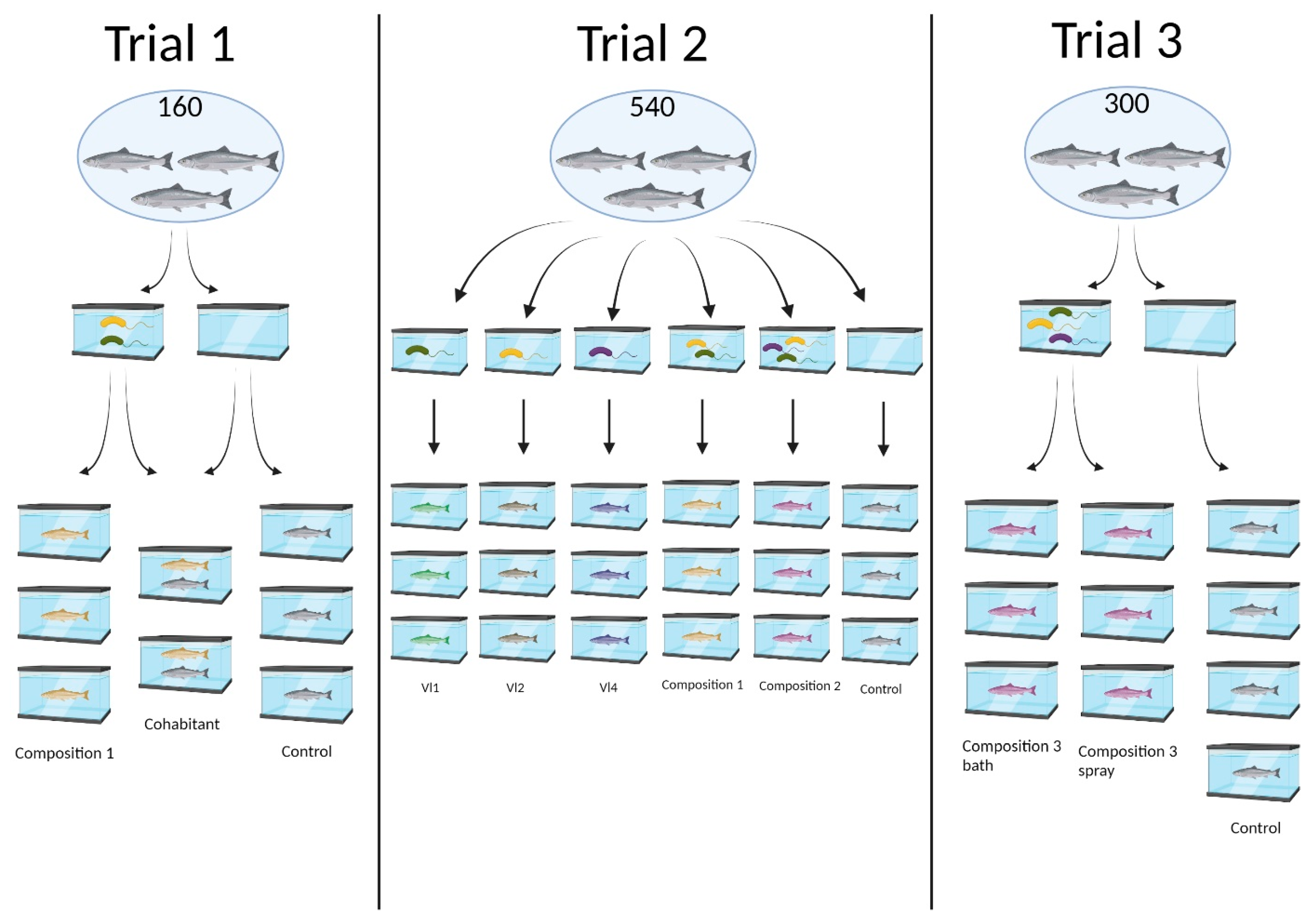
Figure 1. Overview of the three lice challenge trials presented in this study.
Applmicrobiol 03 00090 g001
Figure 2. Lice count per fish, trial 1, sorted by tank. Individual tanks exposed to 30 copepodids per fish. Tanks C, D, and E are controls, while tanks A and B contained fish bathed in probiotic Aliivibrio sp. Vl1 and Vl2. Tanks A and B are significantly different from the control tanks (p < 0.001 by NBMM).
Applmicrobiol 03 00090 g002
Figure 3. Lice count per fish, trial 1, cohabitation tanks, sorted by group. Twenty fish per tank, cohabitated by approximately 50% probiotically exposed fish and 50% control. All fish were exposed to 30 copepodids per fish and counted 34 days post-infestation. The fish exposed to the probiotic Aliivibrio strains are significantly different from the unexposed controls (p < 0.001 by PMM).
Applmicrobiol 03 00090 g003
Figure 4. Mean lice count difference between first and second lice count per group, trial 2.
Applmicrobiol 03 00090 g004
Figure 5. Lice count per fish, trial 3, day 123 post probiotic application with composition 3. Lice count conducted 32 days post infestation with 30 copepodids per fish. Bath group (tank A, B, and C) is significantly different from control group (tank G, H, I, and J) (p < 0.001 by NBMM). Spray group (tank D, E, and F) is not significantly different from bath or control groups (p > 0.05 by NBMM).
Applmicrobiol 03 00090 g005
Table 1. Overview of the three lice challenge trials presented in this study. Administration of probiotic bacteria is considered the trial initiation and marks day 0 in all trials.
TrialsCompositionEffective Dose
(CFU Bacteria mL)
AdministrationChallenge
Trial 1Composition 1 (Aliivibrio spp. VI1/VI2 co-culture)6 × 1082 min immersion30 copepodids/fish
Day 155
Trial 2Aliivibrio sp. Vl1
Aliivibrio sp. Vl2
Aliivibrio sp. Vl4
Composition 1 (Aliivibrio spp. VI1/VI2 co-culture)
Composition 2 (Aliivibrio spp. VI1/VI2/VI3 mixed)
Vl1: 1.4 × 10⁸
Vl2: 2.1 × 10⁸
Vl4: 3.5 × 10⁶
Composition 1: 1.2 × 10⁸
Composition 2: 1.3 × 10⁸
2 min immersion30 copepodids/fish
Day 58
Trial 3Composition 3 (Aliivibrio spp. Vl1/Vl2 co-culture mixed with Vl4)7.1 × 1072 min immersion or spray30 copepodids/fish
Day 91
Table 2. Overview of the results from the three lice challenge trials presented in this study. More information is available in the Supplementary Materials (Tables S1–S4). Attached lice are reported as group averages (SD).
TrialGroupAttached Lice
Probiotic Group
Attached Lice
Control Group
Reduction
Trial 1Composition 1, separate tanks
Composition 1, cohabitation tanks
7.83 (2.4)
4.05 (1.2)
21.38 (3.6)
10.14 (1.8)
63.38%
60.06%
Trial 2Aliivibrio sp. Vl1
Aliivibrio sp. Vl2
Aliivibrio sp. Vl4
Composition 1
Composition 2
Vl1 first count: 23.49 (6.6)
Vl1 second count: 6.69 (3.4)
Vl2 first count: 22.51 (6.1)
Vl2 second count: 5.53 (2.8)
Vl4 first count: 20.61 (6.4)
Vl4 second count: 2.43 (2.1)
Composition 1 first count: 18.02 (7.9)
Composition 1 second count: 2.79 (2.6)
Composition 2 first count: 15.56 (4.4)
Composition 2 second count: 3.14 (2.3)
First count: 15.38 (5.2)
Second count: 4.06 (2.5)
First count: No reduction.
Vl1 second count: No reduction.
Vl2 second count: No reduction.
Vl4 second count: 40.15%
Composition 1 second count: 31.28%
Composition 2 second count: 22.67%
Difference in reduction compared to control (δ):
Vl1: 31.62%
Vl2: 33.34%
Vl4: 37.74%
Composition 1: 25.72%
Composition 2: 8.86%
Trial 3Composition 3 bath
Composition 3 spray
6.77 (3.3)
8.18 (6.8)
19.39 (6.3)Bath: 65.06%
Spray: 57.82%
Table 3. Effect of bacteria on the mortality of pre-adult L. salmonis in glass jars 24 h after exposure.
Attached Lice
at 0 h
Dead Lice
at 24 h
Container250 mL without air250 mL without air
Aliivibrio sp. Vl1100
Aliivibrio sp. Vl2100
Aliivibrio sp. Vl3102
Aliivibrio spp. Vl1, Vl2, Vl3100
A. salmonicida55
A. wodanis Ft 5426100
M. viscosa Ft 542755
Aliivibrio sp. NCIMB 421811010
Aliivibrio spp. NCIMB 42953, 42952, 429541010
Negative control100
Disclaimer/Publisher’s Note: The statements, opinions and data contained in all publications are solely those of the individual author(s) and contributor(s) and not of MDPI and/or the editor(s). MDPI and/or the editor(s) disclaim responsibility for any injury to people or property resulting from any ideas, methods, instructions or products referred to in the content.