Article

Better COVID-19 Outcomes in Children with Good Asthma Control

Department of Paediatric Pulmonology, University Children’s Hospital, University Medical Centre Ljubljana, 1000 Ljubljana, Slovenia
*
Author to whom correspondence should be addressed.
Appl. Microbiol. 2023, 3(4), 1204-1213; https://doi.org/10.3390/applmicrobiol3040083
Submission received: 9 September 2023 / Revised: 10 October 2023 / Accepted: 15 October 2023 / Published: 17 October 2023

Abstract

:
Factors associated with COVID-19 presentation in children with asthma are poorly defined. Our study aimed to assess the clinical course of COVID-19 in children with asthma, with particular attention to possible risk factors for severe disease and long-term sequelae in this group of patients. We assessed the occurrence of SARS-CoV-2 infection in children with asthma six months before their regular outpatient visit to the asthma clinic. Characteristics of patients presenting with signs of SARS-CoV-2 upper (URTI) or lower respiratory tract infection (LRTI) were compared. We focused on factors previously associated with COVID-19 severity. Twenty-seven percent of patients (57/210) reported exposure to SARS-CoV-2 infection. In the symptomatic group, 36% (15/42) reported symptoms of LRTI and 64% (27/42) of URTI. Poorer asthma control was observed in patients with LRTI compared to URTI (80% vs. 7%, p < 0.001). In addition, children with poorer asthma control had a higher risk of presenting with SARS-CoV-2 LRTI in a multiple logistic regression analysis. COVID-19 disease course was not associated with regular ICS use and asthma severity. However, patients on regular ICS had better asthma control (p = 0.026). We found no PFT deterioration post-COVID-19 in either group of patients. Our results suggest good asthma control and treatment adherence prior to infection are associated with better COVID-19 outcomes in children with asthma.

1. Introduction

Asthma is the most prevalent chronic lung condition in childhood [1]. The outbreak of COVID-19, caused by the severe acute respiratory syndrome coronavirus-2 (SARS-CoV-2), thus provoked a major concern among parents of affected children and their healthcare providers. As SARS-CoV-2 is a respiratory virus and predominantly affects the lungs, it was initially feared it could trigger severe asthma attacks. Asthma was also early recognized among pre-existing conditions with a higher risk for severe COVID-19 [2].
Several epidemiologic studies have shown that COVID-19 is usually less severe in children than adults [3,4]. Moreover, children with asthma do not appear disproportionately affected by COVID-19 [5,6,7,8,9,10,11]. Asthma control was reported to be better during the pandemic, with less frequent attacks, probably due to several reasons, including better treatment adherence and reduced exposure to asthma triggers [12].
Although the knowledge about COVID-19 is gradually growing, the published studies on pediatric asthma are still predominantly limited to an epidemiological approach [11,13,14]. Therefore, the role of asthma in susceptibility to COVID-19 and the risk factors for severe disease in children with asthma remain to be elucidated.
The aim of our study was to assess the clinical course of COVID-19 in children with asthma and evaluate potential risk factors for severe disease course with particular attention to their disease control, phenotype, and maintenance treatment. We assessed the long-term consequences of COVID-19 in affected patients, including the impact on lung function.

2. Materials and Methods

2.1. Study Subjects

We assessed the occurrence of COVID-19 in patients with asthma younger than 18 years in a six-month period before their regular outpatient visit to the asthma clinic at University Children’s Hospital Ljubljana from 1 December 2020 to 31 March 2021. Our hospital is the largest children’s referral hospital in Slovenia.

2.2. Study Design

We performed a retrospective observational study. The main objective was to assess the presentation of COVID-19 in the affected patients with asthma, with particular attention to possible risk factors for severe disease course and long-term sequelae in this group of patients. Data on age, gender, body mass index (BMI) Z-score, atopy status, asthma treatment, asthma control at the time of COVID-19, presentation of COVID-19, with attention to symptoms and signs of upper (URTI) or lower respiratory tract infection (LRTI), and the need for treatment escalation, COVID-19 vaccination status, pulmonary function tests (PFTs) before and after COVID-19, and persistence of postinfection complaints unexplainable by uncontrolled asthma were collected in all patients. We compared epidemiological and clinical data of patients presenting with URTI or LRTI symptoms. The clinical impact was assessed by comparing several potential factors for severe disease course, including BMI Z-score, asthma phenotype, regular asthma treatment, and disease control. To assess the possible long-term effect of SARS-CoV-2 infection, we compared PFTs before and after COVID-19. This was an observational study. Since no additional examinations other than regular evaluation were needed and only data regularly collected during a visit to an asthma clinic were used, the study was deemed low risk, and the need for additional ethical permission from the National Medical Ethics Committee was waived. The patients were registered with an encrypted number code. Therefore, individuals could not be matched with their epidemiological and clinical data. The study was conducted according to the principles expressed in the Declaration of Helsinki, the Oviedo Convention on Human Rights and Biomedicine, and the Slovene Code of Medical Deontology.

2.3. Methods

Following the National Institute of Public Health, the diagnosis of COVID-19 was met in a symptomatic subject who tested PCR-positive for SARS-CoV-2 by nasopharyngeal swab or if a patient was symptomatic and was in close contact with a subject with confirmed SARS-CoV-2 infection 14 days prior to symptom initiation. We defined SARS-CoV-2 URTI or LRTI based on the reported symptoms. The degree of asthma control during and before the pandemic was assessed using the Asthma Control Test (ACT), with patients scoring 19 or fewer regarded as having uncontrolled asthma and those scoring 20 points or more regarded as having well-controlled asthma [15]. PFTs were assessed by flow–volume spirometry (Vitalograph, Birmingham, UK). Forced vital capacity (FVC), forced expiratory volume in one second (FEV1), and the relationship between the two was assessed. Results were expressed as percentages of standardized values predicted for children according to sex and height [16].

2.4. Analysis

Categorical variables were described with counts and percentages. Continuous variables were presented as mean (SD) or median (IQR) for normally or non-normally distributed data. Continuous variables were compared using the independent samples t-test or Mann–Whitney U-test, where appropriate, whereas categorical variables were compared using the Pearson chi-square test. A multiple logistic regression analysis was performed to predict SARS-CoV-2 LRTI based on the patient’s age, gender, atopy status, asthma treatment, and asthma control. The paired-samples t-test was used to compare PFT measurements before and after COVID-19. The differences were considered statistically significant when the p value was less than 0.05.

3. Results

Out of 210 screened patients, 27% (57/210) reported exposure to COVID-19 prior to the regular asthma clinic visit. None of the patients received a COVID-19 vaccine prior to the visit in asthma clinic. Forty-two children (74% of all subjects) were symptomatic after the exposure (mean age 11.8 years, SD 3.6 years, 63% boys). A positive nasopharyngeal swab RT-PCR test confirmed the diagnosis in 41% (17/42) of patients; the rest were in close contact with a person with confirmed COVID-19.
The clinical presentation of COVID-19 in our group of symptomatic patients is summarized in Figure 1. The most frequent presentation was fever (50%, 21/42), followed by asthma attack (33%, 14/42) and upper respiratory tract infection (31%, 13/42). Many patients reported having additional nonrespiratory symptoms (48%, 20/42), such as loss of smell or taste (29%, 12/42), headache (24%, 10/42), gastrointestinal symptoms (7%, 3/42), and muscle or joint pain (7%, 3/42). Most patients (60%, 25/42) reported more than one symptom.
Regular asthma treatment at the time of COVID-19 diagnosis is shown in Figure 2. The majority of patients were receiving Global Initiative for Asthma (GINA) Step 1 (41%, 17/42) or Step 2 treatment (41%, 17/42) [17]. One patient with severe asthma on biological treatment with well-controlled disease had a mild disease course, experiencing only fever, with no asthma-related symptoms.
In the symptomatic group, 64% (27/42) reported symptoms of URTI, and 36% (15/42) of LRTI. Fourteen patients with LRTI reported having a COVID-19-related asthma attack and one was diagnosed with viral pneumonia. Patients with an asthma attack all improved on bronchodilator therapy. One patient was seen in an emergency department and received a course of oral steroids, with good clinical improvement. None of the patients with asthma attacks required hospitalization or oxygen supplementation. The patient with pneumonia was hospitalized and received supportive measures only, with no need for supplemental oxygen.
Comparison of characteristics of children with asthma and COVID-19 presenting as URTI or LRTI is summarized in Table 1. We observed poorer asthma control in patients with LRTI compared to URTI. The difference in asthma control remained significant in the group with PCR-confirmed COVID-19 (χ2 = 4.23, p = 0.030). Most patients with LRTI required a step-up in their regular treatment based on the evaluation in the asthma clinic, while children with URTI rarely required such correction. In addition, patients with LRTI were older, more frequently girls, and had a nonallergic asthma phenotype. Regular ICS use and asthma severity were not associated with COVID-19 presentation. However, patients on regular ICS had better asthma control (80% versus 47%, p = 0.026).
In a multiple logistic regression analysis, asthma control assessed by the ACT score had the highest predicted value for SARS-CoV-2 LRTI, with children with poorer asthma control having a higher risk of presenting with SARS-CoV-2 LRTI (Table 2).
Most patients (93%, 39/42) reported no persisting complaint after COVID-19. However, three patients reported pronounced exertional fatigue. Their symptoms persisted for over three months after the infection and entirely resolved by one year after the infection. They were all characterized as having SARS-CoV-2 URTI. Their symptoms were not associated with a decline in PFTs. No long-term consequences, such as multisystem inflammatory syndrome in children (MIS-C), were observed in our group of patients.
When evaluating PFTs before and after COVID-19, we found no decline in lung function among children in our study group (Table 3). In addition, there was no difference in PFTs results between the URTI and LRTI group of patients.

4. Discussion

We performed a retrospective observational study to assess the impact of SARS-CoV-2 on children with asthma. Due to a high number of individuals with confirmed COVID-19 in our country [18], a high percentage of children with asthma reported exposure to SARS-CoV-2 prior to their regular visits to the asthma clinic. Nevertheless, almost a third of those children remained asymptomatic, although they were in close, daily contact with a person with confirmed COVID-19. None of our patients received a COVID-19 vaccine; thus, vaccination status did not affect the severity of the disease course. Forty-two children were diagnosed with COVID-19. Interestingly, although boys were affected by COVID-19 almost twice as often as girls, a more severe disease course was more frequently reported in girls. Even though our hospital is our country’s primary referral pediatric hospital, treating patients with the most severe lung disease, most patients experienced mild disease, most frequently presenting as fever and URTI, similar to previous studies [11,19,20]. Almost half of the patients reported nonrespiratory symptoms, most frequently losing smell or taste. A third of symptomatic patients reported symptoms of LRTI. Among patients with LRTI, the majority reported a COVID-19-related asthma attack. They all improved on bronchodilator therapy with no need for hospitalization or oxygen supplementation. We observed a significant difference in asthma control between patients with URTI and LRTI, with the latter having poorer ACT results. Moreover, patients with severe LRTI were older, more frequently girls, with a nonallergic asthma phenotype. The level of asthma control assessed by ACT score affected the presentation of COVID-19 also based on the logistic regression analysis results, with children with poorer asthma control having a higher risk for SARS-CoV-2 LRTI. We observed no significant change in PFTs results before and after the acute infection, independent of disease severity.
Previous multicenter studies mainly focused on the prevalence of SARS-CoV-2 infection among patients with asthma and the presentation of the disease, with no consideration of possible factors associated with severe disease course and the long-term consequences of SARS-CoV-2 infection in this group of patients [11,21]. Similar to our study, recent research showed that children with poorly controlled asthma are at increased risk of unfavorable COVID-19 outcomes [22]. While this study focused on children with asthma requiring COVID-19 hospitalization, our study showed that the clinical course of COVID-19 also differs in nonhospitalized children with asthma. Therefore, the results of our study stress the importance of special attention to asthma control, as severe disease course was rare in children with well-controlled disease. These results also support findings from previous studies indicating that asthma control influences the risk of future exacerbations and the need for unscheduled healthcare utilization for exacerbations, irrespective of asthma severity and treatment status [23,24]. However, studies assessing asthma control are limited, as only patients with well-controlled asthma are usually included in studies due to ethical reasons [25].
Previous reports predicted that ICS, the cornerstone of asthma treatment, could influence the disease course in children with asthma infected with SARS-CoV-2 [26]. They were shown to promote viral replication, delay viral clearance, and result in local immunosuppression, which could lead to a more severe COVID-19 course. Fortunately, current literature and the results from our study do not support this view [27]. We found no association between the use of ICS as well as their dose and COVID-19 outcome. Moreover, patients with a milder disease course, presenting as URTI, were on regular ICS treatment more often than those with LRTI. In addition, patients on regular ICS had better asthma control, which stresses the need for adherence to regular asthma treatment. In addition, a patient on biological treatment with well-controlled asthma had a mild course of acute infection in our study. Current guidelines on the management of allergic disease during the pandemic recommend that inhaled or oral corticosteroids should be continued for asthmatic patients without SARS-CoV-2 infection to maintain asthma control [17,28]. Moreover, oral corticosteroids and biological treatment should be continued to treat severe asthma exacerbations [28]. Current evidence suggests that treatment with biologicals targeting type 2 inflammation does not increase the risk of SARS-CoV-2 infection, and COVID-19 severity and may have beneficial effects [28]. Therefore, biological treatment may be continued during COVID-19 for asthmatic patients without SARS-CoV-2 infection [17,28]. Moreover, biologicals were associated with lower susceptibility to SARS-CoV-2 infection in asthmatic patients [28]. Omalizumab has been shown to promote the capacity of plasmacytoid dendritic cells to produce interferon γ (IFN-γ), promoting their antiviral activity and potentially contributing to the observed lower susceptibility to COVID-19 [28]. However, it is suggested that allergen immunotherapy (AIT) should be temporarily discontinued until recovery for SARS-CoV-2 positive asthmatic patients or in contact with confirmed cases of COVID-19 [28]. Switching from subcutaneous to sublingual immunotherapy may be considered for AIT during COVID-19 [28].
Interestingly, children with asthma appear to have improved outcomes during the pandemic [12,29,30,31]. This is likely attributed to reduced exposure to asthma triggers, better treatment adherence during the pandemic, wearing of facemasks, and timely adaptation of clinical services by replacing in-person appointments with virtual ones [10,12]. The COVID-19 pandemic has shaped how medical services are conducted to accommodate the social distancing measures and imposed lockdown. Consequently, telemedicine was adopted by many physicians to guide the treatment and follow-up of allergic patients [28]. The results from our study stress the importance of regular follow-up of patients in asthma clinics to maintain and improve asthma control. Moreover, children with asthma tend to have poorer disease control at the time of the initial referral to the dedicated clinic. Therefore, we stress the importance of timely referrals during pandemic times.
Our results support previous reports on the possible preventive influence of atopy on COVID-19 infection and shielding against severe disease. In our study, the allergic asthma phenotype was more frequent in children presenting with symptoms of URTI. This could be explained by the lower expression of angiotensin-converting enzyme 2 (ACE2), a SARS-CoV-2 cellular receptor, in the nasal epithelia in asthma patients with allergic sensitization [32]. Since most children with asthma belong to the allergic phenotype, this could explain the lower risk of severe COVID-19. The different prevalence of the allergic phenotype in children and adults with asthma could explain the different susceptibility to severe COVID-19 between these two groups. The incidence of allergic asthma peaks in early school age and gradually decreases after that. On the other hand, nonallergic asthma has a low prevalence in childhood and peaks in late adulthood [33]. The protective effects of allergic asthma may have been partly attributed to the antiviral effect of eosinophils as well [28]. Studies demonstrated that Th2-high inflammation may be associated with reduced risk, while Th2-low inflammation may be associated with increased risk, for SARS-CoV-2 infection and the severity of COVID-19 [28]. Eosinopenia was associated with worse outcomes of COVID-19, including more prolonged duration of hospitalization, higher severity, and mortality [28]. Monitoring of eosinophils counts as well as other laboratory indices, such as neutrophil-to-lymphocytes ratio, lymphocytopenia, and D-dimer, may be used as predictive biomarkers of the outcomes of COVID-19 [28].
The long-term consequences of COVID-19 are still mainly unknown [28,34]. Many patients recovering from COVID-19 may suffer from long-term complications of COVID-19, including a large variety of symptoms, defined as “post-acute COVID-19 syndrome” or “long COVID-19” [28,34]. In addition, a unique challenge of the COVID-19 pandemic has been the emergence of MIS-C, a rare, recently recognized pediatric hyperinflammatory disorder affecting patients several weeks after infection with SARS-CoV-2 [35]. MIS-C is characterized by overwhelming systemic inflammation, fever, hypotension, and cardiac dysfunction. It has been observed that patients with asymptomatic COVID-19, as well as those with severe COVID-19 illness, have been diagnosed with MIS-C. The majority of patients with MIS-C have detectable antibodies against SARS-CoV-2 in contrast to exhibiting detectable virus via reverse-transcriptase polymerase chain reaction PCR [35]. This suggests that postinfectious immune dysregulation plays a significant role in the pathogenicity of MIS-C rather than a process intrinsic to the acute viral infection [35]. The exact pathophysiology of MIS-C remains unknown. However, postinfectious immune dysregulation, particularly involving the innate immune system, is implicated, given that most patients improve drastically with immunomodulatory agents [35]. A “cytokine storm” plays an important role, with activation of the interleukin 1β (Il-1β) pathway and elevation in levels of proinflammatory cytokines, such as IL-6, IL-8, IL-18, tumor necrosis factor (TNF-α), and IFN-γ, having been reported in patients. This leads to the multiorgan involvement noted in MIS-C patients, particularly cardiac injury [35]. It remains unclear which risk factors predispose some children to develop MIS-C after COVID-19 infection more than others. Few studies have previously assessed possible long-term sequelae of COVID-19 in children with asthma [36,37]. Results from current studies do not indicate an increased risk of long COVID-19 or MIS-C in asthmatic patients, although further, more extensive, studies are warranted [28]. The majority of patients in our study did not report any long-term consequences of COVID-19. However, a few subjects reported significant exertional fatigue several months after the infection, which entirely resolved by one year after the infection. Interestingly, they all had URTI at the time of COVID-19. The observed complaints could be partially associated with children with asthma being less active and spending more time indoors during the COVID-19 pandemic, as observed in some studies [37]. Fortunately, we found no deterioration in PFTs in our group of patients. Similar clinical findings have been reported in previous studies [34,36,38,39].
Our study has several limitations, including not all patients having PCR-confirmed SARS-CoV-2 infection. However, although not all subjects were tested during the pandemic, they exhibited symptoms and signs of COVID-19. In addition, they were in close, daily contact with a person with confirmed COVID-19, usually a household member. Studies also show that COVID-19 is underdiagnosed in most countries due to the limited availability of diagnostic tests, which led most countries to recommend home isolation without testing in case of nonsevere symptoms consistent with COVID-19, as was often the case in our study [10]. In addition, because we were limited to data from a single center, our sample was relatively small, and therefore, one should be careful with interpreting the results. Nevertheless, our sample is still one of the largest to date, and the results are consistent with the expectation that people with better asthma control have a milder disease course. Further, our results show that children with asthma do not seem to require hospital treatment if infected or are predisposed to severe COVID-19, especially if their asthma is well-controlled. The impact of SARS-CoV-2 on children with asthma will need to be further evaluated in more extensive multicenter studies, with particular attention to asthma phenotype, severity, baseline therapy, and concurrent asthma control.

5. Conclusions

Our results indicate favorable outcomes in children with asthma infected by the new coronavirus. The majority experienced a mild disease course. The allergic asthma phenotype, the most prevalent in pediatric age, could potentially shield these patients from severe disease courses.
Good asthma control prior to COVID-19 was associated with a favorable COVID-19 clinical course in children with asthma not requiring hospitalization because of COVID-19. Most children with well-controlled asthma presented with URTI, while children with poorly controlled asthma usually presented with LRTI. Therefore, regular visits to asthma clinics, disease control optimization, and treatment adherence are paramount in asthma management—even more so during the pandemic.

Author Contributions

Conceptualization, J.R.B. and U.K.; methodology, J.R.B.; software, J.R.B.; validation, J.R.B., M.A., D.L., M.P., S.Š.Š., A.Z. and U.K.; formal analysis, J.R.B.; investigation, J.R.B., M.A., D.L., M.P., S.Š.Š., A.Z. and U.K.; data curation, J.R.B.; writing—original draft preparation, J.R.B.; writing—review and editing, J.R.B., M.A., D.L., M.P., S.Š.Š., A.Z. and U.K.; visualization, J.R.B.; supervision, U.K.; project administration, J.R.B. All authors have read and agreed to the published version of the manuscript.

Funding

This research received no external funding.

Data Availability Statement

The data presented in this study are available on request from the corresponding author. The data are not publicly available due to privacy.

Acknowledgments

We are grateful for the assistance provided by our asthma nurse, Sarah Lužar.

Conflicts of Interest

The authors declare no conflict of interest.

References

  1. Barber, A.T.; Loughlin, C.E. Pediatric Pulmonology 2020 year in review: Asthma. Pediatr. Pulmonol. 2021, 56, 2455–2459. [Google Scholar] [CrossRef]
  2. Graff, K.; Smith, C.; Silveira, L.; Jung, S.; Curran-Hays, S.; Jarjour, J.; Carpenter, L.; Pickard, K.; Mattiucci, M.; Fresia, J.; et al. Risk Factors for Severe COVID-19 in Children. Pediatr. Infect. Dis. J. 2021, 40, e137–e145. [Google Scholar] [CrossRef]
  3. Docherty, A.B.; Harrison, E.M.; Green, C.A.; Hardwick, H.E.; Pius, R.; Norman, L.; Holden, K.A.; Read, J.M.; Dondelinger, F.; Carson, G.; et al. Features of 20 133 UK patients in hospital with covid-19 using the ISARIC WHO Clinical Characterisation Protocol: Prospective observational cohort study. BMJ 2020, 369, m1985. [Google Scholar] [CrossRef]
  4. Onder, G.; Rezza, G.; Brusaferro, S. Case-fatality rate and characteristics of patients dying in relation to COVID-19 in Italy. JAMA 2020, 323, 1775–1776. [Google Scholar] [CrossRef]
  5. Zhang, J.J.; Dong, X.; Cao, Y.Y.; Yuan, Y.D.; Yang, Y.B.; Yan, Y.Q.; Akdis, C.A.; Gao, Y.D. Clinical characteristics of 140 patients infected with SARS-CoV-2 in Wuhan, China. Allergy 2020, 75, 1730–1741. [Google Scholar] [CrossRef]
  6. Zhang, J.J.; Cao, Y.Y.; Dong, X.; Wang, B.C.; Liao, M.Y.; Lin, J.; Yan, Y.Q.; Akdis, C.A.; Gao, Y.D. Distinct characteristics of COVID-19 patients with initial rRT-PCR-positive and rRT-PCR-negative results for SARS-CoV-2. Allergy 2020, 75, 1809–1812. [Google Scholar] [CrossRef]
  7. Li, X.; Xu, S.; Yu, M.; Wang, K.; Tao, Y.; Zhou, Y.; Shi, J.; Zhou, M.; Wu, B.; Yang, Z.; et al. Risk factors for severity and mortality in adult COVID-19 inpatients in Wuhan. J. Allergy Clin. Immunol. 2020, 146, 110–118. [Google Scholar] [CrossRef]
  8. Garg, S.; Kim, L.; Whitaker, M.; O’Halloran, A.; Cummings, C.; Holstein, R.; Prill, M.; Chai, S.J.; Kirley, P.D.; Alden, N.B.; et al. Hospitalization Rates and Characteristics of Patients Hospitalized with Laboratory-Confirmed Coronavirus Disease 2019—COVID-NET, 14 States, March 1–30, 2020. MMWR Morb. Mortal. Wkly. Rep. 2020, 69, 458–464. [Google Scholar] [CrossRef]
  9. Halpin, D.M.G.; Faner, R.; Sibila, O.; Badia, J.R.; Agusti, A. Do chronic respiratory diseases or their treatment affect the risk of SARS-CoV-2 infection? Lancet Respir. Med. 2020, 8, 436–438. [Google Scholar] [CrossRef]
  10. Papadopoulos, N.G.; Custovic, A.; Deschildre, A.; Mathioudakis, A.G.; Phipatanakul, W.; Wong, G.; Xepapadaki, P.; Agache, I.; Bacharier, L.; Bonini, M.; et al. Impact of COVID-19 on Pediatric Asthma: Practice Adjustments and Disease Burden. J. Allergy Clin. Immunol. Pract. 2020, 8, 2592–2599.e3. [Google Scholar] [CrossRef]
  11. Moeller, A.; Thanikkel, L.; Duijts, L.; Gaillard, E.A.; Garcia-Marcos, L.; Kantar, A.; Tabin, N.; Turner, S.; Zacharasiewicz, A.; Pijnenburg, M.W.H. COVID-19 in children with underlying chronic respiratory diseases: Survey results from 174 centres. ERJ Open Res. 2020, 6, 00409–02020. [Google Scholar] [CrossRef]
  12. Papadopoulos, N.G.; Mathioudakis, A.G.; Custovic, A.; Deschildre, A.; Phipatanakul, W.; Wong, G.; Xepapadaki, P.; Abou-Taam, R.; Agache, I.; Castro-Rodriguez, J.A.; et al. Childhood asthma outcomes during the COVID-19 pandemic: Findings from the PeARL multi-national cohort. Allergy 2021, 76, 1765–1775. [Google Scholar] [CrossRef]
  13. CDC COVID-19 Response Team. Coronavirus Disease 2019 in Children—United States, February 12-April 2, 2020. MMWR Morb. Mortal. Wkly. Rep. 2020, 69, 422–426. [Google Scholar] [CrossRef]
  14. Parri, N.; Lenge, M.; Buonsenso, D. Children with Covid-19 in pediatric emergency departments in Italy. N. Engl. J. Med. 2020, 383, 187–190. [Google Scholar] [CrossRef]
  15. Banasiak, N.C. Implementation of the Asthma Control Test in Primary Care to Improve Patient Outcomes. J. Pediatr. Health Care 2018, 32, 591–599. [Google Scholar] [CrossRef]
  16. Quanjer, P.H.; Stanojevic, S.; Cole, T.J.; Baur, X.; Hall, G.L.; Culver, B.H.; Enright, P.L.; Hankinson, J.L.; Ip, M.S.; Zheng, J.; et al. Multi-ethnic reference values for spirometry for the 3-95-yr age range: The global lung function 2012 equations. Eur. Respir. J. 2012, 40, 1324–1343. [Google Scholar] [CrossRef]
  17. Global Initiative for Asthma (GINA). Global Strategy for Asthma Management and Prevention. GINA 2023. Available online: http://www.ginasthma.org (accessed on 20 August 2023).
  18. COVID-19 Situation in Slovenia. Available online: https://covid-19.sledilnik.org/en/stats (accessed on 28 August 2023).
  19. Zheng, F.; Liao, C.; Fan, Q.H.; Chen, H.B.; Zhao, X.G.; Xie, Z.G.; Li, X.L.; Chen, C.X.; Lu, X.X.; Liu, Z.S.; et al. Clinical Characteristics of Children with Coronavirus Disease 2019 in Hubei, China. Curr. Med. Sci. 2020, 40, 275–280. [Google Scholar] [CrossRef]
  20. Dong, X.; Cao, Y.Y.; Lu, X.X.; Zhang, J.J.; Du, H.; Yan, Y.Q.; Akdis, C.A.; Gao, Y.D. Eleven faces of coronavirus disease 2019. Allergy 2020, 75, 1699–1709. [Google Scholar] [CrossRef]
  21. Shi, T.; Pan, J.; Katikireddi, S.V.; McCowan, C.; Kerr, S.; Agrawal, U.; Shah, S.A.; Simpson, C.R.; Ritchie, L.D.; Robertson, C.; et al. Risk of COVID-19 hospital admission among children aged 5-17 years with asthma in Scotland: A national incident cohort study. Lancet Respir. Med. 2022, 10, 191–198. [Google Scholar] [CrossRef]
  22. Dolby, T.; Nafilyan, V.; Morgan, A.; Kallis, C.; Sheikh, A.; Quint, J.K. Relationship between asthma and severe COVID-19: A national cohort study. Thorax 2023, 78, 120–127. [Google Scholar] [CrossRef]
  23. Bateman, E.D.; Reddel, H.K.; Eriksson, G.; Peterson, S.; Ostlund, O.; Sears, M.R.; Jenkins, C.; Humbert, M.; Buhl, R.; Harrison, T.W.; et al. Overall asthma control: The relationship between current control and future risk. J. Allergy Clin. Immunol. 2010, 125, 600–608, 608.e1–608.e6. [Google Scholar] [CrossRef]
  24. Bateman, E.D.; Bousquet, J.; Busse, W.W.; Clark, T.J.; Gul, N.; Gibbs, M.; Pedersen, S.; on behalf of the GOAL Steering Committee and Investigators. Stability of asthma control with regular treatment: An analysis of the Gaining Optimal Asthma controL (GOAL) study. Allergy 2008, 63, 932–938. [Google Scholar] [CrossRef]
  25. Message, S.D.; Laza-Stanca, V.; Mallia, P.; Parker, H.L.; Zhu, J.; Kebadze, T.; Contoli, M.; Sanderson, G.; Kon, O.M.; Papi, A.; et al. Rhinovirus-induced lower respiratory illness is increased in asthma and related to virus load and Th1/2 cytokine and IL-10 production. Proc. Natl. Acad. Sci. USA 2008, 105, 13562–13567. [Google Scholar] [CrossRef]
  26. Holley, A.B. Does asthma lead to worse COVID-19 otcomes? Medscape 2020, 10223, 514–523. [Google Scholar]
  27. Lu, X.; Zhang, L.; Du, H.; Zhang, J.; Li, Y.Y.; Qu, J.; Zhang, W.; Wang, Y.; Bao, S.; Li, Y.; et al. SARS-CoV-2 Infection in Children. N. Engl. J. Med. 2020, 382, 1663–1665. [Google Scholar] [CrossRef]
  28. Zhang, H.P.; Sun, Y.L.; Wang, Y.F.; Yazici, D.; Azkur, D.; Ogulur, I.; Azkur, A.K.; Yang, Z.W.; Chen, X.X.; Zhang, A.Z.; et al. Recent developments in the immunopathology of COVID-19. Allergy 2023, 78, 369–388. [Google Scholar] [CrossRef]
  29. Krivec, U.; Kofol Seliger, A.; Tursic, J. COVID-19 lockdown dropped the rate of paediatric asthma admissions. Arch. Dis. Child. 2020, 105, 809–810. [Google Scholar] [CrossRef]
  30. Taquechel, K.; Diwadkar, A.R.; Sayed, S.; Dudley, J.W.; Grundmeier, R.W.; Kenyon, C.C.; Henrickson, S.E.; Himes, B.E.; Hill, D.A. Pediatric Asthma Health Care Utilization, Viral Testing, and Air Pollution Changes During the COVID-19 Pandemic. J. Allergy Clin. Immunol. Pract. 2020, 8, 3378–3387.e11. [Google Scholar] [CrossRef]
  31. Yang, Z.; Wang, X.; Wan, X.G.; Wang, M.L.; Qiu, Z.H.; Chen, J.L.; Shi, M.H.; Zhang, S.Y.; Xia, Y.L. Pediatric asthma control during the COVID-19 pandemic: A systematic review and meta-analysis. Pediatr. Pulmonol. 2022, 57, 20–25. [Google Scholar] [CrossRef]
  32. Jackson, D.J.; Busse, W.W.; Bacharier, L.B.; Kattan, M.; O’Connor, G.T.; Wood, R.A.; Visness, C.M.; Durham, S.R.; Larson, D.; Esnault, S.; et al. Association of respiratory allergy, asthma, and expression of the SARS-CoV-2 receptor ACE2. J. Allergy Clin. Immunol. 2020, 146, 203–206.e3. [Google Scholar] [CrossRef]
  33. Pakkasela, J.; Ilmarinen, P.; Honkamäki, J.; Tuomisto, L.E.; Andersén, H.; Piirilä, P.; Hisinger-Mölkänen, H.; Sovijärvi, A.; Backman, H.; Lundbäck, B.; et al. Age-specific incidence of allergic and non-allergic asthma. BMC Pulm. Med. 2020, 20, 9. [Google Scholar] [CrossRef]
  34. Shaw, B.; Daskareh, M.; Gholamrezanezhad, A. The lingering manifestations of COVID-19 during and after convalescence: Update on long-term pulmonary consequences of coronavirus disease 2019 (COVID-19). Radiol. Med. 2020, 126, 40–46. [Google Scholar] [CrossRef]
  35. Patel, J.M. Multisystem Inflammatory Syndrome in Children (MIS-C). Curr. Allergy Asthma Rep. 2022, 22, 53–60. [Google Scholar] [CrossRef]
  36. Tosca, M.A.; Crocco, M.; Girosi, D.; Olcese, R.; Schiavetti, I.; Ciprandi, G. Unaffected asthma control in children with mild asthma after COVID-19. Pediatr. Pulmonol. 2021, 56, 3068–3070. [Google Scholar] [CrossRef]
  37. Gaietto, K.; Bergum, N.; Acevedo-Torres, N.; Snyder, O.; DiCicco, L.A.; Butler, G.; Rauenswinter, S.; Iagnemma, J.; Wolfson, D.; Kazmerski, T.M.; et al. The Impact of SARS-CoV-2 Infection on Symptom Control and Lung Function in Children with Asthma. Ann. Am. Thorac. Soc. 2023; (submitted; accepted; in press). [Google Scholar] [CrossRef]
  38. Lee, S.; Zhang, A.; Liu, L.; Salvo, D.; Wang, L. Changes in physical activity and sedentary time among children with asthma during the COVID-19 pandemic and influencing factors. J. Asthma 2023, 60, 525–533. [Google Scholar] [CrossRef]
  39. Huang, C.; Huang, L.; Wang, Y.; Li, X.; Ren, L.; Gu, X.; Kang, L.; Guo, L.; Liu, M.; Zhou, X.; et al. 6-month consequences of COVID-19 in patients discharged from hospital: A cohort study. Lancet 2021, 397, 220–232. [Google Scholar] [CrossRef]
Figure 1. Clinical presentation and symptoms of children with asthma and coronavirus disease 2019 (COVID-19). Bars indicate percentages of patients.
Applmicrobiol 03 00083 g001
Figure 2. Distribution of children with asthma and coronavirus disease 2019 (COVID-19) in relation to their basic asthma therapy according to Global Initiative for Asthma (GINA) steps. Bars indicate absolute numbers.
Applmicrobiol 03 00083 g002
Table 1. Characteristics of children with asthma and COVID-19 presenting as upper or lower respiratory tract infection. Significant differences (p < 0.05) are highlighted in bold.
CharacteristicSARS-CoV-2 URTI
(n = 27)
SARS-CoV-2 LRTI
(n = 15)
Test Statistic, p-Value
Age (y), mean (SD)12.0 (SD 4.1)14.6 (SD 3.2)t = 2.10, 0.042
Male, no. (%)20 (74%)6 (40%)χ2 = 4.75, 0.029
BMI Z-score, median (IQR)0.40 (IQR −0.40–1.01)−0.24 (IQR −0.91–1.13)U = 124.00, 0.431
Atopy, no. (%) 24 (89%)8 (53%)χ2 = 5.42, 0.020
Regular ICS, no. (%)18 (67%)7 (47%)χ2 = 1.60, 0.206
Daily ICS dose (budesonide/equivalent) (μg), median (IQR)200 (IQR 0–400)160 (IQR 0–200)U = 147.50, 0.131
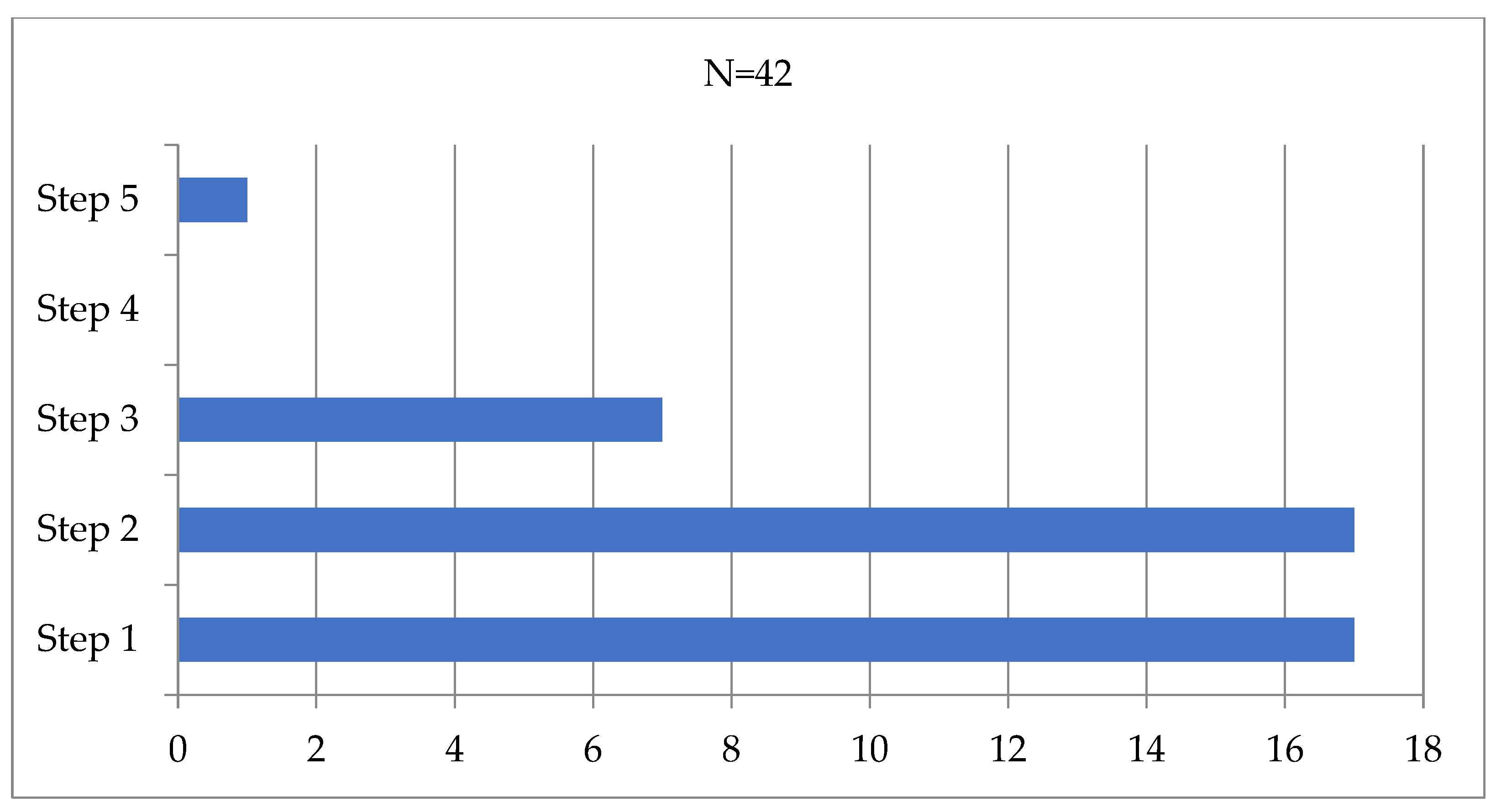
GINA treatment step
Step 1, no. (%)
Step 2, no. (%)
Step 3, no. (%)
Step 5, no (%)

9 (33%)
13 (48%)
4 (15%)
1 (4%)

8 (53%)
4 (27%)
3 (20%)
0 (0%)
χ2 = 2.76, 0.430
Well-controlled asthma, no. (%)25 (93%)3 (20%)χ2 = 22.87, <0.001
ACT score, points, mean (SD)23.6 (SD 1.9)19.7 (SD 1.7)t = 6.39, <0.001
New referrals, no. (%)4 (15%)2 (14%)χ2 = 0.02, 0.895
Therapy step-up, no. (%)3 (11%)7 (47%)χ2 = 6.72, 0.010
Abbreviations: ACT, Asthma Control Test; BMI, body mass index; ICS, inhaled corticosteroids; IQR, interquartile range; LRTI, lower respiratory tract infection; URTI, upper respiratory tract infection. Continuous variables were compared using the independent samples t-test (test statistic t) or Mann–Whitney U-test (Test statistic U), where appropriate, whereas categorical variables were compared by using Pearson chi-square test (test statistic χ2).
Table 2. Multiple logistic regression analysis for SARS-CoV-2 lower respiratory tract infection in children with asthma. Significant differences (p < 0.05) are highlighted in bold.
p ValueOdds Ratio (OR)95% CI for OR
Age
Gender
0.214
0.298
1.22
0.28
0.89–1.67
0.03–3.08
Atopy
Daily ICS dose
0.120
0.242
9.20
1.01
0.56–150.21
1.00–1.01
ACT score0.0030.390.21–0.73
Abbreviations: ACT, Asthma Control Test; CI, confidence interval; ICS, inhaled corticosteroids.
Table 3. Lung function before and after the diagnosis of COVID-19. Lung function measurements are shown as mean (SD).
All Patients
(n = 37)
p ValueSARS-CoV-2 URTI
(n = 27)
p ValueSARS-CoV-2 LRTI
(n = 10)
p Value
FVC, % of predicted
pre-COVID-19
96 (12)0.3094 (11)0.36100 (12)0.62
FVC, % of predicted
post-COVID-19
94 (14)92 (13)99 (16)
FEV1, % of predicted
pre-COVID-19
103 (11)0.32102 (11)0.62104 (13)0.17
FEV1, % of predicted
post-COVID-19
101 (12)101 (13)101 (13)
FEV1/FVC, %
pre-COVID-19
88 (7)0.9589 (8)0.7287 (5)0.42
FEV1/FVC, %
post-COVID-19
88 (5)89 (5)86 (3)
Abbreviations: FEV1, forced expiratory volume in one second; FVC, forced vital capacity; LRTI, lower respiratory tract infection; URTI, upper respiratory tract infection. Continuous variables were compared using the independent samples t-test.
Disclaimer/Publisher’s Note: The statements, opinions and data contained in all publications are solely those of the individual author(s) and contributor(s) and not of MDPI and/or the editor(s). MDPI and/or the editor(s) disclaim responsibility for any injury to people or property resulting from any ideas, methods, instructions or products referred to in the content.

