

Development of Novel Indole and Coumarin Derivatives as Antibacterial Agents That Target Histidine Kinase in S. aureus
 , , , , , , , ,
Abstract
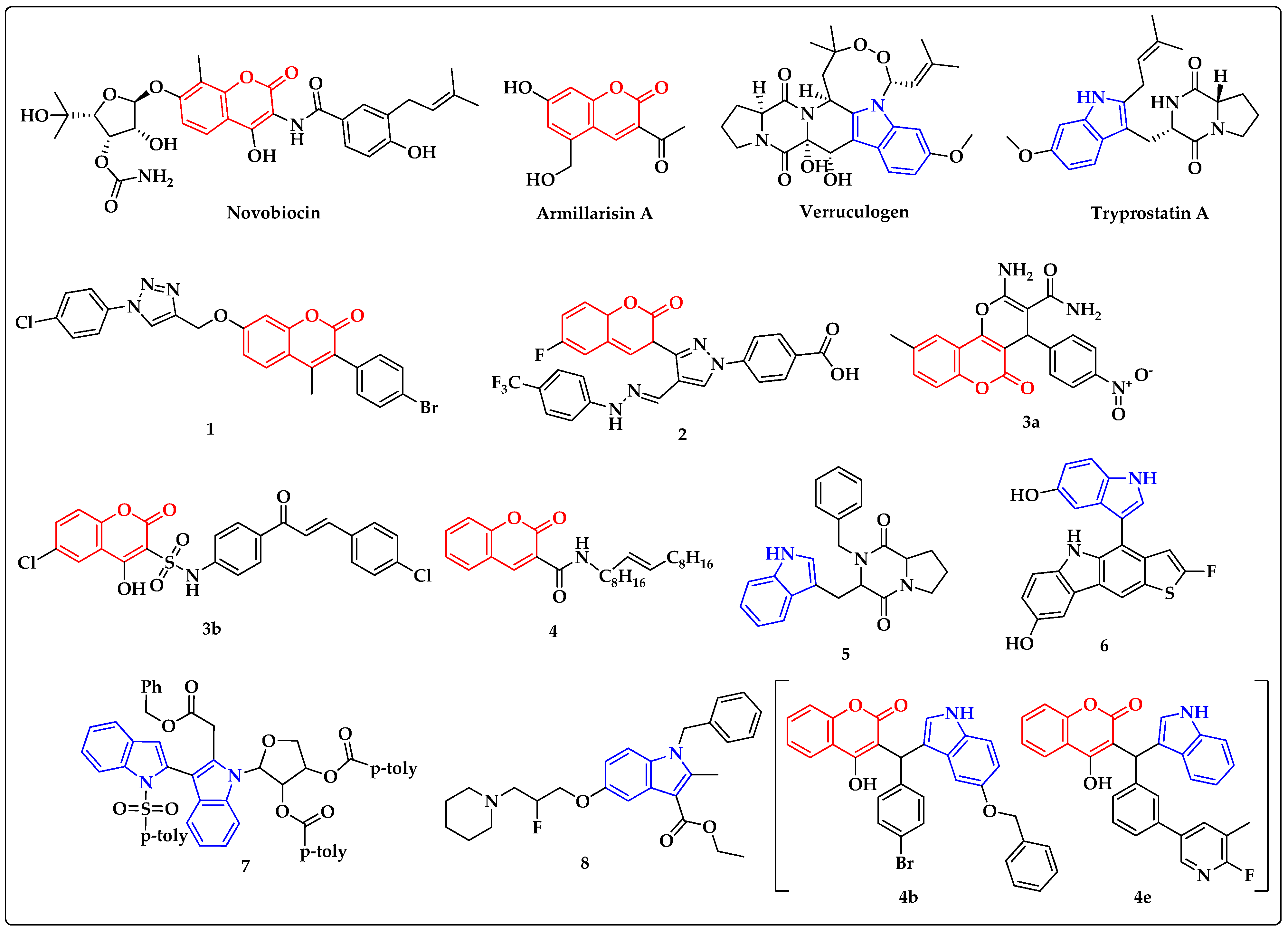
:1. Introduction
2. Materials and Methods
2.1. Chemistry
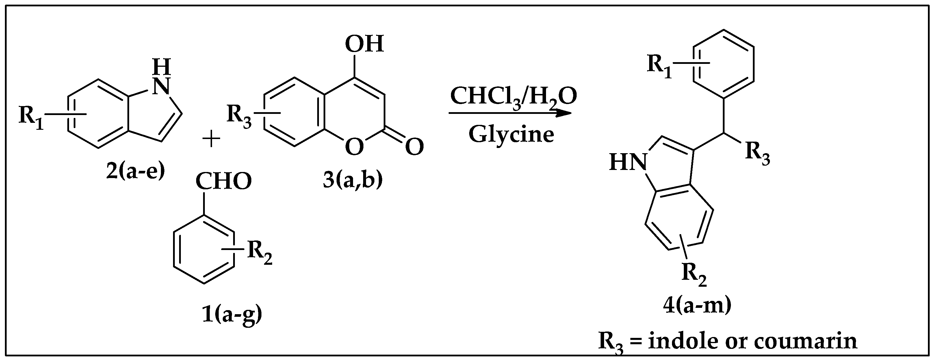
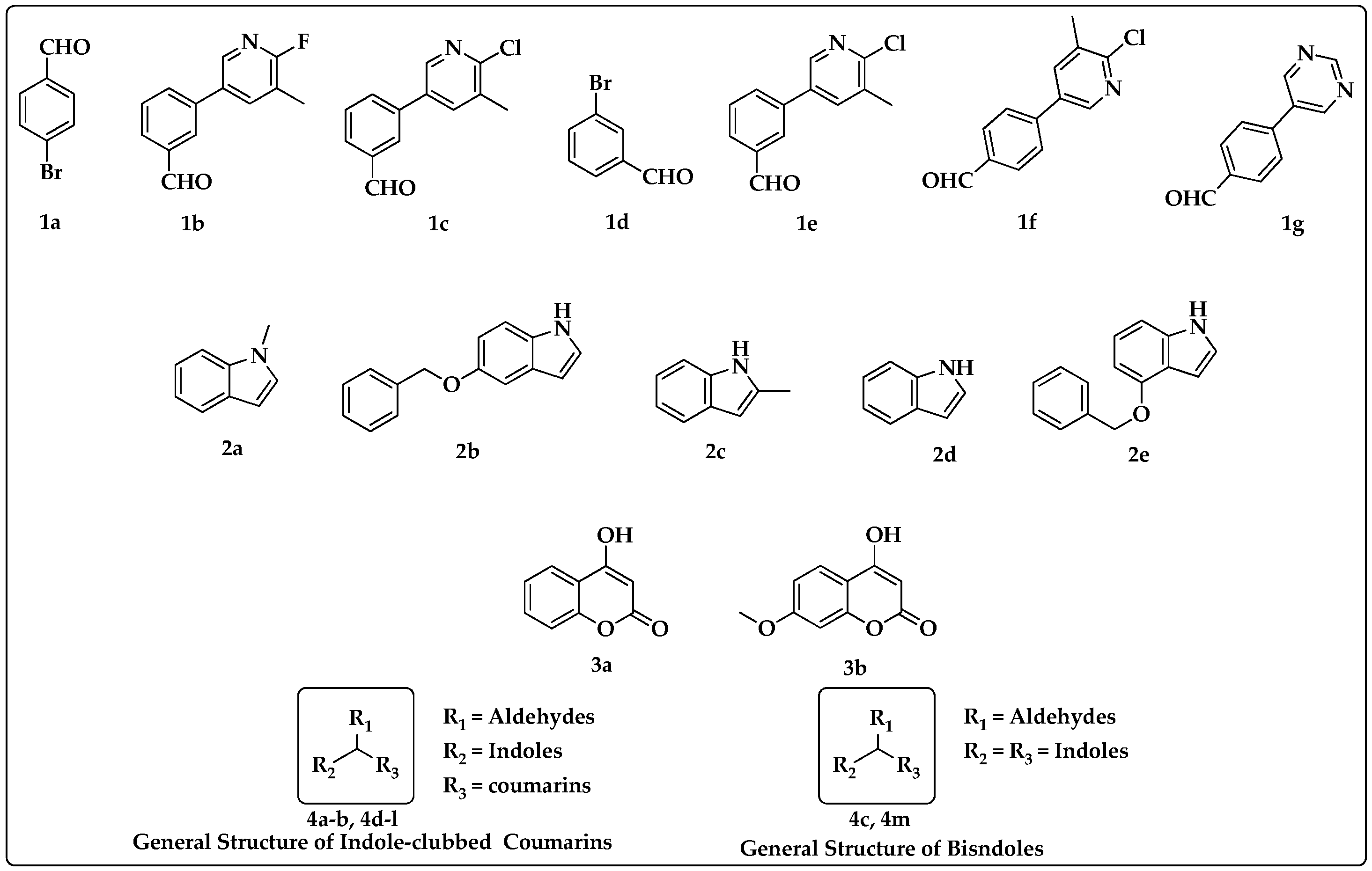
2.1.1. General Procedure for Synthesis of Indole Derivatives 4(a–m)
2.1.2. 3-((4-bromophenyl)(1-methyl-1H-indol-3-yl)methyl)-4-hydroxy-2H-chromen-2-one (4a)
2.1.3. 3-((5-(benzyloxy)-1H-indol-3-yl)(4-bromophenyl)methyl)-4-hydroxy-2H-chromen-2-one (4b)
2.1.4. 3,3′-((4-bromophenyl) methylene) bis(2-methyl-1H-indole) (4c)
2.1.5. 3-((3-(6-fluoro-5-methylpyridin-3-yl)phenyl)(1H-indol-3-yl)methyl)-4-hydroxy-7-meth-oxy-2H-chromen-2-one (4d)
2.1.6. 3-((3-(6-fluoro-5-methylpyridin-3-yl)phenyl)(1H-indol-3-yl)methyl)-4-hydroxy-2H-chromen-2-one (4e)
2.1.7. 3-((3-(6-chloro-5-methylpyridin-3-yl)phenyl)(1H-indol-3-yl)methyl)-4-hydroxy-2H-chromen-2-one (4f)
2.1.8. 3-((3-(6-chloro-5-methylpyridin-3-yl)phenyl)(1H-indol-3-yl)methyl)-4-hydroxy-7-methoxy-2H-chromen-2-one (4g)
2.1.9. 3-((3-bromophenyl)(1-methyl-1H-indol-3-yl)methyl)-4-hydroxy-7-methoxy-2H-chromen-2-one (4h)
2.1.10. 3-((3-bromophenyl)(1-methyl-1H-indol-3-yl)methyl)-4-hydroxy-2H-chromen-2-one (4i)
2.1.11. 3-((3-(6-chloro-5-methylpyridin-3-yl)phenyl)(1-methyl-1H-indol-3-yl)methyl)-4-hydroxy-2H-chromen-2-one (4j)
2.1.12. 3-((4-(benzyloxy)-1H-indol-3-yl) (3-(6-chloro-5-methylpyridin-3-yl) phenyl) methyl)-4-hydroxy-2H-chromen-2-one (4k)
2.1.13. 3-((4-(6-chloro-5-methylpyridin-3-yl)phenyl)(2-methyl-2,7a-dihydro-1H-indol-3-yl)-methyl)-4-hydroxy-2H-chromen-2-one (4l)
2.1.14. 3,3′-((4-(pyrimidin-5-yl) phenyl) methylene)bis(2-methyl-1H-indole (4m)
2.2. Antibacterial Activity of Compounds 4(a–m)
2.3. Minimum Inhibitory Concentration (MIC) of the Active Compounds
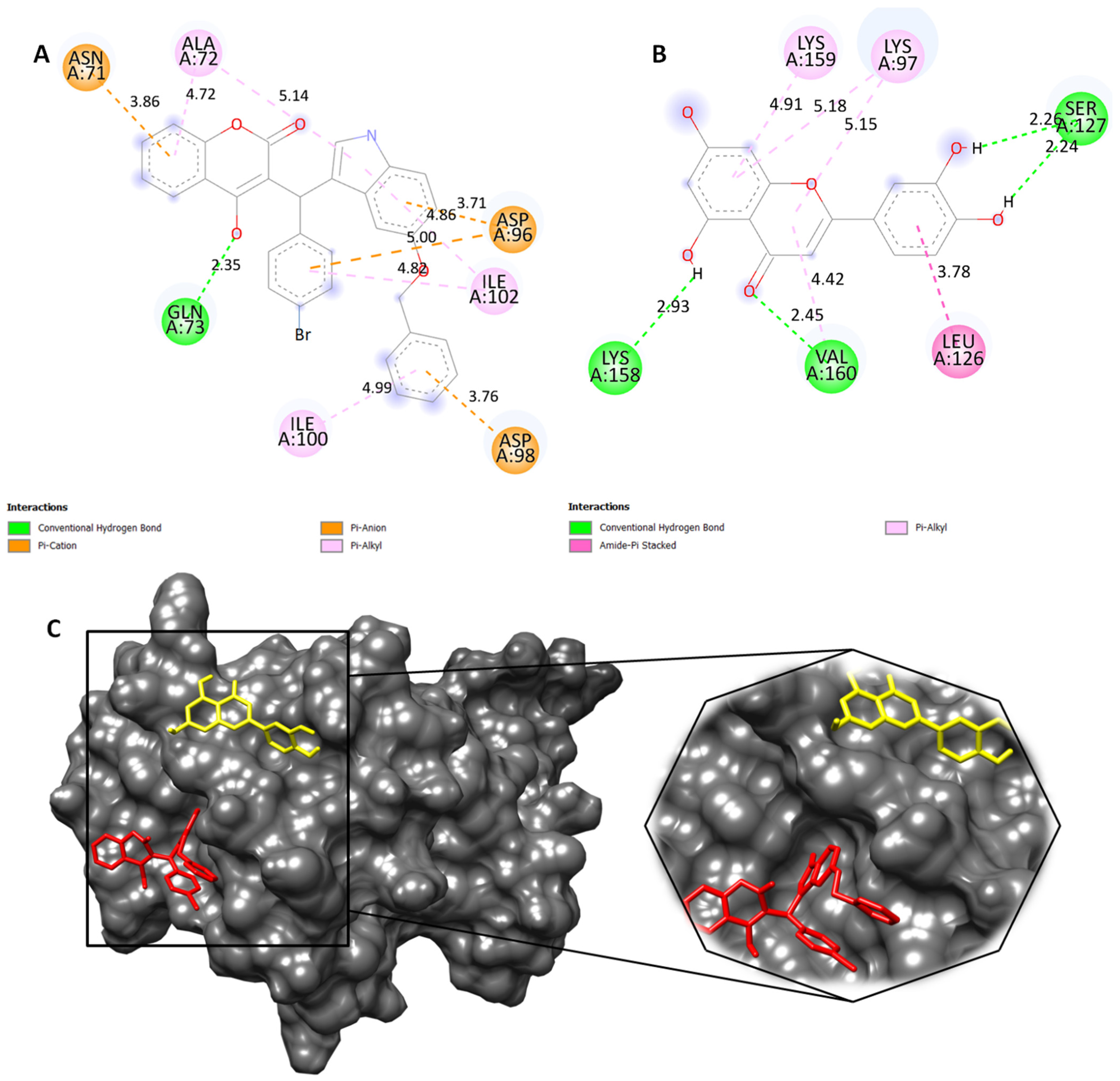
2.4. Molecular Docking Studies
3. Results
3.1. Synthesis of Bisindoles and Heterodimers of Indole-Clubbed Coumarin Derivatives 4a–m
3.2. Antibacterial Activity of Newly Synthesized Heterodimers of Indole-Clubbed Coumarins
3.3. In Silico Molecular Interaction Studies of Novel Compound 4b in Inhibiting Histidine Kinase of S. aureus
4. Discussion
5. Conclusions
Supplementary Materials
Author Contributions
Funding
Institutional Review Board Statement
Informed Consent Statement
Data Availability Statement
Conflicts of Interest
References
- Campbell, I.B.; Macdonald, S.J.F.; Procopiou, P.A. Medicinal Chemistry in Drug Discovery in Big Pharma: Past, Present and Future. Drug Discov. Today 2018, 23, 219–234. [Google Scholar] [CrossRef] [PubMed]
- Aatif, M.; Raza, M.A.; Javed, K.; Nashre-ul-Islam, S.M.; Farhan, M.; Alam, M.W. Potential Nitrogen-Based Heterocyclic Compounds for Treating Infectious Diseases: A Literature Review. Antibiotics 2022, 11, 1750. [Google Scholar] [CrossRef] [PubMed]
- Sheehan, J.C.; Logan, K.R.H. A General Synthesis of the Penicillins. J. Am. Chem. Soc. 1959, 81, 5838–5839. [Google Scholar] [CrossRef]
- Chellat, M.F.; Raguž, L.; Riedl, R. Targeting Antibiotic Resistance. Angew. Chem. Int. Ed. 2016, 55, 6600–6626. [Google Scholar] [CrossRef] [PubMed]
- Grillone, K.; Riillo, C.; Rocca, R.; Ascrizzi, S.; Spanò, V.; Scionti, F.; Polerà, N.; Maruca, A.; Barreca, M.; Juli, G.; et al. The New Microtubule-Targeting Agent SIX2G Induces Immunogenic Cell Death in Multiple Myeloma. Int. J. Mol. Sci. 2022, 23, 10222. [Google Scholar] [CrossRef] [PubMed]
- Barreca, M.; Spanò, V.; Rocca, R.; Bivacqua, R.; Gualtieri, G.; Raimondi, M.V.; Gaudio, E.; Bortolozzi, R.; Manfreda, L.; Bai, R.; et al. Identification of Pyrrolo[3′,4′:3,4]Cyclohepta[1,2-d][1,2]Oxazoles as Promising New Candidates for the Treatment of Lymphomas. Eur. J. Med. Chem. 2023, 254, 115372. [Google Scholar] [CrossRef] [PubMed]
- Lee, B.; Kim, D.G.; Lee, A.; Kim, Y.M.; Cui, L.; Kim, S.; Choi, I. Synthesis and Discovery of the First Potent Proteolysis Targeting Chimaera (PROTAC) Degrader of AIMP2-DX2 as a Lung Cancer Drug. J. Enzym. Inhib. Med. Chem. 2022, 38, 51–66. [Google Scholar] [CrossRef] [PubMed]
- Hajduk, P.J.; Greer, J. A Decade of Fragment-Based Drug Design: Strategic Advances and Lessons Learned. Nat. Rev. Drug Discov. 2007, 6, 211–219. [Google Scholar] [CrossRef]
- Bivacqua, R.; Barreca, M.; Spanò, V.; Raimondi, M.V.; Romeo, I.; Alcaro, S.; Andrei, G.; Barraja, P.; Montalbano, A. Insight into Non-Nucleoside Triazole-Based Systems as Viral Polymerases Inhibitors. Eur. J. Med. Chem. 2023, 249, 115136. [Google Scholar] [CrossRef]
- Ling, L.L.; Schneider, T.; Peoples, A.J.; Spoering, A.L.; Engels, I.; Conlon, B.P.; Mueller, A.; Schäberle, T.F.; Hughes, D.E.; Epstein, S.; et al. A New Antibiotic Kills Pathogens without Detectable Resistance. Nature 2015, 517, 455–459. [Google Scholar] [CrossRef]
- Sass, P.; Josten, M.; Famulla, K.; Schiffer, G.; Sahl, H.-G.; Hamoen, L.; Brötz-Oesterhelt, H. Antibiotic Acyldepsipeptides Activate ClpP Peptidase to Degrade the Cell Division Protein FtsZ. Proc. Natl. Acad. Sci. USA 2011, 108, 17474–17479. [Google Scholar] [CrossRef] [PubMed]
- Smith, P.A.; Koehler, M.F.T.; Girgis, H.S.; Yan, D.; Chen, Y.; Chen, Y.; Crawford, J.J.; Durk, M.R.; Higuchi, R.I.; Kang, J.; et al. Optimized Arylomycins Are a New Class of Gram-Negative Antibiotics. Nature 2018, 561, 189–194. [Google Scholar] [CrossRef] [PubMed]
- Shariati, A.; Arshadi, M.; Khosrojerdi, M.A.; Abedinzadeh, M.; Ganjalishahi, M.; Maleki, A.; Heidary, M.; Khoshnood, S. The Resistance Mechanisms of Bacteria against Ciprofloxacin and New Approaches for Enhancing the Efficacy of This Antibiotic. Front. Public Health 2022, 10, 1025633. [Google Scholar] [CrossRef] [PubMed]
- Shariati, A.; Noei, M.; Chegini, Z. Bacteriophages: The Promising Therapeutic Approach for Enhancing Ciprofloxacin Efficacy against Bacterial Infection. J. Clin. Lab. Anal. 2023, 37, e24932. [Google Scholar] [CrossRef] [PubMed]
- Lowy, F.D. Staphylococcus aureus Infections. N. Engl. J. Med. 1998, 339, 520–532. [Google Scholar] [CrossRef] [PubMed]
- Zimmermann, S.; Klinger-Strobel, M.; Bohnert, J.A.; Wendler, S.; Rödel, J.; Pletz, M.W.; Löffler, B.; Tuchscherr, L. Clinically Approved Drugs Inhibit the Staphylococcus aureus Multidrug NorA Efflux Pump and Reduce Biofilm Formation. Front. Microbiol. 2019, 10, 2762. [Google Scholar] [CrossRef] [PubMed]
- Santajit, S.; Indrawattana, N. Mechanisms of Antimicrobial Resistance in ESKAPE Pathogens. BioMed Res. Int. 2016, 2016, 2475067. [Google Scholar] [CrossRef] [PubMed]
- Mulani, M.S.; Kamble, E.E.; Kumkar, S.N.; Tawre, M.S.; Pardesi, K.R. Emerging Strategies to Combat ESKAPE Pathogens in the Era of Antimicrobial Resistance: A Review. Front. Microbiol. 2019, 10, 539. [Google Scholar] [CrossRef]
- Balouiri, M.; Sadiki, M.; Ibnsouda, S.K. Methods for in Vitro Evaluating Antimicrobial Activity: A Review. J. Pharm. Anal. 2016, 6, 71–79. [Google Scholar] [CrossRef]
- Bem, A.E.; Velikova, N.; Pellicer, M.T.; van Baarlen, P.; Marina, A.; Wells, J.M. Bacterial Histidine Kinases as Novel Antibacterial Drug Targets. ACS Chem. Biol. 2014, 10, 213–224. [Google Scholar] [CrossRef]
- Casino, P.; Rubio, V.; Marina, A. The Mechanism of Signal Transduction by Two-Component Systems. Curr. Opin. Struct. Biol. 2010, 20, 763–771. [Google Scholar] [CrossRef] [PubMed]
- Kenney, L.J. How Important Is the Phosphatase Activity of Sensor Kinases? Curr. Opin. Microbiol. 2010, 13, 168–176. [Google Scholar] [CrossRef] [PubMed]
- Stewart, R.C. Protein Histidine Kinases: Assembly of Active Sites and Their Regulation in Signaling Pathways. Curr. Opin. Microbiol. 2010, 13, 133–141. [Google Scholar] [CrossRef] [PubMed]
- Wolanin, P.M.; Thomason, P.A.; Stock, J.B. Histidine protein kinases: Key signal transducers outside the animal kingdom. Genome Biol. 2002, 3, reviews3013.1. [Google Scholar] [CrossRef] [PubMed]
- Dikiy, I.; Edupuganti, U.R.; Abzalimov, R.R.; Borbat, P.P.; Srivastava, M.; Freed, J.H.; Gardner, K.H. Insights into Histidine Kinase Activation Mechanisms from the Monomeric Blue Light Sensor EL346. Proc. Natl. Acad. Sci. USA 2019, 116, 4963–4972. [Google Scholar] [CrossRef] [PubMed]
- Kenney, L.J. How Can a Histidine Kinase Respond to Mechanical Stress? Front. Microbiol. 2021, 12, 655942. [Google Scholar] [CrossRef] [PubMed]
- Chen, H.; Yu, C.; Wu, H.; Li, G.; Li, C.; Hong, W.; Yang, X.; Wang, H.; You, X. Recent Advances in Histidine Kinase-Targeted Antimicrobial Agents. Front. Chem. 2022, 10, 866392. [Google Scholar] [CrossRef] [PubMed]
- Vo, C.D.; Shebert, H.L.; Zikovich, S.; Dryer, R.A.; Huang, T.P.; Moran, L.J.; Cho, J.; Wassarman, D.R.; Falahee, B.E.; Young, P.D.; et al. Repurposing Hsp90 Inhibitors as Antibiotics Targeting Histidine Kinases. Bioorganic Med. Chem. Lett. 2017, 27, 5235–5244. [Google Scholar] [CrossRef]
- Mascher, T.; Helmann, J.D.; Unden, G. Stimulus Perception in Bacterial Signal-Transducing Histidine Kinases. Microbiol. Mol. Biol. Rev. 2006, 70, 910–938. [Google Scholar] [CrossRef]
- Singh, V.; Dhankhar, P.; Kumar, P. Bacterial Histidine Kinases as Potential Antibacterial Drug Targets. Protein Kinase Inhib. 2022, 26, 711–734. [Google Scholar] [CrossRef]
- Roychoudhury, S.; Zielinski, N.A.; Ninfa, A.J.; Allen, N.E.; Jungheim, L.N.; Nicas, T.I.; Chakrabarty, A.M. Inhibitors of Two-Component Signal Transduction Systems: Inhibition of Alginate Gene Activation in Pseudomonas Aeruginosa. Proc. Natl. Acad. Sci. USA 1993, 90, 965–969. [Google Scholar] [CrossRef] [PubMed]
- Johnson, B.A.; Anker, H.; Meleney, F.L. Bacitracin: A New Antibiotic Produced by a Member of the B. subtilis Group. Science 1945, 102, 376–377. [Google Scholar] [CrossRef] [PubMed]
- Stone, K.J.; Strominger, J.L. Mechanism of Action of Bacitracin: Complexation with Metal Ion and C55-Isoprenyl Pyrophosphate. Proc. Natl. Acad. Sci. USA 1971, 68, 3223–3227. [Google Scholar] [CrossRef] [PubMed]
- Ma, W.; Zhang, D.; Li, G.; Liu, J.; He, G.; Zhang, P.; Yang, L.; Zhu, H.; Xu, N.; Liang, S. Antibacterial Mechanism of Daptomycin Antibiotic against Staphylococcus aureus Based on a Quantitative Bacterial Proteome Analysis. J. Proteom. 2017, 150, 242–251. [Google Scholar] [CrossRef] [PubMed]
- Dinu, V.; Lu, Y.; Weston, N.; Lithgo, R.; Coupe, H.; Channell, G.; Adams, G.G.; Torcello Gómez, A.; Sabater, C.; Mackie, A.; et al. The Antibiotic Vancomycin Induces Complexation and Aggregation of Gastrointestinal and Submaxillary Mucins. Sci. Rep. 2020, 10, 960. [Google Scholar] [CrossRef] [PubMed]
- Shamsudin, N.F.; Ahmed, Q.U.; Mahmood, S.; Ali Shah, S.A.; Khatib, A.; Mukhtar, S.; Alsharif, M.A.; Parveen, H.; Zakaria, Z.A. Antibacterial Effects of Flavonoids and Their Structure-Activity Relationship Study: A Comparative Interpretation. Molecules 2022, 27, 1149. [Google Scholar] [CrossRef] [PubMed]
- Advances in Clinical Chemistry|Book Series|ScienceDirect.com by Elsevier. 1 January 2023. Available online: https://www.sciencedirect.com/bookseries/advances-in-clinical-chemistry (accessed on 20 July 2023).
- Foudah, A.I.; Alqarni, M.H.; Ross, S.A.; Alam, A.; Salkini, M.A.; Kumar, P. Site-Specific Evaluation of Bioactive Coumarin-Loaded Dendrimer G4 Nanoparticles against Methicillin Resistant Staphylococcus aureus. ACS Omega 2022, 7, 34990–34996. [Google Scholar] [CrossRef]
- Basile, A.; Sorbo, S.; Spadaro, V.; Bruno, M.; Maggio, A.; Faraone, N.; Rosselli, S. Antimicrobial and Antioxidant Activities of Coumarins from the Roots of Ferulago campestris (Apiaceae). Molecules 2009, 14, 939–952. [Google Scholar] [CrossRef]
- Yang, X.-C.; Zeng, C.-M.; Avula, S.R.; Peng, X.-M.; Geng, R.-X.; Zhou, C.-H. Novel Coumarin Aminophosphonates as Potential Multitargeting Antibacterial Agents against Staphylococcus aureus. Eur. J. Med. Chem. 2023, 245, 114891. [Google Scholar] [CrossRef]
- Ranjan Sahoo, C.; Sahoo, J.; Mahapatra, M.; Lenka, D.; Kumar Sahu, P.; Dehury, B.; Nath Padhy, R.; Kumar Paidesetty, S. Coumarin Derivatives as Promising Antibacterial Agent(s). Arab. J. Chem. 2021, 14, 102922. [Google Scholar] [CrossRef]
- Vickers, A.A.; Chopra, I.; O’Neill, A.J. Intrinsic Novobiocin Resistance in Staphylococcus Saprophyticus. Antimicrob. Agents Chemother. 2007, 51, 4484–4485. [Google Scholar] [CrossRef] [PubMed]
- Walsh, T.J.; Hansen, S.L.; Tatem, B.A.; Auger, F.; Standiford, H.C. Activity of Novobiocin against Methicillin-resistant Staphylococcus aureus. J. Antimicrob. Chemother. 1985, 15, 435–440. [Google Scholar] [CrossRef] [PubMed]
- Wishnow, R.M.; Strominger, J.L.; Birge, C.H.; Threnn, R.H. Biochemical Effects of Novobiocin on Staphylococcus aureus. J. Bacteriol. 1965, 89, 1117–1123. [Google Scholar] [CrossRef] [PubMed]
- Kashman, Y.; Gustafson, K.R.; Fuller, R.W.; Cardellina, J.H.; McMahon, J.B.; Currens, M.J.; Buckheit, R.W.; Hughes, S.H.; Cragg, G.M.; Boyd, M.R. HIV Inhibitory Natural Products. Part 7. The Calanolides, a Novel HIV-Inhibitory Class of Coumarin Derivatives from the Tropical Rainforest Tree, Calophyllum lanigerum. J. Med. Chem. 1992, 35, 2735–2743. [Google Scholar] [CrossRef]
- Dharavath, R.; Nagaraju, N.; Reddy, M.R.; Ashok, D.; Sarasija, M.; Vijjulatha, M.; Vani, T.; Jyothi, K.; Prashanthi, G. Microwave-Assisted Synthesis, Biological Evaluation and Molecular Docking Studies of New Coumarin-Based 1,2,3-Triazoles. RSC Adv. 2020, 10, 11615–11623. [Google Scholar] [CrossRef] [PubMed]
- Alnufaie, R.; Raj KC, H.; Alsup, N.; Whitt, J.; Andrew Chambers, S.; Gilmore, D.; Alam, M.A. Synthesis and Antimicrobial Studies of Coumarin-Substituted Pyrazole Derivatives as Potent Anti Staphylococcus aureus Agents. Molecules 2020, 25, 2758. [Google Scholar] [CrossRef] [PubMed]
- Alshibl, H.M.; Al-Abdullah, E.S.; Haiba, M.E.; Alkahtani, H.M.; Awad, G.E.A.; Mahmoud, A.H.; Ibrahim, B.M.M.; Bari, A.; Villinger, A. Synthesis and Evaluation of New Coumarin Derivatives as Antioxidant, Antimicrobial, and AntiInflammatory Agents. Molecules 2020, 25, 3251. [Google Scholar] [CrossRef] [PubMed]
- Sharma, R.K.; Singh, V.; Tiwari, N.; Butcher, R.J.; Katiyar, D. Synthesis, Antimicrobial and Chitinase Inhibitory Activities of 3-Amidocoumarins. Bioorganic Chem. 2020, 98, 103700. [Google Scholar] [CrossRef]
- Lee, J.-H.; Cho, H.S.; Kim, Y.; Kim, J.-A.; Banskota, S.; Cho, M.H.; Lee, J. Indole and 7-Benzyloxyindole Attenuate the Virulence of Staphylococcus aureus. Appl. Microbiol. Biotechnol. 2013, 97, 4543–4552. [Google Scholar] [CrossRef]
- Qin, H.-L.; Liu, J.; Fang, W.-Y.; Ravindar, L.; Rakesh, K.P. Indole-Based Derivatives as Potential Antibacterial Activity against Methicillin-Resistance Staphylococcus aureus (MRSA). Eur. J. Med. Chem. 2020, 194, 112245. [Google Scholar] [CrossRef]
- Jia, B.; Ma, Y.; Liu, B.; Chen, P.; Hu, Y.; Zhang, R. Synthesis, Antimicrobial Activity, Structure-Activity Relationship, and Molecular Docking Studies of Indole Diketopiperazine Alkaloids. Front. Chem. 2019, 7, 837. [Google Scholar] [CrossRef] [PubMed]
- Seethaler, M.; Hertlein, T.; Hopke, E.; Köhling, P.; Ohlsen, K.; Lalk, M.; Hilgeroth, A. Novel Effective Fluorinated Benzothiophene-Indole Hybrid Antibacterials against S. aureus and MRSA Strains. Pharmaceuticals 2022, 15, 1138. [Google Scholar] [CrossRef] [PubMed]
- Paudel, A.; Hamamoto, H.; Kobayashi, Y.; Yokoshima, S.; Fukuyama, T.; Sekimizu, K. Identification of Novel Deoxyribofuranosyl Indole Antimicrobial Agents. J. Antibiot. 2011, 65, 53–57. [Google Scholar] [CrossRef] [PubMed]
- Andrews, J.M. Determination of Minimum Inhibitory Concentrations. J. Antimicrob. Chemother. 2001, 48, 5–16. [Google Scholar] [CrossRef] [PubMed]
- Kowalska-Krochmal, B.; Dudek-Wicher, R. The Minimum Inhibitory Concentration of Antibiotics: Methods, Interpretation, Clinical Relevance. Pathogens 2021, 10, 165. [Google Scholar] [CrossRef] [PubMed]
- Huey, R.; Morris, G.M.; Olson, A.J.; Goodsell, D.S. A Semiempirical Free Energy Force Field with Charge-Based Desolvation. J. Comput. Chem. 2007, 28, 1145–1152. [Google Scholar] [CrossRef] [PubMed]
- Schrödinger, L.L.C.; DeLano, W. PyMOL. 2020. Available online: http://www.pymol.org/pymol (accessed on 23 May 2023).
- BIOVIA Dassault Systèmes. Discovery Studio Visualizer, 21.1.0.20298; Dassault Systèmes: San Diego, CA, USA, 2020. [Google Scholar]
- Pettersen, E.F.; Goddard, T.D.; Huang, C.C.; Couch, G.S.; Greenblatt, D.M.; Meng, E.C.; Ferrin, T.E. UCSF Chimera—A visualization system for exploratory research and analysis. J. Comput. Chem. 2004, 25, 1605–1612. [Google Scholar] [CrossRef]
- Lagunin, A.; Stepanchikova, A.; Filimonov, D.; Poroikov, V. PASS: Prediction of Activity Spectra for Biologically Active Substances. Bioinformatics 2000, 16, 747–748. [Google Scholar] [CrossRef]
- Datta, B.; Pasha, M.A. Glycine Catalyzed Convenient Synthesis of 2-Amino-4H-Chromenes in Aqueous Medium under Sonic Condition. Ultrason. Sonochem. 2012, 19, 725–728. [Google Scholar] [CrossRef]
- Valot, L.; Maumus, M.; Montheil, T.; Martinez, J.; Noël, D.; Mehdi, A.; Subra, G. Biocompatible Glycine-Assisted Catalysis of the Sol-Gel Process: Development of Cell-Embedded Hydrogels. ChemPlusChem 2019, 84, 1720–1729. [Google Scholar] [CrossRef]
- Sulaiman, N.B.; Mohan, C.D.; Basappa, S.; Pandey, V.; Rangappa, S.; Bharathkumar, H.; Kumar, A.P.; Lobie, P.E.; Rangappa, K.S. An azaspirane derivative suppresses growth and induces apoptosis of ER-positive and ER-negative breast cancer cells through the modulation of JAK2/STAT3 signaling pathway. Int J Oncol. 2016, 49, 1221–1229. [Google Scholar] [CrossRef]
- Bharathkumar, H.; Mohan, C.D.; Ananda, H.; Fuchs, J.E.; Li, F.; Rangappa, S.; Surender, M.; Bulusu, K.C.; Girish, K.S.; Sethi, G.; et al. Microwave-assisted synthesis, characterization and cytotoxic studies of novel estrogen receptor α ligands towards human breast cancer cells. Bioorg. Med. Chem. Lett. 2015, 25, 1804–1807. [Google Scholar] [CrossRef] [PubMed]
- Blanchard, V.; Chevalier, F.; Imberty, A.; Leeflang, B.R.; Basappa; Sugahara, K.; Kamerling, J.P. Conformational studies on five octasaccharides isolated from chondroitin sulfate using NMR spectroscopy and molecular modeling. Biochemistry 2007, 46, 1167–1175. [Google Scholar] [CrossRef] [PubMed]
- Rangappa, K.S.; Basappa. New cholinesterase inhibitors: Synthesis and structure–activity relationship studies of 1,2-benzisoxazole series and novel imidazolyl-d2-isoxazolines. J. Phys. Org. Chem. 2005, 18, 773–778. [Google Scholar] [CrossRef]
- Sebastian, A.; Pandey, V.; Mohan, C.D.; Chia, Y.T.; Rangappa, S.; Mathai, J.; Baburajeev, C.P.; Paricharak, S.; Mervin, L.H.; Bulusu, K.C.; et al. Novel Adamantanyl-Based Thiadiazolyl Pyrazoles Targeting EGFR in Triple-Negative Breast Cancer. ACS Omega 2016, 1, 1412–1424. [Google Scholar] [CrossRef] [PubMed]
- Srinivas, V.; Mohan, C.D.; Baburajeev, C.P.; Rangappa, S.; Jagadish, S.; Fuchs, J.E.; Sukhorukov, A.Y.; Chandra; Mason, D.J.; Sharath; et al. Synthesis and characterization of novel oxazines and demonstration that they specifically target cyclooxygenase 2. Bioorg. Med. Chem. Lett. 2015, 25, 2931–2936. [Google Scholar] [CrossRef] [PubMed]
- Basappa; Sugahara, K.; Thimmaiah, K.N.; Bid, H.K.; Houghton, P.J.; Rangappa, K.S. Anti-tumor activity of a novel HS-mimetic-vascular endothelial growth factor binding small molecule. PLoS ONE 2012, 7, e39444. [Google Scholar] [CrossRef]
- Priya, B.S.; Swamy, S.N.; Tejesvi, M.V.; Basappa; Sarala, G.; Gaonkar, S.L.; Naveen, S.; Prasad, J.S.; Rangappa, K.S. Synthesis, characterization, antimicrobial and single crystal X-ray crystallographic studies of some new sulfonyl, 4-chloro phenoxy benzene and dibenzoazepine substituted benzamides. Eur. J. Med. Chem. 2006, 41, 1262–1270. [Google Scholar] [CrossRef]
- Rakesh, K.S.; Jagadish, S.; Vinayaka, A.C.; Hemshekhar, M.; Paul, M.; Thushara, R.M.; Sundaram, M.S.; Swaroop, T.R.; Mohan, C.D.; Basappa; et al. A new ibuprofen derivative inhibits platelet aggregation and ROS mediated platelet apoptosis. PLoS ONE 2014, 9, e107182, Erratum in PLoS ONE 2014, 9, e114675. [Google Scholar] [CrossRef]
- Basappa; Rangappa, K.S.; Sugahara, K. Roles of glycosaminoglycans and glycanmimetics in tumor progression and metastasis. Glycoconj. J. 2014, 31, 461–467. [Google Scholar] [CrossRef]


| Entry | Catalyst | Solvent | Time (h) | Yield (%) |
|---|---|---|---|---|
| 1 | Nano Fe2O3 | Ethanol | 12 | 50 |
| 2 | - | Water/Chloroform | 12 | 73 |
| 3 | Glycine | Water/Chloroform | 12 | 92 |
| 4 | I2 | DMSO | 12 | Not formed |
| Entry | Aldehydes (R1) | Indoles (R2) | Coumarin (R3) |
|---|---|---|---|
| 4a | 1a | 2a | 3a |
| 4b | 1a | 2b | 3a |
| 4c | 1a | 2c | - |
| 4d | 1b | 2d | 3b |
| 4e | 1b | 2d | 3a |
| 4f | 1c | 2d | 3a |
| 4g | 1c | 2d | 3b |
| 4h | 1d | 2a | 3b |
| 4i | 1d | 2a | 3a |
| 4j | 1e | 2a | 3a |
| 4k | 1e | 2e | 3a |
| 4l | 1f | 2c | 3a |
| 4m | 1g | 2c | - |
| Compound | MIC (µg/mL) |
|---|---|
| S. aureus | |
| 4b (350 µg/mL) | 0.256 ± 0.005 |
| 4e (160 µg/mL) | 0.290 ± 0.01 |
| Negative control (1:1) Ethanol:distilled water | 0.673 ± 0.011 |
| Positive control Tetracycline | 0.206 ± 0.015 |
| Compound | Diameter of Zone of Inhibition (ZOI) (mm) |
|---|---|
| S. aureus | |
| 4b (350 µg/mL) | 8.0 mm |
| 4e (160 µg/mL) | 7.2 mm |
| Negative control (1:1) Ethanol:distilled water | - |
| Tetracycline (30 µg/disc) | 16.25 mm |
Disclaimer/Publisher’s Note: The statements, opinions and data contained in all publications are solely those of the individual author(s) and contributor(s) and not of MDPI and/or the editor(s). MDPI and/or the editor(s) disclaim responsibility for any injury to people or property resulting from any ideas, methods, instructions or products referred to in the content. |
© 2023 by the authors. Licensee MDPI, Basel, Switzerland. This article is an open access article distributed under the terms and conditions of the Creative Commons Attribution (CC BY) license (https://creativecommons.org/licenses/by/4.0/).
Share and Cite
Poonacha, L.K.; Ramesh, R.; Ravish, A.; Mohan, A.; Uppar, P.M.; Metri, P.K.; Shivananju, N.S.; Gaonkar, S.L.; Gopal, S.; Sukhorukov, A.Y.; et al. Development of Novel Indole and Coumarin Derivatives as Antibacterial Agents That Target Histidine Kinase in S. aureus. Appl. Microbiol. 2023, 3, 1214-1228. https://doi.org/10.3390/applmicrobiol3040084
Poonacha LK, Ramesh R, Ravish A, Mohan A, Uppar PM, Metri PK, Shivananju NS, Gaonkar SL, Gopal S, Sukhorukov AY, et al. Development of Novel Indole and Coumarin Derivatives as Antibacterial Agents That Target Histidine Kinase in S. aureus. Applied Microbiology. 2023; 3(4):1214-1228. https://doi.org/10.3390/applmicrobiol3040084
Chicago/Turabian StylePoonacha, Lisha K., Rashmi Ramesh, Akshay Ravish, Arunkumar Mohan, Pradeep M. Uppar, Prashant K. Metri, Nanjunda Swamy Shivananju, Santosh L. Gaonkar, Shubha Gopal, Alexey Yu Sukhorukov, and et al. 2023. "Development of Novel Indole and Coumarin Derivatives as Antibacterial Agents That Target Histidine Kinase in S. aureus" Applied Microbiology 3, no. 4: 1214-1228. https://doi.org/10.3390/applmicrobiol3040084
APA StylePoonacha, L. K., Ramesh, R., Ravish, A., Mohan, A., Uppar, P. M., Metri, P. K., Shivananju, N. S., Gaonkar, S. L., Gopal, S., Sukhorukov, A. Y., Pandey, V., Shubha, P. B., & Basappa, B. (2023). Development of Novel Indole and Coumarin Derivatives as Antibacterial Agents That Target Histidine Kinase in S. aureus. Applied Microbiology, 3(4), 1214-1228. https://doi.org/10.3390/applmicrobiol3040084