Article

Molecular Identification of Trypanosoma cruzi Isolated from Wild Triatomines and Evaluation of Its Pathogenicity in Experimental Hosts

by
Ana Lucía Torres-Barajas
1,
Melissa Paola Rincón-González
1,
Sandra Luz Martínez-Hernández
1,
Martín Humberto Muñoz-Ortega
2,
David Ibarra-Martínez
2,
Eduardo Sánchez-García
3,
Erick López-Macías
4,
Alberto Aguayo-Acosta
5,
Joel Horacio Elizondo-Luevano
6 and
David Alejandro Hernández-Marín
1,*
1
Departamento de Microbiología, Centro de Ciencias Básicas, Benemérita Universidad Autónoma de Aguascalientes, Aguascalientes 20100, Mexico
2
Departamento de Química, Centro de Ciencias Básicas, Benemérita Universidad Autónoma de Aguascalientes, Aguascalientes 20100, Mexico
3
Departamento de Química, Facultad de Ciencias Biológicas, Universidad Autónoma de Nuevo León, San Nicolás de los Garza 66455, Mexico
4
Área de Microbiología Clínica, Hospital General de Zona No. 3, Instituto Mexicano del Seguro Social, Av. General Prolongación, Zaragoza 20908, Mexico
5
Cell and Molecular Biology Department, University of Mississippi Medical Center, 2500 N State St., Jackson, MS 39216, USA
6
Infectious and Tropical Diseases Group (e-INTRO), IBSAL-CIETUS (Biomedical Research Institute of Salamanca, Centre for Tropical Diseases at the University of Salamanca), Faculty of Pharmacy, Universidad de Salamanca, Campus Miguel de Unamuno s/n, 37007 Salamanca, Spain
*
Author to whom correspondence should be addressed.
Parasitologia 2025, 5(3), 46; https://doi.org/10.3390/parasitologia5030046
Submission received: 24 July 2025 / Revised: 17 August 2025 / Accepted: 28 August 2025 / Published: 2 September 2025

Abstract

Trypanosoma cruzi is a hemoflagellate protozoan and the causative agent of Chagas disease, also known as American trypanosomiasis. Transmission occurs through the feces of triatomine insects, its biological vector. It is estimated that around 7 million people are infected across Mexico, Central America, and South America. This study aimed to identify and characterize T. cruzi isolates obtained from wild triatomine vectors collected in Aguascalientes, Mexico. Molecular identification was performed at different developmental stages—epimastigotes in culture media, metacyclic trypomastigotes in triatomine feces, and amastigotes in mouse cardiac tissue—using endpoint PCR targeting satDNA and mtCytB regions. In addition, next-generation sequencing was employed to analyze variable regions of kinetoplast DNA minicircles. The pathogenicity of the isolated and identified T. cruzi strain was assessed in a murine model, where trypomastigote stages were detected in peripheral blood and amastigote stages in muscle tissue. Molecular analyses confirmed the presence of T. cruzi across different developmental stages from wild vectors, demonstrating that the isolated wild strain possesses pathogenic potential when completing its life cycle in an experimental mammalian host, specifically BALB/c mice.

Graphical Abstract

1. Introduction

The hemoflagellate protozoan Trypanosoma cruzi is the causative agent of Chagas disease, also known as American trypanosomiasis. This parasite is carried by a biological vector called triatomine and transmitted through its feces [1]. A current estimate of the disease in Mexico, Central America, and South America calculates that around 7 million people could be infected. This number is not precise because in the acute stage of the disease, it presents nonspecific and mild symptoms (fever, headache, muscle pain, among others), affecting the accurate diagnosis of the disease, which may even go undetected. Additionally, the disease can be asymptomatic, putting the physical integrity of people who are unaware of being carriers at risk [2].
The vectors of T. cruzi are triatomine bugs, which belong to the family Reduviidae and the order Hemiptera. The specific vector species vary according to the geographic region where the disease is endemic. In Mexico, 34 species of triatomines have been identified, distributed across seven genera, of which the genus Triatoma is the most abundant, comprising 27 species [3,4]. Although the state of Aguascalientes, Mexico, has not traditionally been considered an endemic area for Chagas disease, recent studies have reported the presence of Meccus longipennis and Meccus phyllosoma (previously classified under the genus Triatoma) as carriers of T. cruzi in several localities within the municipality of Calvillo, Aguascalientes [5,6].
The identification of T. cruzi can be performed using various direct and indirect methods, including microscopy (direct visualization of the parasite) and immunological assays such as the detection of specific IgG antibodies against T. cruzi [7]. However, molecular identification remains a more reliable and sensitive approach compared with microscopy and immunological tests [8,9,10]. Molecular techniques can confirm the presence of this microorganism in a wide range of biological samples that are suspected to contain the parasite. The polymerase chain reaction (PCR) has been extensively employed over recent decades as an indispensable molecular tool for the detection of T. cruzi. This approach relies on targeting highly conserved genomic regions. In the case of T. cruzi, the satellite DNA (satDNA) region—composed of tandemly repeated and highly conserved sequences—is commonly used as a species-specific marker. In addition, kinetoplast DNA (kDNA), of extranuclear origin and located within the mitochondria (mtDNA), is also widely utilized for molecular identification [8,11,12,13,14]. After the molecular identification of the microorganism of interest, particularly in non-endemic regions or areas that have been scarcely studied, sequencing its genome or specific genomic regions can offer valuable insights into its potential origin and patterns of distribution [15]. The aim of this study was to determine the incidence of triatomines infected with the Trypanosoma parasite in Calvillo, Aguascalientes, a region not traditionally considered endemic. In addition, molecular analyses were performed to confirm that the isolated parasite corresponds to T. cruzi and to evaluate its pathogenicity in an animal model.

2. Materials and Methods

2.1. Strains Under Study

The T. cruzi Ninoa strain was kindly provided by the Microbiology Department of the Benemérita Universidad Autónoma de Aguascalientes, while the wild strain, designated “Calvillo,” was isolated following the protocol described by Torres-Barajas et al. [16]. Both strains (epimastigotes) were maintained in biphasic NNN culture medium supplemented with gentamicin (0.1% of 40 mg/mL; Laboratorios Química Son’s, Puebla, Mexico) and penicillin (0.5% of 10,000 IU/mL; Pisa®, Guadalajara, Mexico) and subcultured monthly.

2.2. Collection and Storage of Biological Samples

A collection of triatomines (Meccus, syn. Triatoma) was conducted in the locality of Las Cabras, municipality of Calvillo, Aguascalientes, during June and August 2023 [5]. The vectors were fed on a New Zealand rabbit (Oryctolagus cuniculus), after which an abdominal massage was performed to collect their feces. Samples were examined under a light microscope at 400× magnification to detect the presence of the parasite (metacyclic trypomastigotes). Fecal samples testing positive were divided into two portions: one was placed on Whatman No. 1 filter paper and stored at −20 °C until DNA extraction, while the other was suspended in physiological saline (1 mL, ~1 × 10⁴ parasites/mL) and inoculated intraperitoneally into five BALB/c mice [5,16]. Fifteen days post-inoculation, blood was collected from the distal tail vein of each mouse and screened for circulating trypomastigotes. Positive samples were subdivided into three portions: the first was inoculated into biphasic NNN medium for parasite expansion, the second was placed in EDTA tubes (BD®, Franklin Lakes, NJ, USA) and stored at 4 °C until DNA extraction, and the third was Giemsa-stained (Golden Bell®, Ciudad de Mexico, Mexico) for microscopic examination. Thirty days after inoculation, mice were euthanized in a CO2 chamber. Cardiac muscle tissue containing amastigotes was collected, with a portion fixed in neutral buffered formalin for histological sectioning and another stored at −20 °C for DNA extraction [17,18,19,20]. All animal experiments were approved by the Universidad Autónoma de Aguascalientes and conducted in accordance with the institutional Animal Facility guidelines (AUT-B-C-1121-077-Type C) and the official Mexican regulations (NOM-062-ZOO-1999).

2.3. Histological Sections and Stains

Tissue samples fixed in neutral buffered formalin were processed using a tissue processor (Leica TP1020: Histokinette, Leica Microsystems, Wetzlar, Germany) for dehydration, clearing with xylene, and paraffin infiltration. The samples were then embedded in paraffin, allowing the preparation of 5 μm histological sections using a microtome (Leica RM2125 RTS, Leica Microsystems, Wetzlar, Germany). Sections were subsequently deparaffinized in xylene, gradually rehydrated through a series of ethanol solutions followed by distilled water, and stained with hematoxylin and eosin (HE) using Harris hematoxylin [20].

2.4. Obtaining and Quantifying DNA from Biological Samples

2.4.1. Cultures of Epimastigotes

To extract DNA from the parasites in each sample, the protocol provided with the Wizard® Genomic DNA Purification Kit (Promega, Madison, WI, USA) was followed, with specific modifications for each sample type. Total DNA extraction was first performed on the Ninoa and “Calvillo” strains maintained in biphasic NNN medium. Growth tubes containing approximately 1 × 10⁴ parasites/mL were centrifuged to remove the supernatant and washed twice with 400 μL of PBS (pH 7.4) at 3500 rpm for 10 min. The parasites were then resuspended in 200 μL of PBS, transferred to a 1.5 mL Eppendorf tube, and centrifuged at 10,000 rpm for 5 min. After discarding the supernatant, DNA extraction was carried out following the manufacturer’s instructions for tissue culture cells.

2.4.2. Fecal Feces with Metacyclic Trypomastigotes

DNA was extracted from fecal samples impregnated on filter paper and stored at −20 °C. Samples were first allowed to acclimate to room temperature, and the defecation areas were excised using sterile scissors. The cut sections were placed into sterile 15 mL tubes (13 × 100 mm), and 750 µL of sterile distilled water was added to each tube. Samples were then incubated in a water bath at 90–99 °C for 60 min [21,22,23]. After incubation, approximately 500 µL of the fecal suspension was transferred to a clean, sterile Eppendorf tube, and 600 µL of Nuclei Lysis Solution from the Wizard® Genomic DNA Purification Kit was added. DNA extraction was subsequently continued following the manufacturer’s protocol for tissue culture cells.

2.4.3. Sample with Blood Trypomastigotes

A total of 50 μL of blood was collected into an EDTA-containing tube. DNA extraction from this sample was performed using the protocol provided by the manufacturer of the Wizard® Genomic DNA Purification Kit, following the specific instructions for blood samples.

2.4.4. Rodent Muscle with Amastigotes

From the previously frozen sample, 0.5 g of tissue was placed in a sterile 1.5 mL Eppendorf tube, and 600 μL of Nuclei Lysis Solution from the Wizard® Genomic DNA Purification Kit was added. The mixture was then homogenized using a tissue homogenizer. Once lysis was complete, DNA extraction proceeded according to the manufacturer’s protocol for animal tissue samples. Following extraction, the quantity and purity of the DNA were assessed using a BioDrop® spectrophotometer (Isogen Life Science, De Meern, The Netherlands). After determining DNA concentrations, all samples were stored at −20 °C until further use.

2.5. Selection of Genetic Regions and Primer Design

Two genetic regions critical for the identification of T. cruzi were selected: satellite DNA (satDNA) and extranuclear DNA located in the mitochondria, specifically the cytochrome B region (mtCytB) [8,11,24]. Primers for satDNA were designed as Forward-TGTGAATGGGAGTCAGA and Reverse-ATTCCTCCAAGCAGCGGATA, yielding an amplification product of 100 bp. For mtCytB, the primers Forward-TGGAGTGGGGTTCAGTTTAGG and Reverse-CTGGCCGAATACAGGAACAGT were used, producing a 388 bp product. All oligonucleotides were initially designed and evaluated in silico using the BLAST PRIMERS (https://www.ncbi.nlm.nih.gov/tools/primer-blast) online platform and subsequently verified on the Standard Nucleotide BLAST platform (https://www.ncbi.nlm.nih.gov/tools/primer-blast/ (accessed on 14 June 2024).

2.6. Amplification of satDNA and mtCytB in T. cruzi Samples

To perform sequence amplification, reaction tubes were prepared containing 12.5 μL of Promega Master Mix at 2X concentration (Promega, Madison, WI, USA), 9.5 μL of nuclease-free water, 1 μL of each primer (forward and reverse, 0.05 µM) previously designed, and 1 μL of the extracted DNA. The reaction mixtures were then placed in a Techne® 3Prime Thermal Cycler (Techne, Staffordshire, UK) and subjected to amplification under conditions specific to each target sequence.
Standard PCR cycling conditions for satDNA included an initial denaturation at 95 °C for 5 min, followed by 35 cycles of 95 °C for 45 s, 56 °C for 30 s, and 72 °C for 1 s, with a final elongation at 72 °C for 5 min. For mtCytB, conditions consisting of an initial denaturation at 95 °C for 3 min, followed by 35 cycles of 95 °C for 1 min, 54 °C for 40 s, and 72 °C for 30 s, with a final elongation at 72 °C for 5 min. For all amplifications, a negative control was included, containing all reaction components except for DNA. The Ninoa strain was used as a positive control for both satDNA and mtCytB amplification, using epimastigotes from culture media. To minimize the risk of molecular contamination, several precautions were implemented, including replicate controls, use of sterile materials, and all sample handling within a biosafety cabinet.
The amplified products were stored at −20 °C until analysis by electrophoresis. For evaluation, 1.5% agarose gels (Karal®, Ciudad de Mexico, Mexico) were prepared, and electrophoresis was performed at 100 V for 45 min. The gels were subsequently stained with ethidium bromide and visualized using a MiniBIS Pro® transilluminator (Bio-Imaging Systems, Haifa, Israel).

2.7. Selected kDNA Sequence and Amplicon Generation Conditions

The strain “Calvillo” contained in NNN tubes, from which genetic material was extracted, was selected for sequencing [25]. Amplification targeted a conserved region of the mitochondrial DNA (kDNA) minicircle, due to its relevance for diversity and characterization studies [26]. Primers Forward-AATTCCGTTACAAGCCCTGG and Reverse-GACGCATATCACCCACCAAG were used to generate an amplicon of approximately 700 bp. PCR conditions consisted of an initial denaturation at 95 °C for 5 min, followed by 40 cycles of 95 °C for 45 s, 40 °C for 60 s, and 72 °C for 70 s, with a final elongation at 72 °C for 3 min.

2.8. Sequencing of the Amplification Product

The initial sequencing of the kDNA amplification product from the wild T. cruzi strain was performed using the DNA Prep kit (Illumina, San Diego, CA, USA). Both strands were sequenced, except for the terminal nucleotides. Sequencing was carried out on the Illumina MiSeq platform using the v2 300-cycle sequencing kit (2 × 150 bp) with paired-end chemistry, following the manufacturer’s instructions. The resulting libraries were quantified using QUBIT, and fragment sizes were estimated with a fragment analyzer, generating two files labeled R1 and R2. Prior to assembly, read quality was assessed using FastQC version 0.11.9 (Babraham Bioinformatics, Cambridge, UK) and MultiQC version 1.14 (MULTIQC). Adapter sequences were removed using Cutadapt version 4.2 and Trimmomatic version 0.39 [27]. Clean reads were then analyzed with KmerFinder version 3.0.2, comparing k-mers against reference databases to confirm that the sequences originated from T. cruzi and related strains. Finally, a de novo assembly was performed from the filtered reads using the SPAdes Genome Assembler version 3.15.5.

2.9. Alignment and Percentage of Identity

An alignment of the obtained sequence was performed to determine its percentage identity with reference T. cruzi sequences. For this purpose, multiple minicircle sequences of the T. cruzi kinetoplast were retrieved from GenBank (http://www.ncbi.nlm.nih.gov (accessed on 13 December 2024) using the BLAST bioinformatics tool (https://blast.ncbi.nlm.nih.gov/Blast.cgi (accessed on 13 December 2024). Alignments were conducted with Clustal Omega version 1.2.0 (https://www.ebi.ac.uk/jdispatcher/msa/clustalo (accessed on 13 December 2024), including manual adjustments, while gap parameters were managed automatically by the algorithm. The corresponding analyses were then carried out.

3. Results

3.1. Collection of Biological Samples

Ten adult triatomines were collected, including eight M. longipennis and two M. phyllosoma, in addition to three nymphal triatomines. Five adult M. longipennis were found to carry the parasite in their feces, corresponding to a prevalence of 50%, and each was individually impregnated on filter paper (n = 5). Fecal samples were adjusted to a final volume of 100 µL to allow inoculation of five mice. Infection was observed in two of the five mice, corresponding to an infection rate of 40% (blood and cardiac muscle). Regarding the staining results, Giemsa-stained blood smears (Figure 1) revealed the presence of circulating trypomastigotes (arrows) in the infected rodents. Histological sections showed positive proliferation of amastigotes (arrows) in the cardiac muscle of mice that had exhibited parasitemia in the blood (Figure 2). Furthermore, the parasite successfully proliferated from the blood of these mice in three tubes of NNN culture medium, resulting in the establishment of the “Calvillo” strain.

3.2. Quantification of DNA and PCR Analysis of Biological Samples

Prior to molecular analyses, the average DNA concentrations obtained from each type of biological sample were determined, as summarized in Table 1. DNA extracted from culture media exhibited the highest concentration (160 ± 10 µg/mL), with excellent purity (ABS 230/260: 1.62 ± 0.25; ABS 260/280: 1.96 ± 0.10), whereas fecal samples showed the lowest concentration and purity.
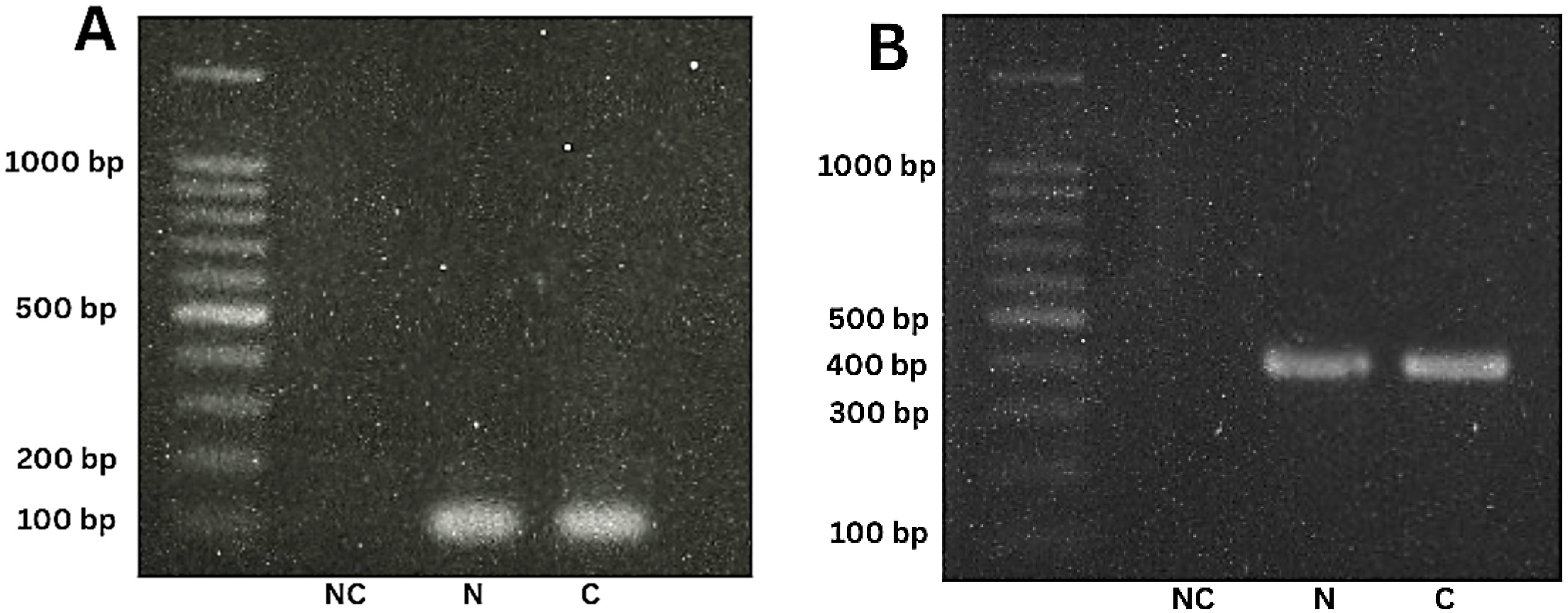
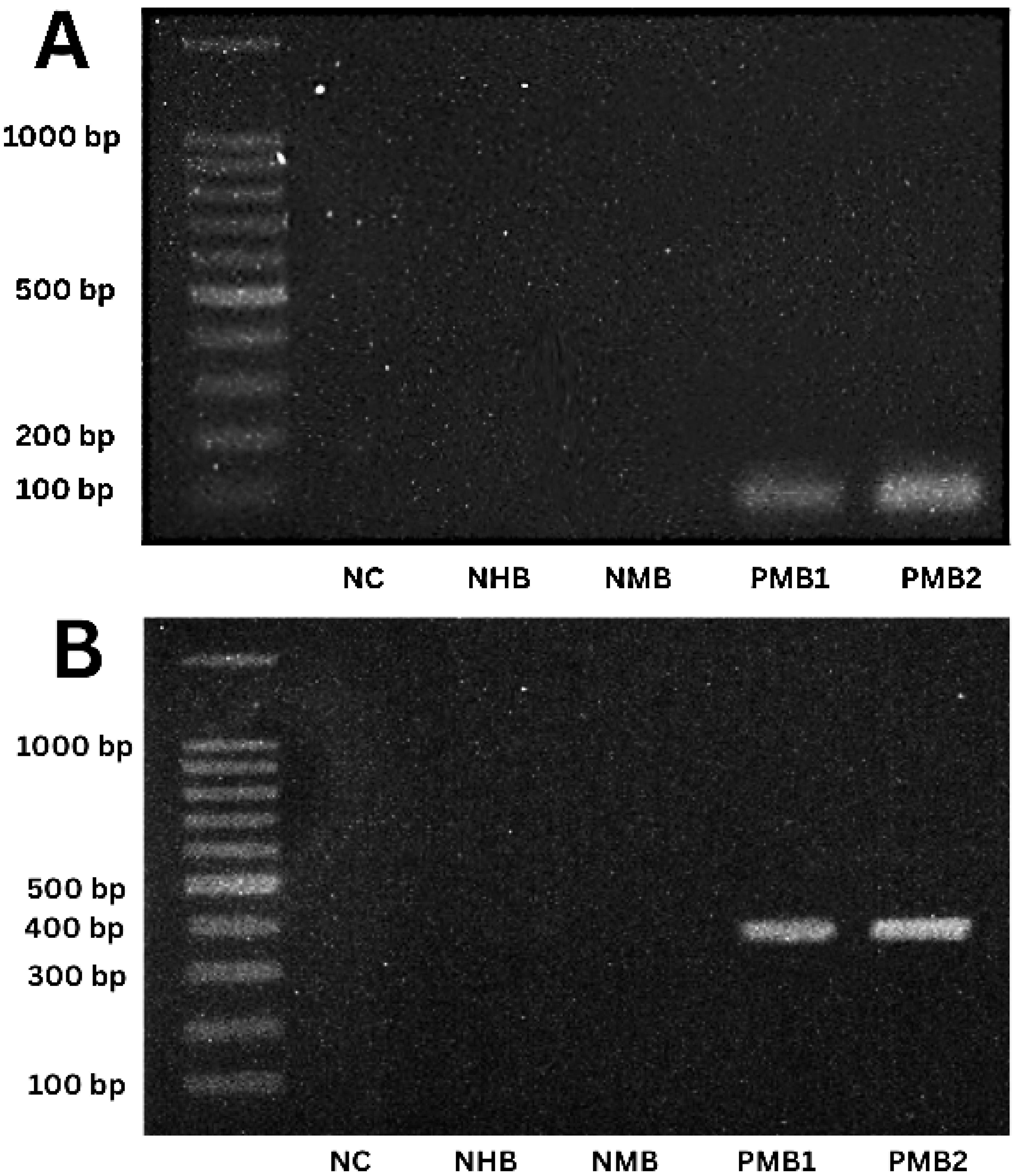
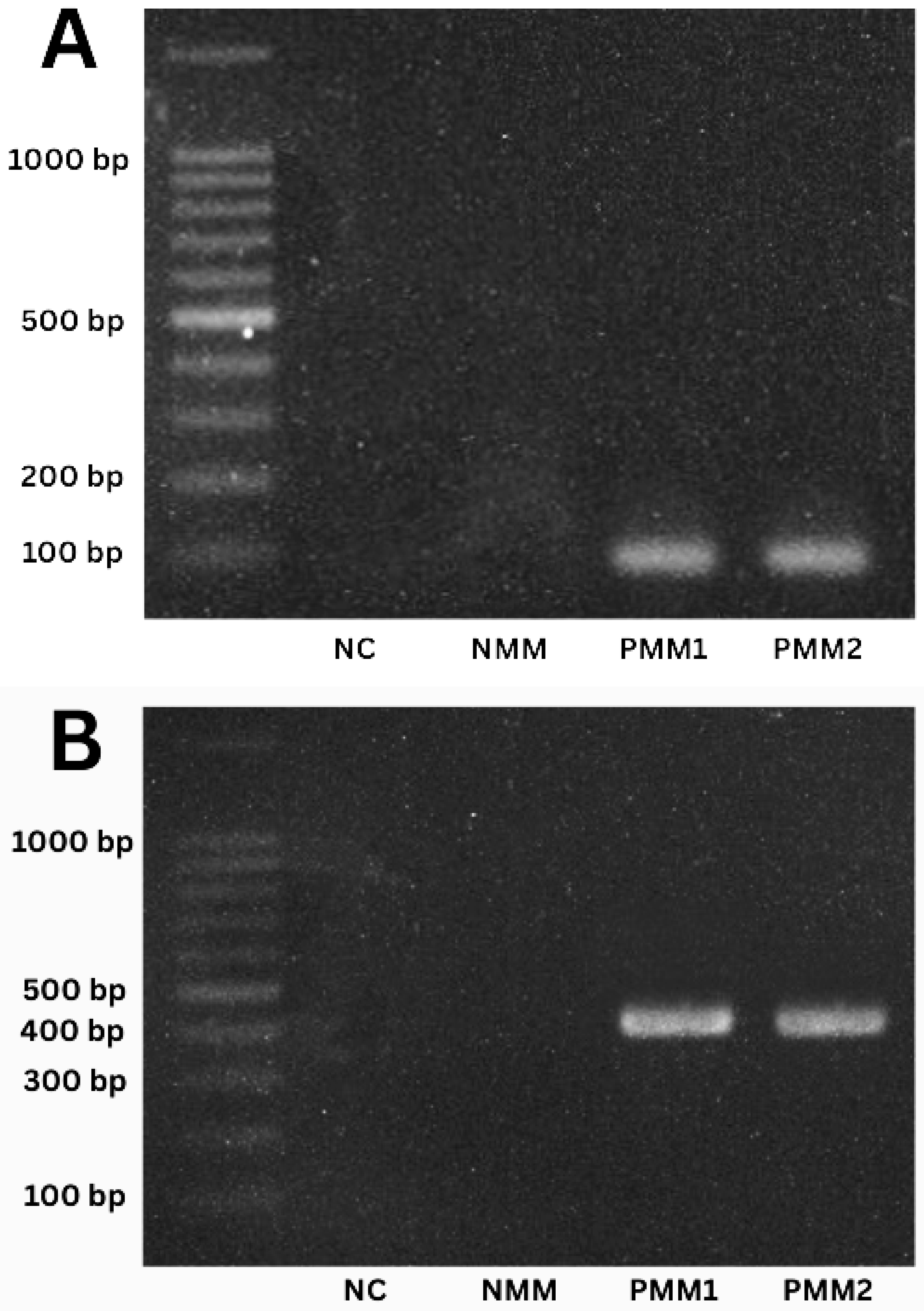
Amplifications were performed for both the satellite DNA (satDNA) and the mitochondrial cytochrome B (mtCytB) sequences. For each amplification, a negative control (NC) without DNA was included, and all reactions were performed in triplicate. Figure 3 shows PCR amplification products from NNN culture media (epimastigotes) containing the control strain Ninoa (N) and the “Calvillo” strain (C); only one of the three positive cultures is displayed. For fecal samples of triatomines (metacyclic trypomastigotes), the five previously obtained samples were analyzed. Similar to culture media, all fecal samples showed amplification of both satDNA and mtCytB (Figure 4). Additionally, a control nymphal fecal sample (NF) was included and yielded a negative result. Figure 5 presents PCR results from blood samples (trypomastigotes), including the negative control (NC), a negative human blood sample (NHB), a negative mouse blood sample (MBN), and the two positive mouse samples. For positive muscle samples (amastigotes), Figure 6 demonstrates the presence of the target genetic sequences, with negative controls consisting of heart muscle from an uninfected mouse and the corresponding NC.

3.3. Sequencing and Analysis of the Selected kDNA

To sequence the wild strain, one of the three NNN culture medium tubes exhibiting epimastigote development was selected. An amplification product of approximately 700 bp of kDNA was obtained from this tube, after which the sequencing process was carried out. A quality control procedure was applied, which reduced the total number of sequenced reads while optimizing the final dataset and removing adapter sequences. Sequencing metrics can be seen in Tables S1 and S2.
Using KmerFinder, the origin of the reads in the dataset was determined. The k-mers most represented corresponded to T. cruzi CL Brener, which also exhibited the greatest sequencing depth. A de novo assembly of the clean reads generated a contiguous sequence of 676 nucleotides.
Subsequently, a multiple alignment was performed between the obtained sequence (BankIt2933897 Seq1 PV256432), the amplification product of the kinetoplast minicircle from Trypanosoma isolates collected in Calvillo, Aguascalientes, and reference kinetoplast minicircle sequences of T. cruzi from GenBank. The wild strain, designated “Problem-Sequence,” was found to be related, showing an approximate 60% identity with reference T. cruzi kinetoplast minicircle sequences. Notably, a higher identity percentage was observed with T. cruzi clones M369r (U07846.1) and M835 (U07845.1) (Figure 7, Table 2).

4. Discussion

As previously mentioned, the parasite T. cruzi is transmitted via the feces of its biological vectors, the triatomines [1]. For this reason, the importance of this premise to detect the presence of the vectors in Mexico geography where the vector is in fact not considered endemic is reinforced by the following investigations. In 2010, triatomines were collected from five different locations in Calvillo, Aguascalientes, between June and September, yielding a total of 121 individuals [28]. Subsequent studies in 2016 demonstrated that these collections are more effective when performed at night [29]. From March to August 2019, a nighttime collection was conducted in nine locations within the municipality of Calvillo, between 8 p.m. and midnight, resulting in 252 specimens [5]. These findings highlight that the timing, schedules, and geographical locations selected for triatomine collection have remained consistent over more than a decade, with variations in the number of individuals captured. Such patterns facilitated a successful collection of the genus Meccus syn. Triatoma, enabling the acquisition of the parasite under study.
In the purity analysis of the extracted genetic material and its corresponding concentrations, it was observed that for all sample types—feces, blood, and muscle—DNA concentrations were obtained with 260/280 nm absorbance ratios close to the ideal range of 1.8–2 (Table 1). However, all samples showed some degree of contamination according to the 260/230 nm ratio, with fecal samples being the most affected. This is likely due to the complex composition of triatomine feces, which can interfere with the efficiency of the DNA extraction process [30,31]. Several studies have reported extractions of genetic material from T. cruzi or similar sample types, with varying results in terms of purity and concentration. For instance, Silva et al. [32] performed DNA extractions from triatomine feces, obtaining 260/280 absorbance values of approximately 1.78 (±0.10) and 260/230 absorbances averaging 1.000 (±0.68) across 15 samples, with maximum concentrations of 92.21 µg/mL. Cardoso et al. [33] reported DNA extracted from parasite culture media with average absorbances of 1.9 ± 1.0 (A260/280) and 2.0 ± 1.4 (A260/230). In 2023, Stanzick et al. [34] obtained blood-derived DNA with a concentration of 32.15 ± 12.45 µg/mL and 260/280 absorbances of 1.875 ± 0.023. Lutz et al. [35] extracted muscle DNA from frozen tissues using the commercial Wizard® Genomic DNA Purification Kit, achieving concentrations of 24 ± 13.4 µg/mL and absorbances of 1.4 ± 0.2 (A260/280) and 0.5 ± 0.2 (A260/230). Finally, Al-Griw et al. [36] performed DNA extractions from mouse muscle using the NucleoSpin® Blood and Tissue kit (QIAGEN), obtaining concentrations of 294.3 ± 38.8 µg/mL and 260/280 absorbances of 1.4 ± 0.5.
The satellite DNA sequence is a key molecular marker for the identification of T. cruzi, as it represents a highly conserved target for parasite amplification and detection [37]. In 2015, Ferrer [8] employed a 188 bp amplicon from this sequence to identify the parasite in blood samples. Similarly, Tavares de Oliveira et al. [38] used a 166 bp amplicon to detect T. cruzi in EDTA-treated blood samples from Latin American migrants living in Spain. More recently, in 2021, Herrera et al. [39] identified T. cruzi in triatomine feces (collected on filter paper) and in mouse tissues (including heart, skeletal muscle, and lungs, among other organs). Using DNA from culture media of the reference strain MDID/VE/1984/Dm28c as a positive control, they consistently amplified a 188 bp fragment in all tested samples, this in the country of Venezuela. In the present study, the 100 bp amplicons were obtained from parasites at different developmental stages: epimastigotes (from culture media), metacyclic trypomastigotes (from triatomine feces), bloodstream trypomastigotes (from mouse blood), and amastigotes (from mouse muscle), thereby confirming the presence of each parasitic form. Comparable findings were reported by Ramírez et al. [40], who analyzed multiple T. cruzi strains supplied by collaborators from Uruguay, Argentina, Colombia, Brazil, and the United States, successfully amplifying the satellite sequence with a 98 bp fragment—similar in size to that used in our investigation. These results highlight the utility of the selected targets and primer design for the identification and diagnosis of Chagas disease. Moreover, the use of multiple molecular markers in PCR assays increases the sensitivity of detection, thereby reducing the risk of false negatives. Importantly, our findings also demonstrated that the T. cruzi strain “Calvillo,” obtained from triatomines, can be considered pathogenic, as it successfully differentiated into each developmental stage depending on the growth substrate—culture media, blood, or tissue [41].
Likewise, mitochondrial DNA regions such as cytochrome B (mtCytB) and kinetoplast DNA (kDNA) have also been widely used as molecular markers for parasite identification [42,43,44]. In this study, we selected a 388 bp sequence within the mtCytB of the isolated Trypanosoma, given the consistent and reliable results reported over time for the molecular characterization of T. cruzi using this region. For instance, Arenas et al. [45] employed cytochrome B as a marker to identify T. cruzi in both vectors and human patients in Chile, obtaining a 573 bp amplicon. Later, in 2015, cytochrome B was studied as a potential therapeutic target when GNF7686 was shown to inhibit the parasite’s respiratory chain [46]. More recently, Balasubramanian et al. [47] monitored triatomines in Texas, USA, targeting this genomic region and amplifying a 228 bp fragment. In 2025, Gomez-Palacios et al. [48] conducted surveillance in the Mexican state of Durango, extracting intestinal contents from triatomine vectors to amplify a 382 bp fragment of cytochrome B DNA.
Regarding the sequencing of the amplification product, a fragment of one of the minicircles within the kinetoplast (kDNA) was selected. These regions are highly valuable in genetic research, as they provide insights into the diversity and evolutionary dynamics of the parasite when compared across different clones [13]. After assembling the sequence, a BLAST analysis was performed, followed by a multiple alignment with the sequences showing the highest identity. This analysis revealed that the clones M369r (U07846.1) and M835 (U07845.1) of T. cruzi share a closer relationship with the “Calvillo” strain under study. These clones have previously been employed as representative models for genetic studies of kDNA minicircle cloning and transcription analysis, and to further explore their functional roles [49]. Specifically, sequence U07846.1 has been used as a target to detect T. cruzi kDNA in the cardiac tissue of Rhesus macaques [50], as well as in samples from patients undergoing treatment for Chagas disease in Colombian communities [51]. Altogether, these findings underscore the clinical and research relevance of the variable region of the kDNA minicircle.
Sequencing enables the identification of the discrete typing unit (DTU) to which the parasite belongs. The DTUs, ranging from TcI to TcVI and TcBat, represent groups of strains that share certain genetic, molecular, or immunological markers, though they are not necessarily genetically identical. Their classification is as follows: DTUs I and II (TcI and TcII) are considered ancestral lineages; DTUs III and IV (TcIII and TcIV) result from at least one recombination event between DTUs I and II, making them homozygous hybrids; and DTUs V and VI (TcV and TcVI) are heterozygous hybrids derived from DTUs II and III. Additionally, a lineage associated with bats, TcBat, has been described and is now recognized as the seventh DTU (DTU VII) [14,52]. According to the classification proposed by Telleria et al. [53], the sequence ATGAGGGGTAGTT was exclusively associated with DTU I, while the sequences TGGGTAG and GGTAGTAT were found only in DTU II. In contrast, the sequences TAGTGGT, TATAGTTT, and TGTTTGA were detected across nearly all DTUs of T. cruzi. Based on this framework, the assembled sequence in our study is most likely classified within DTUs III or IV, as the characteristic markers for DTUs I and II were absent. A similar pattern was observed in the clones M369r (U07846.1) and M835 (U07845.1), suggesting that both may share a common evolutionary origin.
This study represents a preliminary approach to identifying a new potential endemic region of Trypanosoma cruzi in Aguascalientes, Mexico. Given the limited number of samples and replicates, the analysis remains primarily descriptive and qualitative, constrained by the difficulties of collecting triatomines, the availability of resources, and the specific conditions required for handling both vectors and parasites. Nonetheless, these findings establish a foundation for expanding research to other regions and contribute to strengthening the understanding of Chagas disease.

5. Conclusions

This study identified and genetically characterized Trypanosoma cruzi from wild triatomines (Meccus syn. Triatoma) in Calvillo, Aguascalientes, Mexico, thereby confirming the presence of the parasite in the region. End-point PCR targeting satDNA and mtCytB successfully detected T. cruzi at different developmental stages in both vectors and in the mouse model.
Next-generation sequencing of kinetoplast DNA minicircles revealed that the strain “Calvillo” belongs to Discrete Typing Units (DTUs) III and IV, showing phylogenetic relatedness to clones M369r and M835. These DTUs are commonly associated with sylvatic cycles and variable pathogenicity, which may represent a potential public health risk in this region.
An experimental infection in mice reproduced the complete life cycle of the parasite and confirmed the pathogenic potential of the strain. Altogether, these findings provide molecular, phylogenetic, and pathological evidence for a pathogenic T. cruzi strain circulating in local vectors, underscoring the need to strengthen epidemiological surveillance in the study region.

Supplementary Materials

The following supporting information can be downloaded at: https://www.mdpi.com/article/10.3390/parasitologia5030046/s1, Table S1: Basic metrics of assemblies before and after quality control; Table S2: Metrics of the assemblies before and after quality control performed with QUAST.

Author Contributions

Conceptualization: A.L.T.-B., M.H.M.-O. and D.A.H.-M.; data curation: J.H.E.-L., E.S.-G., A.A.-A. and S.L.M.-H.; formal analysis: E.L.-M. and A.A.-A.; funding acquisition: M.H.M.-O. and D.A.H.-M.; investigation: A.L.T.-B., M.P.R.-G. and S.L.M.-H.; resources, D.A.H.-M.; methodology: M.H.M.-O., E.S.-G. and D.A.H.-M.; validation: E.L.-M. and J.H.E.-L.; visualization: M.P.R.-G., S.L.M.-H. and J.H.E.-L.; writing—original draft: M.H.M.-O. and D.A.H.-M.; writing—review and editing: A.L.T.-B., M.H.M.-O., D.I.-M. and D.A.H.-M. All authors have read and agreed to the published version of the manuscript.

Funding

This work was supported by the Secretariat of Science, Humanities, Technology and Innovation (SECIHTI) under grant numbers 241312, A1-S-21375, CVU Scholarship 556587 (D.A.H.-M), UAA PIBB19-11N and PIBB24-7.

Institutional Review Board Statement

Not applicable.

Informed Consent Statement

Not applicable.

Data Availability Statement

Data present in this study are available upon request from the corresponding author.

Acknowledgments

We are grateful to Norma Adela Carrasco Esparza for preserving the strains used in this study and to Dionisio Vera Miramontes for assisting with the triatomine collection. For the sequencing process, which was conducted by Anastacio Palacios Marmolejo and Brian Muñoz, special gratitude is extended to Laboratorio Estatal de Salud Pública del Estado de Aguascalientes. We are also grateful to Rosa Isela Chávez Gómez, for allowing us to use their facilities. Antonio Ovalle Carcaño is especially appreciated for his assistance in the bioinformatics analysis of the data gathered throughout the sequencing procedure. Lastly, we acknowledge the English evaluation of Efraín Alejandro Luna Gutiérrez.

Conflicts of Interest

The authors declare no conflicts of interest.

Abbreviations

The following abbreviations are used in this manuscript:
satDNAsatellite DNA
mtCytBcytochrome B gene
kDNAKinetoplast DNA

References

  1. Nguyen, T.; Waseem, M.; Winters, R. Chagas Disease. In StatPearls [Internet]; StatPearls Publishing: Treasure Island, FL, USA, 2025. [Google Scholar]
  2. Parasites—American Trypanosomiasis (Also Known as Chagas Disease). Available online: https://www.cdc.gov/chagas/index.html (accessed on 15 May 2025).
  3. Álvarez-Hernández, D.A.; Franyuti-Kelly, G.A.; Díaz-López-Silva, R.; González-Chávez, A.M.; González-Hermosillo-Cornejo, D.; Vázquez-López, R. Chagas disease: Current perspectives on a forgotten disease. Rev. Med. Hosp. Gen. Méx. 2018, 81, 154–164. [Google Scholar] [CrossRef]
  4. Enfermedad de Chagas (Tripanosomiasis Americana). Available online: https://www.who.int/es/news-room/fact-sheets/detail/chagas-disease-(american-trypanosomiasis) (accessed on 15 May 2025).
  5. Morales-Moran, S.; Sánchez-García, E.; Chávez-Gómez, R.; Carrasco-Esparza, N.; Aguayo-Acosta, A.; Hernández-Marín, D. Distribution of Triatoma (Meccus) phyllosoma and Triatoma (Meccus) longipennis as vectors of Trypanosoma cruzi in the state of Aguascalientes, Mexico and surroundings. Rev. Alianzas Tend. BUAP 2021, 6, 1–15. [Google Scholar] [CrossRef]
  6. Cesaretto, N.R.; de Oliveira, J.; Ravazi, A.; Madeira, F.F.; Dos Reis, Y.V.; de Oliveira, A.B.B.; Vicente, R.D.; Cristal, D.C.; Galvão, C.; de Azeredo-Oliveira, M.T.V.; et al. Trends in taxonomy of Triatomini (Hemiptera, Reduviidae, Triatominae): Reproductive compatibility reinforces the synonymization of Meccus Stål, 1859 with Triatoma Laporte, 1832. Parasites Vectors 2021, 14, 340. [Google Scholar] [CrossRef] [PubMed]
  7. Rivera, H.N.; McAuliffe, I.; Aderohunmu, T.; Wiegand, R.E.; Montgomery, S.P.; Bradbury, R.S.; Handali, S. Evaluation of the Performance of Ortho T. cruzi ELISA Test System for the Detection of Antibodies to Trypanosoma cruzi. J. Clin. Microbiol. 2022, 60, e0013422. [Google Scholar] [CrossRef]
  8. Ferrer, E. Técnicas moleculares para el diagnóstico de la enfermedad de Chagas. Saber 2015, 27, 359–371. [Google Scholar]
  9. Manual de Procedimientos Para la Enfermedad de Chagas en México; Gobierno de México: Ciudad de México, Mexico, 2019; pp. 1–109.
  10. Domínguez, J.L.; Ligonio, A.R.; Mendoza, A.C.; Díaz, M.C.M.; Cruz, V.A.R.; Monteón, A.L. El diagnóstico para la enfermedad de Chagas: A más de 110 años de su descubrimiento. JETERAPS 2021, 27, 31–39. [Google Scholar] [CrossRef]
  11. Ferreira Filho, J.C.R.; Braz, L.M.A.; Andrino, M.L.A.; Yamamoto, L.; Kanunfre, K.A.; Okay, T.S. Mitochondrial and satellite real time-PCR for detecting T. cruzi DTU II strain in blood and organs of experimentally infected mice presenting different levels of parasite load. Exp. Parasitol. 2019, 200, 13–15. [Google Scholar] [CrossRef]
  12. Kann, S.; Concha, G.; Weinreich, F.; Hahn, A.; Rückert, C.; Kalinowski, J.; Landt, O.; Frickmann, H. Comparative Assessment of Two Commercial Real-Time PCR Assays for the Diagnosis of Trypanosoma cruzi DNA in Serum. Microorganisms 2023, 11, 901. [Google Scholar] [CrossRef]
  13. Gómez-Palacio, A.; Cruz-Saavedra, L.; Van den Broeck, F.; Geerts, M.; Pita, S.; Vallejo, G.; Carranza, J.C.; Ramírez, J.D. High-throughput analysis of the Trypanosoma cruzi minicirculome (mcDNA) unveils structural variation and functional diversity. Sci. Rep. 2024, 14, 5578. [Google Scholar] [CrossRef]
  14. Silvestrini, M.M.A.; Alessio, G.D.; Frias, B.E.D.; Sales Júnior, P.A.; Araújo, M.S.S.; Silvestrini, C.M.A.; Brito Alvim de Melo, G.E.; Martins, H.R. New insights into Trypanosoma cruzi genetic diversity, and its influence on parasite biology and clinical outcomes. Front. Immunol. 2024, 15, 1342431. [Google Scholar] [CrossRef]
  15. Kann, S.; Kunz, M.; Hansen, J.; Sievertsen, J.; Crespo, J.J.; Loperena, A.; Arriens, S.; Dandekar, T. Chagas Disease: Detection of Trypanosoma cruzi by a New, High-Specific Real Time PCR. J. Clin. Med. 2020, 9, 1517. [Google Scholar] [CrossRef]
  16. Torres-Barajas, A.L.; Salas-Baéz, K.D.; Chávez-Gómez, R.I.; Carrasco-Esparza, N.A.; Muñoz-Ortega, M.H.; Sánchez-García, E.; Hernández-Marín, D.A. Actividad tripanocida de cinco plantas latinoamericanas. Rev. Biol. Trop. 2024, 72, e54026. [Google Scholar] [CrossRef]
  17. Davies, C.; Poma, R.H.; Marino Cardozo, R.; Mora, M.C.; Ramos, F.; Rajal, V.B.; Basombrío, M.A. Detección de Trypanosoma cruzi en tejido y sangre murina por PCR convencional y en tiempo real. Acta Bioquím. Clín. Latinoam. 2014, 48, 421–428. [Google Scholar]
  18. Ucan-Euan, F.; Hernández-Betancourt, S.; Arjona-Torres, M.; Panti-May, A.; Torres-Castro, M. Estudio histopatológico en tejido cardiaco de roedores infectados con Trypanosoma cruzi, capturados en barrios suburbanos, Mérida, México. Biomédica 2019, 39, 32–43. [Google Scholar] [CrossRef] [PubMed]
  19. Reboreda-Hernandez, O.A.; Gonzalez-Rodriguez, N.; Cruz-Gonzalez, A.R.; Roman-Cedillo, A.; Ortiz-Butron, R. Influencia de la inoculación oral en la enfermedad de Chagas en modelo murino. Horiz. Sanit. 2021, 20, 198–206. [Google Scholar] [CrossRef]
  20. Serra-Campos, A.O.; Abreu-Junior, A.N.; Nascimento, A.A.; Abidu-Figueiredo, M.; Lima, M.S.C.S.; Machado-Santos, C. Gastroesophageal tube of the Iguana iguana (Iguanidae): Histological description, histochemical and immunohistochemical analysis of 5-HT and SS cells. Braz. J. Biol. 2023, 83, e242086. [Google Scholar] [CrossRef]
  21. Braz, L.M.; Raiz-Júnior, R.; Alárcon, R.S.; Gakiya, E.; Amato-Neto, V.; Okay, T.S. Suitability of a rapid DNA isolation and amplification for detection of Trypanosoma cruzi in Triatoma infestans dry fecal spots collected on filter paper. Parasite 2008, 15, 595–598. [Google Scholar] [CrossRef]
  22. López, M.; Herrera, L.; Morocoima, A.; Rivera, M.G.; Viettri, M.; Lares, M.; Ferrer, E. Utility of a Fluid Library with Samples of Humans, Reservoirs and Vectors Collected in Filter Paper, for Retrospective Diagnosis of American Trypanosomiasis in Endemic Areas of Venezuela. Acta Parasitol. 2020, 66, 287–293. [Google Scholar] [CrossRef] [PubMed]
  23. Gómez-Bravo, A.; Cirignoli, S.; Wehrendt, D.; Schijman, A.; León, C.M.; Flores-Chaves, M.; Nieto, J.; Kieran, T.J.; Abril, M.; Guhl, F. Zoonotic Cycle of American Trypanosomiasis in an Endemic Region of the Argentine Chaco, Factors That Influenced a Paradigm Shift. Insects 2024, 15, 471. [Google Scholar] [CrossRef]
  24. Rueda-Concha, K.L.; Payares-Mercado, A.; Guerra-Castillo, J.; Melendrez, J.; Arroyo-Munive, Y.; Martínez-Abad, L.; Cochero, S.; Bejarano, E.E.; Paternina, L.E. Circulación de Leishmania infantum y Trypanosoma cruzi en perros domésticos de áreas urbanas de Sincelejo, región Caribe de Colombia. Biomédica 2022, 42, 633–649. [Google Scholar] [CrossRef]
  25. Probst, C.M.; Melo, M.F.A.D.; Pavoni, D.P.; Toledo, M.J.O.; Galdino, T.S.; Brandão, A.A.; Britto, C.; Krieger, M.A. A new Trypanosoma cruzi genotyping method enables high resolution evolutionary analyses. Mem. Inst. Oswaldo Cruz 2021, 116, e200538. [Google Scholar] [CrossRef]
  26. Callejas-Hernández, F.; Herreros-Cabello, A.; Del Moral-Salmoral, J.; Fresno, M.; Gironès, N. The Complete Mitochondrial DNA of Trypanosoma cruzi: Maxicircles and Minicircles. Front. Cell. Infect. Microbiol. 2021, 11, 672448. [Google Scholar] [CrossRef]
  27. Marcel, M. Cutadapt removes adapter sequences from high-throughput sequencing reads. EMBnet J. 2011, 17, 10–12. [Google Scholar] [CrossRef]
  28. Licón-Trillo, A.; Balsimelli-De La Peña, K.; Acosta-Legarda, M.; Leal-Berumen, I.; Nogueda-Torres, B.; Martínez-Ibarra, J. Infección natural por Trypanosoma cruzi en triatominos del Centro y Norte de México. Bol. Malariol. Sal. Amb. 2010, 50, 311–314. [Google Scholar]
  29. Hylton, A.; Fitzpatrick, D.M.; Suepaul, R.; Dobson, A.P.; Charles, R.A.; Peterson, J.K. Preliminary characterization of triatomine bug blood meals on the island of Trinidad reveals opportunistic feeding behavior on both human and animal hosts. Trop. Med. Infect. Dis. 2020, 5, 166. [Google Scholar] [CrossRef]
  30. Hart, M.L.; Meyer, A.; Johnson, P.J.; Ericsson, A.C. Comparative evaluation of DNA extraction methods from feces of multiple host species for downstream next-generation sequencing. PLoS ONE 2015, 10, e0143334. [Google Scholar] [CrossRef]
  31. Galla, G.; Praeg, N.; Rzehak, T.; Sprecher, E.; Colla, F.; Seeber, J.; Illmer, P.; Hauffe, H.C. Comparison of DNA extraction methods on different sample matrices within the same terrestrial ecosystem. Sci. Rep. 2024, 14, 8715. [Google Scholar] [CrossRef]
  32. Silva, A.N.B.D.; Souza, R.D.C.M.D.; Honorato, N.R.M.; Martins, R.R.; Câmara, A.C.J.D.; Galvão, L.M.D.C.; Chiari, E. Comparison of phenol-chloroform and a commercial deoxyribonucleic acid extraction kit for identification of bloodmeal sources from triatomines (Hemiptera: Reduviidae). Rev. Soc. Bras. Med. Trop. 2020, 53, e20200189. [Google Scholar] [CrossRef] [PubMed]
  33. Cardoso, G.V.F.; Oliveira, A.C.S.; Silva, A.S.; Silva, M.C.L.; Lima, J.S.; Roos, T.B.; Moraes, C.M. Protocol optimization for the detection of Trypanosoma cruzi DNA in açai (Euterpe oleraceae) pulp. Acta Amazon. 2021, 51, 79–84. [Google Scholar] [CrossRef]
  34. Stanzick, K.J.; Simon, J.; Zimmermann, M.E.; Schachtner, M.; Peterhoff, D.; Niller, H.H.; Überla, K.; Wagner, R.; Heid, I.M.; Stark, K.J. DNA extraction from clotted blood in genotyping quality. Biotechnol. Tech. 2023, 74, 23–29. [Google Scholar] [CrossRef] [PubMed]
  35. Lutz, Í.; Miranda, J.; Santana, P.; Martins, T.; Ferreira, C.; Sampaio, I.; Vallinoto, M.; Gomes, G.E. Quality analysis of genomic DNA and authentication of fisheries products based on distinct methods of DNA extraction. PLoS ONE 2023, 18, e0282369. [Google Scholar] [CrossRef]
  36. Al-Griw, H.H.; Zraba, Z.A.; Al-Muntaser, S.K.; Draid, M.M.; Zaidi, A.M.; Tabagh, R.M.; Al-Griw, M.A. Effects of storage temperature on the quantity and integrity of genomic DNA extracted from mice tissues: A comparison of recovery methods. Open Vet. J. 2017, 7, 239–243. [Google Scholar] [CrossRef]
  37. Chacón, F.; Bacigalupo, A.; Álvarez-Duhart, B.; Cattan, P.E.; Solís, R.; Muñoz-San Martín, C. The parasite load of Trypanosoma cruzi modulates feeding and defecation patterns of the Chagas disease vector Triatoma infestans. Microorganisms 2022, 10, 1003. [Google Scholar] [CrossRef]
  38. de Oliveira, M.T.; Sulleiro, E.; Silgado Gimenez, A.; de Lana, M.; Zingales, B.; Santana da Silva, J.; Marin-Neto, J.A.; Molina, I. Quantification of parasite burden of Trypanosoma cruzi and identification of Discrete Typing Units (DTUs) in blood samples of Latin American immigrants residing in Barcelona, Spain. PLoS Negl. Trop. Dis. 2020, 14, e0008311. [Google Scholar] [CrossRef]
  39. Herrera, L.; Aguilar, C.M.; Morocoima, A.; Viettri, M.; Lares, M.; Ferrer, E. Detection of Trypanosoma cruzi DNA in false negative samples of collected triatomines, xenodiagnosis material, and biopsies of experimentally infected animals. Int. Microbiol. 2021, 24, 141–147. [Google Scholar] [CrossRef] [PubMed]
  40. Ramírez, J.C.; Torres, C.; Curto, M.L.A.; Schijman, A.G. New insights into Trypanosoma cruzi evolution, genotyping and molecular diagnostics from satellite DNA sequence analysis. PLoS Negl. Trop. Dis. 2017, 11, e0006139. [Google Scholar] [CrossRef]
  41. Rossi, I.V.; de Souza, D.A.S.; Ramirez, M.I. The end justifies the means: Chagas disease from a perspective of the host–Trypanosoma cruzi interaction. Life 2024, 14, 488. [Google Scholar] [CrossRef] [PubMed]
  42. Muñoz-San Martín, C.; Solari, A.; Apt, W.; Zulantay, I. Caracterización de las Unidades Discretas de Tipificación de Trypanosoma cruzi según sus marcadores moleculares. Rev. Ibero Latinoam. Parasitol. 2013, 72, 5–21. [Google Scholar]
  43. Rodrigues, M.S.; Morelli, K.A.; Jansen, A.M. Cytochrome c oxidase subunit 1 gene as a DNA barcode for discriminating Trypanosoma cruzi DTUs and closely related species. Parasit. Vectors 2017, 10, 488. [Google Scholar] [CrossRef]
  44. de Souza, M.L.; Lapierre, T.J.W.J.D.; de Lima Marques, G.V.; Ferraz, W.R.; Penteado, A.B.; Trossini, H.G.G.; Murta, S.M.F.; de Oliveira, R.B.; de Oliveira Rezende, C., Jr.; Ferreira, R.S. Molecular targets for Chagas disease: Validation, challenges and lead compounds for widely exploited targets. Expert. Opin. Ther. Targets 2023, 27, 911–925. [Google Scholar] [CrossRef] [PubMed]
  45. Arenas, M.; Campos, R.; Coronado, X.; Ortiz, S.; Solari, A. Trypanosoma cruzi genotypes of insect vectors and patients with Chagas of Chile studied by means of cytochrome b gene sequencing, minicircle hybridization, and nuclear gene polymorphisms. Vector-Borne Zoonotic Dis. 2012, 12, 196–205. [Google Scholar] [CrossRef] [PubMed]
  46. Khare, S.; Roach, S.L.; Barnes, S.W.; Hoepfner, D.; Walker, J.R.; Chatterjee, A.K.; Neitz, R.J.; Arkin, M.R.; McNamara, C.W.; Ballard, J.; et al. Utilizing chemical genomics to identify cytochrome b as a novel drug target for Chagas disease. PLoS Pathog. 2015, 11, e1005058. [Google Scholar] [CrossRef] [PubMed]
  47. Balasubramanian, S.; Curtis-Robles, R.; Chirra, B.; Auckland, L.D.; Mai, A.; Bocanegra-Garcia, V.; Clark, P.; Clark, W.; Cottingham, M.; Fleurie, G.; et al. Characterization of triatomine bloodmeal sources using direct Sanger sequencing and amplicon deep sequencing methods. Sci. Rep. 2022, 12, 10234. [Google Scholar] [CrossRef]
  48. Gómez-Palacios, R.L.; Ruiz-Tovar, K.; Huerta, H.; González-Reyes, M.O.; Padilla-Medina, I.; De la Rosa-Arana, J.L.; Fonseca-Coronado, S. Natural infection with Trypanosoma cruzi and feeding habits of Triatominae (Hemiptera: Reduviidae) from the state of Durango, Mexico. Acta Trop. 2025, 251, 107555. [Google Scholar] [CrossRef] [PubMed]
  49. Thertulien, R.; Simpson-Haidaris, P.J.; Haidaris, C.G. Intracellular localization of a Trypanosoma cruzi kDNA minicircle transcript using RNA:RNA in situ hybridization. J. Eukaryot. Microbiol. 1994, 41, 402–407. [Google Scholar] [CrossRef]
  50. DeLorenzo, M.; Carias, E.; Mustonen, A.; Gonzalez, O.; Dick, E.J., Jr.; Kumar, S. In situ hybridization assay for the diagnosis of Chagas myocarditis and orchitis in a rhesus macaque (Macaca mulatta): A case report. J. Med. Primatol. 2019, 48, 182–185. [Google Scholar] [CrossRef]
  51. Kann, S.; Concha, G.; Frickmann, H.; Hagen, R.M.; Warnke, P.; Molitor, E.; Hoerauf, A.; Backhaus, J. Chagas disease: Comparison of therapy with nifurtimox and benznidazole in indigenous communities in Colombia. J. Clin. Med. 2024, 13, 2565. [Google Scholar] [CrossRef]
  52. Herreros-Cabello, A.; Callejas-Hernández, F.; Gironès, N.; Fresno, M. Trypanosoma cruzi genome: Organization, multi-gene families, transcription, and biological implications. Genes 2020, 11, 1196. [Google Scholar] [CrossRef]
  53. Telleria, J.; Lafay, B.; Virreira, M.; Barnabé, C.; Tibayrenc, M.; Svoboda, M. Trypanosoma cruzi: Sequence analysis of the variable region of kinetoplast minicircles. Exp. Parasitol. 2006, 114, 279–288. [Google Scholar] [CrossRef]
Figure 1. Giemsa-stained mouse blood samples at 100×. (A) Presence of T. cruzi in mouse 1 and (B) presence of T. cruzi in mouse 2.
Parasitologia 05 00046 g001
Figure 2. Amastigotes in cardiac muscle (A) mouse 1 and (B) and (C) mouse 2 at 100×.
Parasitologia 05 00046 g002
Figure 3. Amplification of satDNA (A) and mtCytB (B) in samples from culture media (epimastigotes). N: Ninoa, C: Calvillo, NC: negative control.
Parasitologia 05 00046 g003
Figure 4. Amplification of satDNA (A) and mtCytB (B) in samples from the feces of triatomines (metacyclic trypomastigotes). NC: negative control, NF: negative nymph feces. Samples 1 to 5 belong to samples positive for T. cruzi in triatomine feces.
Parasitologia 05 00046 g004
Figure 5. Amplification of satDNA (A) and mtCytB (B) from blood samples (blood trypomastigotes). NC: negative control, NHB: negative human blood, NMB: negative mouse blood, PMB1: positive mouse blood 1, PMB2: positive mouse blood 2.
Parasitologia 05 00046 g005
Figure 6. Amplification of satDNA (A) and mtCytB (B) in samples from muscles (amastigotes). NC: negative control, NMM: negative mouse muscle, PMM 1: positive mouse muscle 1, PMM2: positive mouse muscle 2.
Parasitologia 05 00046 g006
Figure 7. Alignment between the sequences obtained from GenBank and the experimental sequence of interest obtained through sequencing, where based on the results of the multiple alignment, a 60% identity is presented, with the sequences from the minicircle of kinetoplast of U07846.1 and U07845.1 of T. cruzi. The image shows the alignment, where the region with the highest similarity between the sequences coincides with ORF1 and ORF2. Problem-Sequence: The sequence obtained from the sequencing aligns with the kinetoplast minicircle of the parasite genus Trypanosoma, is appointed.
Parasitologia 05 00046 g007
Table 1. Average concentration and purity of genetic material obtained from biological samples.
Sample TypeConcentrationAbsorbance 230/260Absorbance 260/280
Culture media (n = 3)160 ± 10 µg/mL1.62 ± 0.251.96 ± 0.10
Triatomine feces (n = 5)13.5 ± 4 µg/mL0.41 ± 102.05 ± 0.33
Blood (n = 2)34.3 ± 6.5 µg/mL0.96 ± 341.88 ± 0.11
Muscle (n = 2)34.5 ± 9.5 µg/mL1.37 ± 412.02 ± 0.21
Table 2. Sequence information obtained by alignment from GenBank with the experimental sequence.
Access NumberSequence InformationPercent Identity with the Problem-Sequence
M15511.1T.cruzi kinetoplast minicircle pTckAWP-2, complete33.54
M15512.1T.cruzi kinetoplast minicircle pTckCA1-73 DNA fragment33.71
HG008629.1Homo sapiens genomic DNA containing Trypanosoma cruzi kinetoplast minicircle DNA, family B patient 028, clone FH09333.42
M19177.1T.cruzi kinetoplast minicircle DNA, clone cl1 cst 433.21
M18815.1T.cruzi kinetoplast minicircle DNA, clone pTc-2133.23
U07846.1Trypanosoma cruzi Y kinetoplast minicircle sequence, clone M369r, orf, complete cds60.94
U07845.1Trypanosoma cruzi Y kinetoplast minicircle sequence, clone M835, orf, complete cds60.46
X56188.1T. cruzi cDNA52 minicircle transcript (highly homologous to kinetoplast DNA minicircle sequences)60.19
Disclaimer/Publisher’s Note: The statements, opinions and data contained in all publications are solely those of the individual author(s) and contributor(s) and not of MDPI and/or the editor(s). MDPI and/or the editor(s) disclaim responsibility for any injury to people or property resulting from any ideas, methods, instructions or products referred to in the content.