Abstract
Background: The present exploratory study aimed to investigate the relationship between arginine vasopressin (AVP) secretion and cerebral edema and evaluate its usefulness as an index for assessing severity of cerebral edema. Methods: Forensic autopsy was performed within 96 h after death in 143 cases, and samples of right heart blood and cerebrospinal fluid (CSF) were collected. Serum AVP levels and CSF were then measured and relationships with brain weight (BW) and normalized BW were investigated. Results: No relationships between serum AVP levels or CSF and age, sex, survival, or postmortem period were identified. A comparison in regard to cause of death revealed lower CSF AVP concentrations in cases of blunt head injury and cerebrovascular disease. In addition, a correlation was observed between serum AVP levels and non-normalized or normalized BW with blunt head injury or asphyxia. Receiver operating characteristic analysis revealed that neither serum nor CSF AVP concentrations yielded cutoff values readily applicable to the diagnosis of cerebral edema. Conclusions: The present findings suggest that postmortem AVP concentrations remain relatively stable and may be involved in the formation of cerebral edema. The findings also highlighted issues such as the influence of confounding factors associated with individual pathologies and the verification of AVP stability in postmortem samples. Thus, the usefulness of AVP as an indicator of cerebral edema in forensic evaluation remains unclear.
    1. Introduction
The formation of cerebral edema is one of the major factors leading to high mortality and morbidity in affected individuals []. Cerebral edema is often found under various pathological conditions encountered in the field of legal medicine, such as in cases involving head injury, heat stroke, and intoxication [,,]. The formation of cerebral edema may depress vital brain centers, such as those involved in respiratory and cardiac functions, resulting from the compression of blood vessels, reduced tissue blood flow, and reduced oxygenation []. This can lead to varying degrees of neurological impairment and even death. Confirming the presence of cerebral edema at autopsy is thus of forensic significance.
Arginine vasopressin (AVP), also known as antidiuretic hormone, is secreted from the posterior pituitary gland and principally produced by neurons that have their cell bodies within the supraoptic nuclei of the hypothalamus []. It is derived from preprovasopressin, a larger, 164-amino acid precursor peptide consisting of the signal peptide AVP, along with neurophysin II and copeptin []. Copeptin, a 39-amino acid peptide derived from the C-terminal portion of the precursor protein preprovasopressin, is released into circulation in amounts equivalent to AVP []. Copeptin is sometimes measured as a surrogate biomarker for AVP based on its ex vivo stability []. Arginine vasopressin has been reported to play essential roles in the control of body water homeostasis and associated disorders [], and in modulating stress, behavior, pituitary functions, and the immune response []. Excessive secretion of AVP promotes the formation of cerebral edema in cerebrovascular disease by disrupting the hydromineral balance in the neurovascular unit []. Based on this, AVP has been suspected in cerebral edema formation in medico-legal autopsy cases. On the other hand, although AVP is reportedly unstable ex vivo, few reports have described its measurement using postmortem specimens, and the extent to which it is actually related to the pathology of forensic autopsy cases remains unclear.
Generally, the gold standard for assessing cerebral edema is subjective evaluation based on macroscopic characteristics or histopathological examination [,]. Brain weight (BW) is also measured to assess cerebral edema []. Normalized BW corrects BW for intracranial measurements or volume, and has been reported to be a more reliable method for assessing cerebral edema without relying on subjective impressions [,]. However, these methods require mathematical calculations and the acquisition of morphometric parameters. By contrast, postmortem biochemistry can corroborate pathological evidence through the “visualization” of functional changes []. Arginine vasopressin levels can easily be evaluated quantitatively and objectively using body fluid samples. Therefore, the measurement of AVP as part of a forensic laboratory investigation may be useful in diagnosing or assessing the severity of cerebral edema.
Given this background, the present exploratory study aimed to investigate the relationship between AVP secretion and cerebral edema, and to evaluate its utility as an index for assessing the severity of cerebral edema.
2. Materials and Methods
2.1. Ethics Statement
This study was approved by the independent ethics committee of the Osaka Metropolitan University Graduate School of Medicine (approval No.: 2749). Informed consent for the analysis of autopsy data was considered to have been obtained in the form of an opt-out.
2.2. Autopsy Samples
Serial forensic autopsy samples were used within 96 h after death at our institute, except in putrefactive or highly destructive cases. We analyzed autopsy cases in which serum and cerebrospinal fluid (CSF) samples could be collected, resulting in a total of 143 cases (110 males, 33 females; median age, 66 years; range, 21–93 years). The cause of death was classified as follows according to the findings of the completed autopsy and macromorphological, micropathological, and toxicological examinations: hemorrhagic shock due to sharp instrument injury (hereinafter, “sharp instrument injury”; n = 8), blunt head injury (n = 15), blunt injury excluding head injury (hereinafter, “non-head injury”; n = 18), asphyxia (n = 19), drowning (n = 14: fresh water, n = 10; salt water, n = 4), intoxication (n = 13: methamphetamine, n = 6; psychotropic drug, n = 7), fire fatality (n = 24), acute cardiac death (n = 25), and cerebrovascular disease (n = 7). The case profiles are shown in Table 1. For each cause of death, clearly verifiable cases with well-established pathological evidence without any significant complications were included. For the present examination, the survival period was established based on witnesses and circumstantial evidence, and the postmortem period was estimated based on pathological findings. Demographic and autopsy data (including BW) were collected from autopsy documents.
       
    
    Table 1.
    Case profiles (n = 143).
  
2.3. Biochemical Analysis
Specimens were collected aseptically using syringes to obtain CSF and blood from the right heart chambers. Samples were subsequently stored at −80 °C until use. CSF that was highly contaminated with blood at the time of collection was excluded from the analysis. Concentrations of AVP in serum and CSF were measured using a radioimmunoassay (RIA) kit (AVP kit YAMASA; Yamasa Corporation, Chiba, Japan). For these measurements, the clinical serum reference range was <2.8 pg/mL.
2.4. Normalization of BW Using Computed Tomography (CT) Scan Data
Because BW is affected by individual differences such as body size, it was normalized to brain volume as determined from CT data. The method proposed by Bauer et al. [] was used to normalize BW to intracranial volume. Normalization of BW by intracranial volume was performed by calculating the quotient of BW (in grams) and intracranial volume (in milliliters). Intracranial volume was measured using the automatic brain (intracranial) extraction function of the CT image-analyzing system (Volume Analyzer SYNAPSE VINCENT version 3; FUJIFILM Medical, Tokyo, Japan) []. In this method, the threshold for classifying edema was set to 0.9332, with calculated values below this limit taken as indicating cerebral edema, and higher values indicating no edema []. Postmortem CT was performed immediately before autopsy within the framework of routine casework using a CT scanner (ECLOS; Hitachi Medical, Tokyo, Japan). The CT settings were as follows: tube voltage, 120 kVp; tube current, 200 mAs; pitch factor, 1.25; collimation, 1.25 × 16 mm; and section thickness, 1.25 mm.
2.5. Statistical Analysis
The Shapiro–Wilk and Kolmogorov–Smirnov tests were used to analyze the data distribution. Both tests similarly demonstrated that our data set did not follow a normal distribution. Spearman’s rank correlation coefficient was used to compare two values, including AVP concentrations, subject age, postmortem period, survival, and BW. For comparisons between groups, we used the nonparametric Kruskal–Wallis test, and this analysis was only performed if at least three cases were in one group. In the data for this test, the line in each box represents the median, and lines outside each box indicate the 90% confidence interval. Maximum AVP concentrations in serum and CSF were log-transformed for graphical presentation only. Diagnostic relevance was estimated based on the calculated sensitivity, specificity, and accuracy values (i.e., the proportion of correctly predicted cases), and the usefulness of serum AVP levels for differentiating between edema and non-edema was evaluated using receiver operating characteristic (ROC) curves and areas under the ROC curves. Youden’s index (sensitivity + specificity − 1) was used to determine the optimal cutoff. All analyses were performed using the SPSS 9.0 statistical package (SPSS Inc., Chicago, IL, USA).
3. Results
3.1. Relationship of AVP Concentrations with Sex, Age, Survival, Postmortem Period, and Sampling Site
Serum and CSF AVP levels did not differ between male and female subjects. Neither AVP level correlated with age, postmortem period, or survival. No correlation was observed between serum and CSF AVP levels, and CSF AVP levels (median, 8.5 pg/mL) were significantly higher than serum AVP levels (median, 2.0 pg/mL; p < 0.01).
3.2. Relationship Between AVP Levels and Cause of Death
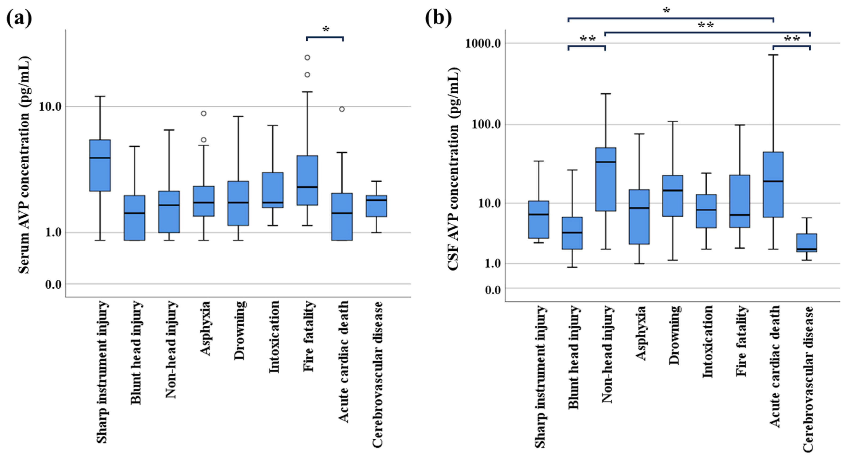
In comparisons between each cause-of-death group, serum AVP levels were significantly higher in fire deaths than in acute cardiac deaths (p < 0.05), whereas no significant differences were observed among other causes of death (Figure 1a). On the other hand, CSF AVP levels were significantly lower in cases involving blunt head injury and cerebrovascular disease than in those involving non-head injury or acute cardiac death (p < 0.01–0.05) (Figure 1b). Due to the possibility of bias resulting from blood contamination or other confounding factors, subsequent analyses were performed distinguishing CSF AVP concentrations in blunt head injury and cerebrovascular disease from other cause-of-death groups.
      
    
    Figure 1.
      Postmortem concentrations of arginine vasopressin (AVP) in serum (a) and cerebrospinal fluid (CSF) (b) in relation to cause of death. Serum AVP levels were significantly higher in cases involving fire fatality than in those involving acute cardiac death (p < 0.05). In addition, CSF AVP levels in cases involving blunt head injury and cerebrovascular disease were significantly lower than in those involving non-head injury or acute cardiac death (p < 0.01–0.05). * p < 0.05; ** p < 0.01. Circles: outliers.
  
3.3. Relationship Between AVP Concentrations and BW
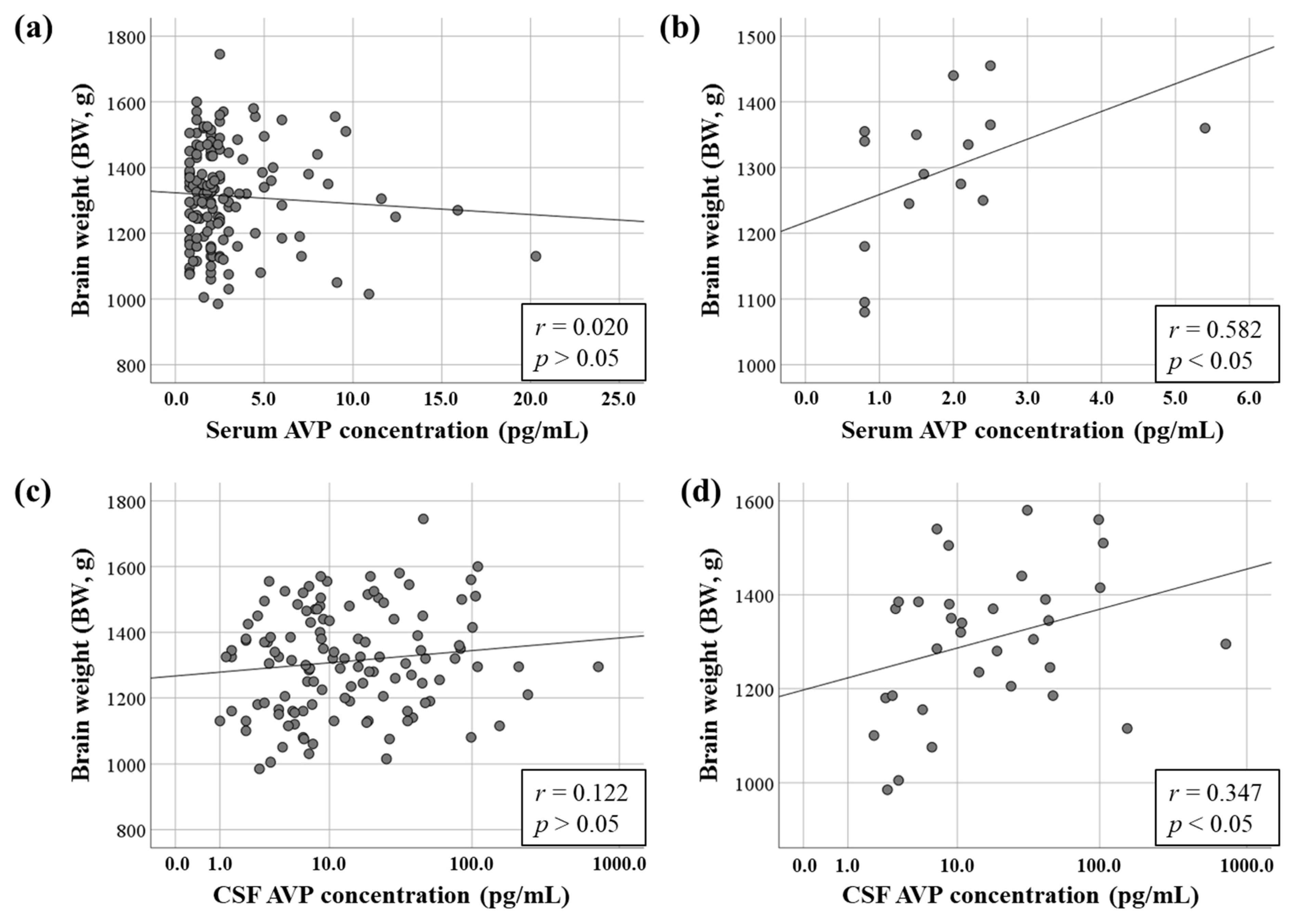
No correlation was found between serum AVP concentration and BW, including in all cause-of-death groups (r = 0.020, p > 0.05) (Figure 2a). To eliminate confounding factors for each individual pathology, hierarchical analysis was performed to analyze correlations for each cause of death. As a result, tendencies toward moderate correlation were seen between serum AVP concentration and BW with blunt head injury (r = 0.582, p < 0.05) (Figure 2b). The results of correlation analyses between serum AVP concentration and BW for other causes of death were as follows: sharp instrument injury (r = −0.217, p > 0.05), non-head injury (r = 0.158, p > 0.05), asphyxia (r = 0.028, p > 0.05), drowning (r = 0.175, p > 0.05), intoxication (r = 0.119, p > 0.05), fire fatality (r = −0.242, p > 0.05), acute cardiac death (r = 0.052, p > 0.05), and cerebrovascular disease (r = −0.214, p > 0.05).
      
    
    Figure 2.
      Relationships between postmortem serum and cerebrospinal fluid (CSF) levels of arginine vasopressin (AVP) and brain weight (BW). (a) Correlations between serum AVP concentration and BW in all cases (n = 143). (b) Correlations between serum AVP concentration and BW in cases involving blunt head injury (n = 15). (c) Correlations between CSF AVP concentration and BW with causes of death except for blunt head injury and cerebrovascular disease (n = 121). (d) Correlations between CSF AVP concentration and BW in cases involving sharp instrument injury and acute cardiac death (n = 33).
  
No correlation was found between CSF AVP concentration and BW among all cause-of-death groups, except for blunt head injury and cerebrovascular disease (r = 0.122, p > 0.05) (Figure 2c). Even in the mixed group combining blunt head injury and cerebrovascular disease, no correlation was observed between CSF AVP concentration and BW (r = 0.148, p > 0.05). Hierarchical analysis of the correlation between causes of death revealed no significant correlation between CSF AVP concentration and BW. Tendencies toward a correlation were seen between CSF AVP concentration and BW with sharp instrument injury (r = 0.419, p > 0.05), acute cardiac death (r = 0.371, p > 0.05), and cerebrovascular disease (r = 0.523, p > 0.05). When these cause-of-death groups, except for cerebrovascular disease, were combined and reanalyzed, a significant correlation was identified between BW and CSF AVP concentration (r = 0.347, p < 0.05) (Figure 2d). The results of correlation analyses between CSF AVP concentration and BW for each cause of death were as follows: blunt head injury (r = 0.182, p > 0.05), non-head injury (r = 0.084, p > 0.05), asphyxia (r = 0.190, p > 0.05), drowning (r = −0.104, p > 0.05), intoxication (r = −0.077, p > 0.05), and fire fatality (r = −0.276, p > 0.05).
3.4. Relationship Between AVP Concentration and Normalized BW
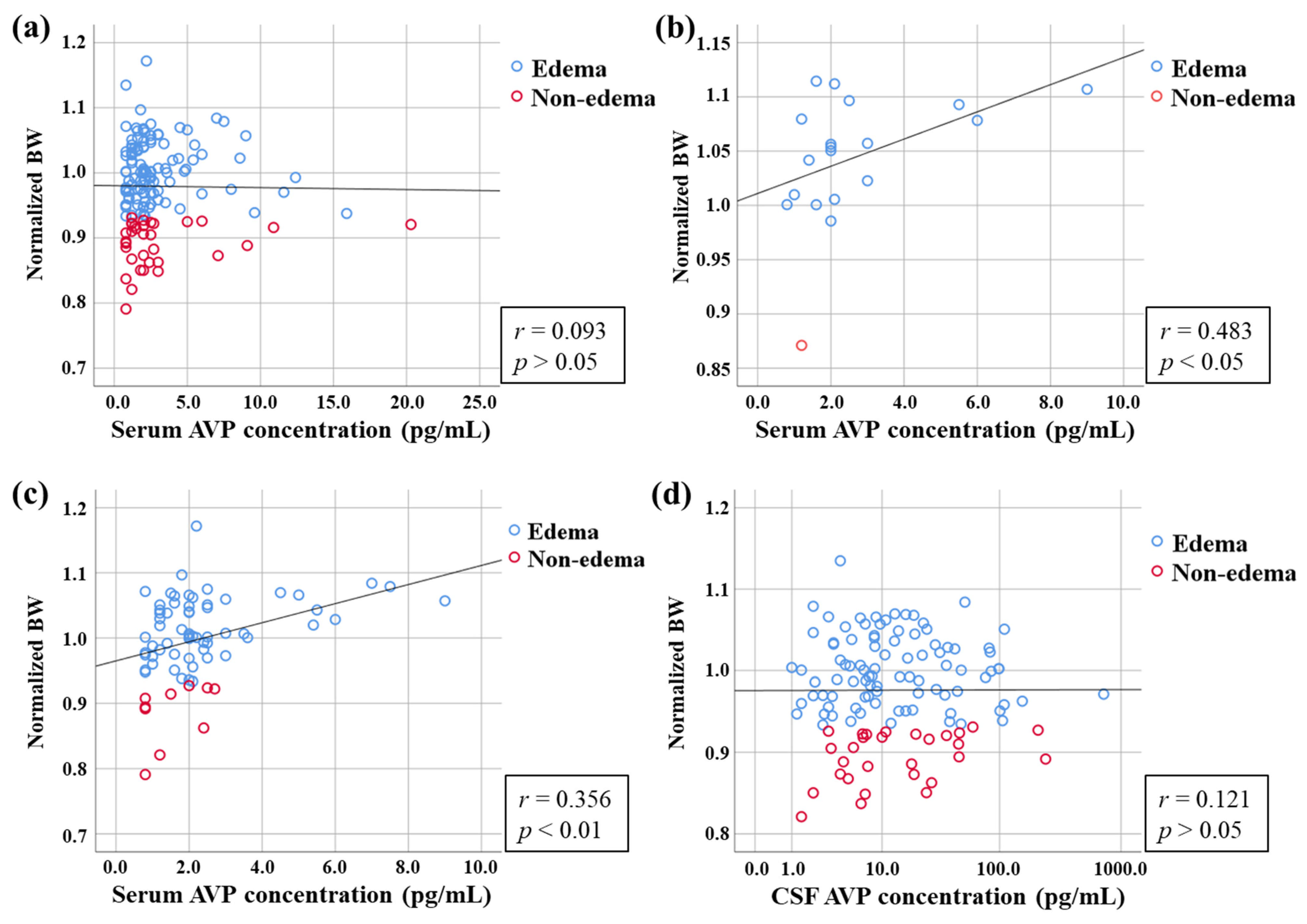
No correlation was found between serum AVP concentration and normalized BW, including all cause-of-death groups (r = 0.093, p > 0.05) (Figure 3a). To eliminate confounding factors for each individual pathology, hierarchical analysis was performed to analyze correlations for each cause of death. As a result, tendencies toward a moderate correlation were seen between serum AVP concentration and BW with asphyxia (r = 0.483, p < 0.05) (Figure 3b). Tendencies toward a correlation were identified between serum AVP concentration and normalized BW in cases involving blunt head injury (r = 0.278, p > 0.05), non-head injury (r = 0.283, p > 0.05), intoxication (r = 0.315, p > 0.05), and cerebrovascular disease (r = 0.321, p > 0.05). When these cause-of-death groups were combined and reanalyzed, a significant correlation was found between normalized BW and serum AVP concentration (r = 0.356, p < 0.01) (Figure 3c). The results of correlation analysis between serum AVP concentration and BW for other causes of death were as follows: sharp instrument injury (r = −0.060, p > 0.05), drowning (r = −0.270, p > 0.05), fire fatality (r = 0.062, p > 0.05), and acute cardiac death (r = 0.124, p > 0.05).
      
    
    Figure 3.
      Relationships between postmortem serum and cerebrospinal fluid (CSF) levels of arginine vasopressin (AVP) and normalized brain weight (BW). (a) Correlations between serum AVP concentration and normalized BW in all cases (n = 143). (b) Correlations between serum AVP concentration and normalized BW in asphyxia (n = 19). (c) Correlations between serum AVP concentration and normalized BW in cases involving blunt head injury, non-head injury, asphyxia, intoxication, and cerebrovascular disease (n = 72). (d) Correlations between CSF AVP concentration and normalized BW in all cause-of-death groups except blunt head injury and cerebrovascular disease (n = 121).
  
Conversely, no correlation was found between CSF AVP concentration and normalized BW among all cause-of-death groups except for blunt head injury and cerebrovascular disease (r = 0.121, p > 0.05) (Figure 3d). Even in the mixed group of blunt head injury and cerebrovascular disease, no correlation was observed between CSF AVP concentration and normalized BW (r = 0.025, p > 0.05). Hierarchical analysis of the correlation between causes of death revealed no significant correlation between CSF AVP concentration and normalized BW. Tendencies toward correlations were seen between CSF AVP concentration and normalized BW in cases involving sharp instrument injury (r = 0.323, p > 0.05) and cerebrovascular disease (r = 0.505, p > 0.05). The results of correlation analyses between CSF AVP concentration and normalized BW for other causes of death were as follows: non-head injury (r = 0.018, p > 0.05), asphyxia (r = −0.009, p > 0.05), drowning (r = 0.160, p > 0.05), intoxication (r = 0.033, p > 0.05), fire fatality (r = −0.040, p > 0.05), and acute cardiac death (r = −0.029, p > 0.05).
3.5. Relationship Between AVP Concentration and Classification of Edema by Normalized BW
The method of normalizing BW to intracranial volume proposed by Bauer et al. [] was used. Normalization of BW by intracranial volume was achieved by calculating the quotient of BW (in grams) and intracranial volume (in milliliters). In this method, the threshold for classifying edema was set as 0.9332, with calculated values below this limit taken as indicating cerebral edema, and higher values indicating no edema. According to the edema classification as reported by Bauer et al. [], 115 cases (76.9%) were classified as edema and 33 (23.1%) as non-edema in all cause-of-death groups (Table 2). No significant differences in serum or CSF AVP levels were seen between these edema and non-edema groups. In addition, ROC analysis revealed that the cutoff value for serum AVP concentration to classify edema and non-edema was 1.55 pg/mL (sensitivity 0.718; specificity 0.424). For CSF AVP concentrations in the cause-of-death groups excluding blunt head injury and cerebrovascular disease, ROC analysis revealed that the optimal cutoff for classifying edema and non-edema was 7.05 pg/mL (sensitivity 0.641; specificity 0.414).
       
    
    Table 2.
    Assessment of edema by normalized brain weight and arginine vasopressin (AVP) concentration.
  
4. Discussion
In the present study, postmortem AVP levels in serum and CSF did not correlate with survival or postmortem period, and were therefore considered to remain relatively stable after death. Postmortem AVP levels also did not differ by sex, age, or survival period. Although AVP levels in CSF and serum were not correlated, CSF AVP levels were revealed to be higher than serum AVP levels. The clinical reference range for serum AVP concentrations is <2.8 pg/mL, and serum AVP concentrations in most study subjects remained within this range. The half-life of plasma AVP is reportedly short, at approximately 24 min []. However, in this study, most values were within the clinical reference range, and although the influence of the half-life cannot be denied, no differences in the course of the disease were seen, suggesting that measuring AVP concentrations may be useful in diagnosing the final disease condition. In addition, AVP has been reported to show low ex vivo stability []. However, AVP is reportedly relatively stable when frozen at −80 °C. Because ex vivo stability was not verified in this study, the extent to which post-collection storage may be involved is unclear. However, no correlation with time since death was identified, and no significant decrease was observed within a few hours after death in vivo.
Serum AVP levels were significantly higher in fire-related deaths than in acute cardiac deaths, but no significant differences were observed among other causes of death. On the other hand, CSF AVP levels tended to be lower after blunt head injury and cerebrovascular disease. Decreases in AVP levels are reportedly attributable to damage to the hypothalamus or posterior pituitary gland [,]. However, although cases in which the CSF was highly contaminated with blood were excluded, the possibility remains that the CSF was contaminated with blood to some extent in cases of death caused by head injury or cerebrovascular disease, and completely excluding these potential contaminations is difficult. Therefore, we cannot exclude the possibility of contamination by hemorrhage or its confounding factors, and whether the decreased CSF AVP concentration in this study represents an important finding of impaired AVP secretion remains unclear. In addition, CSF AVP may need to be evaluated separately for these causes of death.
It has been suggested that AVP regulates brain water permeability, and that increased AVP may play a role in edema formation [,,]. In addition, BW is often used in addition to macroscopic observation to assess the presence or absence of cerebral edema []. In the present study, a correlation was identified between serum AVP and non-normalized BW in the group comprising death due to blunt head injury. Further, normalized BW (calculated by dividing BW measured at autopsy by intracranial volume segmented from CT) is reportedly a method for assessing cerebral edema that agrees well with macroscopic assessment []. The present study identified a correlation between serum AVP and normalized BW in cases involving asphyxia. Further, serum AVP tended to correlate with normalized BW in the groups comprising death due to blunt head injury, non-head injury, intoxication, or cerebrovascular disease. Serum AVP concentrations did not differ between causes of death, and in cases where a correlation with BW was observed, the concentrations may be able to be evaluated comprehensively, although a possibility of confounding factors exists. In addition, with these causes of death, AVP concentrations were suggested to be related to the amount of water in the brain. By contrast, although CSF AVP concentrations correlated with BW in some cause-of-death groups, no significant correlation was observed with normalized BW. This suggests the possibility of confounding factors such as blunt trauma, acute hypoxia due to asphyxiation, water/mineral imbalance in drowning, drug toxicity, and burn shock in cases of fire injury in CSF AVP. Thus, CSF AVP concentrations were suggested to be more susceptible to these influences than serum AVP concentrations.
In addition, to distinguish between higher and lower serum and CSF AVP levels, estimated cutoff values were calculated based on ROC analysis, resulting in cutoff values of 1.55 pg/mL and 7.05 pg/mL, respectively. The values calculated in this study represent exploratory findings, and the lack of significant differences in AVP concentrations between the two groups (edema and non-edema) should be noted. Further, many limitations exist regarding the measurement of AVP in this study, and its use as an indicator of cerebral edema remains controversial. Postmortem studies based on body samples are inherently complex, and the etiologies of brain edema are diverse. Nevertheless, the findings of this study suggest that AVP concentrations may serve as a potential biomarker for evaluating the severity of cerebral edema, in consideration of various confounding factors.
The present study has some limitations. The classification reported by Bauer et al. [] was used to compare results for the quantitative evaluation of the presence or absence of edema. In this report, the presence or absence of cerebral edema was quantitatively evaluated by dividing brain weight by intracranial volume. As a result, 76.9% of cases were classified as edema, which may have been a slightly lenient evaluation of the edema classification for the study population. In addition, a significant imbalance existed in the number of samples between the edema and non-edema groups, which may have biased the results. Further, the formation of cerebral edema reportedly shows a complicated relationship with various factors, such as dysfunction of transporters and ion channels, disruption of the blood–brain barrier, and activation of immune cells []. Various factors are believed to influence the formation of edema, and factors other than AVP may also need to be considered, as do the stability of AVP and the possibility of blood contamination in CSF. The exclusion of contaminated CSF samples may have introduced a potential selection bias, as the contamination itself may be related to the underlying pathophysiology. Although the results of this study did not reveal any significant decrease in AVP concentration with time after death or survival, the AVP concentration reportedly decreases with repeated freezing and thawing and extended storage []. These conditions may have potentially affected the measured AVP concentrations. To address these issues, simultaneously validating the evaluation of sampling sites may be useful, such as peripheral blood samples and measurement of copeptin, a more stable alternative peptide released into the blood in amounts similar to AVP [,,].
5. Conclusions
The present study aimed to explore the potential of AVP for diagnosing and assessing the severity of cerebral edema. We were unable to demonstrate superior results compared with previous methods for assessing cerebral edema. On the other hand, the present findings suggest that postmortem AVP levels are relatively stable. In addition, the AVP concentration was suggested to be involved in the formation of cerebral edema. These findings also highlight issues such as the influence of confounding factors associated with individual pathologies and the verification of AVP stability in postmortem samples. However, the utility of measuring AVP as an indicator of cerebral edema in forensic evaluation remains controversial, and further validation of the limitations and measurement of alternative peptides are needed to establish biochemical markers such as AVP as indicators of cerebral edema.
Author Contributions
Conceptualization, T.I.; methodology, N.T.; validation, N.T.; formal analysis, N.T. and K.M.; investigation, N.T. and K.M.; resources, N.T. and T.I.; data curation, N.T., K.M. and T.I.; writing—original draft preparation, N.T.; writing—review and editing, T.I.; visualization, N.T.; supervision, T.I.; project administration, N.T. All authors have read and agreed to the published version of the manuscript.
Funding
This research received no external funding.
Institutional Review Board Statement
This study was conducted according to the tenets of the Declaration of Helsinki and its later amendments, and was approved by the Independent Ethics Committee of the Osaka Metropolitan University Graduate School of Medicine (no. 2749, 30 January 2014).
Informed Consent Statement
This study was evaluated by the independent ethics committee of the Osaka Metropolitan University Graduate School of Medicine. Informed consent from bereaved family members was considered by the ethics committee to have been obtained in the form of providing an opt-out for autopsy data analysis and the study protocol was approved (authorization no. 2749, 30 January 2014). These practices are part of the routine in the author’s institute and country.
Data Availability Statement
The original data presented in this study are included in the article. Further inquiries can be directed to the corresponding author.
Conflicts of Interest
The authors declare no conflicts of interest.
References
- Marmarou, A. Pathophysiology of traumatic brain edema: Current concepts. Acta Neurochir. Suppl. 2003, 86, 7–10. [Google Scholar]
 - Lundesgaard Eidahl, J.M.; Opdal, S.H.; Rognum, T.O.; Stray-Pedersen, A. Postmortem evaluation of brain edema: An attempt with measurements of water content and brain-weight-to-inner-skull-circumference ratio. J. Forensic Leg. Med. 2019, 64, 1–6. [Google Scholar] [CrossRef] [PubMed]
 - Du, Y.; Xu, J.T.; Jin, H.N.; Zhao, R.; Zhao, D.; Du, S.H.; Xue, Y.; Xie, X.L.; Wang, Q. Increased cerebral expressions of MMPs, CLDN5, OCLN, ZO1 and AQPs are associated with brain edema following fatal heat stroke. Sci. Rep. 2017, 7, 1691. [Google Scholar] [CrossRef] [PubMed]
 - Wang, Q.; Ishikawa, T.; Michiue, T.; Zhu, B.L.; Guan, D.W.; Maeda, H. Molecular pathology of brain matrix metalloproteases, claudin5, and aquaporins in forensic autopsy cases with special regard to methamphetamine intoxication. Int. J. Legal Med. 2014, 128, 469–474. [Google Scholar] [CrossRef] [PubMed]
 - Donkin, J.J.; Vink, R. Mechanisms of cerebral edema in traumatic brain injury: Therapeutic developments. Curr. Opin. Neurol. 2010, 23, 293–299. [Google Scholar] [CrossRef]
 - Neumann, I.; Russell, J.A.; Landgraf, R. Oxytocin and vasopressin release within the supraoptic and paraventricular nuclei of pregnant, parturient and lactating rats: A microdialysis study. Neuroscience 1993, 53, 65–75. [Google Scholar] [CrossRef]
 - Palmiere, C.; Augsburger, M. Copeptin as a diagnostic biomarker for sepsis-related deaths. Peptides 2014, 59, 75–78. [Google Scholar] [CrossRef]
 - Katan, M.; Müller, B.; Christ-Crain, M. Copeptin: A new and promising diagnostic and prognostic marker. Crit. Care 2008, 12, 117. [Google Scholar] [CrossRef]
 - Morgenthaler, N.G.; Struck, J.; Alonso, C.; Bergmann, A. Assay for the measurement of copeptin, a stable peptide derived from the precursor of vasopressin. Clin. Chem. 2006, 52, 112–119. [Google Scholar] [CrossRef]
 - Rotondo, F.; Butz, H.; Syro, L.V.; Yousef, G.M.; Di Ieva, A.; Restrepo, L.M.; Quintanar-Stephano, A.; Berczi, I.; Kovacs, K. Arginine vasopressin (AVP): A review of its historical perspectives, current research and multifunctional role in the hypothalamo-hypophysial system. Pituitary 2016, 19, 345–355. [Google Scholar] [CrossRef]
 - Bankir, L.; Bichet, D.G.; Morgenthaler, N.G. Vasopressin: Physiology, assessment and osmosensation. J. Intern. Med. 2017, 282, 284–297. [Google Scholar] [CrossRef] [PubMed]
 - Jia, S.W.; Liu, X.Y.; Wang, S.C.; Wang, Y.F. Vasopressin Hypersecretion-Associated Brain Edema Formation in Ischemic Stroke: Underlying Mechanisms. J. Stroke Cerebrovasc. Dis. 2016, 25, 1289–1300. [Google Scholar] [CrossRef] [PubMed]
 - Hausmann, R.; Vogel, C.; Seidl, S.; Betz, P. Value of morphological parameters for grading of brain swelling. Int. J. Legal Med. 2006, 120, 219–225. [Google Scholar] [CrossRef] [PubMed]
 - Bauer, M.; Deigendesch, N.; Wittig, H.; Scheurer, E.; Lenz, C. Tissue sample analysis for post mortem determination of brain edema. Forensic Sci. Int. 2021, 323, 110808. [Google Scholar] [CrossRef]
 - Radojevic, N.; Radnic, B.; Vucinic, J.; Cukic, D.; Lazovic, R.; Asanin, B.; Savic, S. Mathematical model in post-mortem estimation of brain edema using morphometric parameters. J. Forensic Leg. Med. 2017, 4, 21–28. [Google Scholar] [CrossRef]
 - Bauer, M.; Gerlach, K.; Scheurer, E.; Lenz, C. Analysis of different post mortem assessment methods for cerebral edema. Forensic Sci. Int. 2020, 308, 110164. [Google Scholar] [CrossRef]
 - Kutlu, E.; Çil, N.; Avci, E.; Bir, F.; Kiliç, İ.D.; Dereli, A.K.; Acar, K. Significance of postmortem biomarkers and multimarker strategy in sudden cardiac death. Leg. Med. 2023, 61, 102212. [Google Scholar] [CrossRef]
 - Michiue, T.; Hishmat, A.M.; Oritani, S.; Miyamoto, K.; Amin, M.F.; Ishikawa, T.; Maeda, H. Virtual computed tomography morphometry of the patella for estimation of sex using postmortem Japanese adult data in forensic identification. Forensic Sci. Int. 2018, 285, 206.e1–206.e6. [Google Scholar] [CrossRef]
 - Baumann, G.; Dingman, J.F. Distribution, blood transport, and degradation of antidiuretic hormone in man. J. Clin. Investig. 1976, 57, 1109–1116. [Google Scholar] [CrossRef]
 - Dick, M.; Catford, S.R.; Kumareswaran, K.; Hamblin, P.S.; Topliss, D.J. Persistent syndrome of inappropriate antidiuretic hormone secretion following traumatic brain injury. Endocrinol. Diabetes Metab. Case Rep. 2015, 2015, 150070. [Google Scholar] [CrossRef]
 - Hadjizacharia, P.; Beale, E.O.; Inaba, K.; Chan, L.S.; Demetriades, D. Acute diabetes insipidus in severe head injury: A prospective study. J. Am. Coll. Surg. 2008, 207, 477–484. [Google Scholar] [CrossRef] [PubMed]
 - Dóczi, T.; László, F.A.; Szerdahelyi, P.; Joó, F. Involvement of vasopressin in brain edema formation: Further evidence obtained from the Brattleboro diabetes insipidus rat with experimental subarachnoid hemorrhage. Neurosurgery 1984, 14, 436–441. [Google Scholar] [CrossRef] [PubMed]
 - Jenkins, J.S.; Mather, H.M.; Ang, V. Vasopressin in human cerebrospinal fluid. J. Clin. Endocrinol. Metab. 1980, 50, 364–367. [Google Scholar] [CrossRef] [PubMed]
 - Bauer, M.; Berger, C.; Gerlach, K.; Scheurer, E.; Lenz, C. Post mortem evaluation of brain edema using quantitative MRI. Forensic Sci. Int. 2022, 337, 111376. [Google Scholar] [CrossRef]
 - Han, W.; Song, Y.; Rocha, M.; Shi, Y. Ischemic brain edema: Emerging cellular mechanisms and therapeutic approaches. Neurobiol. Dis. 2023, 178, 106029. [Google Scholar] [CrossRef]
 - Palmiere, C.; Tettamanti, C.; Bonsignore, A.; De Stefano, F.; Vanhaebost, J.; Rousseau, G.; Scarpelli, M.P.; Bardy, D. Cardiac troponins and NT-proBNP in the forensic setting: Overview of sampling site, postmortem interval, cardiopulmonary resuscitation, and review of the literature. Forensic Sci. Int. 2018, 282, 211–218. [Google Scholar] [CrossRef]
 - Cao, Z.; Zhao, M.; Xu, C.; Zhang, T.; Jia, Y.; Wang, T.; Zhu, B. Evaluation of agonal cardiac function for sudden cardiac death in forensic medicine with postmortem brain natriuretic peptide (BNP) and NT-proBNP: A meta-analysis. J. Forensic Sci. 2020, 65, 686–691. [Google Scholar] [CrossRef]
 
Disclaimer/Publisher’s Note: The statements, opinions and data contained in all publications are solely those of the individual author(s) and contributor(s) and not of MDPI and/or the editor(s). MDPI and/or the editor(s) disclaim responsibility for any injury to people or property resulting from any ideas, methods, instructions or products referred to in the content.  | 
© 2025 by the authors. Licensee MDPI, Basel, Switzerland. This article is an open access article distributed under the terms and conditions of the Creative Commons Attribution (CC BY) license (https://creativecommons.org/licenses/by/4.0/).