Abstract
To ensure the country’s sustainable health recovery, viruses like COVID-19 need faster detection and sampling than the rate at which they spread. Blood plasma has proven to be an important and better clinical sample for the detection and diagnosis of various medical conditions as compared to whole blood. For in situ and in vivo health monitoring, plasma can be easily processed through microfluidic Lab-on-Chip (LOC) devices without the clotting that shortens the turnaround time and using minimum amounts of sample and reagents. The present review discusses the key properties of blood plasma as a perfect sample for the microfluidic LOC devices and the importance of passive plasma separators within any kind of LOC device as an embedded unit. The passive LOC plasma separators offer rapid extraction without external forces in the form of a miniaturized automated unit. This article compares various plasma separators on the basis of plasma extraction efficiency, fabrication techniques, and separation science utilized for hemolysis-free extraction. Recent developments in the area of passive bioseparators based on microfiltration and self-driven hydrodynamic and flow-cytometric approaches are discussed in detail.
1. Introduction
The separation of plasma from whole blood has been the topic of sustainable research owing to its potential for the rapid and early diagnosis of critical medical conditions such as Alzheimer’s disease [1], kidney damage [2], cancer [3], acute stroke [4], malaria [5], diabetes mellitus [6], and many other diseases such as viral infections; as well as the identification of the success rates of anti-tumor therapies. Plasma has now been clinically opted as a new standard analyte in the laboratory testing and diagnosis of biomarkers such as free-DNAs [7], enzymes, and other hormones.
The key properties of plasma [8], such as its Newtonian behavior and its higher fluidity due to a lower viscosity and clotting profile [9], as opposed to whole blood samples (heavier and larger cells [10] other than biomarkers), support easier sample handling, preparation with a minimum number of reagents, and a free-flow operation through microfluidics with on-chip detection or Lab-on-Chip Testing (LOCT). These advantages result in short turnaround times, the low probability of false detection, and compatibility with POCT (Point-of-Care Testing). Filtering out the unwanted and interfering cells such as RBCs (red blood cells—erythrocytes), WBCs (white blood cells—leukocytes), and platelets (thrombocytes) from whole blood, supports a clog-free operation. Blood rheology, RBC clotting, and the viscoelastic physiology of whole blood [10] makes plasma separation the initial protocol for either subsequent lab-testing or chip testing. Furthermore, on-chip plasma extraction develops the base of rapid extraction extended to LOCT, µ-FT (microfluidics testing), and µTAS (Micro Total Analysis System), to carry-out POCT successfully for the rapid and early detection of biomarkers associated with chronic diseases including HIV/AIDS (human immunodeficiency virus and acquired immune deficiency syndrome), COVID-19, and HBVs/HCVs (hepatitis B and C), etc., for both antibody (Ab)- and antigen (Ag)-based testing, where Ag-based testing is preferable for the early detection of rampant diseases.
This review paper, therefore, covers the importance of LOC microfluidics’ and microfiltration approaches for efficient and RBC-free plasma extraction in brief. We conclude with a discussion on selected plasma separation devices fabricated with different subtractive, additive, and replicative techniques for LOCT.
2. Plasma Separation Techniques
RBCs, WBCs, and platelets are generally heavier and larger compared to plasma, as a result they can coagulate due to the RBCs’ rheology [11] or the sediment due to the gravitational or inertial forces as shown in Figure 1a. The properties of these cells are exploited in the microfluidics-based active separation techniques [12]. Generally, plasma (the watery part of blood) mostly consists of smaller cells below 500 nm as shown in Table 1, and key biomarkers, as shown in Figure 1b, which aid in the diagnosis of various illnesses. Microfiltration-based approaches [13] utilize the cell size to design and optimize plasma filters to extract target biomarkers accordingly; while, on the other hand, the hydrodynamics properties of plasma, which are better than the whole blood as shown in Table 2, are exploited for microfluidics-based plasma separation [14].
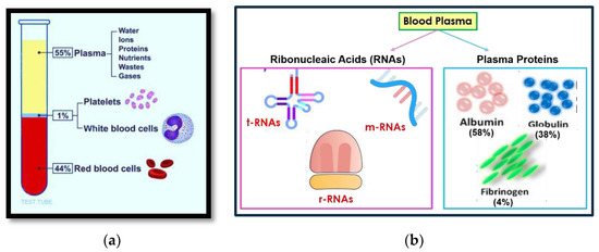
Figure 1.
(a) The components of human whole blood depicting the RBCs settling at the bottom of a test-tube due to the gravitationally assisted sedimentation; (b) the constituents of blood plasma depicting the volumetric percentage of plasma proteins and various RNAs.

Table 1.
The Physiology of Whole Blood.
Table 2.
A Comparison of Whole Blood and Plasma Characteristics.
2.1. Force-Driven Active Plasma Separation
Plasma is extracted conventionally from the whole blood contained inside the centrifuge, or a sedimentation chamber of compact discs as per Stoke’s Law, by employing centrifugation, electromechanically, at a high rotational velocity (3800 rpm), to release pure plasma at the output [15]. The advancement from CD-based microfluidics towards slanted-spiral microchannels and multiplexed slanted-spirals [16] has assisted ultra-fast rapid extraction, improving the flow rates from 1.5 mL/min to 24 mL/min.
Microfluidics-based cell sorting techniques through activated cell sorting (ACS) exploit flow cytometry via magnetic (MACS) [17], dielectrophoresis [18], and acoustic [19] forces for the microscale extraction of plasma based on cell properties—such as supermagnetic (RBCs) and paramagnetic (WBCs), cell-interaction with the fluid, and cell shape, size, stiffness, and weight, etc., to guide the target cells towards the dedicated direction or position within 25 min of operation with almost 100% purity and label-free detection.
2.2. Self-Driven Passive Plasma Separation
Self-driven passive plasma separation, also referred to as passive cell-sorting techniques, makes use of the internal fluid properties and physical sizes of the cells for the self-separation process rather than the external forces. A schematic of various self-driven passive mechanisms for plasma separation is shown in Figure 2.
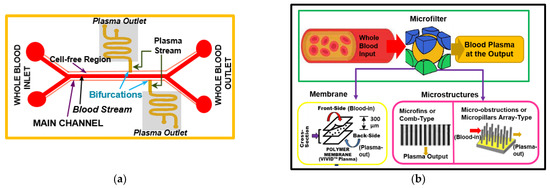
Figure 2.
A schematic showing the operating principal of microfluidics and microfiltration-based plasma separation: (a) microfluidics-based self-driven passive plasma separation through the cell-free layer and bifurcations; (b) microfiltration-based self-driven passive plasma separation through membrane-assisted and microstructure-assisted plasma filtration.
Microfluidics-based approaches make use of the Newtonian characteristics of blood plasma and hydrodynamic effects. The plasma is extracted via a capillary force-driven followability assisted hydrophobic µ-fluidic channel. The unwanted cells are separated through the hydrophilic or main channel. Microfiltration employs passive cell sorting-based approaches, which operate according to the size-selection trapping of larger blood cells and the microfiltration of plasma either through microstructures or through microporous separation membranes.
Some of the passive cell sorting techniques based on flow cytometry are gravitation-assisted [20], sedimentation [21], deterministic lateral displacement [22], pinched-flow fractionation [23], and biomimetic separation methods [24]. Passive cell sorting through microstructures is based on various filter designs with pores and nano-fibers [25], and comb-like [26] and mesh-type [27] structures. Microporous separation membranes and micro-obstructions oriented for different filter modes such as cross-flow filtration [28], dead-end filtration [29], and tangential-flow filtration [30]—pertaining to blood flow—provides the liberty for the designers to optimize new and better passive plasma separators.
3. Passive Lab-on-Chip Plasma Separation
The field of LOC plasma separators is a recent phenomenon that has cleverly integrated the principles of separation science, flow cytometry, plasma physiology, and blood rheology, to fabricate a rapid, compact, and POC device compatible with LOC architecture. A brief comparison between them on the basis of fabrication technology, device structure, extraction efficiency, and separation technology is depicted in Table 3.
Table 3.
A comparison between the passive microfluidic Lab-on-Chip plasma separators.
4. Conclusions
In vivo monitoring of diseases is a critical issue, especially in the severe stages of an infection. LOC plasma separators can improve the survival rate of patients by achieving an early and rapid diagnosis. We have briefly reviewed the prominent techniques and devices for the passive microfluidic LOC plasma separators developed to date; however, while detailed elaboration is beyond the scope of this paper, the presented information sequentially covers their key aspects in terms of the fabrication technology, extraction efficiency, and the detected analytes.
Author Contributions
Conceptualization, S.K.; Investigations, S.K.; Original draft preparation, S.K.; Supervision, G.A.; Review, G.A.; Editing, G.A. All authors have read and agreed to the published version of the manuscript.
Funding
This research received no external funding.
Informed Consent Statement
Not applicable for studies not involving humans or animals.
Data Availability Statement
No new data were created or analyzed in this study. Data sharing is not applicable to this article.
Conflicts of Interest
The authors declare no conflict of interest.
Abbreviations
| DNA | Deoxyribonucleic acid |
| COVID 19 | Corona Virus Disease 2019 |
| SARS-CoV2 | Severe Acute Respiratory Syndrome Coronavirus2 |
| CHIKV | Chickengunya Virus |
| PDMS | Polydimethylsiloxane |
| PMMA | Polymethylmethacrylate |
| DLP | Digital Light processing |
| SLA | Stereolithography |
| SU 8 | Epoxy based Negative Photoresist |
References
- Eke, C.S.; Jammeh, E.; Li, X.; Carroll, C.; Pearson, S.; Ifeachor, E. Early Detection of Alzheimer’s Disease with Blood Plasma Proteins Using Support Vector Machines. IEEE J. Biomed. Health Informatics 2020, 25, 218–226. [Google Scholar] [CrossRef] [PubMed]
- Kazi, R.N. Early detection of kidney function in diabetic kidney disease: An approach to prevent end stage renal disease. J. Interv. Nephrol. 2018, 1, 15–18. [Google Scholar]
- Schwarzenbach, H.; Hoon, D.S.B.; Pantel, K. Cell-free nucleic acids as biomarkers in cancer patients. Nat. Rev. Cancer 2011, 11, 426–437. [Google Scholar] [CrossRef] [PubMed]
- Rainer, T.; Wong, K.S.L.; Lam, W.; Yuen, E.; Lam, N.Y.; Metreweli, C.; Lo, Y.D. Prognostic Use of Circulating Plasma Nucleic Acid Concentrations in Patients with Acute Stroke. Clin. Chem. 2003, 49, 562–569. [Google Scholar] [CrossRef] [PubMed]
- Franklin, B.S.; Vitorino, B.L.F.; Coelho, H.C.; Menezes-Neto, A.; Santos, M.L.S.; Campos, F.M.F.; Brito, C.F.; Fontes, C.; Lacerda, M.V.; Carvalho, L.H. Plasma Circulating Nucleic Acids Levels Increase According to the Morbidity of Plasmodium vivax Malaria. PLoS ONE 2011, 6, e19842. [Google Scholar] [CrossRef]
- Rhee, M.K.; Ho, Y.-L.; Raghavan, S.; Vassy, J.L.; Cho, K.; Gagnon, D.; Staimez, L.R.; Ford, C.N.; Wilson, P.W.F.; Phillips, L.S. Random plasma glucose predicts the diagnosis of diabetes. PLoS ONE 2019, 14, e0219964. [Google Scholar] [CrossRef]
- Wagner, J. Free DNA–new potential analyte in clinical laboratory diagnostics? Biochem. Medica 2012, 22, 24–38. [Google Scholar] [CrossRef]
- Benjamin, R.J.; McLaughlin, L.S. Plasma components: Properties, differences, and uses. Transfusion 2012, 52, 9S–19S. [Google Scholar] [CrossRef]
- Nader, E.; Skinner, S.; Romana, M.; Fort, R.; Lemonne, N.; Guillot, N.; Gauthier, A.; Antoine-Jonville, S.; Renoux, C.; Hardy-Dessources, M.-D.; et al. Blood Rheology: Key Parameters, Impact on Blood Flow, Role in Sickle Cell Disease and Effects of Exercise. Front. Physiol. 2019, 10, 1329. [Google Scholar] [CrossRef] [PubMed]
- Kalmokoff, M.L.; Koval, S.F.; Jarrell, K.F. Relatedness of the flagellins from methanogens. Arch. Microbiol. 1992, 157, 481–487. [Google Scholar] [CrossRef]
- Cripps, C.M. Rapid method for the estimation of plasma haemoglobin levels. J. Clin. Pathol. 1968, 21, 110–112. [Google Scholar] [CrossRef] [PubMed]
- Wang, Y.; Nunna, B.; Talukder, N.; Etienne, E.; Lee, E. Blood Plasma Self-Separation Technologies during the Self-Driven Flow in Microfluidic Platforms. Bioengineering 2021, 8, 94. [Google Scholar] [CrossRef] [PubMed]
- Jørgensen, M.K.; Eriksen, K.B.; Christensen, M.L. Particle Track and Trace during Membrane Filtration by Direct Observation with a High Speed Camera. Membranes 2020, 10, 68. [Google Scholar] [CrossRef]
- Mateen, S.A.; Bhole, K.S. A review on microfluidic devices for separation of blood constituents. IOP Conf. Ser. Mater. Sci. Eng. 2020, 810, 012024. [Google Scholar] [CrossRef]
- Amasia, M.; Madou, M. Large-volume centrifugal microfluidic device for blood plasma separation. Bioanalysis 2010, 2, 1701–1710. [Google Scholar] [CrossRef] [PubMed]
- Rafeie, M.; Zhang, J.; Asadnia, M.; Li, W.; Warkiani, M.E. Multiplexing slanted spiral microchannels for ultra-fast blood plasma separation. Lab Chip 2016, 16, 2791–2802. [Google Scholar] [CrossRef]
- Civelekoglu, O.; Frazier, A.B.; Sarioglu, A.F. The Origins and the Current Applications of Microfluidics-Based Magnetic Cell Separation Technologies. Magnetochemistry 2022, 8, 10. [Google Scholar] [CrossRef]
- Yasukawa, T.; Yamada, J.; Shiku, H.; Matsue, T.; Suzuki, M. Microfluidic Separation of Blood Cells Based on the Negative Dielectrophoresis Operated by Three Dimensional Microband Electrodes. Micromachines 2020, 11, 833. [Google Scholar] [CrossRef]
- Nair, K.P.P.R.; Veettil, T.C.P.; Wood, B.R.; Paul, D.; Alan, T. Haemoprocessor: A Portable Platform Using Rapid Acoustically Driven Plasma Separation Validated by Infrared Spectroscopy for Point-of-Care Diagnostics. Biosensors 2022, 12, 119. [Google Scholar] [CrossRef]
- Zhang, X.-B.; Wu, Z.-Q.; Wang, K.; Zhu, J.; Xu, J.-J.; Xia, X.-H.; Chen, H.-Y. Gravitational Sedimentation Induced Blood Delamination for Continuous Plasma Separation on a Microfluidics Chip. Anal. Chem. 2012, 84, 3780–3786. [Google Scholar] [CrossRef]
- Garcia-Rey, S.; Nielsen, J.B.; Nordin, G.P.; Woolley, A.T.; Basabe-Desmonts, L.; Benito-Lopez, F. High-Resolution 3D Printing Fabrication of a Microfluidic Platform for Blood Plasma Separation. Polymers 2022, 14, 2537. [Google Scholar] [CrossRef] [PubMed]
- Chien, W.; Zhang, Z.; Gompper, G.; Fedosov, D.A. Deformation and dynamics of erythrocytes govern their traversal through microfluidic devices with a deterministic lateral displacement architecture. Biomicrofluidics 2019, 13, 044106. [Google Scholar] [CrossRef]
- Rodríguez-Villarreal, A.I.; Arundell, M.; Carmona, M.; Samitier, J. High flow rate microfluidic device for blood plasma separation using a range of temperatures. Lab Chip 2009, 10, 211–219. [Google Scholar] [CrossRef] [PubMed]
- Namgung, B.; Tan, J.K.S.; Wong, P.A.; Park, S.-Y.; Leo, H.L.; Kim, S. Biomimetic Precapillary Flow Patterns for Enhancing Blood Plasma Separation: A Preliminary Study. Sensors 2016, 16, 1543. [Google Scholar] [CrossRef]
- Lopresti, F.; Keraite, I.; Ongaro, A.E.; Howarth, N.M.; La Carrubba, V.; Kersaudy-Kerhoas, M. Engineered Membranes for Residual Cell Trapping on Microfluidic Blood Plasma Separation Systems: A Comparison between Porous and Nanofibrous Membranes. Membranes 2021, 11, 680. [Google Scholar] [CrossRef] [PubMed]
- Moorthy, J.; Beebe, D.J. In situ fabricated porous filters for microsystems. Lab Chip 2003, 3, 62–66. [Google Scholar] [CrossRef]
- Andersson, H.; van der Wijngaart, W.; Enoksson, P.; Stemme, G. Micromachined flow-through filter-chamber for chemical reactions on beads. Sens. Actuators B Chem. 2000, 67, 203–208. [Google Scholar] [CrossRef]
- Zheng, S.; Lin, H.; Liu, J.-Q.; Balic, M.; Datar, R.; Cote, R.J.; Tai, Y.-C. Membrane microfilter device for selective capture, electrolysis and genomic analysis of human circulating tumor cells. J. Chromatogr. A 2007, 1162, 154–161. [Google Scholar] [CrossRef]
- Faustino, V.; Catarino, S.O.; Pinho, D.; Lima, R.A.; Minas, G. A Passive Microfluidic Device Based on Crossflow Filtration for Cell Separation Measurements: A Spectrophotometric Characterization. Biosensors 2018, 8, 125. [Google Scholar] [CrossRef]
- Wang, Y.; Keller, K.; Cheng, X. Tangential Flow Microfiltration for Viral Separation and Concentration. Micromachines 2019, 10, 320. [Google Scholar] [CrossRef]
- Park, S.; Shabani, R.; Schumacher, M.; Kim, Y.-S.; Bae, Y.M.; Lee, K.-H.; Cho, H.J. On-chip whole blood plasma separator based on microfiltration, sedimentation and wetting contrast. Microsyst. Technol. 2015, 22, 2077–2085. [Google Scholar] [CrossRef]
- Madadi, H.; Casals-Terré, J.; Mohammadi, M. Self-driven filter-based blood plasma separator microfluidic chip for point-of-care testing. Biofabrication 2015, 7, 025007. [Google Scholar] [CrossRef] [PubMed]
- Kim, H.; Park, H.; Chung, D.R.; Kim, T.; Park, E.; Kang, M. A self-pressure-driven blood plasma-separation device for point-of-care diagnostics. Talanta 2022, 247, 123562. [Google Scholar] [CrossRef] [PubMed]
- Su, X.; Zhang, J.; Zhang, D.; Wang, Y.; Chen, M.; Weng, Z.; Wang, J.; Zeng, J.; Zhang, Y.; Zhang, S.; et al. High-Efficiency Plasma Separator Based on Immunocapture and Filtration. Micromachines 2020, 11, 352. [Google Scholar] [CrossRef]
- Maria, M.S.; Rakesh, P.E.; Chandra, T.S.; Sen, A.K. Capillary flow of blood in a microchannel with differential wetting for blood plasma separation and on-chip glucose detection. Biomicrofluidics 2016, 10, 054108. [Google Scholar] [CrossRef] [PubMed]
Disclaimer/Publisher’s Note: The statements, opinions and data contained in all publications are solely those of the individual author(s) and contributor(s) and not of MDPI and/or the editor(s). MDPI and/or the editor(s) disclaim responsibility for any injury to people or property resulting from any ideas, methods, instructions or products referred to in the content. |
© 2022 by the authors. Licensee MDPI, Basel, Switzerland. This article is an open access article distributed under the terms and conditions of the Creative Commons Attribution (CC BY) license (https://creativecommons.org/licenses/by/4.0/).