Article

Wedge Osteotomy of the Maxilla for the Treatment of Obstructive Sleep Apnea (OSA) Through Virtual Surgical Planning, CAD/CAM Technology: Consecutive Case Series

1
Department of Innovative Technologies in Medicine & Dentistry, University of Chieti-Pescara, Via Dei Vestini 31, 66100 Chieti, Italy
2
Maxillofacial Surgery Unit, San 2 Camillo-Forlanini Hospital, 00152 Rome, Italy
3
Independent Researcher, 00185 Roma, Italy
4
Postgraduate School in Maxillofacial Surgery–Sapienza University of Rome, 00185 Rome, Italy
5
Otolaryngology Unit, San Camillo-Forlanini Hospital, 00152 Rome, Italy
6
Sub-Intensive Respiratory Therapy Unit (STIRS), San Camillo-Forlanini Hospital, 00152 Rome, Italy
*
Author to whom correspondence should be addressed.
Surgeries 2025, 6(3), 74; https://doi.org/10.3390/surgeries6030074 (registering DOI)
Submission received: 22 July 2025 / Revised: 18 August 2025 / Accepted: 29 August 2025 / Published: 30 August 2025

Abstract

Orthognathic surgery, particularly maxillomandibular advancement (MMA), has emerged as an effective therapeutic option for patients with moderate to severe OSA who are refractory to conventional treatments. The wedge osteotomy of the maxilla, often performed in combination with mandibular surgery, can be a surgical treatment for obstructive sleep apnea (OSA). This case series report describes 6 OSA patients without anteroposterior maxillary deficiency who were treated with wedge osteotomy of the maxilla. Material and Methods: We conducted a retrospective analysis of 6 patients who underwent maxillomandibular advancement (MMA) for obstructive sleep apnea (OSA), all operated on consecutively by the same surgeon between 2018 and 2024 at the Maxillofacial Surgery of San Camillo-Forlanini Hospital, in Rome, Italy. Patients were evaluated using a CAD/CAM-assisted approach. A pre- and postoperative comparative analysis was conducted to assess the effectiveness of the surgical treatment in improving OSA-related parameters. Maxillary wedge osteotomy and bilateral sagittal split osteotomies (BSSO) of the mandibular ramus were digitally planned. Results: The comparison between preoperative and postoperative CT scans, along with 3D reconstructions generated using dedicated software, revealed a counterclockwise rotation of the occlusal plane, resulting in a mandibular advancement of approximately 13 mm. The CT shows a significant increase in airway volume following the skeletal repositioning. The airway volume increased from 20.665 ± 546 mm3 to 27.177 ± 446 mm3. Conclusions: Counterclockwise rotational orthognathic surgery without maxillary advancement has been shown to effectively enlarge the posterior pharyngeal space while also delivering excellent esthetic outcomes.

1. Introduction

Obstructive sleep apnea (OSA) is a chronic respiratory disorder characterized by repeated episodes of partial or complete upper airway obstruction during sleep, resulting in intermittent hypoxemia and sleep fragmentation [1]. This condition is associated with an increased risk of hypertension, cardiovascular disease, metabolic disorders, stroke and impaired quality of life.

1.1. Clinical Signs

Insomnia and obstructive sleep apnea (OSA) are both associated with an increased risk of hypertension and cardiovascular disease in older adults and younger military veterans [2].
Obstructive sleep apnea (OSA) has been associated with an increased risk of difficult tracheal intubation due to anatomical alterations of the upper airway and a greater tendency for pharyngeal collapse. Conversely, an increased neck circumference—often linked to obesity—has been identified as an independent predictive factor for difficult mask ventilation, further complicating airway management in patients with OSA [3]. The underlying pathophysiological mechanisms include intermittent hypoxemia, chronic activation of the sympathetic nervous system, systemic inflammation, and elevated nocturnal blood pressure [4]. These factors act synergistically to promote vascular damage and endothelial dysfunction, significantly contributing to overall cardiovascular risk. Ischemic stroke and obstructive sleep apnea (OSA) are both highly prevalent conditions with significant clinical, social, and economic impact. A growing body of evidence indicates that OSA is an independent risk factor for cerebrovascular events, including ischemic stroke.

1.2. Therapeutic Approach

Although conservative treatment, such as continuous positive airway pressure (CPAP), is considered the first-line therapy, many patients exhibit poor long-term adherence or intolerance [5,6]. In this context, orthognathic surgery—particularly maxillomandibular advancement (MMA)—has emerged as an effective therapeutic option for patients with moderate to severe OSA who are refractory to conventional treatments. The maxillomandibular advancement (MMA) technique was introduced by Riley and Powell in the mid-1980s as a surgical treatment for obstructive sleep apnea (OSA) [7]. This procedure has proven particularly effective in patients with severe or treatment-resistant OSA, especially when both mandibular and maxillary advancement are required [8]. MMA works by increasing the volume of the upper airway through anterior repositioning of the skeletal segments, thereby improving airway patency during sleep. Over time, the technique has evolved thanks to the introduction of digital planning tools and a deeper understanding of OSA pathophysiology, expanding its indications to include patients without evident skeletal dysmorphisms. Numerous studies have shown that MMA can lead to a significant reduction in the apnea-hypopnea index (AHI), while also improving oxygen saturation and sleep quality. Additionally, the procedure can provide both esthetic and functional benefits, contributing to greater patient satisfaction. However, the complexity of the surgery requires careful patient selection, multidisciplinary preoperative planning, and thorough postoperative follow-up. The wedge osteotomy of the maxilla, often performed in combination with mandibular advancement, allows for three-dimensional repositioning of the facial skeletal structures, with the goal of enlarging the retropharyngeal space and improving upper airway patency. The wedge osteotomy of the maxilla, often performed in combination with mandibular surgery, can be a surgical treatment for obstructive sleep apnea (OSA). This case series report describes three OSA patients without anteroposterior maxillary deficiency who were treated with wedge osteotomy of the maxilla.

2. Materials and Methods

We conducted a retrospective analysis of patients who underwent maxillomandibular advancement (MMA) for obstructive sleep apnea (OSA), all operated on consecutively by the same surgeon between 2018 and 2024 at the Maxillofacial Surgery of San Camillo-Forlanini Hospital, in Rome, Italy. A dedicated informed consent form was developed for this study. All procedures were conducted in accordance with the ethical principles outlined in the Declaration of Helsinki.
Patients were included in the study based on the following criteria:
  • Diagnosis of moderate to severe obstructive sleep apnea (OSA), confirmed by polysomnography with an apnea-hypopnea index (AHI) > 15.
  • Multidisciplinary evaluation by the institutional OSA team, including a maxillofacial surgeon, dentist, pulmonologist, ENT specialist, and bariatric surgeon, with a consensus recommendation for mandibular advancement (MA).
  • Preoperative drug-induced sleep endoscopy (DISE) demonstrating potential benefit from MA, confirmed through mandibular advancement maneuvers.
  • Good general health status with no contraindications to general anesthesia (ASA physical status classification ≤ 3).
  • Patients not requiring maxillary advancement.
Exclusion Criteria
Patients were excluded if they met any of the following conditions:
  • Pediatric age group (<18 years).
  • Presence of respiratory diseases or specific craniofacial malformations.
  • Severe systemic conditions contraindicating general anesthesia (ASA ≥ 4).
  • History of severe cardiovascular disease.
  • Presence of psychiatric disorders.
  • Syndromic conditions.
  • Previous orthognathic surgery.
  • Requirement for maxillary advancement.
  • Refusal to provide informed consent.
For each patient, demographic and clinical data were collected, including age, sex, and preoperative body mass index (BMI). Polysomnographic evaluations were conducted by the Pneumology Unit within six months prior to surgery and repeated six months postoperatively to assess treatment outcomes. A comparative analysis was conducted to assess the effectiveness of each technique in improving OSA-related parameters. Patients were evaluated using a CAD/CAM-assisted approach. A pre- and postoperative comparative analysis was conducted to assess the effectiveness of the surgical treatment in improving OSA-related parameters (Figure 1).
This study included 6 patients with severe obstructive sleep apnea syndrome (OSAS) who underwent mandibular advancement (MA) surgery without advance the maxilla. The patients had a mean age of 41 ± 3 years; 5 were male and 1 were female.
For all patients Helical CT scans were acquired for virtual surgical planning, enabling the design of patient-specific cutting guides and fixation plates. Scans of the head and neck were performed with the patient in a supine position and the head in a natural posture. These imaging datasets, combined with occlusal records derived from digital dental models and clinical photographs, were used to generate a three-dimensional reconstruction of the craniofacial skeleton, accurately aligned with the patient’s natural head position. Preoperative and postoperative assessments included full-night polysomnography, performed within six months before surgery and repeated six months after the procedure. The following respiratory and oxygenation parameters were collected and analyzed:
Apnea–Hypopnea Index (AHI)
Apnea Index (AI)
Hypopnea Index (HI)
Mean Apnea Duration
Oxygen Desaturation Index (ODI)
Mean Oxygen Saturation (SaO2)
Lowest Oxygen Saturation (Nadir SaO2)
Mean Desaturation Events
Percentage of Time with SaO2 < 90%
Pre- and postoperative cephalometric tracings were generated from CT scan images using Dolphin 3D software (version 11.7; Dolphin Imaging & Management Solutions, Chatsworth, CA, USA).
Maxillary wedge osteotomy and bilateral sagittal split osteotomies (BSSO) of the mandibular ramus were digitally planned. Virtual simulations of the maxillary wedge osteotomy and mandibular movements were performed, and both intermediate and final surgical splints were designed according to a maxilla-first approach (Figure 1; Figure 2). These splints were subsequently fabricated using 3D printing. Following virtual surgical planning, CAD/CAM technology was employed to create polyamide cutting guides tailored to the planned osteotomies, along with custom-made titanium fixation plates. Both the guides and plates were manufactured through 3D printing techniques. CT images were segmented to simulate osteotomies and evaluate the airway. Additionally, patient-specific cutting guides and fixation plates were designed as part of the surgical planning process.

2.1. Surgical Procedures

Maxillary wedge osteotomy, an intraoral incision was made in the upper vestibule, above the dental apices, extending from the right first molar to the left first molar. A mucoperiosteal flap was elevated to expose the maxillary bone. The bone-supported cutting guide was then positioned on the maxilla. Both cutting guides were secured in place using 2.0 mm screws. A Le Fort I osteotomy was performed using a piezosurgical device. An additional osteotomy was carried out in a more cranial position, extending distally to connect with the Le Fort I osteotomy. The intervening bone wedge with an anterior base was removed. The maxilla was then repositioned according to the preoperative plan and stabilized using custom-made titanium plates. The repositioning involved a counterclockwise rotation of the maxilla, aligning its basal portion with the superior osteotomy line. This procedure allowed us to elevate the anterior portion of the maxilla, inducing a counterclockwise rotation to create a new inclination angle of the occlusal and maxillary planes. Following this newly established occlusal plane, also rotated counterclockwise, the mandible was repositioned more anteriorly. This approach contrasts with conventional MMA procedures, in which the maxilla is often advanced excessively, potentially leading to an overly acute nasolabial angle and an unnatural facial appearance. This rotation resulted in a 3 mm advancement of the maxilla, which significantly contributed to creating the necessary space for a full mandibular advancement of up to 13 mm. This approach facilitated greater mandibular projection, optimizing the effectiveness of the treatment. Pharyngeal airway evaluation based on DISE findings was limited to a morphological assessment only.
In our surgical protocol, we adopted a “maxilla first” approach for maxillomandibular advancement (MMA), based on both anatomical and functional considerations. One of the primary reasons for this choice is the need to limit maxillary advancement for esthetic purposes, particularly to avoid an excessively acute nasolabial angle and an unnatural facial appearance. By performing the maxillary osteotomy first and applying a counter-clockwise rotation, we were able to redefine the occlusal plane. This newly established plane then served as a precise guide for mandibular advancement and rotation, allowing for accurate repositioning of the mandible in harmony with the maxilla. This sequence simplifies the occlusal realignment process and enhances surgical precision. Attempting mandibular advancement first, without the reference of the rotated maxilla, would make it significantly more difficult to achieve the desired functional and esthetic outcomes. Moreover, this approach minimizes the risk of over-advancing the maxilla, which is a common concern in conventional MMA procedures. The controlled rotation and limited advancement (3 mm) of the maxilla created sufficient space for a full mandibular advancement (up to 13 mm), optimizing airway expansion while preserving facial balance.

2.2. Bilateral Sagittal Split Osteotomy (BSSO)

The same surgical technique was used for both groups. A vestibular mucosal incision was made along the mandible, extending from the first molar to the anterior region of the mandibular ramus, following the oblique ridge. A mucoperiosteal flap was elevated to expose the lateral surface of the mandibular ramus, allowing for identification of the lingula (Spix spine) and the inferior alveolar nerve. A tooth- and bone-supported cutting guide was then positioned, and a bilateral sagittal split osteotomy (BSSO) was performed using a piezoelectric surgical device. The mandible was repositioned according to the preoperative plan and stabilized with custom-made titanium plates, aligning the occlusal plane with that of the maxilla. This maneuver also involved a counterclockwise rotation of the mandible. All patients received antibiotic prophylaxis with bacampicillin, which was continued orally for six days postoperatively.

2.3. Statistical Analysis

The experimental data has been collected and elaborated using the Graphpad 9 software package (Prism, San Diego, CA, USA). The Kolmogorov–Smirnov test has been applied to test the normal distribution of the continuous variables followed by the t-Student test to evaluate the statistical significance among the variables investigated. The Mann–Whitney test has been applied to evaluate the significance of the categorical data pre-treatment and post-treatment. The level of significance was considered for p value < 0.05.
The experimental data were collected and analyzed using GraphPad Prism 9 software (GraphPad Software, San Diego, CA, USA). The Kolmogorov–Smirnov test was applied to assess the normal distribution of continuous variables. Prior to performing Student’s t-test, Levene’s test for equality of variances was conducted to verify the assumption of homogeneity. When variances were equal (p > 0.05), the standard t-test was used; otherwise, Welch’s correction was applied. The Mann–Whitney test was used to evaluate the significance of categorical data before and after treatment. A p-value < 0.05 was considered statistically significant.

3. Results

The comparison between preoperative and postoperative CT scans, along with 3D reconstructions generated using dedicated software, revealed a counterclockwise rotation of the occlusal plane, resulting in a mandibular advancement of approximately 13 mm. The CT shows a significant increase in airway volume following the skeletal repositioning. The airway volume increased from 20.665 ± 546 mm3 to 27.177 ± 446 mm3 (Figure 3; Figure 4).
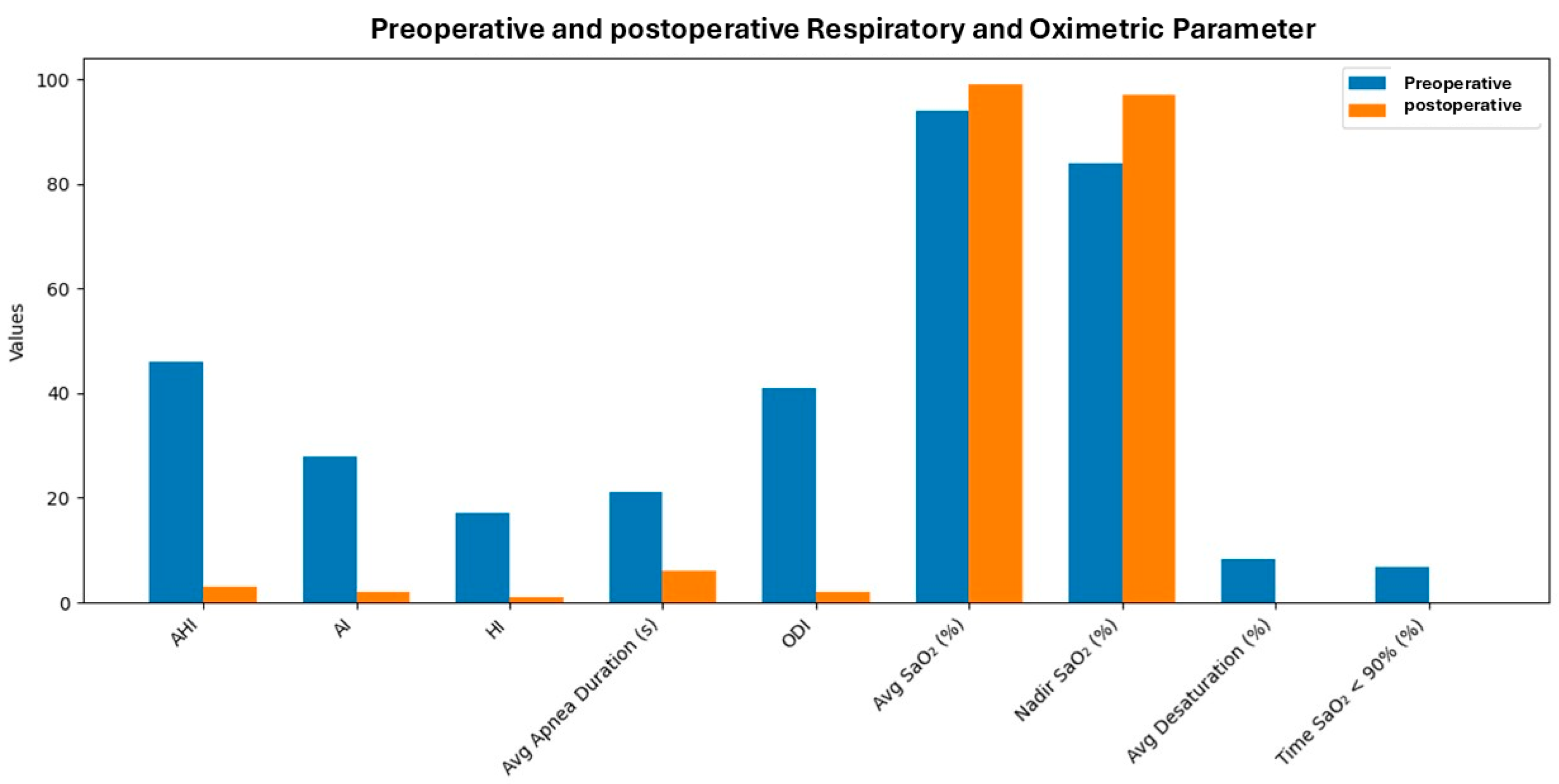
We observed a clinically significant improvement across all evaluated respiratory and oximetric parameters. The apnea-hypopnea index (AHI) showed a reduction greater than 90%, transitioning from a severe OSAS profile to a normal respiratory pattern (<5 events/hour). A significant decrease in the apnea index (AI) was observed, reflecting a near-complete elimination of obstructive apnea episodes. Similarly, the hypopnea index (HI) was almost entirely resolved, suggesting the normalization of airflow during sleep. The average duration of obstructive events decreased from 21 ± 6 s to just 6 s, thereby reducing the hemodynamic burden and the risk of prolonged oxygen desaturation (Figure 5).
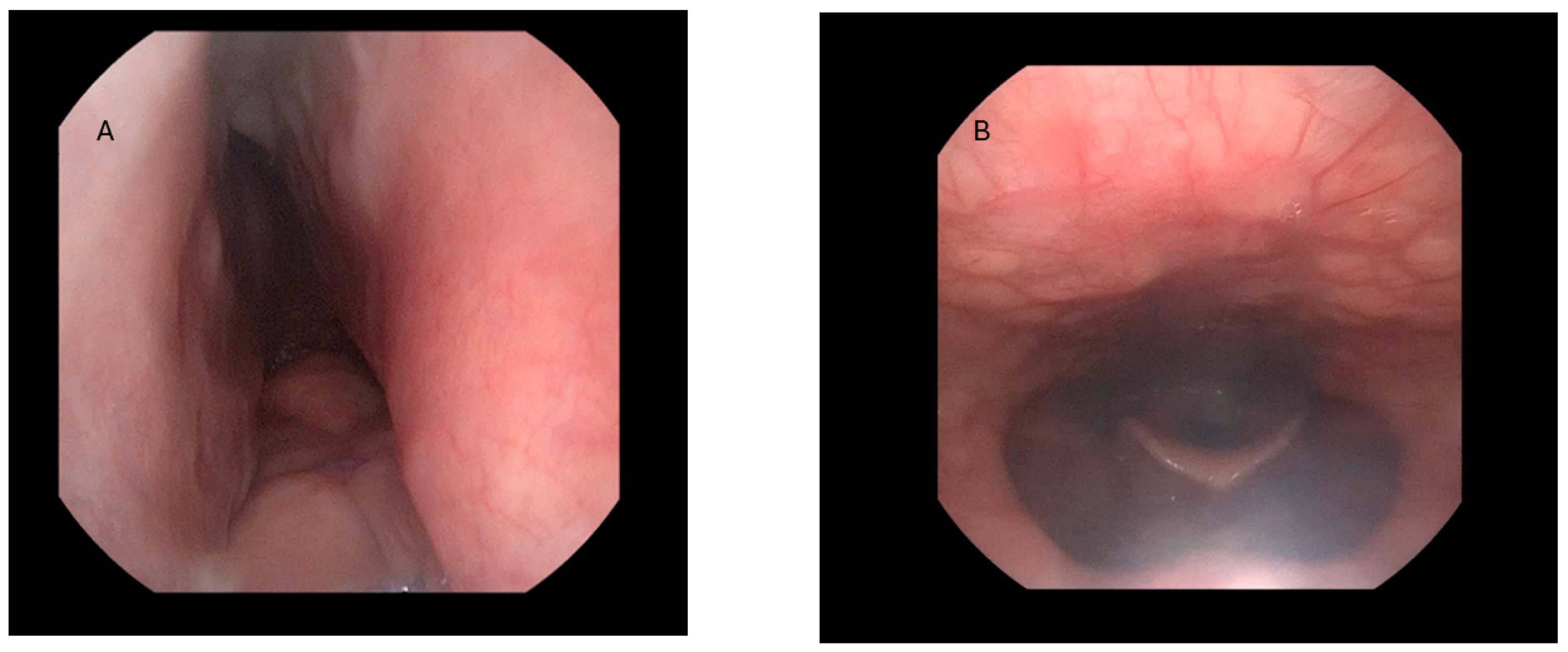
Respiratory and oxygenation parameters were collected and analyzed pre- and postoperatively, as summarized in Table 1; Table 2. Laryngoscopy revealed a change in the shape of the pharyngeal area, which transitioned from a flattened to an elliptical configuration (Figure 4). The statistical analysis revealed a normalization of respiratory parameters and a significant improvement in systemic oxygenation, with p-values < 0.0001 for all variables analyzed.

4. Discussion

The combination of maxillary wedge osteotomy and mandibular advancement resulted in a marked and clinically significant improvement across all evaluated respiratory and oximetric parameters, demonstrating the efficacy of this surgical approach in the treatment of obstructive sleep apnea syndrome (OSAS), particularly in cases where maxillary advancement is not required.

4.1. Impact on Respiratory and Oximetric Parameters

The procedure led to the complete resolution of snoring, indicating a substantial reduction in pharyngeal airway obstruction. The apnea-hypopnea index (AHI) showed a reduction greater than 90%, transitioning from a severe OSAS profile to a normal respiratory pattern (<5 events/hour). A significant decrease in the apnea index (AI) was observed, reflecting a near-complete elimination of obstructive apnea episodes. Similarly, the hypopnea index (HI) was almost entirely resolved, suggesting a normalization of airflow during sleep. The average duration of obstructive events decreased from 21 ± 6 s to just 6 s, thereby reducing the hemodynamic burden and the risk of prolonged oxygen desaturation. These findings support the use of maxillary wedge osteotomy, in combination with mandibular advancement, as an effective surgical strategy in selected OSAS patients, particularly when full maxillary advancement is not indicated but anterior repositioning of the mandible is necessary to restore upper airway patency.

4.2. Advantages of Wedge Osteotomy

Advantages of Wedge Osteotomy of the maxilla, a technical variant of the Le Fort I osteotomy, allows for a selective and controlled advancement of the mandible without anteroposterior displacement of the upper maxilla, thereby improving the patency of the retropharyngeal airway. This technique is often combined with surgical mandibular advancement to maximize the airway opening effect and is used in cases where there is no anteroposterior deficiency of the maxilla. In patients affected by obstructive sleep apnea (OSA) without evident anteroposterior maxillary deficiency, wedge osteotomy of the maxilla, often combined with other surgical techniques, can represent a valid therapeutic option. Although traditionally indicated in cases of maxillary hypoplasia, advancements in surgical techniques and a deeper understanding of OSA pathophysiology have broadened the indications to include patients without obvious skeletal abnormalities. In these cases, the goal is not so much esthetic or occlusal correction, but rather the functional improvement of airway patency. Ideal candidates for MMA surgery are patients with moderate to severe OSA, or those with mild OSA associated with a congenital dentofacial deformity or discrepancy [9]. Counterclockwise rotation of the occlusal plane is a surgical technique that can be employed to achieve proper occlusion, even in the absence of a sagittal maxillary deficiency or when mandibular and maxillary advancements are not perfectly aligned [10]. Counterclockwise rotation of the maxilla results in advancement of the pogonion, contributing both to an improved facial profile and to an increase in upper airway volume.

4.3. Comparisons with Other Studies

The study of Christino et al. [10] found that for each degree of rotation, the anterior mandible advanced 0.71 mm. When comparing individuals who received MMA with (n = 19) and without (n = 19) counterclockwise rotation, the study found that there was greater reduction in AHI (80% vs. 62%), total volume increase (45% vs. 30%), retropalatal volume increase (49% vs. 43%), minimum axial area increase in the retropalatal region (92% vs. 76%), and minimum axial area increase in the retrolingual region (97% vs. 31%) in those that had counterclockwise rotation. Counterclockwise rotational orthognathic surgery has proven particularly effective not only in improving airway patency and reducing OSA, but also in enhancing facial esthetics, thus offering a dual benefit: both functional and morphological [11]. The combination of a Le Fort I maxillary wedge osteotomy with counterclockwise rotation and mandibular advancement has been shown to significantly enhance upper airway volume and reduce the Apnea-Hypopnea Index (AHI) in patients with obstructive sleep apnea [12,13]. Counterclockwise rotation of the occlusal plane, especially when combined with maxillomandibular advancement (MMA), leads to substantial increases in total airway volume and minimum axial area, with effect sizes indicating marked improvements in upper airway patency [14,15]. Patients who underwent counterclockwise rotation of the occlusal plane showed a significant reduction in AHI and an improvement in minimum oxygen saturation during sleep, indicating better control of obstructive sleep apnea (OSA).
Our results are consistent with those reported by other authors [16].

4.4. Impact on Airspace

Counterclockwise rotation advances the mandible and simultaneously repositions the hyoid bone in an anterosuperior direction, further contributing to the expansion of the upper airway [3]. The improvements observed in all studied parameters and airway dimensions, achieved through counterclockwise rotation and mandibular advancement, are generally stable over time. Both the enlargement of the upper airway and the reduction in OSA symptoms tend to persist postoperatively, as also reported by other authors [11]. The effectiveness of the anterior repositioning of the maxilla and mandible, which results in forward traction of the soft tissues and muscular structures involved in maintaining airway patency. Specifically, the advancement of the muscular insertions of the anterior belly of the digastric muscle, the mylohyoid, the genioglossus, and the geniohyoid muscles contributes to the stabilization of the tongue base and the floor of the mouth. A mandibular advancement of at least 10 mm is generally recommended to achieve a clinically significant benefit, while maintaining occlusal balance and acceptable facial esthetics. Three-dimensional surgical planning and the use of digital technologies (CAD/CAM, guided surgery) have improved the precision and predictability of outcomes. Recent systematic reviews and clinical studies confirm the efficacy of MMA in the treatment of OSAS, with success rates exceeding 80% in appropriately selected patients. Moreover, the surgical sequence (maxilla-first vs. mandible-first) remains a topic of debate, although it does not appear to significantly affect functional outcomes, despite potential technical and occlusal implications. This approach helps harmonize the relationship between the dental arches and optimize airway patency, contributing to the overall success of the procedure [17]. Although the results of this study are promising, several limitations must be acknowledged. First, the sample size was relatively small, which may affect the generalizability of the findings. Second, the follow-up period was limited, preventing a comprehensive assessment of long-term stability and relapse rates. Future studies with larger cohorts and extended follow-up durations are needed to validate these preliminary outcomes.

5. Conclusions

Counterclockwise rotational orthognathic surgery without maxillary advancement has been shown to effectively enlarge the posterior pharyngeal space while also delivering excellent esthetic outcomes. When carefully planned, this technique represents a valid alternative to conventional orthognathic approaches for the surgical correction of obstructive sleep apnea (OSA).
Authors should discuss the results and how they can be interpreted from the perspective of previous studies and of the working hypotheses. The findings and their implications should be discussed in the broadest context possible. Future research directions may also be highlighted.

Author Contributions

Conceptualization, V.A.M. and A.S.; methodology, R.P.; software, F.A.G.; validation, S.D.N., L.T. and F.P.; formal analysis, A.C.; investigation, V.A.M. and A.S.; resources, V.M.; data curation, L.T.; writing—original draft preparation, A.S., L.D.P. and V.A.M.; writing—review and editing, A.S. and V.M.; visualization, G.B.; supervision, R.P.; project administration, L.T.; funding acquisition, A.S. All authors have read and agreed to the published version of the manuscript.

Funding

This research received no external funding.

Institutional Review Board Statement

The Hospital San Camillo-Forlanini, in Rome, Italy, classified the study to be exempt from ethical review as it carries only negligible risk and involves the use of existing data that contains only non-identifiable data about human beings.

Informed Consent Statement

Written consent has been obtained.

Data Availability Statement

The data presented in this study are available on request from the corresponding author. The data are not publicly available due to Personal Data Protection Law in our country.

Acknowledgments

The authors would like to thank Mauro Di Beradino for providing the illustrations included in this work.

Conflicts of Interest

The authors declare no conflicts of interest.

References

  1. Locke, B.W.; Lee, J.J.; Sundar, K.M. OSA and Chronic Respiratory Disease: Mechanisms and Epidemiology. Int. J. Environ. Res. Public Health 2022, 19, 5473. [Google Scholar] [CrossRef] [PubMed]
  2. Gaffey, A.E.; Burg, M.M.; Yaggi, H.K.; Wang, K.; Brandt, C.A.; Haskell, S.G.; Bastian, L.A.; Chang, T.E.; Levine, A.; Skanderson, M.; et al. Insomnia, Sleep Apnea, and Incidence of Hypertension and Cardiovascular Disease. medRxiv 2025. [Google Scholar] [CrossRef] [PubMed]
  3. Seet, E.; Chung, F.; Wang, C.Y.; Tam, S.; Kumar, C.M.; Ubeynarayana, C.U.; Yim, C.C.; Chew, E.F.F.; Lam, C.K.M.; Cheng, B.C.P.; et al. Association of Obstructive Sleep Apnea With Difficult Intubation: Prospective Multicenter Observational Cohort Study. Anesth. Analg. 2021, 133, 196–204. [Google Scholar] [CrossRef] [PubMed]
  4. He, J.-M.; Yang, Y. Association between Neutrophil-Lymphocyte Ratio and All-Cause and Cardiovascular Mortality in Patients with Diabetes or Prediabetes with Comorbid Obstructive Sleep Apnea Symptoms: Evidence from NHANES 2005–2008 and 2015–2018. Front. Endocrinol. 2025, 16, 1512621. [Google Scholar] [CrossRef] [PubMed]
  5. Weaver, T.E.; Maislin, G.; Dinges, D.F.; Bloxham, T.; George, C.F.P.; Greenberg, H.; Kader, G.; Mahowald, M.; Younger, J.; Pack, A.I. Relationship between Hours of CPAP Use and Achieving Normal Levels of Sleepiness and Daily Functioning. Sleep 2007, 30, 711–719. [Google Scholar] [CrossRef] [PubMed]
  6. Knauert, M.P.; Adekolu, O.; Xu, Z.; Deng, A.; Chu, J.-H.; Baldassarri, S.R.; Kushida, C.; Yaggi, H.K.; Zinchuk, A. Morning Chronotype Is Associated with Improved Adherence to Continuous Positive Airway Pressure among Individuals with Obstructive Sleep Apnea. Ann. Am. Thorac. Soc. 2023, 20, 1182–1191. [Google Scholar] [CrossRef] [PubMed]
  7. Riley, R.W.; Powell, N.B.; Guilleminault, C.; Nino-Murcia, G. Maxillary, Mandibular, and Hyoid Advancement: An Alternative to Tracheostomy in Obstructive Sleep Apnea Syndrome. Otolaryngol. Head. Neck Surg. 1986, 94, 584–588. [Google Scholar] [CrossRef] [PubMed]
  8. Varghese, R.; Adams, N.G.; Slocumb, N.L.; Viozzi, C.F.; Ramar, K.; Olson, E.J. Maxillomandibular Advancement in the Management of Obstructive Sleep Apnea. Int. J. Otolaryngol. 2012, 2012, 373025. [Google Scholar] [CrossRef] [PubMed]
  9. Yu, M.S.; Ibrahim, B.; Riley, R.W.; Liu, S.Y.-C. Maxillomandibular Advancement and Upper Airway Stimulation: Extrapharyngeal Surgery for Obstructive Sleep Apnea. Clin. Exp. Otorhinolaryngol. 2020, 13, 225–233. [Google Scholar] [CrossRef] [PubMed]
  10. Christino, M.; Vinha, P.P.; Faria, A.C.; Garcia, D.M.; de Mello-Filho, F.V. Impact of Counterclockwise Rotation of the Occlusal Plane on the Mandibular Advancement, Pharynx Morphology, and Polysomnography Results in Maxillomandibular Advancement Surgery for the Treatment of Obstructive Sleep Apnea Patients. Sleep. Breath. 2021, 25, 2307–2313. [Google Scholar] [CrossRef] [PubMed]
  11. Choi, J.W.; Jeong, W.S.; Kang, M.K.; Lee, J.Y.; Chung, Y.S. Counterclockwise Rotational Orthognathic Surgery for the Obstructive Sleep Apnea in Class II Dentofacial Deformity: Polysomnography and 3D Computed Tomographic Analysis. Ann. Plast. Surg. 2021, 86, 640–646. [Google Scholar] [CrossRef] [PubMed]
  12. Shah, N.; Waite, P.D.; Kau, C.H. A Combined Orthodontic / Orthognathic Approach in the Management of Obstructive Sleep Apnoea: Balancing Treatment Efficacy and Facial Aesthetics. J. Orthod. 2020, 47, 354–362. [Google Scholar] [CrossRef] [PubMed]
  13. Jeong, W.S.; Kim, Y.C.; Chung, Y.S.; Lee, C.Y.; Choi, J.W. Change in Posterior Pharyngeal Space After Counterclockwise Rotational Orthognathic Surgery for Class II Dentofacial Deformity Diagnosed With Obstructive Sleep Apnea Based on Cephalometric Analysis. J. Craniofac Surg. 2017, 28, e488–e491. [Google Scholar] [CrossRef] [PubMed]
  14. Louro, R.S.; Calasans-Maia, J.A.; Mattos, C.T.; Masterson, D.; Calasans-Maia, M.D.; Maia, L.C. Three-Dimensional Changes to the Upper Airway after Maxillomandibular Advancement with Counterclockwise Rotation: A Systematic Review and Meta-Analysis. Int. J. Oral. Maxillofac. Surg. 2018, 47, 622–629. [Google Scholar] [CrossRef] [PubMed]
  15. Zinser, M.J.; Zachow, S.; Sailer, H.F. Bimaxillary “rotation Advancement” Procedures in Patients with Obstructive Sleep Apnea: A 3-Dimensional Airway Analysis of Morphological Changes. Int. J. Oral. Maxillofac. Surg. 2013, 42, 569–578. [Google Scholar] [CrossRef] [PubMed]
  16. Knudsen, T.B.; Laulund, A.S.; Ingerslev, J.; Homøe, P.; Pinholt, E.M. Improved Apnea-Hypopnea Index and Lowest Oxygen Saturation after Maxillomandibular Advancement with or without Counterclockwise Rotation in Patients with Obstructive Sleep Apnea: A Meta-Analysis. J. Oral. Maxillofac. Surg. 2015, 73, 719–726. [Google Scholar] [CrossRef] [PubMed]
  17. Trevisiol, L.; Bersani, M.; Lobbia, G.; Scirpo, R.; D’Agostino, A. Sequencing in Orthognathic Bimaxillary Surgery: Which Jaw Should Be Operated First? A Scoping Review. J. Clin. Med. 2023, 12, 6826. [Google Scholar] [CrossRef] [PubMed]
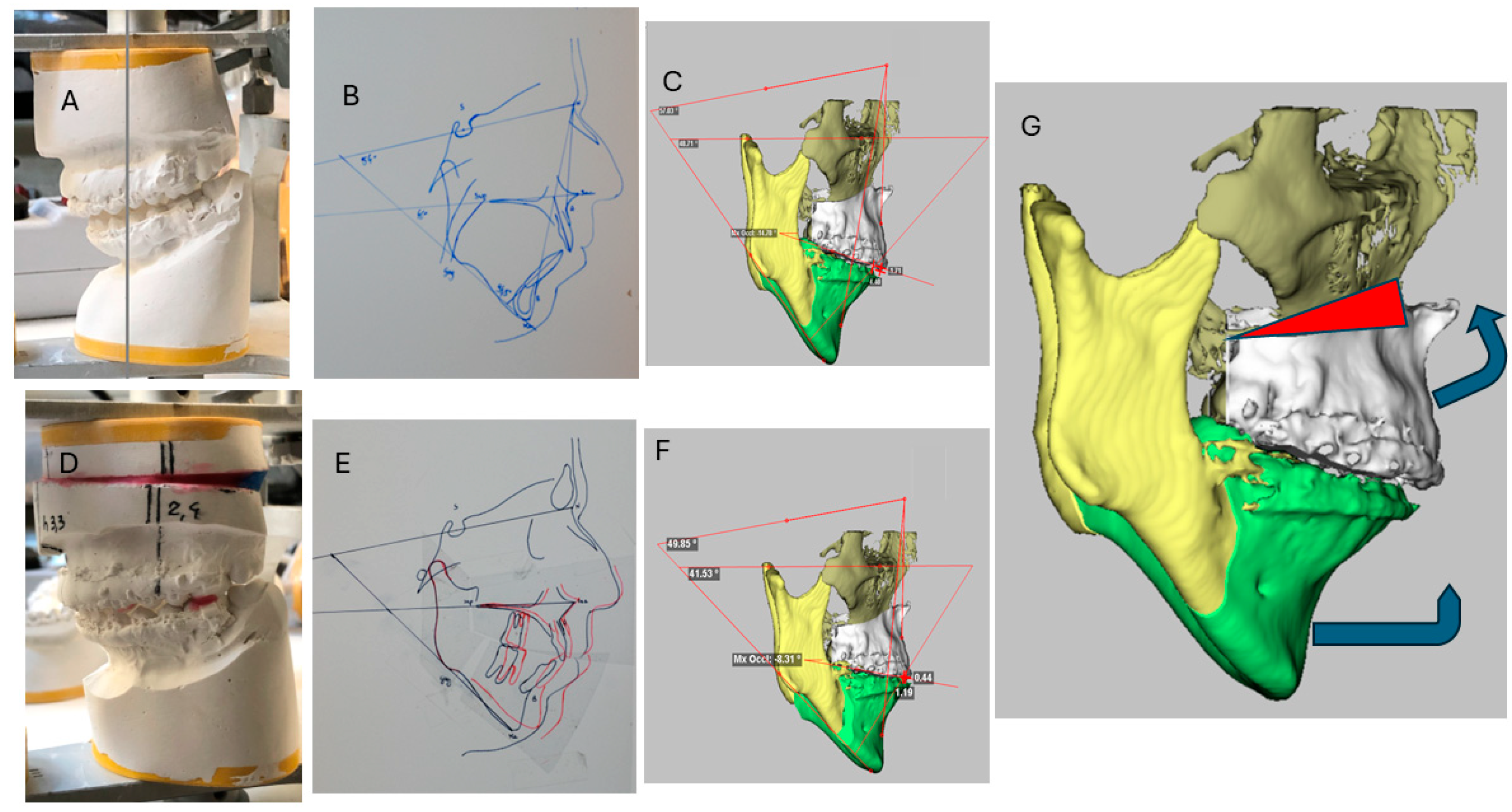
Figure 1. (A) The intermaxillary relationship was initially assessed through model mounting on a semi-adjustable articulator, allowing for a detailed evaluation of occlusal dynamics and skeletal harmony. (B,C) A preoperative cephalometric analysis confirmed the need for mandibular advancement, without any indication for maxillary advancement. (E) A simulation of the postoperative outcome was performed by mounting the models in the articulator, reproducing the anticipated skeletal and occlusal changes. (F) In parallel, a software-based cephalometric simulation was conducted, incorporating mandibular rotation and advancement to predict the three-dimensional effects on the upper airway and facial profile. (G) A surgical plan developed through dedicated planning software, which integrated skeletal movements and occlusal goals.
Surgeries 06 00074 g001
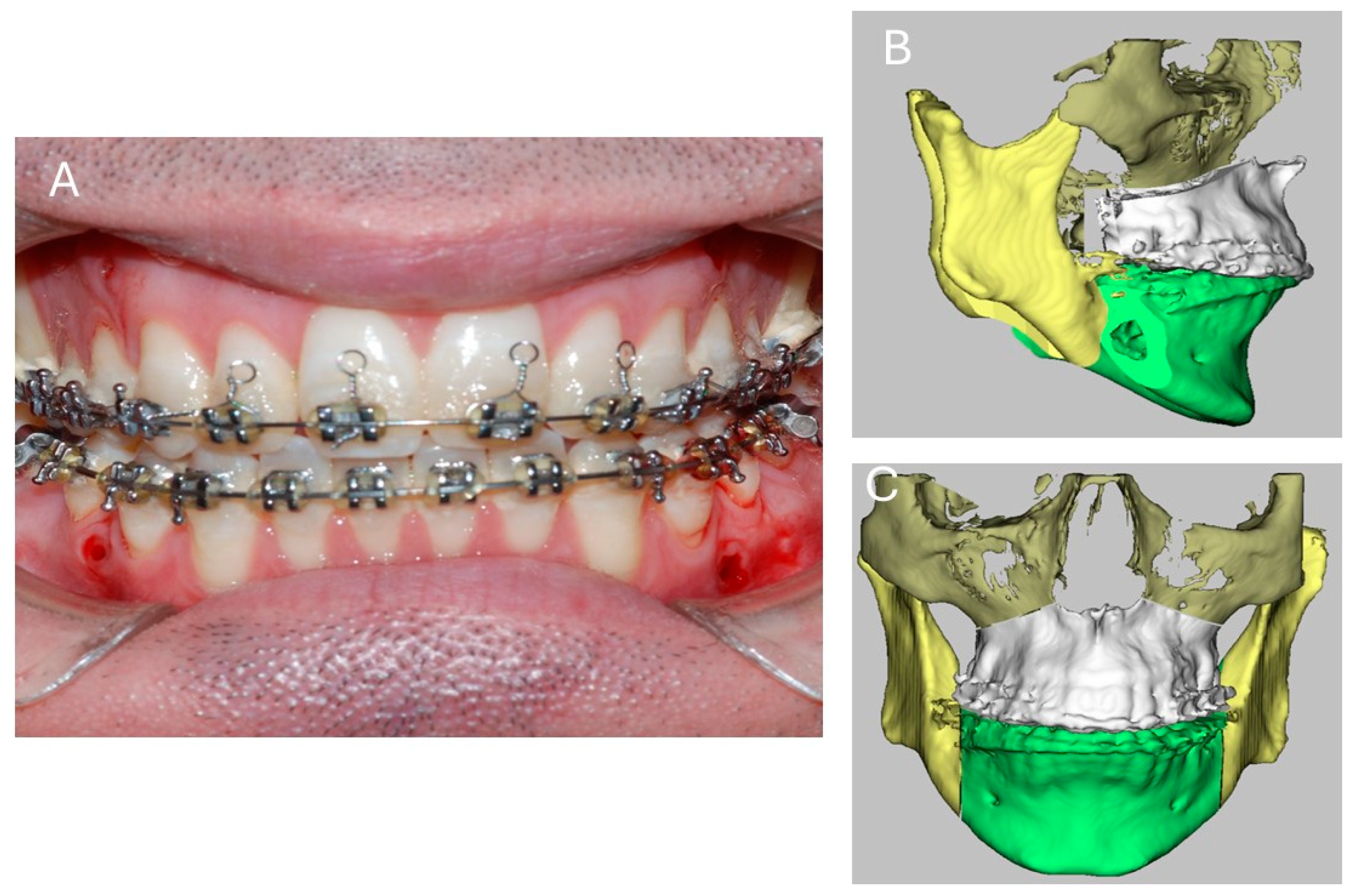
Figure 2. (A) Occlusal aspect after surgery. (B) Frontal view of the intermaxillary relationships achieved postoperatively. (C) Lateral view of the intermaxillary relationships achieved postoperatively.
Surgeries 06 00074 g002
Figure 3. (AC) Preoperative computed tomography (CT) scans were analyzed using dedicated surgical planning software, which enabled three-dimensional visualization and quantification of the upper airway spaces. (DF) Postoperative computed tomography (CT) scans were analyzed using dedicated surgical planning software; there has been a significant increase in the upper airspace.
Surgeries 06 00074 g003
Figure 4. Laryngoscopy reveals a change in the shape of the posterior airspace, from an elliptical shape (A) to a round shape (B) with an increase in volume.
Surgeries 06 00074 g004
Figure 5. Pre- and postoperative respiratory and oximetric parameters.
Surgeries 06 00074 g005
Table 1. Respiratory and oximetric parameters before and after maxillary wedge osteotomy and mandibular advancement surgery.
ParameterPreoperativePostoperativeClinical Interpretation
Snoring68%0%Complete resolution of snoring, indicating reduced obstruction.
Apnea-Hypopnea Index (AHI)46 events/hour3 events/hour>90% reduction; normalization of respiratory pattern.
Apnea Index (AI)28 events/hour2 events/hourSignificant reduction in complete apneas.
Hypopnea Index (HI)17 events/hour1 event/hourNear-complete resolution of hypopneas.
Average Apnea Duration21 ± 6 s6 sShorter events, reduced hemodynamic impact.
Table 2. Oximetric parameters before and after maxillary wedge osteotomy and mandibular advancement surgery.
ParameterPreoperativePostoperativeClinical Interpretation
Oxygen Desaturation Index (ODI)41 events/hour2 events/hourMarked reduction in desaturation episodes.
Average Oxygen Saturation (SaO2)94 ± 2%99%Improved systemic oxygenation.
Nadir Oxygen Saturation (SaO2)84%97%Higher minimum saturation; reduced hypoxic burden.
Average Desaturation8.4%0%Elimination of oxygen desaturation events.
Time with SaO2 < 90%6.9%0%No time spent in clinically significant hypoxemia.
Disclaimer/Publisher’s Note: The statements, opinions and data contained in all publications are solely those of the individual author(s) and contributor(s) and not of MDPI and/or the editor(s). MDPI and/or the editor(s) disclaim responsibility for any injury to people or property resulting from any ideas, methods, instructions or products referred to in the content.