Abstract
Cancer is one of the leading causes of illness and death in the world. It is observed that the main reason for the low effectiveness of cancer treatment is limited bioavailability. Another noted cause is the lack of specificity of conventional chemotherapeutics, which contributes to the destruction of not only cancer cells, but also normal cells, and consequently leads to serious adverse effects. In recent years, researchers have paid special attention to the use of photodynamic therapy. Another major step in this progress is turning to photosensitizing natural compounds, which we present in this review. Natural photosensitizers are being investigated for their potential to treat central nervous system (CNS) tumors using photodynamic therapy (PDT). These compounds, derived from natural sources, offer an alternative to synthetic photosensitizers, potentially minimizing toxicity and enhancing therapeutic efficacy. Research focuses on isolating, synthesizing, and evaluating these natural photosensitizers for their ability to selectively accumulate in tumor cells and be activated by light to produce cytotoxic reactive oxygen species, leading to tumor cell death.
1. Introduction
Cancer continues to be one of the main health problems in most regions of the world [1]. According to the 2020 GLOBOCON report, it is one of the leading causes of death [2,3]. Despite the advanced diagnostic and therapeutic methods currently used, it is estimated that within 15 to 35 years, cancer incidence and mortality rates will double [1]. Moreover, surgical treatment, chemotherapy or radiotherapy are not ideal; they are associated with a large number of side effects, the development of drug resistance, limited therapeutic effects, the occurrence of secondary cancers, or the local recurrence of the disease [2,4]. An alternative to conventional treatment for many cancers is photodynamic therapy (PDT) [5]. Its main features are minimal invasiveness, high effectiveness, the possibility of combining with other forms of treatment, high selectivity, and limited susceptibility to the development of drug resistance [6]. Moreover, its beneficial effects are also used in the fight against non-oncological diseases caused by fungi, viruses [7], and bacteria [8].
1.1. History of Photodynamic Therapy (PDT)
The health effects of light on the human body have been known for thousands of years. The origins of heliotherapy in Egypt, Greece, and India date back to as early as 3000 BCE. The first medical document on the healing effects of sunlight, called the Ebers Papyrus, dates back to around 1550 BCE. Using powders of plant origin, sunlight was supposed to be effective at treating skin lesions. Similar conclusions were also reached by inhabitants of the Indian, Roman, Greek, and Chinese civilizations, using sunlight to treat diseases of other systems. It is believed to have healing effects in diseases such as rickets, pulmonary tuberculosis, acne, vitiligo, or lupus, often in combination with locally or orally applied plant powders and extracts [7]. The fathers of photodynamic therapy are considered to be Herman Von Tappeiner and Oscar Raab, who in 1900 noticed the effect of light on the photoactivation of acridine pigment and, as a consequence, cell death [9]. The next breakthrough discoveries were made by the Danish physician Niels Finsen, whose achievements were recognized with the Nobel Prize in 1903. He noticed that, in addition to sunlight, red and ultraviolet light also had therapeutic properties [7]. In the 1960s, the first photosensitizer (PS) was isolated—a derivative of hematoporphyrin from hemoglobin [10], and in the 1970s, the first photodynamic therapy using it in laboratory conditions was performed in the USA [1]. In 1993, Photofrin was approved in Canada for the treatment of bladder tumors, which finally gave photodynamic therapy the status of a therapeutic method [7,11]. In oncology, PDT is currently in the treatment of brain cancer [12], liver cancer [7], breast cancer [13], gastrointestinal cancer [14], pancreas cancer [15], prostate cancer [16], head and neck cancer [17], lung cancer [18], and used mainly in the treatment of skin cancer [6,19].
1.2. Mechanism of Photodynamic Therapy (PDT)
The essence is the occurrence of a photochemical reaction between photosensitizer molecules, selectively accumulating in pathological tissue, light of a specific wavelength and oxygen [8]. There are two types of photodynamic therapy—I and II. The photosensitizer, under the influence of light of an appropriate wavelength, is excited from the ground state S0 to the unstable excited singlet states S1, S2, or other Sn. Then the Sn state relaxes to the S1 state, and the S1 state to the ground state S0”, according to basic Jablonski. Type I PDT involves the direct interaction of PS in the T1 state with surrounding substances by generating free radicals as a result of electron transfer. Then the free radical anions react with triplet oxygen and H2O to form a hydroxyl radical and a superoxide anion. The essence of type II PDT is the transformation of triplet oxygen into singlet oxygen by the photosensitizer into the T1 state through energy transfer [20]. The newly formed singlet oxygen interacts with electron-rich molecules, causing their oxidation and, as a consequence, damage to some cellular organelles, including mitochondria, the cell membrane, the nucleus, lysosomes, and tissue necrosis [21]. Therefore, type I PDT is more resistant to oxygen deficiency than type II, which is a common phenomenon in cancer tumors [22].
Unlike conventional cancer treatment methods, i.e., chemotherapy and radiotherapy, PDT does not induce immunosuppressive effects, but stimulates the immune system. However, due to tumor hypoxia, incomplete selectivity and PS penetration into the tumor, or poor light penetration into deeper tissues, this method still requires improvement before it can be used on a large scale [3,10].
1.3. Photosensitizers (PSs)
Photosensitizers (PSs) are one of the three mandatory components needed to perform PDT [23]. When exposed to light of the appropriate wavelength, they induce photophysical or photochemical processes [9]. In PDT, PSs are crucial for generating reactive oxygen species (ROS) upon light activation, leading to cell damage and death. However, a critical view of photosensitizers in PDT reveals several limitations and areas for improvement. These include the need for better selectivity, deeper tissue penetration, and enhanced efficacy (Table 1).

Table 1.
Limitations of PS and research areas for improvements.
PS used in PDT are categorized into generations based on their development and characteristics (Table 2). First-generation photosensitizers, like Photofrin®, had issues with prolonged photosensitivity and limited tissue penetration due to weak absorption at longer wavelengths. Second-generation photosensitizers, such as benzoporphyrins and phthalocyanines, were developed to address these limitations, offering improved tissue penetration and reduced photosensitivity. Third-generation photosensitizers utilize nanotechnology to enhance tumor targeting and delivery, improving overall PDT efficacy.
Table 2.
Characteristics of individual generations of photosensitizers.
2. Natural Photosensitizers
Natural photosensitizers absorb light at specific wavelengths, enabling them to be used in photodynamic therapy (PDT) and other applications. Many natural photosensitizers have peak absorption in the red-to-near-infrared region (650–800 nm), allowing for deeper tissue penetration. Other photosensitizers, like curcumin, absorb in the blue light range (405–435 nm).
2.1. Quinonoids
Quinonoids are a group of substances, almost all of which are photosensitive. Many of them have anticancer properties, which is why they are often used in oncology research. They are divided into three main groups: benzoquinone, anthraquinone, and perylenequinones [24]. Anthraquinones are a group of compounds with natural phototoxic properties [25]. They have been shown to have antibacterial [26,27], antifungal [28], and anticancer effects, including in the treatment of breast cancer [29].
Hypericin (Figure 1) (HYP; 4,5,7,4′,5′,7′-hexahydroxy-2,2′-dimethylnaphthodianthrone) is a hydroxylated phenatroperylenequinone [30], a derivative of anthraquinones [4]. It is isolated from the plant Hipericum perforatum, also known as St. John’s wort [4,24,31].
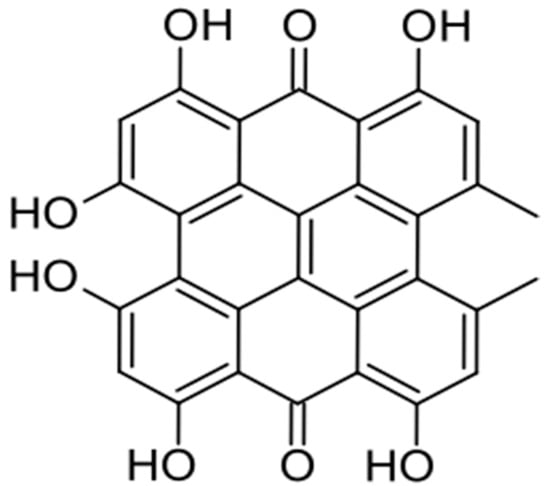
Figure 1.
Chemical structure of hypericin.
The methods of synthesizing hypericin are presented in Table 3. Hypericin synthesis typically involves a multi-step process, often starting from emodin or emodin anthrone. A key step is the dimerization of emodin anthrone to form protohypericin, which is then converted to hypericin through irradiation with visible light. Various methods exist, including chemical synthesis and biosynthesis pathways, with the latter often involving polyketide synthase (PKS) enzymes (Table 4).
Table 3.
The characteristics of lasers.
Table 4.
Synthesis and biosynthesis of Hypericin.
Since ancient times, hypericin has been used both as an antidepressant and an antiviral agent [31,37]. Its beneficial effects are also noticeable in the treatment of endocrine diseases, e.g., in relieving menopausal symptoms or those related to polycystic ovary syndrome, and skin diseases—including plaque psoriasis, wounds and scars after surgery, or changes caused by HSV-1 and HSV-1 viruses [38].
Its anticancer effects have also long been known and it is used in the treatment of cancers, including those of the urinary bladder, colon, breast, cervix, nasopharynx, liver, melanoma, leukemia, and lymphoma [30].
Its use in PDT of glioma has been confirmed by studies that were among the first to be conducted by Miccoli et al. They showed that by influencing the energy metabolism of SNB-19 cells, HYP-PDT inhibits the binding of hexokinase to mitochondria [39]. In turn, Ritz et al. obtained effective inactivation of three glioma cells—U373 MG, LN229, and T98G—after a short incubation and exposure to low-dose light [40]. While HYP-PDT has shown efficacy in treating various cancers and other conditions, some studies suggest potential “bed” issues, particularly regarding skin reactions and the timing of light application in relation to drug administration (Table 5).
Table 5.
Hypericin’s Positive and Negative Aspects in PDT.
Perylenequinolones are dark pigments containing an oxidized pentacyclic core, represented by the parent perylenequinolone 1 [41]. In 1956, the first, simplest perylenequinolone—Diol 1—was isolated from the fungus Daldinia [42].
Hypocrelin (Figure 2) belongs to the perylenequinone derivatives [4,24], and hypocrelin A (HA), B (HB), C (HBC), and D (HBD) are distinguished [43]. They are characterized by the same perylene–quinone structure and similar properties, but contain different side rings [43,44]. Both hypocrelin A and B are characterized by photodynamic activity. Hypocrelin is isolated from parasitic bamboo fungi—Hypocrella bambusea and Shiraia bambusicola [43].
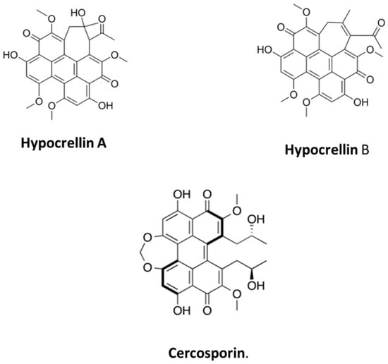
Figure 2.
Chemical structure of hypocrelin A, B, and Cercosporin.
The installation of axial chirality of perylenequinone enables the synthesis of more complex perylenequinone natural products. The stereochemical transfer is complicated due to the dynamic state of the perylenequinone helical axis. The stereochemical helix of perylenequinone is stable at room temperature, but after the formation of the seven-membered ring it loses its integrity, leading to rapid atropisomerization. The synthetic process includes several steps, such as enantioselective biaryl coupling, deacylation, and protection of free naphthols. Then, after hydroxylation, the allyl groups are transformed to form a diketone. The key step is the aldol cyclization of the diketone, which allows the formation of a seven-membered ring. After the synthesis is developed, the aldol reaction is carried out, leading to the formation of hypocrelin A as the main product. This product has the desired stereochemical properties, and a small amount of the E enolate gives the anti-aldol product, leading to diastereomers, including shiraiachrome A [41,45,46,47,48]. Hypocrelin has long been used as a traditional medicinal compound in Asia. It is primarily known for its antiviral, antidepressant [43], and antifungal [49] effects. Both hypocrelin A and B are characterized by photodynamic activity [50].
Hypocrelin has long been used as a traditional medicinal compound in Asia. It is primarily known for its antiviral, antidepressant [43], and antifungal [49] effects. Both hypocrelin A and B are characterized by photodynamic activity [50]. Hypocrelin A is an excellent photosensitizer and is characterized by selective accumulation in cancer cells, which leads to their death [43].
The main anticancer application of hypocrelin is related to the treatment of breast cancer [51] and skin cancer [52]. There is also evidence of the effectiveness of PDT using hypocrelin as a PS in the treatment of lung cancer [53], ovarian cancer [54], bladder tumor [55], and gastric adenocarcinoma [56]. However, some studies show that both hypocrelin A and B can also lead to the death of human brain cancer cells by inhibiting angiogenesis via photodynamic action [57].
Cercosporin (Figure 3), a deep red pigment, was first isolated by Kuyama et al. in 1957 from Cercospora kikuchii Gardner, a pathogenic fungus on the purple spot of Japanese soybeans [58,59,60]. Chemically, it is a dihydroxy–perilenequinone [56]. The structure of this phytotoxin was originally proposed by Kuyama in 1962, but it was modified by Lousber et al. [60,61] and confirmed by Yamazaki et al. Based on mass spectrometry and elemental analysis, the chemical formula was C29H2O10 and the molecular weight was 534 [62]. Some naturally occurring red pigments, such as elsinochromes, fagopyrin, and erythroaffin, exhibit photodynamic activity. In connection with the above, studies have been conducted which have shown that in an oxygen environment it exerts a photosensitizing effect on mice or microbiomes, which makes it a photodynamic pigment [60]. Its photosensitizing function has also been demonstrated in the photooxygenation reaction, including 2,5-dimethylfuran and amino acid residues [59].
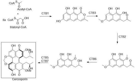
Figure 3.
Cercosporin synthesis.
The synthesis of cercosporin is shown in the scheme (Figure 3) below.
Cercosporin is known primarily for its harmful effect on the leaves of cultivated plants [63]. It plays a key role in the development of white leaf spot disease [64,65]. It may be useful in the fight against harmful blooms of cyanobacteria, taking care of water resources [66,67]. Scientific studies conducted by Cadelis et al. have shown that isolates of Cercospora beticola exhibit strong antibacterial activity against methicillin-resistant Staphylococcus aureus strains [68]. In the study conducted by Mastrangelopoulou, Grigalavicius et al., two human glioma cell lines (T98G and U87) were used in 2D cultures, obtaining clear bioenergetic breakdowns, which confirmed its anticancer activity [69]. These results were confirmed in their subsequent work in 3D cell culture [70].
2.2. Curcumin
Turmeric, a plant related to the ginger family, has active polyphenolic compounds in its structure, which include curcumin, bisdemethoxycurcumin, and demethoxycurcumin, called curcuminoids [71]. They constitute up to 6% of the dry weight of turmeric [72]. Traditionally, the powdered rhizome of Curcuma longa is used in Asian and Indian cuisine as a curry spice, and also as an antimicrobial, insect repellent or dye [71,72]. In the traditional medicines of some cultures, turmeric is also used as a preparation for treating fresh wounds and bruises, scabs in smallpox and chickenpox, insect bites, anthelmintics or in the treatment of urological, liver, and biliary tract diseases [72].
Chemically, curcumin (Figure 4) is 1,7-bis-(4-hydroxy-3-methoxyphenyl)-hepta-1,6-diene-3,5-dione, with the chemical formula C21H20O6. It is insoluble in water at neutral and acidic pH, but soluble in acetone, methanol, dimethyl sulfoxide, and ethanol [73].
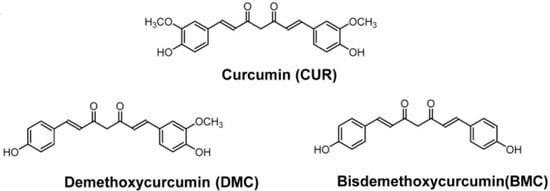
Figure 4.
Curcumin and derivatives.
Curcumin was first isolated in 1815 by Vogel and Pelletier, and its pure structure was isolated by Vogel in 1842. Its chemical structure was first described by Milobedzka et al. in 1910, and its synthesis by Lampe and Milobedzka in 1913 [73].
Anderson et al. in 2000 proposed a method for isolating the compound curcumin from ground turmeric plants [73]. The first step was to stir magnetically ground turmeric in dichloromethane, then heat it for about 1 h at boiling temperature. Then the obtained mixture was filtered under suction. The remaining filtrate was concentrated in a water bath at 50 degrees Celsius. The last steps were to combine the oily substance with hexane, collect the obtained precipitate under suction, and analyze the sample [73].
Chemically, curcumin is diferuloylmethane or 1,6-heptadiene-3,5-dione-1,7-bis(4-hydroxy-3-methoxyphenyl)-(1E, 6E) [73]. Curcumin is characterized by low bioavailability, which results from its rapid metabolism, poor water solubility, low chemical stability, and a negatively charged state [74,75]. Therefore, various techniques are being tested to modify these features. One of them is the modification of curcumin compounds from the basic form to the nanometric form [76,77]. Nanocomplexation consists of dissolving the substance in a suitable solvent and then mixing it with a protein solution [76]. Another potential modification is gelation, the essence of which is to combine the compound with a protein or protein/polysaccharide and then with a gelling agent. Gelling agents include aerogels and emulsion-filled gels, emulsion gels, organogels, and hydrogels [74,78]. Encapsulation of curcumin is achieved by electrospraying, which involves the generation of monodisperse particles ranging in size from submicrometers to hundreds of micrometers. It involves exposing a liquid droplet in a capillary nozzle to a high electric field, under the influence of which it deforms and then disintegrates into small droplets due to varicose instability [79]. Changing the pH is also considered to increase the bioavailability of curcumin [74]. Studies conducted using curcumin in PDT show that it is particularly useful in the treatment of cancers such as breast [79], uterine [80], ovarian [81], skin [82], colon [83], liver [84], lung [85], and leukemia [86]. However, studies have also been conducted to demonstrate the effect of PDT using curcumin nanoparticles on glioma cells [86,87,88,89]. The studies conducted by Kielbik et al. focused on the effect of PDT using curcumin nanoparticles on human glioblastoma multiforme cells. They showed that over 90% of the cancer cells underwent apoptosis. Within approx. 2 h, curcumin was distributed in the cytoplasm, suggesting that curcumin is a promising substance for use as a photosensitizer in the therapy of glioblastoma multiforme [89].
As for the other properties of curcumin, numerous studies indicate that it can also exhibit anti-aging, [90] anti-inflammatory [91], and healing effects on skin diseases [91], diabetes [92], circulatory system diseases [93], eye diseases [94], osteoarthritis [95], or neurological diseases [96].
2.3. Chlorophyll Derivatives and Pheophorbide A
Chlorophyll derivatives instead of magnesium as the core atom contain palladium, zinc, copper, nickel, cobalt, and iron [24]. Pheophorbide A (PBA, (3S,4S)-9-ethenyl-14-ethyl-21(methoxycarbonyl)-4,8,13,18-tetramethyl-20-oxo-3-phorbinepropanoic acid) [97] is a chlorophyll derivative [4]. Pheophorbide A (Figure 5) is isolated from the Chinese medicinal herb Scutellaria barbarta and silkworm excrement [4].
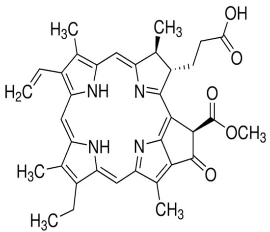
Figure 5.
Chemical structure of pheophorbide A.
Treatment of ethanolic solution of chlorophyll a under acidic conditions led to obtaining crude pheophytin, thanks to the possibility of easy removal of Mg2+ ion. Hydrolysis of pheophytin with 80% TFA in water made it possible to obtain pheophorbide-a in the form of a fine powder. The synthesis of pheophorbide is shown in the scheme below (Figure 6) [98,99,100].
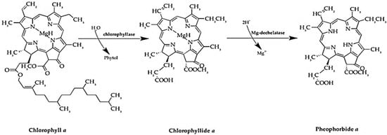
Figure 6.
Pheophorbide A synthesis.
Pheophorbide A is classified as a compound with various properties—antiviral [101,102], anti-inflammatory [103], antioxidant [104], immunostimulating [105], and antiparasitic [106]. Additionally, one of the latest applications of pheophorbide A is the treatment of lymphatic vessel failure, which prevents the development of its complications, such as lymphedema, chronic inflammation, or impaired wound healing [107].
Pheophorbide A is also used in the treatment of neoplastic diseases, such as hepatocellular carcinoma [108], squamous-cell carcinoma of the oral cavity [109], uterine sarcoma [110], prostate cancer [111], cancer [112], and breast tumors [113]. Cho et al. demonstrated its inhibitory effect on glioblastoma multiforme cells using U87MG in dark conditions. In addition, they observed a lack of cytotoxic effects on normal cells [114].
2.4. Alkaloids and Berberine
Alkaloids are organic chemical compounds that have one or more basic nitrogen atoms in their structure. They are characterized by a cyclic ring structure. Natural photoactive alkaloids can be divided into five groups (Figure 7) [115].
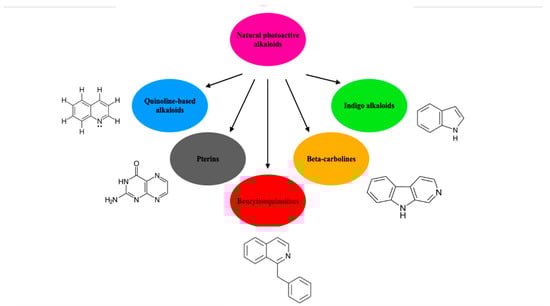
Figure 7.
Classification of natural photoactive alkaloids.
Alkaloids are secondary metabolites of plants and animals. They can also be found in leaves, stems, roots, and seeds of plants of families such as Papaveraceae, Amaryllidaceae, Menispermaceae, Loganiaceae, Ranunculaceae, or Solanaceae. In the plant, they probably perform defensive functions against pathogens that threaten the host [115].
Berberine (Figure 8) is an isoquinoline alkaloid, based on quinoline, isolated from the Chinese herb Coptis chinensis, as well as other Berberis plants [116,117].
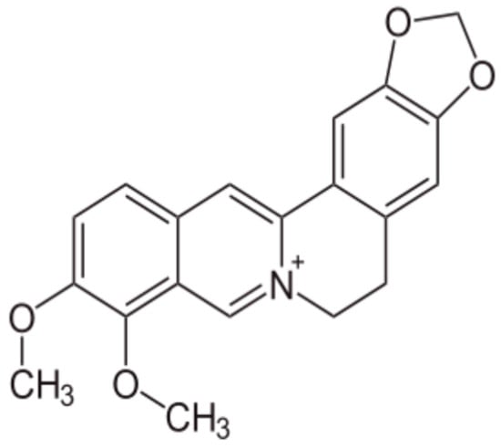
Figure 8.
Chemical structure of berberine.
Berberine is credited with a number of health benefits [118]. Many studies confirm that it has a cholesterol-lowering effect [119], prevents obesity, and helps in the treatment of obesity [120], atherosclerosis [121], liver diseases [122], neurodegenerative diseases [123], ischemic stroke [124], inflammation and cancer of the pancreas [125], inflammatory diseases and cancers of the digestive tract [126,127], lung cancer [128], and ovarian cancer [129].
Cancer cells are characterized by a high cholesterol metabolism and the expression of a large amount of receptors for low-density lipoproteins (LDLs) [130]. Since berberine has an affinity for low-density lipoproteins, Andreazza et al. experimentally demonstrated that it accumulates in larger amounts in cancer cells than in normal cells. The above findings resulted in berberine being used as a photosensitizer, including in the therapy of central nervous system tumors [131]. In this regard, studies conducted by Carriero et al. showed that the use of PDT with berberine as PS on human astrocytoma cell lines resulted in increased activation of apoptosis pathways, increased ROS production, increased mitochondrial depolarization, and increased activation of caspases [130]. Glial tumor cells are also characterized by having N-acetyltransferase activity. Wang et al. showed that this activity was inhibited in a dose-dependent manner after the use of PDT with berberine on these cells, confirming the efficacy of this substance as a PS [132].
A future research direction includes Photon Upconverting Nanoparticles (PUNPs); these nanoparticles convert infrared light into higher energy visible light, allowing for deeper tissue penetration and the activation of photosensitizers. The next future research direction is to synthetize inorganic photosensitizers and use them like quantum dots and plasmonic nanoparticles. Also, researchers can attach photosensitizers to targeting molecules or use stimuli-responsive systems to control their release at the target site. And finally, combining PDT with other therapies like chemotherapy or immunotherapy to enhance treatment efficacy can be used. Rhe literature reports the synthesis of curcumin and symmetric curcuminoids of the aromatic (bisdemethoxycurcumin) and heterocyclic types, with yields ranging from good to excellent using the cyclic difluoro-boronate derivative of acetylacetone prepared by a reaction of 2,4-pentanedione with boron trifluoride in THF (ca. 95%) [133]. Also, the total syntheses of berberine hydrochloride and its analogs was achieved by a convergent strategy from available meconine derivatives, which were based on base mediated isoquinoline annulation followed by a trifluoroacetic anhydride-promoted decarbonylative elimination protocol [134].
3. Conclusions
The search for new photosensitizers and the modification of existing ones shows therapeutic potential. The search for effective drugs makes natural compounds the future and a challenge for modern oncology. The development of photodynamic therapy is one of the main and promising directions of research. To meet these requirements, current cancer therapy and diagnostics focus on the use of nanotechnology achievements. Ongoing research focuses on developing more effective and selective photosensitizers, as well as improving their delivery and activation. The development of new photosensitizers with improved properties and targeted delivery systems is an active area of research. Further studies are needed to optimize PDT protocols and understand the mechanisms of action in different disease settings. The exploration of new PDT applications beyond cancer treatment, such as antimicrobial therapy and wound healing, is also promising.
Author Contributions
Conceptualization, J.I. (Julia Inglot), J.S., J.I. (Jadwiga Inglot), D.B.-A., and D.A.; methodology, J.I. (Julia Inglot), J.S., J.I. (Jadwiga Inglot), D.B.-A., and D.A.; software, J.I. (Julia Inglot), J.S., J.I. (Jadwiga Inglot), D.B.-A., and D.A.; validation, J.I. (Julia Inglot), J.S., J.I. (Jadwiga Inglot), D.B.-A., and D.A.; formal analysis, J.I. (Julia Inglot), J.S., J.I. (Jadwiga Inglot), D.B.-A., and D.A.; investigation, J.I. (Julia Inglot), J.S., J.I. (Jadwiga Inglot), D.B.-A., and D.A.; resources, J.I. (Julia Inglot), J.S., J.I. (Jadwiga Inglot), D.B.-A., and D.A.; data curation, J.I. (Julia Inglot), J.S., J.I. (Jadwiga Inglot), D.B.-A., and D.A.; writing—original draft preparation, J.I. (Julia Inglot), J.S., J.I. (Jadwiga Inglot), D.B.-A., and D.A.; writing—review and editing, J.I. (Julia Inglot), J.S., J.I. (Jadwiga Inglot), D.B.-A., and D.A.; visualization, J.I. (Julia Inglot), J.S., J.I. (Jadwiga Inglot), D.B.-A., and D.A.; supervision, J.I. (Julia Inglot), J.S., J.I. (Jadwiga Inglot), D.B.-A., and D.A.; funding acquisition J.I. (Julia Inglot), J.S., J.I. (Jadwiga Inglot), D.B.-A., and D.A. All authors have read and agreed to the published version of the manuscript.
Funding
This research received no external funding.
Conflicts of Interest
The authors declare no conflicts of interest.
References
- Mfouo-Tynga, I.S.; Dias, L.D.; Inada, N.M.; Kurachi, C. Features of third generation photosensitizers used in anticancer photodynamic therapy: Review. Photodiagnosis Photodyn. Ther. 2021, 34, 102091. [Google Scholar] [CrossRef] [PubMed]
- Jia, J.; Wu, X.; Long, G.; Yu, J.; He, W.; Zhang, H.; Wang, D.; Ye, Z.; Tian, J. Revolutionizing cancer treatment: Nanotechnology-enabled photodynamic therapy and immunotherapy with advanced photosensitizers. Front. Immunol. 2023, 14, 1219785. [Google Scholar] [CrossRef] [PubMed] [PubMed Central]
- Sarbadhikary, P.; George, B.P.; Abrahamse, H. Recent Advances in Photosensitizers as Multifunctional Theranostic Agents for Imaging-Guided Photodynamic Therapy of Cancer. Theranostics 2021, 11, 9054–9088. [Google Scholar] [CrossRef] [PubMed] [PubMed Central]
- Kubrak, T.P.; Kołodziej, P.; Sawicki, J.; Mazur, A.; Koziorowska, K.; Aebisher, D. Some Natural Photosensitizers and Their Medicinal Properties for Use in Photodynamic Therapy. Molecules 2022, 27, 1192. [Google Scholar] [CrossRef] [PubMed] [PubMed Central]
- Kawauchi, K.; Urano, R.; Kinoshita, N.; Kuwamoto, S.; Torii, T.; Hashimoto, Y.; Taniguchi, S.; Tsuruta, M.; Miyoshi, D. Photosensitizers Based on G-Quadruplex Ligand for Cancer Photodynamic Therapy. Genes 2020, 11, 1340. [Google Scholar] [CrossRef] [PubMed] [PubMed Central]
- Wang, Y.; Staudinger, J.N.; Mindt, T.L.; Gasser, G. Theranostics with photodynamic therapy for personalized medicine: To see and to treat. Theranostics. Theranostics 2023, 13, 5501–5544. [Google Scholar] [CrossRef] [PubMed] [PubMed Central]
- Lima, E.; Reis, L.V. Photodynamic Therapy: From the Basics to the Current Progress of N-Heterocyclic-Bearing Dyes as Effective Photosensitizers. Molecules. Molecules 2023, 28, 5092. [Google Scholar] [CrossRef] [PubMed] [PubMed Central]
- Kolarikova, M.; Hosikova, B.; Dilenko, H.; Barton-Tomankova, K.; Valkova, L.; Bajgar, R.; Malina, L.; Kolarova, H. Photodynamic therapy: Innovative approaches for antibacterial and anticancer treatments. Med. Res. Rev. 2023, 43, 717–774. [Google Scholar] [CrossRef] [PubMed]
- Aires-Fernandes, M.; Botelho Costa, R.; Rochetti do Amaral, S.; Mussagy, C.U.; Santos-Ebinuma, V.C.; Primo, F.L. Development of Biotechnological Photosensitizers for Photodynamic Therapy: Cancer Research and Treatment-From Benchtop to Clinical Practice. Molecules 2022, 27, 6848. [Google Scholar] [CrossRef] [PubMed] [PubMed Central]
- Liu, M.; Li, C. Recent Advances in Activatable Organic Photosensitizers for Specific Photodynamic Therapy. Chempluschem 2020, 85, 948–957. [Google Scholar] [CrossRef] [PubMed]
- Rahman, K.M.M.; Giram, P.; Foster, B.A.; You, Y. Photodynamic Therapy for Bladder Cancers, A Focused Review. Photochem. Photobiol. 2023, 99, 420–436. [Google Scholar] [CrossRef] [PubMed] [PubMed Central]
- Bartusik-Aebisher, D.; Serafin, I.; Dynarowicz, K.; Aebisher, D. Photodynamic therapy and associated targeting methods for treatment of brain cancer. Front. Pharmacol. 2023, 14, 1250699. [Google Scholar] [CrossRef] [PubMed] [PubMed Central]
- Gustalik, J.; Aebisher, D.; Bartusik-Aebisher, D. Photodynamic therapy in breast cancer treatment. J. Appl. Biomed. 2022, 20, 98–105. [Google Scholar] [CrossRef] [PubMed]
- Yano, T.; Wang, K.K. Photodynamic Therapy for Gastrointestinal Cancer. Photochem. Photobiol. 2020, 96, 517–523. [Google Scholar] [CrossRef] [PubMed]
- Wang, Y.; Wang, H.; Zhou, L.; Lu, J.; Jiang, B.; Liu, C.; Guo, J. Photodynamic therapy of pancreatic cancer: Where have we come from and where are we going? Photodiagnosis Photodyn. Ther. 2020, 31, 101876. [Google Scholar] [CrossRef] [PubMed]
- Osuchowski, M.; Bartusik-Aebisher, D.; Osuchowski, F.; Aebisher, D. Photodynamic therapy for prostate cancer—A narrative review. Photodiagnosis Photodyn. Ther. 2021, 33, 102158. [Google Scholar] [CrossRef] [PubMed]
- Mosaddad, S.A.; Mahootchi, P.; Rastegar, Z.; Abbasi, B.; Alam, M.; Abbasi, K.; Fani-Hanifeh, S.; Amookhteh, S.; Sadeghi, S.; Soufdoost, R.S.; et al. Photodynamic Therapy in Oral Cancer: A Narrative Review. Photobiomodulation Photomed. Laser Surg. 2023, 41, 248–264. [Google Scholar] [CrossRef] [PubMed]
- El-Hussein, A.; Manoto, S.L.; Ombinda-Lemboumba, S.; Alrowaili, Z.A.; Mthunzi-Kufa, P. A Review of Chemotherapy and Photodynamic Therapy for Lung Cancer Treatment. Anti Cancer Agents Med. Chem. 2020, 21, 149–161. [Google Scholar] [CrossRef] [PubMed]
- Adnane, F.; El-Zayat, E.; Fahmy, H.M. The combinational application of photodynamic therapy and nanotechnology in skin cancer treatment: A review. Tissue Cell 2022, 77, 101856. [Google Scholar] [CrossRef] [PubMed]
- Oskroba, A.; Bartusik-Aebisher, D.; Myśliwiec, A.; Dynarowicz, K.; Cieślar, G.; Kawczyk-Krupka, A.; Aebisher, D. Photodynamic Therapy and Cardiovascular Diseases. Int. J. Mol. Sci. 2024, 25, 2974. [Google Scholar] [CrossRef] [PubMed] [PubMed Central]
- Gao, J.; Chen, Z.; Li, X.; Yang, M.; Lv, J.; Li, H.; Yuan, Z. Chemiluminescence in Combination with Organic Photosensitizers: Beyond the Light Penetration Depth Limit of Photodynamic Therapy. Int. J. Mol. Sci. 2022, 23, 12556. [Google Scholar] [CrossRef] [PubMed] [PubMed Central]
- Verger, A.; Brandhonneur, N.; Molard, Y.; Cordier, S.; Kowouvi, K.; Amela-Cortes, M.; Dollo, G. From molecules to nanovectors: Current state of the art and applications of photosensitizers in photodynamic therapy. Int. J. Pharm. 2021, 604, 120763. [Google Scholar] [CrossRef] [PubMed]
- Lu, B.; Wang, L.; Tang, H.; Cao, D. Recent advances in type I organic photosensitizers for efficient photodynamic therapy for overcoming tumor hypoxia. J. Mater. Chem. B 2023, 11, 4600–4618. [Google Scholar] [CrossRef] [PubMed]
- An, J.; Tang, S.; Hong, G.; Chen, W.; Chen, M.; Song, J.; Li, Z.; Peng, X.; Song, F.; Zheng, W.H. An unexpected strategy to alleviate hypoxia limitation of photodynamic therapy by biotinylation of photosensitizers. Nat. Commun. 2022, 13, 2225. [Google Scholar] [CrossRef] [PubMed] [PubMed Central]
- Zhou, X.; Ying, X.; Wu, L.; Liu, L.; Wang, Y.; He, Y.; Han, M. Research Progress of Natural Product Photosensitizers in Photodynamic Therapy. Planta Med. 2024, 90, 368–379. [Google Scholar] [CrossRef] [PubMed]
- Montoya, S.C.N.; Comini, L.R.; Sarmiento, M.; Becerra, C.; Albesa, I.; Argüello, G.A.; Cabrera, J.L. Natural anthraquinones probed as Type I and Type II photosensitizers: Singlet oxygen and superoxide anion production. J. Photochem. Photobiol. B Biol. 2005, 78, 77–83. [Google Scholar] [CrossRef]
- Walter, A.B.; Simpson, J.; Jenkins, J.L.; Skaar, E.P.; Jansen, E.D. Optimization of optical parameters for improved photodynamic therapy of Staphylococcus aureus using endogenous coproporphyrin III. Photodiagnosis Photodyn. Ther. 2020, 29, 101624. [Google Scholar] [CrossRef] [PubMed]
- Wang, Y.; Li, J.; Geng, S.; Wang, X.; Cui, Z.; Ma, W.; Yuan, M.; Liu, C.; Ji, Y. Aloe-emodin-mediated antimicrobial photodynamic therapy against multidrug-resistant Acinetobacter baumannii: An in vivo study. Photodiagnosis Photodyn. Ther. 2021, 34, 102311. [Google Scholar] [CrossRef] [PubMed]
- Ma, W.; Liu, C.; Li, J.; Hao, M.; Ji, Y.; Zeng, X. The effects of aloe emodin-mediated antimicrobial photodynamic therapy on drug-sensitive and resistant Candida albicans. Photobiol. Sci. 2020, 19, 485–494. [Google Scholar] [CrossRef]
- Comini, L.R.; Fernandez, I.M.; Vittar, N.B.R.; Nunez Montoya, S.C.; Cabrera, J.L.; Rivarola, V.A. Photodynamic activity of anthraquinones isolated from Heterophyllaea pustulata Hook f.(Rubiaceae) on MCF-7c3 breast cancer cells. Phytomedicine 2011, 18, 1093–1095. [Google Scholar] [CrossRef]
- Dong, X.; Zeng, Y.; Zhang, Z.; Fu, J.; You, L.; He, Y.; Hao, Y.; Gu, Z.; Yu, Z.; Qu, C.; et al. Hypericin-mediated photodynamic therapy for the treatment of cancer: A review. J. Pharm. Pharmacol. 2021, 73, 425–436. [Google Scholar] [CrossRef] [PubMed]
- Liu, X.; Jiang, C.; Li, Y.; Liu, W.; Yao, N.; Gao, M.; Ji, Y.; Huang, D.; Yin, Z.; Sun, Z.; et al. Evaluation of hypericin: Effect of aggregation on targeting biodistribution. J. Pharm. Sci. 2015, 104, 215–222. [Google Scholar] [CrossRef] [PubMed]
- Motoyoshiya, J.; Masue, Y.; Nishi, Y.; Aoyama, H. Synthesis of Hypericin via Emodin Anthrone Derived from a Two-fold Diels-Alder Reaction of 1,4-Benzoquinone. Nat. Prod. Commun. 2007, 2, 67–70. [Google Scholar] [CrossRef]
- Huang, L.F.; Wang, Z.H.; Chen, S.L. Hypericin: Chemical synthesis and biosynthesis. Chin. J. Nat. Med. 2014, 12, 81–88. [Google Scholar] [CrossRef]
- Tobia, A.J.; Cabana, B.E.; Vadlapatla, V.; Connolly, R.H. US Patent Methods for Preparing Hypericin. U.S. Patent WO2011034922A1, 24 March 2011. [Google Scholar]
- Pillai, P.P.; Nair, A.R. Hypericin biosynthesis in Hypericum hookerianum Wight and Arn: Investigation on biochemical pathways using metabolite inhibitors and suppression subtractive hybridization. Comptes Rendus Biol. 2014, 337, 571–580. [Google Scholar] [CrossRef]
- Priyadarshini, M.; S, K.; P, T.; Murugesan, S.; S, V.; Nayak, S.; Roopan, S.M.; Raj, N.A.N. Green synthesis of hypericin from Hypericum perforatum (St. John’s Wort) for photodynamic Antibacterial treatment against Staphylococcus aureus and Escherichia coli. Nat. Prod. Res. 2025, 1–8. [Google Scholar] [CrossRef]
- Nobakht, S.Z.; Akaberi, M.; Mohammadpour, A.H.; Tafazoli Moghadam, A.; Emami, S.A. Hypericum perforatum: Traditional uses, clinical trials, and drug interactions. Iran. J. Basic Med. Sci. 2022, 25, 1045–1058. [Google Scholar] [CrossRef] [PubMed] [PubMed Central]
- Miccoli, L.; Beurdeley-Thomas, A.; De Pinieux, G.; Sureau, F.; Oudard, S.; Dutrillaux, B.; Poupon, M.F. Light-induced Photoactivation of Hypericin Affects the Energy Metabolism of Human Glioma Cells by Inhibiting Hexokinase Bound to Mitochondria. Cancer Res. 1998, 58, 5777–5786. [Google Scholar] [PubMed]
- Ritz, R.; Wein, H.T.; Dietz, K.; Schenk, M.; Roser, F.; Tatagiba, M.; Strauss, W.S. Photodynamic therapy of malignant glioma with hypericin: Comprehensive in vitro study in human glioblastoma cell lines. Int. J. Oncol. 2007, 30, 659–667. [Google Scholar] [CrossRef] [PubMed]
- Mulrooney, C.A.; O’BRien, E.M.; Morgan, B.J.; Kozlowski, M.C. Perylenequinones: Isolation, Synthesis, and Biological Activity. Eur. J. Org. Chem. 2012, 2012, 3887–3904. [Google Scholar] [CrossRef] [PubMed] [PubMed Central]
- Anderson, J.M.; Murray, J. Isolation of 4,9-dihydroxyperylene-3,10-quinone from a fungus. In book Chemistry and Industry Society of Chemical Industry. May 12 1956. Available online: https://books.google.pl/books/about/Chemistry_and_Industry.html?id=HpFhdYTE7P4C&redir_esc=y (accessed on 6 February 2025).
- Cai, Y.; Liang, X.; Liao, X.; Ding, Y.; Sun, J.; Li, X. High-yield hypocrellin A production in solid-state fermentation by Shiraia sp. SUPER-H168. Appl. Biochem. Biotechnol. 2010, 160, 2275–2286. [Google Scholar] [CrossRef] [PubMed]
- Nenghui, W.; Zhiyi, Z. Relationship between photosensitizing activities and chemical structure of hypocrellin A and B. J. Photochem. Photobiol. B Biol. 1992, 14, 207–217. [Google Scholar] [CrossRef] [PubMed]
- Mulrooney, C.A.; Li, X.; DiVirgilio, E.S.; Kozlowski, M.C. General approach for the synthesis of chiral perylenequinones via catalytic enantioselective oxidative biaryl coupling. J. Am. Chem. Soc. 2003, 125, 6856–6857. [Google Scholar] [CrossRef] [PubMed]
- O’Brien, E.M.; Morgan, B.J.; Kozlowski, M.C. Dynamic Sterochemistry Transfer in a Transannular Aldol Reaction: Total Synthesis of Hypocrellin A. Angew. Chem. Int. Ed. 2008, 47, 6877–6880. [Google Scholar] [CrossRef] [PubMed]
- Mulrooney, C.A.; Morgan, B.J.; Li, X.; Kozlowski, M.C. Perylenequinone natural products: Enantioselective synthesis of the oxidized pentacyclic core. J. Org. Chem. 2010, 75, 16–29. [Google Scholar] [CrossRef]
- O’Brien, E.M.; Morgan, B.J.; Carroll, P.J.; Kozlowski, M.C. Perylenequinone natural products: Total synthesis of hypocrellin A. J. Org. Chem. 2010, 75, 57–68. [Google Scholar] [CrossRef]
- Yang, Y.; Wang, C.; Zhuge, Y.; Zhang, J.; Xu, K.; Zhang, Q.; Zhang, H.; Chen, H.; Chu, M.; Jia, C. Photodynamic antifungal activity of hypocrellin A against Candida albicans. Front. Microbiol. 2019, 10, 1810. [Google Scholar] [CrossRef]
- Xiao, Q.; Wu, J.; Pang, X.; Jiang, Y.; Wang, P.; Leung, A.W.; Gao, L.; Jiang, S.; Xu, C. Discovery and Development of Natural Products and their Derivatives as Photosensitizers for Photodynamic Therapy. Curr. Med. Chem. 2018, 25, 839–860. [Google Scholar] [CrossRef] [PubMed]
- Jiang, Y.; Xia, X.; Leung, A.W.; Xiang, J.; Xu, C. Apoptosis of breast cancer cells induced by hypocrellin B under light-emitting diode irradiation. Photodiagnosis Photodyn. Ther. 2012, 9, 337–343. [Google Scholar] [CrossRef] [PubMed]
- Nkosi, P.W.G.; Chandran, R.; Abrahamse, H. Hypocrellin: A Natural Photosensitizer and Nano-Formulation for Enhanced Molecular Targeting of PDT of Melanoma. Wiley Interdiscip. Rev. Nanomed. Nanobiotechnol. 2024, 16, e1997. [Google Scholar] [CrossRef] [PubMed] [PubMed Central]
- Chang, J.E.; Cho, H.J.; Yi, E.; Kim, D.D.; Jheon, S. Hypocrellin B and paclitaxel-encapsulated hyaluronic acid-ceramide nanoparticles for targeted photodynamic therapy in lung cancer. J. Photochem. Photobiol. B Biol. 2016, 158, 113–121. [Google Scholar] [CrossRef] [PubMed]
- Rajan, S.S.; Chandran, R.; Abrahamse, H. Advancing Photodynamic Therapy with Nano-Conjugated Hypocrellin: Mechanisms and Clinical Applications. Int. J. Nanomed. 2024, 19, 11023–11038. [Google Scholar] [CrossRef] [PubMed] [PubMed Central]
- Chin, W.; Lau, W.; Cheng, C.; Olivo, M. Evaluation of Hypocrellin B in a human bladder tumor model in experimental photodynamic therapy: Biodistribution, light dose and drug-light interval effects. Int. J. Oncol. 2004, 25, 623–629. [Google Scholar] [CrossRef] [PubMed]
- Zhang, W.G.; Ma, L.P.; Wang, S.W.; Zhang, Z.Y.; Cao, G.D. Antisense bcl-2 retrovirus vector increases the sensitivity of a human gastric adenocarcinoma cell line to photodynamic therapy. Photochem Photobiol. 1999, 69, 582–586. [Google Scholar] [PubMed]
- Deininger, M.H.; Weinschenk, T.; Morgalla, M.H.; Meyermann, R.; Schluesener, H.J. Release of regulators of angiogenesis following Hypocrellin-A and -B photodynamic therapy of human brain tumor cells. Biochem. Biophys. Res. Commun. 2002, 298, 520–530. [Google Scholar] [CrossRef] [PubMed]
- Ebermann, R.; Alth, G.; Kreitner, M.; Kubin, A. Natural products derived from plants as potential drugs for the photodynamic destruction of tumor cells. J. Photochem. Photobiol. B Biol. 1996, 36, 95–97. [Google Scholar] [CrossRef] [PubMed]
- Okubo, A.; Yamazaki, S.; Fuwa, K. Biosynthesis of Cercosporin. Agric. Biol. Chem. 1975, 39, 1173–1175. [Google Scholar]
- Yamazaki, S.; Okubo, A.; Akiyama, Y.; Fuwa, K. Cercosporin, a novel photodynamic pigment isolated from Cercospora kikuchii. Agric. Biol. Chem. 1975, 39, 287–288. [Google Scholar] [CrossRef]
- Macrì, F.; Vianello, A. Photodynamic activity of compounds structurally related to cercosporin. Agric. Biol. Chem. 1980, 44, 2967–2970. [Google Scholar] [CrossRef] [PubMed]
- Yamazaki, S.; Ogawa, T. The chemistry and stereochemistry of cercosporin. Agric. Biol. Chem. 1972, 36, 1707–1718. [Google Scholar] [CrossRef][Green Version]
- Marchiando, N.C.; Zón, M.A.; Fernández, H. Determination of cercosporin (CER) phytotoxin isolated from infected peanut leaves by using adsorptive stripping square wave voltammetry. Anal. Chim. Acta 2005, 550, 199–203. [Google Scholar] [CrossRef]
- Gunasinghe, N.; You, M.P.; Cawthray, G.R.; Barbetti, M.J. Cercosporin From Pseudocercosporella capsellae and its Critical Role in White Leaf Spot Development. Plant Dis. 2016, 100, 1521–1531. [Google Scholar] [CrossRef] [PubMed]
- Koh, E.; Chaturvedi, A.K.; Javitt, G.; Brandis, A.; Fluhr, R. Multiple paths of plant host toxicity are associated with the fungal toxin cercosporin. Plant Cell Environ. 2023, 46, 2542–2557. [Google Scholar] [CrossRef] [PubMed]
- Liu, M.; Zhang, Y.; Yuan, Z.; Lu, L.; Liu, X.; Zhu, X.; Wang, L.; Liu, C.; Rao, Y. Cercosporin-bioinspired photoinactivation of harmful cyanobacteria under natural sunlight via bifunctional mechanisms. Water Res. 2022, 215, 118242. [Google Scholar] [CrossRef] [PubMed]
- Yuan, Z.; Liu, M.; Su, Z.; Xu, H.; Liu, C.; Lu, L.; Wang, L.; Zhu, X.; Zhang, Y.; Rao, Y. Designing a cercosporin-bioinspired bifunctional algicide with flocculation and photocatalysis for efficiently controlling harmful cyanobacterial blooms. J. Hazard. Mater. 2023, 459, 132110. [Google Scholar] [CrossRef] [PubMed]
- Cadelis, M.M.; Li, S.A.; van de Pas, S.J.; Grey, A.; Mulholland, D.; Weir, B.S.; Copp, B.R.; Wiles, S. Antimicrobial Natural Products from Plant Pathogenic Fungi. Molecules 2023, 28, 1142. [Google Scholar] [CrossRef] [PubMed] [PubMed Central]
- Mastrangelopoulou, M.; Grigalavicius, M.; Berg, K.; Ménard, M.; Theodossiou, T.A. Cytotoxic and Photocytotoxic Effects of Cercosporin on Human Tumor Cell Lines. Photochem. Photobiol. 2019, 95, 387–396. [Google Scholar] [CrossRef] [PubMed]
- Grigalavicius, M.; Mastrangelopoulou, M.; Arous, D.; Juzeniene, A.; Ménard, M.; Skarpen, E.; Berg, K.; Theodossiou, T.A. Photodynamic Efficacy of Cercosporin in 3D Tumor Cell Cultures. Photochem. Photobiol. 2020, 96, 699–707. [Google Scholar] [CrossRef] [PubMed]
- Kotha, R.R.; Luthria, D.L. Curcumin: Biological, Pharmaceutical, Nutraceutical, and Analytical Aspects. Molecules 2019, 24, 2930. [Google Scholar] [CrossRef] [PubMed] [PubMed Central]
- Nelson, K.M.; Dahlin, J.L.; Bisson, J.; Graham, J.; Pauli, G.F.; Walters, M.A. The Essential Medicinal Chemistry of Curcumin. J. Med. Chem. 2017, 60, 1620–1637. [Google Scholar] [CrossRef] [PubMed] [PubMed Central]
- Prasad, S.; Gupta, S.C.; Tyagi, A.K.; Aggarwal, B.B. Curcumin, a component of golden spice: From bedside to bench and back. Biotechnol. Adv. 2014, 32, 1053–1064. [Google Scholar] [CrossRef] [PubMed]
- Abd El-Hack, M.E.; El-Saadony, M.T.; Swelum, A.A.; Arif, M.; Abo Ghanima, M.M.; Shukry, M.; Noreldin, A.; Taha, A.E.; El-Tarabily, K.A. Curcumin, the active substance of turmeric: Its effects on health and ways to improve its bioavailability. J. Sci. Food Agric. 2021, 101, 5747–5762. [Google Scholar] [CrossRef] [PubMed]
- Wang, X.; Wang, L.; Fekrazad, R.; Zhang, L.; Jiang, X.; He, G.; Wen, X. Polyphenolic natural products as photosensitizers for antimicrobial photodynamic therapy: Recent advances and future prospects. Front. Immunol. 2023, 14, 1275859. [Google Scholar] [CrossRef] [PubMed] [PubMed Central]
- Reda, F.M.; El-Saadony, M.T.; Elnesr, S.S.; Alagawany, M.; Tufarelli, V. Effect of Dietary Supplementation of Biological Curcumin Nanoparticles on Growth and Carcass Traits, Antioxidant Status, Immunity and Caecal Microbiota of Japanese Quails. Animals 2020, 10, 754. [Google Scholar] [CrossRef] [PubMed] [PubMed Central]
- Hashemi, B.; Madadlou, A.; Salami, M. Functional and in vitro gastric digestibility of the whey protein hydrogel loaded with nanostructured lipid carriers and gelled via citric acid-mediated crosslinking. Food Chem. 2017, 237, 23–29. [Google Scholar] [CrossRef] [PubMed]
- Nikoo, A.M.; Kadkhodaee, R.; Ghorani, B.; Razzaq, H.; Tucker, N. Electrospray-assisted encapsulation of caffeine in alginate microhydrogels. Int. J. Biol. Macromol. 2018, 116, 208–216. [Google Scholar] [CrossRef] [PubMed]
- Khorsandi, K.; Hosseinzadeh, R.; Shahidi, F.K. Photodynamic treatment with anionic nanoclays containing curcumin on human triple-negative breast cancer cells: Cellular and biochemical studies. J. Cell. Biochem. 2019, 120, 4998–5009. [Google Scholar] [CrossRef] [PubMed]
- de Matos, R.P.A.; Calmon, M.F.; Amantino, C.F.; Villa, L.L.; Primo, F.L.; Tedesco, A.C.; Rahal, P. Effect of Curcumin-Nanoemulsion Associated with Photodynamic Therapy in Cervical Carcinoma Cell Lines. BioMed Res. Int. 2018, 2018, 4057959. [Google Scholar] [CrossRef] [PubMed] [PubMed Central]
- Duse, L.; Agel, M.R.; Pinnapireddy, S.R.; Schäfer, J.; Selo, M.A.; Ehrhardt, C.; Bakowsky, U. Photodynamic Therapy of Ovarian Carcinoma Cells with Curcumin-Loaded Biodegradable Polymeric Nanoparticles. Pharmaceutics 2019, 11, 282. [Google Scholar] [CrossRef] [PubMed] [PubMed Central]
- Fadeel, D.A.A.; Kamel, R.; Fadel, M. PEGylated lipid nanocarrier for enhancing photodynamic therapy of skin carcinoma using curcumin: In-vitro/in-vivo studies and histopathological examination. Sci. Rep. 2020, 10, 10435. [Google Scholar] [CrossRef] [PubMed] [PubMed Central]
- de Freitas, C.F.; Kimura, E.; Rubira, A.F.; Muniz, E.C. Curcumin and silver nanoparticles carried out from polysaccharide-based hydrogels improved the photodynamic properties of curcumin through metal-enhanced singlet oxygen effect. Mater. Sci. Eng. C 2020, 112, 110853. [Google Scholar] [CrossRef] [PubMed]
- Li, S.; Yang, S.; Liu, C.; He, J.; Li, T.; Fu, C.; Meng, X.; Shao, H. Enhanced Photothermal-Photodynamic Therapy by Indocyanine Green and Curcumin-Loaded Layered MoS2 Hollow Spheres via Inhibition of P-Glycoprotein. Int. J. Nanomed. 2021, 16, 433–442. [Google Scholar] [CrossRef] [PubMed] [PubMed Central]
- Baghdan, E.; Duse, L.; Schüer, J.J.; Pinnapireddy, S.R.; Pourasghar, M.; Schäfer, J.; Schneider, M.; Bakowsky, U. Development of inhalable curcumin loaded Nano-in-Microparticles for bronchoscopic photodynamic therapy. Eur. J. Pharm. Sci. 2019, 132, 63–71. [Google Scholar] [CrossRef] [PubMed]
- Kouhpeikar, H.; Butler, A.E.; Bamian, F.; Barreto, G.E.; Majeed, M.; Sahebkar, A. Curcumin as a therapeutic agent in leukemia. J. Cell. Physiol. 2019, 234, 12404–12414. [Google Scholar] [CrossRef] [PubMed]
- Ailioaie, L.M.; Ailioaie, C.; Litscher, G. Latest Innovations and Nanotechnologies with Curcumin as a Nature-Inspired Photosensitizer Applied in the Photodynamic Therapy of Cancer. Pharmaceutics 2021, 13, 1562. [Google Scholar] [CrossRef] [PubMed] [PubMed Central]
- Jamali, Z.; Khoobi, M.; Hejazi, S.M.; Eivazi, N.; Abdolahpour, S.; Imanparast, F.; Moradi-Sardareh, H.; Paknejad, M. Evaluation of targeted curcumin (CUR) loaded PLGA nanoparticles for in vitro photodynamic therapy on human glioblastoma cell line. Photodiagnosis Photodyn. Ther. 2018, 23, 190–201. [Google Scholar] [CrossRef] [PubMed]
- Kielbik, A.; Wawryka, P.; Przystupski, D.; Rossowska, J.; Szewczyk, A.; Saczko, J.; Kulbacka, J.; Chwiłkowska, A. Effects of Photosensitization of Curcumin in Human Glioblastoma Multiforme Cells. In Vivo 2019, 33, 1857–1864. [Google Scholar] [CrossRef] [PubMed] [PubMed Central]
- Sadeghi, M.; Dehnavi, S.; Asadirad, A.; Xu, S.; Majeed, M.; Jamialahmadi, T.; Johnston, T.P.; Sahebkar, A. Curcumin and chemokines: Mechanism of action and therapeutic potential in inflammatory diseases. Inflammopharmacology 2023, 31, 1069–1093. [Google Scholar] [CrossRef] [PubMed] [PubMed Central]
- Vollono, L.; Falconi, M.; Gaziano, R.; Iacovelli, F.; Dika, E.; Terracciano, C.; Bianchi, L.; Campione, E. Potential of Curcumin in Skin Disorders. Nutrients 2019, 11, 2169. [Google Scholar] [CrossRef] [PubMed] [PubMed Central]
- Marton, L.T.; Pescinini-E-Salzedas, L.M.; Camargo, M.E.C.; Barbalho, S.M.; Haber, J.F.d.S.; Sinatora, R.V.; Detregiachi, C.R.P.; Girio, R.J.S.; Buchaim, D.V.; Bueno, P.C.d.S. The Effects of Curcumin on Diabetes Mellitus: A Systematic Review. Front. Endocrinol. 2021, 12, 669448. [Google Scholar] [CrossRef] [PubMed] [PubMed Central]
- Yang, C.; Zhu, Q.; Chen, Y.; Ji, K.; Li, S.; Wu, Q.; Pan, Q.; Li, J. Review of the Protective Mechanism of Curcumin on Cardiovascular Disease. Drug Des. Dev. Ther. 2024, 18, 165–192. [Google Scholar] [CrossRef] [PubMed] [PubMed Central]
- Kao, Y.W.; Hsu, S.K.; Chen, J.Y.F.; Lin, I.L.; Chen, K.J.; Lee, P.Y.; Ng, H.S.; Chiu, C.C.; Cheng, K.C. Curcumin Metabolite Tetrahydrocurcumin in the Treatment of Eye Diseases. Int. J. Mol. Sci. 2020, 22, 212. [Google Scholar] [CrossRef] [PubMed] [PubMed Central]
- Bideshki, M.V.; Jourabchi-Ghadim, N.; Radkhah, N.; Behzadi, M.; Asemani, S.; Jamilian, P.; Zarezadeh, M. The efficacy of curcumin in relieving osteoarthritis: A meta-analysis of meta-analyses. Phytother. Res. 2024, 38, 2875–2891. [Google Scholar] [CrossRef] [PubMed]
- Gagliardi, S.; Morasso, C.; Stivaktakis, P.; Pandini, C.; Tinelli, V.; Tsatsakis, A.; Prosperi, D.; Hickey, M.; Corsi, F.; Cereda, C. Curcumin Formulations and Trials: What’s New in Neurological Diseases. Molecules 2020, 25, 5389. [Google Scholar] [CrossRef] [PubMed] [PubMed Central]
- Xodo, L.E.; Rapozzi, V.; Zacchigna, M.; Drioli, S.; Zorzet, S. The chlorophyll catabolite pheophorbide a as a photosensitizer for the photodynamic therapy. Curr. Med. Chem. 2012, 19, 799–807. [Google Scholar] [CrossRef] [PubMed]
- You, H.; Yoon, H.E.; Yoon, J.H.; Ko, H.; Kim, Y.C. Synthesis of pheophorbide-a conjugates with anticancer drugs as potential cancer diagnostic and therapeutic agents. Bioorg. Med. Chem. 2011, 19, 5383–5391. [Google Scholar] [CrossRef] [PubMed]
- Jones, G.B.; Mitchell, M.O.; Weinberg, J.S.; D’Amico, A.V.; Bubley, G.J. Towards enzyme activated antiprostatic agents. Bioorg. Med. Chem. Lett. 2000, 10, 1987–1989. [Google Scholar] [CrossRef]
- Pickaert, G.; Cesario, M.; Ziessel, R. A convenient protocol for the synthesis of ligands from a 4-methyl-3, 5-diacylaminophenyl platform. J. Org. Chem. 2004, 69, 5335. [Google Scholar] [CrossRef]
- Ohta, S.; Ono, F.; Shiomi, Y.; Nakao, T.; Aozasa, O.; Nagate, T.; Kitamura, K.; Yamaguchi, S.; Nishi, M.; Miyata, H. Anti-herpes simplex virus substances produced by the marine green alga, Dunaliella primolecta. J. Appl. Phycol. 1998, 10, 349–356. [Google Scholar] [CrossRef]
- Ratnoglik, S.L.; Aoki, C.; Sudarmono, P.; Komoto, M.; Deng, L.; Shoji, I.; Fuchino, H.; Kawahara, N.; Hotta, H. Antiviral activity of extracts from Morinda citrifolia leaves and chlorophyll catabolites, pheophorbide a and pyropheophorbide a, against hepatitis C virus. Microbiol. Immunol. 2014, 58, 188–194. [Google Scholar] [CrossRef] [PubMed]
- Lauritano, C.; Helland, K.; Riccio, G.; Andersen, J.H.; Ianora, A.; Hansen, E.H. Lysophosphatidylcholines and Chlorophyll-Derived Molecules from the Diatom Cylindrotheca closterium with Anti-Inflammatory Activity. Mar. Drugs 2020, 18, 166. [Google Scholar] [CrossRef] [PubMed] [PubMed Central]
- Lanfer-Marquez, U.M.; Barros, R.M.; Sinnecker, P. Antioxidant activity of chlorophylls and their derivatives. Food Res. Int. 2005, 38, 885–891. [Google Scholar] [CrossRef]
- Bui-Xuan, N.H.; Tang, P.M.K.; Wong, C.K.; Chan, J.Y.W.; Cheung, K.K.Y.; Jiang, J.L.; Fung, K.P. Pheophorbide a: A photosensitizer with immunostimulating activities on mouse macrophage RAW 264.7 cells in the absence of irradiation. Cell. Immunol. 2011, 269, 60–67. [Google Scholar] [CrossRef] [PubMed]
- Miranda, N.; Volpato, H.; da Silva Rodrigues, J.H.; Caetano, W.; Ueda-Nakamura, T.; de Oliveira Silva, S.; Nakamura, C.V. The photodynamic action of pheophorbide a induces cell death through oxidative stress in Leishmania amazonensis. J. Photochem. Photobiol. B Biol. 2017, 174, 342–354. [Google Scholar] [CrossRef]
- Kim, J.; Gousopoulos, E.; Faleschini, T.M.; Hamburger, M.; Potterat, O.; Detmar, M. Pheophorbide a identified in an Eupatorium perfoliatum extract is a novel lymphatic vascular activator. Biomed. Pharmacother. 2022, 147, 112664. [Google Scholar] [CrossRef] [PubMed]
- Tang, P.M.K.; Chan, J.Y.W.; Au, S.W.N.; Kong, S.K.; Tsui, S.K.W.; Waye, M.M.Y.; Mak, T.C.W.; Fong, W.P.; Fung, K.P. Pheophorbide a, an active compound isolated from Scutellaria barbata, possesses photodynamic activities by inducing apoptosis in human hepatocellular carcinoma. Cancer Biol. Ther. 2006, 5, 1111–1116. [Google Scholar] [CrossRef] [PubMed]
- Ahn, M.Y.; Yoon, H.; Kwon, S.; Lee, J.; Min, S.; Kim, Y.; Ahn, S.; Yoon, J. Synthesized Pheophorbide a-mediated photodynamic therapy induced apoptosis and autophagy in human oral squamous carcinoma cells. J. Oral Pathol. Med. 2013, 42, 17–25. [Google Scholar] [CrossRef]
- Tang, P.M.K.; Liu, X.Z.; Zhang, D.M.; Fong, W.P.; Fung, K.P. Pheophorbide a based photodynamic therapy induces apoptosis via mitochondrial-mediated pathway in human uterine carcinosarcoma. Cancer Biol. Ther. 2009, 8, 533–539. [Google Scholar] [CrossRef] [PubMed]
- Gheewala, T.; Skwor, T.; Munirathinam, G. Photodynamic therapy using pheophorbide and 670nm LEDs exhibits anti-cancer effects in-vitro in androgen dependent prostate cancer. Photodiagnosis Photodyn. Ther. 2018, 21, 130–137. [Google Scholar] [CrossRef] [PubMed]
- Bui-Xuan, N.H.; Tang, P.M.K.; Wong, C.K.; Fung, K.-P. Photo-activated pheophorbide-a, an active component of Scutellaria barbata, enhances apoptosis via the suppression of ERK-mediated autophagy in the estrogen receptor-negative human breast adenocarcinoma cells MDA-MB-231. J. Ethnopharmacol. 2010, 131, 95–103. [Google Scholar] [CrossRef]
- Hoi, S.W.; Wong, H.M.; Chan, J.Y.; Yue, G.G.L.; Tse, G.M.; Law, B.K.; Fong, W.P.; Fung, K.P. Photodynamic Therapy of Pheophorbide a Inhibits the Proliferation of Human Breast Tumour via Both Caspase-dependent and-independent Apoptotic Pathways in In Vitro and In Vivo Models. Phytother. Res. 2012, 26, 734–742. [Google Scholar] [CrossRef]
- Cho, M.; Park, G.M.; Kim, S.N.; Amna, T.; Lee, S.; Shin, W.S. Glioblastoma-specific anticancer activity of pheophorbide a from the edible red seaweed Grateloupia elliptica. Microbiol. Biotechnol. 2014, 24, 346–353. [Google Scholar] [CrossRef] [PubMed]
- Olofinsan, K.; Abrahamse, H.; George, B.P. Therapeutic Role of Alkaloids and Alkaloid Derivatives in Cancer Management. Molecules 2023, 28, 5578. [Google Scholar] [CrossRef] [PubMed] [PubMed Central]
- Polat, E.; Kang, K. Natural Photosensitizers in Antimicrobial Photodynamic Therapy. Biomedicines 2021, 9, 584. [Google Scholar] [CrossRef] [PubMed] [PubMed Central]
- Song, D.; Hao, J.; Fan, D. Biological properties and clinical applications of berberine. Front. Med. 2020, 14, 564–582. [Google Scholar] [CrossRef] [PubMed]
- Och, A.; Podgórski, R.; Nowak, R. Biological Activity of Berberine-A Summary Update. Toxins 2020, 12, 713. [Google Scholar] [CrossRef] [PubMed] [PubMed Central]
- Wang, L.; Peng, L.Y.; Wei, G.H.; Ge, H. Therapeutic Effects of Berberine Capsule on Patients with Mild Hyperlipidemia. Zhongguo Zhong Xi Yi Jie He Za Zhi Zhongguo Zhongxiyi Jiehe Zazhi Chin. J. Integr. Tradit. West. Med. 2016, 36, 681–684. [Google Scholar] [PubMed]
- Ilyas, Z.; Perna, S.; Al-Thawadi, S.; Alalwan, T.A.; Riva, A.; Petrangolini, G.; Gasparri, C.; Infantino, V.; Peroni, G.; Rondanelli, M. The effect of Berberine on weight loss in order to prevent obesity: A systematic review. Biomed. Pharmacother. 2020, 127, 110137. [Google Scholar] [CrossRef] [PubMed]
- Rui, R.; Yang, H.; Liu, Y.; Zhou, Y.; Xu, X.; Li, C.; Liu, S. Effects of Berberine on Atherosclerosis. Front. Pharmacol. 2021, 12, 764175. [Google Scholar] [CrossRef] [PubMed] [PubMed Central]
- Shan, M.Y.; Dai, Y.; Ren, X.D.; Zheng, J.; Zhang, K.B.; Chen, B.; Yan, J.; Xu, Z.H. Berberine mitigates nonalcoholic hepatic steatosis by downregulating SIRT1-FoxO1-SREBP2 pathway for cholesterol synthesis. J. Integr. Med. 2021, 19, 545–554. [Google Scholar] [CrossRef] [PubMed]
- Cai, Z.; Wang, C.; Yang, W. Role of berberine in Alzheimer’s disease. Neuropsychiatr. Dis. Treat. 2016, 12, 2509–2520. [Google Scholar] [CrossRef] [PubMed] [PubMed Central]
- Luo, D.; Yu, B.; Sun, S.; Chen, B.; Harkare, H.V.; Wang, L.; Pan, J.; Huang, B.; Song, Y.; Ma, T.; et al. Effects of adjuvant berberine therapy on acute ischemic stroke: A meta-analysis. Phytother. Res. 2023, 37, 3820–3838. [Google Scholar] [CrossRef] [PubMed]
- Vlavcheski, F.; O’neill, E.J.; Gagacev, F.; Tsiani, E. Effects of Berberine against Pancreatitis and Pancreatic Cancer. Molecules 2022, 27, 8630. [Google Scholar] [CrossRef] [PubMed] [PubMed Central]
- Zhu, C.; Li, K.; Peng, X.X.; Yao, T.J.; Wang, Z.Y.; Hu, P.; Cai, D.; Liu, H.Y. Berberine a traditional Chinese drug repurposing: Its actions in inflammation-associated ulcerative colitis and cancer therapy. Front. Immunol. 2022, 13, 1083788. [Google Scholar] [CrossRef] [PubMed] [PubMed Central]
- Xu, M.; Ren, L.; Fan, J.; Huang, L.; Zhou, L.; Li, X.; Ye, X. Berberine inhibits gastric cancer development and progression by regulating the JAK2/STAT3 pathway and downregulating IL-6. Life Sci. 2022, 290, 120266. [Google Scholar] [CrossRef] [PubMed]
- Hsu, C.; Pallathadka, H.; Gupta, J.; Ma, H.; Al-Shukri, H.H.K.; Kareem, A.K.; Zwamel, A.H.; Mustafa, Y.F. Berberine and berberine nanoformulations in cancer therapy: Focusing on lung cancer. Phytotherapy Res. 2024, 38, 4336–4350. [Google Scholar] [CrossRef] [PubMed]
- Liu, L.; Fan, J.; Ai, G.; Liu, J.; Luo, N.; Li, C.; Cheng, Z. Berberine in combination with cisplatin induces necroptosis and apoptosis in ovarian cancer cells. Biol. Res. 2019, 52, 37. [Google Scholar] [CrossRef] [PubMed] [PubMed Central]
- Carriero, F.; Martinelli, C.; Gabriele, F.; Barbieri, G.; Zanoletti, L.; Milanesi, G.; Casali, C.; Azzalin, A.; Manai, F.; Paolillo, M.; et al. Berberine Photo-Activation Potentiates Cytotoxicity in Human Astrocytoma Cells through Apoptosis Induction. J. Pers. Med. 2021, 11, 942. [Google Scholar] [CrossRef] [PubMed] [PubMed Central]
- Luiza Andreazza, N.; Vevert-Bizet, C.; Bourg-Heckly, G.; Sureau, F.; José Salvador, M.; Bonneau, S. Berberine as a photosensitizing agent for antitumoral photodynamic therapy: Insights into its association to low density lipoproteins. Int. J. Pharm. 2016, 510, 240–249. [Google Scholar] [CrossRef] [PubMed]
- Wang, D.Y.; Yeh, C.C.; Lee, J.H.; Hung, C.F.; Chung, J.G. Berberine inhibited arylamine N-acetyltransferase activity and gene expression and DNA adduct formation in human malignant astrocytoma (G9T/VGH) and brain glioblastoma multiforms (GBM 8401) cells. Neurochem. Res. 2002, 27, 883–889. [Google Scholar] [CrossRef] [PubMed]
- Obregón-Mendoza, M.A.; Meza-Morales, W.; Alvarez-Ricardo, Y.; Estévez-Carmona, M.M.; Enríquez, R.G. High Yield Synthesis of Curcumin and Symmetric Curcuminoids: A “Click” and “Unclick” Chemistry Approach. Molecules 2023, 28, 289. [Google Scholar] [CrossRef] [PubMed]
- Yan, X.; Zheng, J.; Li, W.D.Z. Concise total syntheses of berberine and its analogues enabled by trifluoroacetic anhydride-promoted decarbonylative-elimination reaction. Tetrahedron Lett. 2023, 132, 154826. [Google Scholar] [CrossRef]


Disclaimer/Publisher’s Note: The statements, opinions and data contained in all publications are solely those of the individual author(s) and contributor(s) and not of MDPI and/or the editor(s). MDPI and/or the editor(s) disclaim responsibility for any injury to people or property resulting from any ideas, methods, instructions or products referred to in the content. |
© 2025 by the authors. Licensee MDPI, Basel, Switzerland. This article is an open access article distributed under the terms and conditions of the Creative Commons Attribution (CC BY) license (https://creativecommons.org/licenses/by/4.0/).