Abstract
(1) Background. The aim of this work is to combine non-invasive imaging with chemical characterization analyses to study original and restoration materials of a late 16th-century painting on a canvas representing the “Coronation of the Virgin with the Saints Ambrose and Jerome”, preserved in the Diocesan archive of Orte, a town in the district of Viterbo (Italy). The diagnostic campaign was addressed to support the restoration activities and the choice of the most suitable cleaning operations. (2) Methods. Both traditional analytical techniques and innovative multispectral imaging were applied to solve the diagnostic issues and best address the restoration of the painting. Specifically, hypercolorimetric multispectral imaging (HMI), X-ray fluorescence spectroscopy (XRF), Fourier transform infrared spectroscopy (FT-IR), optical microscopy, and gas chromatography coupled with mass spectrometry (GC-MS) were combined to obtain information on the general conservation state of the artwork and the characterization of pigments, organic binders, and superimposed materials, these last being particularly important to identify ancient and not-documented restoration intervention, enabling the correct choice of the most suitable and effective cleaning intervention. (3) Results. Multispectral data allowed us to differentiate and map original materials through infrared and ultraviolet false color images and spectral reflectance-based similarity maps, suggesting pigment attribution and focusing point analysis for characterization. This approach was particularly successful to identify and locate the presence of unaltered smalt blue in the first painting coat, which had been covered with other pigments, and to suggest the use of organic dye in mixtures with cinnabar and ochres. Spectroscopic and chromatographic techniques enabled us to identify the painting palette and confirm the use of oil-based binder for the pigments and characterize the altered top layers, made with a natural resin and an animal glue. (4) Conclusions. The characterization of the artwork’s materials was essential to select the most suitable methods and materials for the bio-cleaning, based on bacteria, experimented with during the restoration activities.
1. Introduction
This work is focused on the scientific investigation of a painting on canvas preserved in the Diocesan Museum of Orte (Viterbo, Italy) with the aim of supporting the restoration activities, recently concluded, especially the cleaning operation [1].
The artwork is classified by art historians as an oil painting, dating from the late 16th or early 17th century [1,2]. It represents a scene of the Coronation of the Virgin between angels and the saints Ambrose and Jerome. The author is unknown, but it is possible to trace the area of origin: It is, in all probability, an author from the Tuscan-Roman area. The canvas comes from the Cathedral of Santa Maria Assunta in Orte, and it is currently kept inside the Diocesan Archive of Orte. The dimensions of the work are 240 cm high by 136 cm wide and, from the first on-site examination, it seemed that the painting has never been restored (Figure 1).
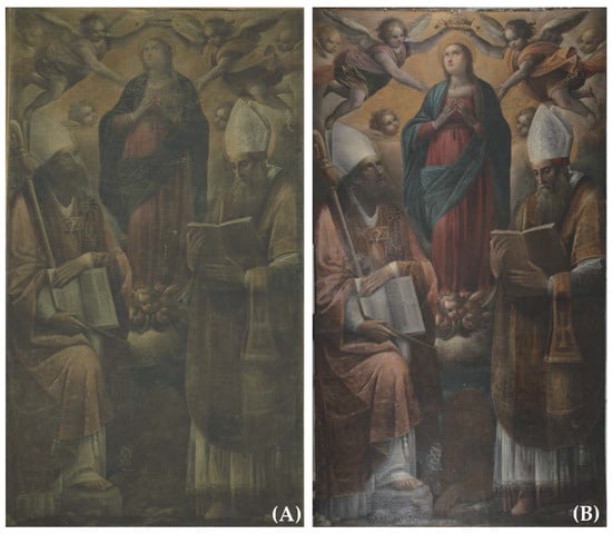
Figure 1.
Calibrated images of the canvas painting before (A) and after the cleaning (B).
It should be stressed that the restoration treatment was essential for the proper legibility of the painting. In fact, the comprehension of the iconographic elements was drastically compromised by the chromatic alteration of the superimposed materials. Through the removal of these materials, it was possible to start the art-historical research in order to comprehend the subject of the painting and the identity of the saints. Furthermore, the cleaning operations made it possible to formulate a hypothesis regarding the geographic area where the artwork was originally painted. The cost of not restoring might have led to a lack of precious information about the history, iconography, and technique of the painting. The synergy between restoration treatment, the diagnostic campaign, and the art-historical research shed light on this painting that has never been studied before.
The cleaning, aimed at removing the altered surface layers that reduced the visibility of the painting, was performed by combining traditional solvent-based solutions and innovative bacteria-based systems, as detailed in Section 3.5.
From a technical point of view, the painting presents a fixed wooden frame, likely original, nailed around the support. On the upper crosspiece of the frame, a metal hook is nailed, which presumably represents the original system for anchoring the painting to the wall. The support has an ancient lining whose adhesive is likely starch-based (perhaps due to the possible dating of the work, it is a glue–paste mixture). The relining canvas still seems to be well adhered to the original support. On the front there is a wooden frame, molded and gilded, integral with the textile support by means of a nailing system.
On the occasion of the recently concluded restoration, a diagnostic campaign was requested in order to characterize the original materials and the possible superimposed layers, also with the aim of selecting the most effective cleaning approach.
Between a wide variety of analytic methods available for art diagnostics, non-invasive techniques have been preferred, enhanced, and widely applied in conservation science and art restoration over the years, due to the possibility of using complementary methods and obtaining more information from the same investigated areas or samples of artwork, such as Fiber Optic Reflectance Spectroscopy (FORS), X-ray Fluorescence Spectroscopy (XRF), Fourier Transform Infrared Spectroscopy (FTIR), and portable Raman Spectroscopy [3,4,5,6,7]. Although these methods are commonly used for scientific art investigation as well as other applications, there is still a need for new, non-invasive techniques that could extend the amount of information obtained from the first diagnostic investigation of artwork analysis and limit the amount of eventual invasive testing required [8]. In this perspective, non-invasive imaging techniques are fundamental for obtaining complete knowledge of the surfaces, addressing the possible sampling of micro-chips of materials for laboratory analysis and, in the case of impossibility, taking samples from the investigated artworks [9,10], as well as supporting sensitive restoration interventions such as cleaning [11,12].
In a methodological approach using a rapid contactless imaging technique for comprehensive screening of an artwork’s surface to later focus micro analyses, multispectral imaging has been successfully used in scientific diagnostics over the last thirty years and is being enhanced in terms of resolution, portability, and cost-efficiency thanks to engineering research reducing dimensions and costs and refining the sensitivity of sensors [13].
In this work, traditional analytic techniques were applied in combination with an innovative multispectral imaging method named Hypercolorimetric Multispectral Imaging (HMI), allowing us to investigate the general state of conservation of the artwork, as well as characterizing painting and restoration materials [14].
The HMI technique is based on the simultaneous exploitation of the electromagnetic spectrum from ultraviolet to near-infrared regions. The acquisition, made under a standard metric, allows for characterizing the investigated surfaces in a more detailed way than standard colorimetry. The system transforms any spectra in the range of 300–1000 nm into sevenfold hypercolorimetric coordinates. HMI guarantees very high radiometric (better than 95%) and colorimetric precision (better than ΔE = 2) [14,15]. This technique has been previously tested on other case studies—among which are prestigious paintings of the Italian Renaissance—demonstrated to be a valid, helpful, and totally non-invasive diagnostic approach to map the conservation status, detect hidden details, compare different areas of the paintings, and address the choice of the reasoned sampling points to reduce the number of micro-samples from artworks [16,17,18].
In the present paper, this innovative approach was successfully applied using the capability of HMI to supply a great deal of information about painting materials, especially organics, so as to drastically reduce the number of samples necessary for detailed characterizations of original and possible superimposed compounds, safeguarding the conservation of the painting. This is particularly relevant in the present paper due to the old age of the painting and the different altered superimposed layers and dimensions. The large size of the canvas, in fact, represented a challenge for the diagnostics, making the choice of the area for point analysis and sampling difficult. For this reason, the HMI technique was very useful to guide the selection of the on-site point analysis by XRF spectroscopy and micro-sampling for laboratory investigation. The information gathered by HMI, combined with that from other non-invasive and laboratory analyses, greatly helped the cleaning intervention, especially in the choice of the most effective bacteria strains, used for the first time to bio-clean a canvas painting.
2. Materials and Methods
2.1. Hypercolorimetric Multispectral Imaging (HMI)
The HMI system consists of three main steps, all quick and executable on site: (1) Acquisition of the images, (2) calibration of the acquired images, and (3) processing of the multispectral outputs derived from the calibration.
- (1)
- Acquisition. This step is performed using a Nikon (Nital SpA, Moncalieri Torino, Italy) D800FR 36 Megapixel camera, modified to obtain full-range spectral reflectance measurements, a Nikon (Nital SpA, Moncalieri Torino, Italy) 17–35 zoom lens, and two filters named A (UV-Vis) and B (Vis-IR), whose spectra are shown in [19]. The filters are screwed in front of the camera lens before each shot is taken. Lighting is obtained by two NEEWER (Neewer, Shenzhen, China) 750II Flash Speedlite TTL with an LCD Display and Wireless Triggers. The flashes were modified by removing their front plastic lenses, thus allowing emissions in the 300–1000 nm region. Two photographic shots (one with filter A and another with filter B) were needed to cover the entire spectral range (300–1000 nm). To produce radiometrically and colorimetric calibrated images, white patches and a color-checker were positioned in the scene around the painting. The color-checker consists of 36 color samples from the NCS (Natural Color System® ©, NCS, Milan, Italy) catalog.
- (2)
- Calibration. This step is performed using the proprietary software SpectraPick® (Version 1.1, created by Profilocolore, Rome, Italy). Through a series of guided steps, it is possible, starting from two images acquired with filter A and filter B, to obtain seven tiff files representing the multispectral monochromatic images centered at 350 nm (UVR), 450 nm, 550 nm, 650 nm, 750 nm (IR1), 850 nm (IR2), and 960 nm (IR3) and the RGB 16-bit color image.
- (3)
- Processing. After the acquisition and calibration steps, the obtained multispectral images were processed through the HMI software PickViewer® (Version 1.0, created by Profilocolore, Rome, Italy), which provides powerful tools such as producing infrared and ultraviolet false color images by simply combining the calibrated channel; reading pixelwise colorimetry and spectral reflectance; creating similarity maps according to color or spectral data; applying principal component analysis (PCA), convolutional neural network (CNN)-based clustering, and other digital image processing; and querying and producing a dedicated color and spectral signature database. Each relevant result can be saved as an image in tiff, png, or jpeg format.
2.2. Ultraviolet Fluorescence Photography (UVF)
UVF images were acquired with the above-described digital camera and two filters screwed in front of the lens: Filter A and the UV-IR cut filter [19].
The UV-induced fluorescence (UVF) is obtained by irradiating the painting with UV LED lamps CR230B-HP (365 nm) positioned at 45° with respect to the artwork. After having completed the HMI acquisition, with a third shot coupling A and UV-IR cut filters the UVF images were obtained.
2.3. XRF Spectroscopy
X-Ray Fluorescence spectra of selected points of the painting were acquired with a portable XRF spectrometer (Surface Monitor II, Assing, Rome, Italy). The instrument is based on a silver anode as the excitation source and detector X-123 Si Pin that allow the detection of elements from atomic number 16 (S) to 92 (U) with an energy resolution of 146 eV. The X-ray beam is collimated to a spot diameter of approximately 2.0 mm. For all measurements on the painting, the following experimental conditions were employed: Tube voltage of 40 kV, tube anode current of 76 μA, and acquisition time of 60 s. XRF measurements were performed in 22 points, the positions of which are marked in Figure 2.
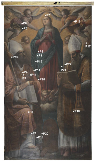
Figure 2.
Calibrated image of the painting after the cleaning with the points of XRF analysis.
2.4. FTIR Spectroscopy and Micro-Stratigraphic Analysis
Laboratory analyses were performed on four micro-samples named S1, S2, S3, and S4. S1 is located on the back of the painting in correspondence with the relining canvas (Figure 3); the other three were sampled from the painting surface in correspondence with the dark blue mantle of the Virgin (S2), a white cloud at the foot of the Virgin (S3), and the red of the Virgin dress (S4), as shown in Figure 3.
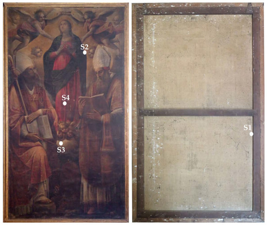
Figure 3.
Front and back of the painting with the sampling points for laboratory analysis.
Fourier transform infrared (FTIR) spectroscopy was applied using a Nicolet Avatar 360 instrument equipped with a DTGS (Deuterated TriGlycine Sulphate) detector.
The FTIR spectrometer operates in the 400–4000 cm−1 spectral range with a resolution of 4 cm−1. Sample powder (S2) without any treatment was grounded in agate mortar with potassium bromide (KBr), also used as background material. Moreover, to verify the presence of mastic, a further spectrum was gathered from the cotton swab used for the cleaning test. The resin, removed with the swab embedded with the solvent mixture, was re-solubilized with the same solvent based on acetone, and, after the liquid phase evaporation, the solid fraction was examined by FTIR.
For each sample, 128 scans were acquired in diffuse reflectance modality (DRIFT).
On the same sample (S2), examined through FTIR spectroscopy, a cross-section was also obtained after embedding a micro-fragment in resin and cutting it with a diamond blade. The cross-section was polished with ultrafine abrading paper and observed under a Zeiss (Zeiss, Oberkochen, Germany) Axioskope Polarizing microscope equipped with an AxioCam (Zeiss, Oberkochen, Germany) digital camera and a Hg-vapor lamp (supplied by Leica Microsystems, Wetzlar, Germany) for UVF observation.
Both analyses were performed on a micro-sample taken from the Virgin mantle, chosen after the non-invasive in situ investigation.
2.5. Gas Chromatography-Mass Spectrometry (GC-MS)
In order to characterize the binder, the superimposed materials of the painting, and the adhesive of the relining on the back, so as to support the cleaning intervention, samples S1, S2, S3, and S4 were considered. A Hewlett Packard HP 6890 series equipped with an MSD-HP 5973 detector with a single quadrupole and a split-splitless injector was used. The mass spectrometer was operated in the EI positive mode (70 eV). The carrier gas was used in the constant flow mode (He, purity 99.995%) at 20 mL/min. The separation of components was performed by means of an SLB-5 fused-silica capillary column (5% phenyl, 95% methyl), 30 m in length with a film thickness of 0.25 µm.
The method chosen, based on the work of recent years, allows us to obtain two chromatograms for each sample: The first one from fatty acid derivatives and the second one from amino acid derivatives [20].
3. Results and Discussion
3.1. Ultraviolet Fluorescence Photography
The first significant result was obtained by UVF photography performed before and during the cleaning tests (Figure 4). This technique is, in fact, a fundamental step in the restoration of paintings and other kinds of artefacts because it provides information about the conservation status, the presence of superimposed materials, and previous interventions, supplying the conservators with relevant knowledge to make decisions about cleaning [19].
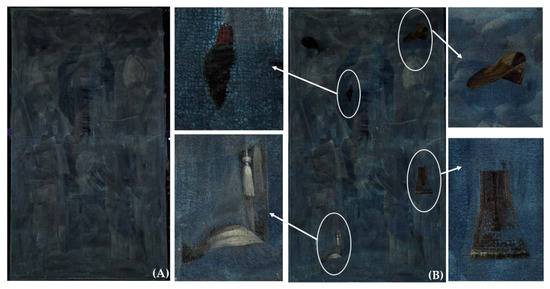
Figure 4.
UVF image before cleaning (A) and during the first cleaning tests (B). Details of the cleaned areas are highlighted by the white circles and arrows.
The UVF images shown in Figure 3 were acquired after the first step of cleaning was performed by first applying a water solution of tribasic ammonium citrate (TAC) at 2% (w/v) and then a solvent mixture made of ligroin/acetone (10:90 v/v), as detailed in Section 3.5.
The presence of a diffuse pale blue fluorescence suggested that a superimposed restoration material was present. The fluorescence, in fact, is not uniformly distributed and appears to be associated with residues of a probable protein glue applied by large brush strokes [21]. The UVF image also shows a pale-yellow fluorescence under the pale blue one that could be associated with the varnish and the binder of the painting. This first piece of diagnostic evidence provided relevant information about the state of conservation and the previous interventions of the artwork. Together with close observation of the painting surface and the support canvas, possible thanks to the restoration work, the UVF images showed how, unlike what was originally supposed, the artwork had instead undergone at least two previous undocumented interventions: One with the application of a varnishing resin and the second of re-lining and the application of a surface facing.
The results of UVF photography were also useful to address the preliminary cleaning tests that demonstrated their effectiveness in removing the altered surface materials that clearly reduced the clarity of the painting, as visible in the details in Figure 4.
3.2. HMI Data
The seven calibrated HMI spectral images were processed in PickViewer® software to identify elements of interest useful for answering diagnostic questions about the main conservation issues, painting materials and techniques, and ancient restoration evidence.
Most significant details were observed in the IR images, with this being a diagnostic spectral range to identify underdrawings and investigate the nature of pigments according to their transparency to IR radiation and distinguish painting materials based on their spectral behavior in false-color IR images (IRFC). HMI processing software can provide high-quality IR images and add significant further information through the application of statistical tools such as PCA, which can increase the readability of the painted layers or reveal hidden features not apparent in any of the individual spectral images [22]. Moreover, in the studying of underdrawings and potential pentimenti, a good number of pigments commonly used in Medieval and Renaissance painting palettes show good transparency already in the near-infrared (NIR) [21,23] spectral range between 780 and 1100 nm, registered by a CCD (Charge Coupled Device)-modified digital camera such as the one used in this study.
In IRFC, the Virgin’s mantle, in the lower right part, takes on a false wine-red color while the rest appears very dark blue. The same false wine-red color is also observed in the bottom right, near the edge of the Virgin’s mantle (Figure 5).
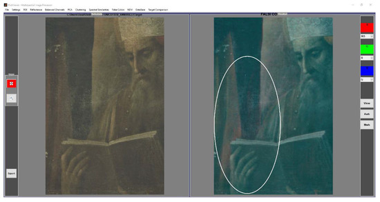
Figure 5.
Graphic user interface (GUI) of HMI software showing the result of IR false color processing of the detail of the Virgin’s mantle. The white circle highlights the possible pentimento.
This result suggests the possible use of blue smalt to make the background, and a probable pentimento in painting the mantle of the Virgin (see the detail in the white circle of Figure 5). It seems that the Virgin’s mantle was enlarged and overlapped the background made of blue smalt. Another piece of evidence from the IRFC image, obtained before and after the cleaning intervention, is the yellow color assumed by the Virgin’s dress and the angel’s drapery that can be associated with vermilion/cinnabar (Figure 6). The IRFC image served to hypothesize possible pigments and, above all, address the punctual micro-sampling and XRF analysis to identify the painting materials.
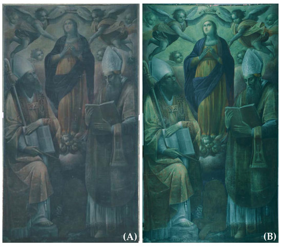
Figure 6.
IRFC images obtained through HMI software before (A) and after (B) cleaning. The removal of the superimposed layers increased the clarity of the painting surface and also of the IR false colors.
Still exploiting the HMI bands in the infrared, reflectography images were used to investigate the preparatory drawing, and principal component analysis (PCA) was applied to the IR1, IR2, and IR3 channels, i.e., the three infrared calibrated bands produced by SpectraPick® software (Version 1.1, created by Profilocolore, Rome, Italy) and centered at 750 nm (IR1), 850 nm (IR2), and 950 nm (IR3), to better highlight possible hidden details. PC1, and above all PC2, which are the first and the second principal components obtained from PCA, clearly confirm the pentimento in correspondence with the Virgin mantle and some changes to the hands of St. Jerome (Figure 7A,B). The IR3 band clearly shows the preparatory drawing in correspondence with the two angels holding the crown, particularly evident in their arms’ contour and the drapery (Figure 7C).
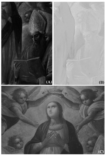
Figure 7.
(A) First PC and (B) second PC resulting from the application of PCA to the three infrared channels; (C) IR3 channel showing the drawing, well-visible in the angels’ arms and drapery.
Further details of interest were added by the UV reflected (UVR) image acquired in HMI analysis, such as the UV-calibrated band at 350 nm. Since UV light interaction with an artwork’s surface is limited to the very upper layer of the painting, UVR photography is particularly useful to detect the topmost pigment and layers related to original layers, as well as later interventions, such as some white restoration pigments. In this case, the UVR image (Figure 8A) provided an interesting map of some white pigment likely used in finishing brushstrokes given to enhance brighter areas, as in the miter of both Saints and in some spots of their cuffs and vests, which could be attributed to upper coats of lead white [24]. Another interesting result, obtained by using the UVR channel combined with the three IR channels through the algorithm NDVI (Normalized Difference Vegetation Index) of PickViewer®, concerns a detail of St. Jerome’s chasuble (Figure 8B). The normalized difference between diagnostic spectral bands is a graphic indicator, generally applied in agriculture red and near-infrared bands, to process remote sensing measurements and assess whether the observed subject is healthy or not and, therefore, whether it contains living green vegetation. In the case of its application on the Crowned Virgin painting, the NDVI algorithm makes the details of the drawing on the chasuble incredibly clear, as observable in Figure 6B, in comparison to the same detail in the visible calibrated image (Figure 8C).
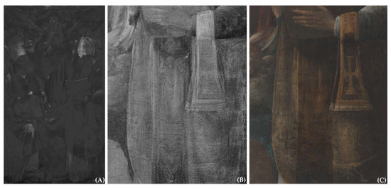
Figure 8.
(A) UVR channel highlighting the lead white coats on the surface. (B) Result of the application of NDVI algorithm to the UVR, IR1, IR2, and IR3 calibrated channels and (C) the same detail in the visible calibrated image after cleaning.
The last process presented in this paper is related to the use of the database implemented in PickViewer® software to check the presence of organic dyes that could be hypothesized in the pink cope of St. Ambrose and in the lighter areas of the Virgin’s dress characterized by a light green false color in IRFC (see Figure 6B).
The pink color was first checked through the multispectral similarity (MS) algorithm of PickViewer®, which allows one to map the pixels with the same multispectral characteristics (Figure 9).
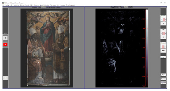
Figure 9.
Multispectral similarity tool applied to a pink area (white dot in the RGB image of the painting visible in the left window of PickViewer® GUI) showing the map of distribution of such pigment/dye (the white pixels visible in the image on the right window of PickViewer® GUI).
The result of the MS algorithm confirms the same composition of painting materials in the St. Ambrose cope and in the highlighting of the Virgin’s dress.
The pink color was compared with those contained in the database available in PickViewer®, and a close similarity was found with alizarin, which can be hypothesized as dye used to obtain the pink color.
3.3. XRF Spectroscopy
Results of XRF spectroscopy on selected analysis points of the painting are provided in Table 1 in terms of the main detected elements and are synthetically resumed in the followings:

Table 1.
Results of punctual XRF spectroscopy applied to the painting. The main detected elements are reported with the proposed pigments associated with those elements.
- (i)
- In all analysis points, we detected Pb, Ca, and Fe, suggesting the presence of ground and preparation layers mainly made of lead white or lead oxide, gypsum or calcite, and iron-based pigments.Concerning the coloring agents, the palette appears to be quite simple, and most of the identified pigments can be related to the traditional artist materials employed in the late 16th century:
- (ii)
- Red hues have been achieved by employing vermillion and red ochre.
- (iii)
- Blue hues have been achieved by employing azurite and blue smalt, the latter in the background.
- (iv)
- Yellow was obtained by mixing yellow ochre and possibly lead oxide.
- (v)
- The gilding of the frame is likely original, and it is made of gold applied to a mordant based on lead iron compounds. The presence of Ca and Sr suggests the use of a gypsum-based ground layer.
The presence of Cu in dark areas of the painting suggests it may be related to darkened copper acetate or resinate, pigments particularly used with linseed oil as a binder in easel paintings between the 15th and 17th centuries [25].
Copper acetate and resinate had the tendency to darken in an irreversible way according to a mechanism that was recently investigated [26].
3.4. FTIR Spectroscopy, Micro-Stratigraphy, and GC-MS
These techniques were applied to micro-samples taken from the Virgin dress and mantle (right side, close to the background), from the white cloud at the foot of the Virgin, and from the back of the painting, in correspondence with the glue used to reline the original canvas, in order to investigate the organic composition. Specifically, sample S2, used for FTIR analysis and optical microscopy on the cross-section, was selected in an area where the HMI has shown the possible presence of unaltered blue smalt protected by the layer used for creating the Virgin mantle (Figure 5). Here, the IRFC highlighted a wine-red response typical of blue smalt. Samples S3 and S4 were selected in correspondence with areas where UVF has shown a particularly evident light blue fluorescence attributable to the superimposed proteinaceous material, as visible in Figure 4A. The choice of the sampling points was also made considering the presence of lacunae on the painting surface.
These data are particularly relevant for the cleaning intervention as the knowledge of surface and back material composition is fundamental to address the choice of solvent mixtures and, above all, the effective bacteria strains for bio-cleaning.
The FTIR spectrum (Figure 10) is quite complex and exhibits the signatures of several possible materials.
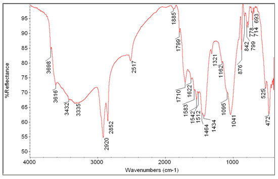
Figure 10.
FTIR spectrum of P2 micro-sample taken from the Virgin mantle, right outline.
The wavenumbers at cm−1 2920, 2852, 1710 (this signature can also be associated with terpene resin), 1464, 1162, and 1095 identify the binder based on aged siccative oil, with the carboxylate’s reflectance at 1622 cm−1 and 1542 cm−1, which has been attributed to the formation of those anions coordinated with lead [27]. Calcium carbonate signatures are also unequivocally attributed in the spectrum (calcite signatures at cm−1 2517, 1799, 1434, 876, and 714) [28]. Some reflectance values may be attributed to azurite such as those at cm−1 3432, 1885, 1583–1434 (overlapped with the calcite signature), 799, and 693. This attribution, even if difficult due to the presence of several compounds in the examined sample, may be considered a possibility and compatible with the presence of Cu as detected by XRF spectroscopy.
The presence of terpene resin and protein glue, hypothesized on the basis of the UVF image, cannot be excluded, but their eventual signatures may be hidden by or overlap with those of other materials. The bands at 3416 cm−1, 1710 cm−1, and 1162 cm−1 are associated with mastic, which may be the component of the brownish varnish; those at 1650 cm−1 (visible as a shoulder of the 1622 cm−1 band), 1542 cm−1, and 1464 cm−1 may be the three typical bands of the amide group of proteins [29].
Some other reflectance signatures in the FTIR spectrum can be attributed to iron-based earth that contains iron oxides but also silicates and calcium carbonate. The reflectance values attributable to these materials are visible at cm−1 3698, 3616, 1041, 525, and 472. Due to the wine-red color of the IRFC image obtained through the HMI technique, the presence of blue smalt could also be hypothesized. This pigment has FTIR signatures in the region of Si-O stretching silicates at approximately 1000–1100 cm−1 (very strong), 778 cm−1 (medium), 693 cm−1 (weak), and 459 cm−1 (strong) [30]. All these signatures are overlapped with those of silicates of earth and azurite, making it difficult to doubtlessly confirm the presence of smalt. For this reason, a cross-section was obtained from the sample whose images under visible reflected light and UV fluorescence are shown in Figure 11. The cross-section confirms that the ground layer is colored and contains iron oxide, red–orange grains likely of minium (layer 1). On this ground, a darker red layer can be observed (layer 2) containing red and black particles together with glassy light blue-grey grains that could be associated with smalt; this is the point corresponding to the pentimento highlighted by the IRFC image, which, in this area, revealed a wine-red false color under the Virgin’s mantle. A very thin layer (3) with pale yellow fluorescence under UV is present over layer 2, likely consisting of a coat of binder. The painting layer (4) is very dark and contains blue particles that can be associated with azurite. On the surface, a transparent inhomogeneous layer characterized by a light blue fluorescence can be observed (layer 6), applied over a brownish varnish (layer 5) without any fluorescence under UV. The surface layer, exhibiting light blue fluorescence, was the same as that observed in the UVF image (see Figure 4).
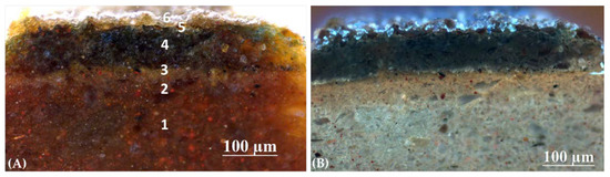
Figure 11.
Cross-section of micro-sample S2 from the outline of the Virgin’s mantle under reflected visible light (A) and ultraviolet radiation (B).
To verify the presence of mastic, a further spectrum was gathered from the cotton swab used for the cleaning test. The obtained spectrum shows all the main signatures of mastic resin, therefore confirming this material was used to obtain the varnish layer (Figure 12). Specifically, the mastic signals are those at cm−1 3460–3327 (O-H stretching band), 2948–2854 (C-H stretching bands), 1713 (C=O stretching band), 1460–1379 (C-H bending bands), and 1232–1080 (C-O stretching bands) [29] (p. 189).
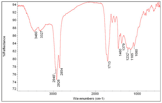
Figure 12.
FTIR spectrum of the material solubilized from the cotton swab.
It was important to confirm the presence of mastic in order to prepare mock-ups for the cleaning tests with bacteria with the same stratigraphy and materials as the painting.
To obtain a precise characterization of the protein glue, hypothesized thanks to the light blue fluorescence under UV, but not well-highlighted by FTIR analysis, and the binder of the pigments, GC-MS was applied to sample S2 as well as to the other two samples (S3 and S4) from the surface of the painting, and to a further sample (S1) taken from the back in correspondence with the adhesive used for relining in a previous undocumented intervention.
Therefore, gas chromatography, coupled with a mass spectrometry detector, was used to identify the lipidic and the proteinaceous materials. Both the lipid and proteinaceous components were found in the four examined samples. It was observed that, except for the S1 sample, which is also of different types, the protein component resulted in being greater than the lipid component.
GC-MS analysis of the proteinaceous fractions consisted of the quantitative assessment of the amino acids’ composition. To identify the proteinaceous typology, the relative percentage content of amino acids in each sample was compared to those from a dataset of 42 reference samples of egg, casein, and animal glue, belonging to the reference collection of the Opificio delle Pietre Dure of Florence [31]
Principal component analysis (PCA) was performed on the correlation matrix of the relative percentage contents of eight amino acid (alanine, glycine, leucine, proline, hydroxyproline, aspartic acid, glutamic acid, and phenylalanine) components [32].
The resulting score plot for the examined samples is reported in Figure 13.
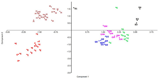
Figure 13.
Score plot of reference materials and painting samples S1, S2, S3, and S4. (G: Animal glue, in green; M: Milk, in brown; E: Egg, in red; GE: Animal glue and egg, blue; GM: Animal glue and milk, violet).
The evaluation by means of PCA, whose score plot is reported in Figure 13, locates samples S2, S3, and S4 in a new cluster suggesting the presence of animal glue. Sample S1 shows the presence of animal glue and milk proteins.
Lipidic fraction gas chromatograms of every sample are characterized by dicarboxylic acids, particularly azelaic acid, and saturated fatty acids, principally palmitic and stearic acids. The low amount of azelaic acid, whose formation is due to the oxidation of unsaturated acids, suggests that the observed lipidic fraction cannot be attributed to only siccative oil. It is assumed that there is a mixture of vegetable oils, presumably consisting of a siccative oil in addition to one or more non-siccative or semi-siccative oils.
It was found that in the S1 sample, for textile support, taken for the identification of the components of the relining adhesive, the presence of an oil mixture, animal glue, and milk was found, and peaks were identified with odd fragmentation such as amine derivatives, likely due to substances with hydrolyzed polyamine or polyamide groups, which could be due to the canvas material.
3.5. A Synthesis of the Cleaning Process Helped by Diagnostics and Laboratory Analysis
The above-presented analyses were highly useful to address the choice of the cleaning systems to remove the altered surface layers that reduced the clarity of the artwork. The preliminary cleaning tests used a solution of tribasic ammonium citrate (TAC) at 2% (w/v) to remove the surface glue residues and a solvent mixture made of ligroin/acetone (10:90 v/v) to solubilize the oxidized varnish based on mastic resin. In the area where the altered restoration varnish layer was thicker, the cleaning was completed by applying a gel system made up of 2 g of Carbopol Ultrez 21® (supplied by CTS Europe, Altavilla Vicentina, Vicenza, Italy), 1.5 g of citric acid, 6 mL of triethanolamine (TEA), and 100 mL of demineralized water. Lastly, in two areas of the painting surface and one on the back, bio-cleaning with selected bacteria strains was successfully applied, after laboratory tests aimed to choose the most effective system and time of application [1]. This is the first time that bacteria have been used for cleaning glue and mastic resin from a canvas painting, and the details of the test on mocks-up and the bio-cleaning on the real artwork will be presented in a separate paper.
The advantages of bio-cleaning, over traditional chemical methods based on solvents, are environmental sustainability, selectivity for the artwork, and safety for operators’ health since they are non-pathogenic and non-toxic. Sustainability results from the environment from which the bacterial strains are isolated: They are wild-type bacteria selected from hypogea, harsh environments, and artworks. Being non-toxic and isolated from natural environments, waste produced at the end of bio-cleaning operations does not need particular precautions regarding their disposal [33].
Specifically, preliminary tests were performed on mocks-up prepared with the same materials and stratigraphy found on the artwork thanks to the analysis. On these mock-ups, some bacteria strains tested were chosen after having verified their effectiveness in vitro to degrade animal glue and mastic resin (Table 2).
Table 2.
The bacterial strains selected for the cleaning tests.
Bacteria strains were applied with different kinds of gelling agents selected to obtain the micro-packs that were applied to the surfaces to be cleaned. The tests demonstrated that the most effective system, both in terms of effectiveness and ease of application, was that based on LAM 21 applied with the Vanzan® NF-C (supplied by CTS Europe, Altavilla Vicentina, Vicenza, Italy) gelling agent (composed of xanthan gum) for 4 h.
The combined use of traditional solvent systems and innovative bio-cleaning through bacteria has proven successful in significantly improving the painting quality, as shown in Figure 1B.
4. Conclusions
The great potentiality of multispectral imaging, performed through the innovative HMI system, combined with UVF photography, has been demonstrated, especially the processing software, which allowed us to gather extensive information about hidden details, pigment composition, superimposed materials, preparatory drawing, etc.
The imaging analysis was also particularly useful to address the sampling points in order to obtain significant samples and limit their number to a minimum.
XRF spectroscopy performed on-site during the restoration provided information about the chemical elements, allowing us to hypothesize the pictorial palette.
Analysis of micro-samples (FTIR, micro-stratigraphy and GC-MS) completed the information about materials: Organic binders, superimposed layers, adhesive, providing the data useful for preparing the mocks-up to test bacteria, and, above all, choosing the strains effectiveness in removing the protein glue and the mastic resin.
In conclusion, the synergy between restoration and diagnostics proved, once again, effective in completing the intervention successfully, thus restoring the full readability of the painting.
Author Contributions
Conceptualization, S.A., S.S. and C.P.; methodology, C.P., L.L., C.C., A.C. and A.M.; software, L.L. and C.C.; validation, L.L., C.C., C.P. and S.A.; formal analysis, C.P., L.L., C.C., A.C. and A.M.; investigation, L.L., C.C., A.C. and A.M.; resources, C.P. and A.C.; data curation, L.L., C.C., C.P., A.C. and A.M.; writing—original draft preparation, C.P. and C.C.; writing—review and editing, all authors; visualization, S.A., L.L. and C.C.; supervision, S.S., C.P. and A.C.; project administration, S.S.; funding acquisition, C.P. and A.C. All authors have read and agreed to the published version of the manuscript.
Funding
This research received no external funding.
Institutional Review Board Statement
Not applicable.
Informed Consent Statement
Not applicable.
Data Availability Statement
Not applicable.
Acknowledgments
The authors would like to thank Giorgia Agresti for the realization of the cross-section and for having supported Sofia Annarilli during the development of her thesis work.
Conflicts of Interest
The authors declare no conflict of interest.
References
- Annarilli, S. La Tela Dei Santi Ambrogio E Girolamo Con la Vergine Incoronata. Un Nuovo Contributo per la Biopulitura E la Sostenibilità Nel Restauro. Master’s Thesis, University of Tuscia, Viterbo, Italy, 6 June 2022. [Google Scholar]
- Anselmi, S.E.; Principi, L. Il Museo d’Arte Sacra di Orte; Centro Studi Tuscia: Orte, Italy, 2014; pp. 77–78. [Google Scholar]
- Bersani, D.; Berzioli, M.; Caglio, S.; Casoli, A.; Lottici, P.P.; Medeghini, L.; Poldi, G.; Zannini, P. An integrated multi-analytical approach to the study of the dome wall paintings by Correggio in Parma cathedral. Microchem. J. 2014, 114, 80–88. [Google Scholar] [CrossRef]
- Miliani, C.; Rosi, F.; Burnstock, A.; Brunetti, B.G.; Sgamellotti, A. Non-invasive in situ investigations versus micro-sampling: A comparative study on a Renoirs painting. Mater. Sci. Proces. 2007, 89, 849–856. [Google Scholar] [CrossRef]
- Ford, T.; Rizzo, A.; Hendriks, E.; Frøysaker, T.; Caruso, F. A non-invasive screening study of varnishes applied to three paintings by Edvard Munch using portable diffuse reflectance infrared Fourier transform spectroscopy (DRIFTS). Herit. Sci. 2019, 7, 1–13. [Google Scholar] [CrossRef]
- Brunetti, B.; Miliani, C.; Rosi, F.; Doherty, B.; Monico, L.; Romani, A.; Sgamellotti, A. Non-invasive Investigations of Paintings by Portable Instrumentation: The MOLAB Experience. Review. In Analytical Chemistry for Cultural Heritage. Topics in Current Chemistry Collections; Mazzeo, R., Ed.; Springer: Cham, Switzerland, 2016; Volume 374, pp. 41–75. [Google Scholar] [CrossRef]
- Vagnini, M.; Gabrieli, F.; Daveri, A.; Sali, D. Handheld new technology Raman and portable FT-IR spectrometers as complementary tools for the in situ identification of organic materials in modern art. Spectroch. Acta A 2017, 176, 174–182. [Google Scholar] [CrossRef]
- Cavaleri, T.; Pelosi, C.; Ricci, M.; Laureti, S.; Romano, F.P.; Caliri, C.; Ventura, B.; De Blasi, S.; Gargano, M. IR Reflectography, Pulse-Compression Thermography, MA-XRF, and Radiography: A Full-Thickness Study of a 16th-Century Panel Painting Copy of Raphael. J. Imaging 2022, 8, 150. [Google Scholar] [CrossRef]
- Luciani, G.; Pelosi, C.; Agresti, G.; Monaco, A.L. How to reveal the invisible: The fundamental role of diagnostics for religious painting investigation. Eur. J. Sci. Theol. 2019, 15, 209–220. [Google Scholar]
- Lanteri, L.; Agresti, G.; Pelosi, C. A new practical approach for 3D documentation in ultraviolet fluorescence and infrared reflectography of polychromatic sculptures as fundamental step in restoration. Heritage 2019, 2, 207–215. [Google Scholar] [CrossRef]
- Colantonio, C.; Lanteri, L.; Ciccola, A.; Serafini, I.; Postorino, P.; Censorii, E.; Rotari, D.; Pelosi, C. Imaging Diagnostics Coupled with Micro-Invasive Analyses for the Restoration Artifacts from French Polynesia. Heritage 2022, 5, 215–232. [Google Scholar] [CrossRef]
- Fontana, R.; Fovo, A.D.; Striova, J.; Pezzati, L.; Pampaloni, E.; Raffaelli, M.; Barucci, M. Application of non-invasive optical monitoring methodologies to follow and record painting cleaning processes. Appl. Phys. A 2015, 121, 957–966. [Google Scholar] [CrossRef]
- Liang, H. Advances in multispectral and hyperspectral imaging for archaeology and art conservation. Appl. Phys. A 2012, 106, 309–323. [Google Scholar] [CrossRef]
- Melis, M.; Miccoli, M.; Quarta, D. Multispectral hypercolorimetry and automatic guided pigment identification: Some masterpieces case studies. In Proceedings of the SPIE 8790, Optics for Arts, Architecture, and Archaeology IV, Munich, Germany, 30 May 2013; Pezzati, L., Targowski, P., Eds.; Volume 33, pp. 1–14. [Google Scholar] [CrossRef]
- Samarelli, M.; Melis, M.; Miccoli, M.; Vigl, E.E.; Zink, A.R. Complete mapping of the tattoos of the 5300-year-old Tyrolean Iceman. J. Cult. Herit. 2015, 16, 753–758. [Google Scholar] [CrossRef]
- Laureti, S.; Colantonio, C.; Burrascano, P.; Melis, M.; Calabrò, G.; Malekmohammadi, H.; Sfarra, S.; Ricci, M.; Pelosi, C. Development of integrated innovative techniques for paintings examination: The case studies of The Resurrection of Christ attributed to Andrea Mantegna and the Crucifixion of Viterbo attributed to Michelangelo’s workshop. J. Cult. Herit. 2019, 40, 1–16. [Google Scholar] [CrossRef]
- Colantonio, C.; Pelosi, C.; D’Alessandro, L.; Sottile, S.; Calabrò, G.; Melis, M. Hypercolorimetric Multispectral Imaging (HMI) system for cultural heritage diagnostics: An innovative study for copper painting examination. Eur. Phys. J. Plus 2018, 133, 526. [Google Scholar] [CrossRef]
- Ricci, M.; Laureti, S.; Malekmohammadi, H.; Sfarra, S.; Lanteri, L.; Colantonio, C.; Calabrò, G.; Pelosi, C. Surface and Interface Investigation of a 15th Century Wall Painting Using Multispectral Imaging and Pulse-Compression Infrared Thermography. Coatings 2021, 11, 546. [Google Scholar] [CrossRef]
- Lanteri, L.; Pelosi, C. 2D and 3D ultraviolet fluorescence applications on cultural heritage paintings and objects through a low-cost approach for diagnostics and documentation. In Proceedings of the SPIE 11784, Optics for Arts, Architecture, and Archaeology IV, Munich, Germany, 20–21 June 2021; Liang, H., Groves, R., Eds.; pp. 1–10. [Google Scholar] [CrossRef]
- Cosentino, A. Effects of Different Binders on Technical Photography and Infrared Reflectography of 54 Historical Pigments. Int. J. Conserv. Sci. 2015, 6, 287–298. [Google Scholar]
- Casoli, A.; Santoro, S. Organic materials in the wall paintings in Pompei: A case study of Insula del Centenario. Chem. Cent. J. 2012, 6, 107. [Google Scholar] [CrossRef] [PubMed]
- Legnaioli, S.; Grifoni, E.; Lorenzetti, G.; Marras, L.; Pardini, L.; Palleschi, V.; Salerno, E.; Tonazzini, A. Enhancement of hidden patterns in paintings using statistical analysis. J. Cult. Herit. 2013, 14, S66–S70. [Google Scholar] [CrossRef]
- Attas, M.; Cloutis, E.; Collins, C.; Goltz, D.; Majzels, C.; Mansfield, J.; Mantsch, H. Near-infrared spectroscopic imaging in art conservation: Investigation of drawing constituents. J. Cult. Herit. 2003, 4, 127–136. [Google Scholar] [CrossRef]
- Pronti, L.; Felici, A.C.; Ménager, M.; Vieillescazes, C.; Piacentini, M. Spectral Behavior of White Pigment Mixtures Using Reflectance, Ultraviolet-Fluorescence Spectroscopy and Multispectral Imaging. Appl. Spectrosc. 2017, 71, 2616–2625. [Google Scholar] [CrossRef]
- Kühn, H. Verdigris and Copper Resinate. In Artists’Pigments. A Handbookof Their History and Characteristics; Roy, A., Ed.; National Gallery of Art: Washington, DC, USA, 1993; Volume 2, pp. 131–158. [Google Scholar]
- Alter, M.; Binet, L.; Touati, N.; Lubin-Germain, N.; Hô, A.-S.L.; Mirabet, F.; Gourier, D. Photochemical Origin of the Darkening of Copper Acetate and Resinate Pigments in Historical Paintings. Inorg. Chem. 2019, 58, 13115–13128. [Google Scholar] [CrossRef]
- Meilunas, R.J.; Bentsen, J.G.; Steinberg, A. Analysis of aged paint binders by FTIR spectroscopy. Stud. Conserv. 1990, 35, 33–51. [Google Scholar] [CrossRef]
- Mazzocchin, G.A.; Orsega, E.F.; Baraldi, P.; Zannini, P. Aragonite in Roman wall paintings of the VIIIa Regio Aemilia, and Xa Regio, Venetia et Histria. Ann. Di Chim. 2006, 96, 377–387. [Google Scholar] [CrossRef] [PubMed]
- Derrick, M.R.; Stulik, D.; Landry, J.M. Infrared Spectroscopy in Conservation Science, 1st ed.; The Getty Conservation Institute: Los Aneles, CA, USA, 1999; pp. 178–200. [Google Scholar]
- IRUG Database. Available online: http://www.irug.org/search-spectral-database (accessed on 12 August 2022).
- Lanterna, G.; Mairani, A.; Matteini, M.; Rizzi, M.; Vigato, P.A. Characterisation of Decay Markers on Pictorial Models Simulating Ancient Polychromies. In Proceedings of the 2nd International Congress on Science and Technology for the Safeguard of Cultural Heritage in the Mediterranean Basin, Paris, France, 5–9 July 1999; Ferrari, A., Ed.; Elsevier: Amsterdam, The Netherlands, 2000; pp. 487–489. [Google Scholar]
- Casoli, A.; Montanari, A.; Palla, L. Painted Models Simulating Ancient Polychromies: A Statistical Analysis of Chemical Results. In Proceedings of the 3rd International Congress on Science and Technology for the Safeguard of Cultural Heritage in the Mediterranean Basin, Alcalá De Henares, Spain, 9–14 July 2021; Guarino, A., Ed.; Elsevier: Amsterdam, The Netherlands, 2001; pp. 839–845. [Google Scholar]
- Sprocati, A.R.; Alisi, C.; Migliore, G.; Marconi, P.; Tasso, F. Sustainable Restoration Through Biotechnological Processes: A Proof of Concept. In Microorganisms in the Deterioration and Preservation of Cultural Heritage; Joseph, E., Ed.; Springer: Cham, Switzerland, 2021; pp. 235–261. [Google Scholar] [CrossRef]


Publisher’s Note: MDPI stays neutral with regard to jurisdictional claims in published maps and institutional affiliations. |
© 2022 by the authors. Licensee MDPI, Basel, Switzerland. This article is an open access article distributed under the terms and conditions of the Creative Commons Attribution (CC BY) license (https://creativecommons.org/licenses/by/4.0/).