You are currently viewing a new version of our website. To view the old version click .
Neuroglia
  • Review
  • Open Access

6 November 2025

Stress-Induced Transcriptional and Epigenetic Plasticity of Astrocytes, Microglia and Oligodendrocytes in the Pathophysiology of Depression

,
,
,
and
1
Department of Applied Biology, CSIR-Indian Institute of Chemical Technology (CSIR-IICT), Hyderabad 500007, Telangana, India
2
Academy of Scientific and Innovative Research (AcSIR), Ghaziabad 201002, Uttar Pradesh, India
3
CSIR-Centre for Cellular and Molecular Biology (CSIR-CCMB), Hyderabad 500007, Telangana, India
*
Author to whom correspondence should be addressed.
This article belongs to the Special Issue The Multifaceted Roles of Glia: From Cellular Functions to Neurological Implications, 2nd Edition

Abstract

Major Depressive Disorder (MDD) remains a leading cause of disability worldwide, perpetuated by an incomplete understanding of its pathophysiology and the limited efficacy of conventional antidepressants. Historically, research has focused on neuron-centric models, particularly the monoamine hypothesis. However, the field is now recognizing the critical role of glial cells such as astrocytes, microglia, and oligodendrocytes, establishing them as key contributors to the molecular basis of depression. Rather than serving solely supportive roles, these cells actively modulate neuroinflammation, synaptic plasticity, neurotransmitter homeostasis, and metabolic regulation, processes disrupted in MDD. We discuss how stress-induced epigenetic modifications such as histone acetylation, methylation, and DNA methylation are linked to alterations in astrocytic glutamate transport, microglial inflammatory states, and oligodendrocyte-mediated myelination. Special emphasis is placed on the concept of glial transcriptional plasticity, whereby environmental adversity induces durable and cell type specific gene expression changes that underlie neuroinflammation, excitatory–inhibitory imbalance, and white matter deficits observed in MDD. By integrating findings from postmortem human tissue, single-cell omics, and stress-based animal models, this review highlights converging molecular mechanisms linking stress to glial dysfunction. We further outline how targeting glial transcriptional regulators may provide new therapeutic avenues beyond conventional monoaminergic approaches.

1. Introduction

The neurobiological understanding of Major Depressive Disorder (MDD) has long been connected to the neuronal dysregulation. The monoamine hypothesis, which proposes that depression arises from deficiency in synaptic serotonin, norepinephrine, or dopamine, has long guided antidepressant development for over half a century []. While transformative, the delayed therapeutic onset of monoaminergic drugs, their limited efficacy in a substantial subset of patients, and the complex, multifactorial nature of MDD all point to pathological mechanisms that extend beyond the synapse. A fundamental shift in perspective is imperative, moving from a neuron-centric view to one that regards the central nervous system (CNS) as a complex ecosystem. Glial cells such as astrocytes, microglia, and oligodendrocytes are increasingly recognized as important contributors to brain health and disease []. Glial cells collectively outnumber neurons in many brain regions and are indispensable for nearly every aspect of CNS function, including synaptic transmission, metabolic support, immune surveillance, and the formation of myelin insulation []. Glial dysfunction has been implicated as a possible factor in the pathology of MDD []. Post-mortem and neuroimaging studies consistently reveal changes in glial cell numbers and density, particularly astrocytes and oligodendrocytes, in key mood-regulating brain regions of individuals with MDD, such as the prefrontal cortex (PFC), hippocampus, and amygdala [,]. The marked interindividual variability in antidepressant response [] may, in part, stem from stress-induced transcriptional and epigenetic reprogramming of glial cells, which shapes neuroinflammation, synaptic support, and myelin integrity in depression. Concurrently, microglial activation and the ensuing neuroinflammatory cascade are increasingly understood to implicate in ischemia, Alzheimer’s disease and other comorbid neurological disorders [,,].
A key challenge is to understand what mechanisms translate environmental risk factors, most notably chronic psychosocial stress, into these profound and lasting states of glial dysfunction? The answer likely involves epigenetic regulation. Epigenetic modifications, such as histone post-translational modifications, are heritable changes in gene expression that occur without altering the underlying DNA sequence. These mechanisms provide a molecular interface between the environment and the genome, allowing experiences like early-life adversity or chronic stress to induce stable changes in cellular function []. Histone modifications, catalyzed by a diverse enzymatic network of “writers” (acetyltransferases, methyltransferases) and “erasers” (deacetylases, demethylases), alter chromatin architecture to control the accessibility of genes to the transcriptional machinery. Epigenetic changes in microglia can prime them for a chronic, pro-inflammatory state that perpetuates a pathological milieu throughout the brain [,,]. It is hypothesized that stress-induced changes in glial transcriptional plasticity may serve as an important mechanism in the pathophysiology of depression. Understanding histone-dependent transcriptional control in glia is not just an incremental advance; it establishes a direct link between the diverse cellular and synaptic dysfunctions of MDD and offers rational design of novel antidepressant strategies. Given that depression is among the leading causes of global disability with marked gender-specific manifestations [], understanding stress-induced glial transcriptional remodeling provides a critical bridge between molecular mechanisms and clinical disability outcomes.
To ensure a comprehensive and current synthesis, this review is based on a structured literature search of the PubMed, Scopus, and Google Scholar databases. Our search strategy primarily focused on publications from the last five years (2020–2025) to capture the most recent advances in glial epigenetics, while also including some foundational studies (2015–2020) and few articles from earlier periods for historical context. Key search terms included combinations of ‘MDD’, ‘depression’, ‘chronic stress’ with ‘glia’, ‘astrocyte’, ‘microglia’, ‘oligodendrocyte’, and ‘epigenetics’, ‘transcriptional plasticity’, ‘histone modifications’, ‘DNA methylation’, ‘neuroinflammation’, ‘myelin plasticity’, and ‘non-coding RNA’. We prioritized primary research articles from human post-mortem studies, single-cell omics, and preclinical stress-based animal models, as well as high-impact research articles to provide a coherent narrative.

2. Epigenetic Codes of Glial Identity and CNS Homeostasis

The foundation of CNS stability rests heavily on the coordinated functions of astrocytes and microglia. Astrocytes are the principal homeostatic cells of the CNS. They regulate the extracellular environment by controlling ion concentrations clearing neurotransmitters like glutamate, providing metabolic substrates to neuron via the astrocyte-neuron lactate shuttle, and maintaining the integrity of the blood–brain barrier. Complementing this role, microglia function as the resident immune cells of the brain. In the healthy state, they are not merely quiescent but are highly active, continuously surveying their local microenvironment with motile processes. This surveillance is critical for normal brain development, where microglia mediate neurogenesis and synaptic pruning by phagocytosing apoptotic neural progenitors and eliminating redundant or weak synaptic connections, thereby shaping the architecture of nascent neural circuits [].
The functional identity of an astrocyte is not merely a product of its genetic code but is actively established and maintained by a specific epigenetic landscape. During neurodevelopment, the promoter of the canonical astrocyte marker gene, glial fibrillary acidic protein (GFAP), is demethylated, a crucial step that permits its expression and solidifies the astrocytic fate []. This process is governed by the activity of DNA methyltransferases (DNMTs) such as DNMT1 and DNMT3a, which ensure that astrocyte-specific genes are transcriptionally accessible while genes associated with other neural lineages remain silenced. Recent single-cell multi-omics analyses have reinforced this principle, demonstrating that distinct DNA methylation profiles, dependent on DNMT3A, separate functional astrocytes from neural stem cells by selectively methylating stemness-related genes and demethylating astrocyte-specific genes [,]. While DNA methylation provides a stable foundation for cell identity, histone modifications offer a dynamic functional regulation.
Microglia, too, are defined by a unique epigenetic signature that distinguishes them from peripheral macrophages and other CNS cells. The establishment of microglial identity is driven by lineage-determining transcription factors, most notably PU.1, which binds to specific enhancer regions across the genome. The accessibility of the PU.1 gene locus itself is epigenetically controlled; histone H4 acetylation at its promoter is required for its transcription, and this can be modulated by HDAC inhibitors []. In the homeostatic state, the functional poise of microglia is maintained by a delicate balance of repressive and activating histone marks. The histone methyltransferase EZH2 (Enhancer of Zeste Homolog 2) deposits the repressive mark H3K27me3 at the promoters of pro-inflammatory genes, keeping them silenced. This is counteracted by the demethylase JMJD3 (Jumonji domain-containing protein 3), which removes this mark to permit gene expression. In the healthy brain, this balance is tightly regulated to maintain a state of quiescent readiness [].
The stable homeostasis of the CNS is not simply the sum of independent astrocyte and microglial functions but arises from their continuous, bidirectional communication, which is itself highly influenced by epigenetic mechanisms. This forms a reciprocal feedback loop that maintains the quiescent, supportive state of both cell types. The process begins with the astrocyte’s epigenetically controlled secretome. In a healthy state, a specific pattern of histone acetylation and DNA methylation in astrocytes ensures the baseline expression of factors that promote microglial quiescence and suppresses those that would provoke activation. These astrocytic signals are received by microglia, whose response is predetermined by their own epigenetic landscape. For instance, the methylated, repressed state of pro-inflammatory gene promoters in homeostatic microglia ensures that they do not overreact to minor fluctuations in their environment [,]. The molecular mechanism maintaining this homeostatic loop is complex and involves multiple signaling systems that are themselves subject to epigenetic control. Purinergic signaling is a fundamental and rapid mode of reciprocal communication. Astrocytes can release adenosine triphosphate (ATP), which acts as a crucial signaling molecule. In the extracellular space, ATP can directly stimulate ionotropic (P2X) and metabotropic (P2Y) purinergic receptors on microglia. The microglial P2Y12 receptor is critical for sensing ATP and guiding microglial process motility toward sites of release. The signal is tightly regulated by ecto-nucleotidases like CD39 and CD73 on the cell surface, which sequentially hydrolyze ATP to adenosine. Adenosine then acts on P1 receptors to exert distinct, often opposing, modulatory effects, creating a finely tuned system for reciprocal communication [,].
The generation of mature, myelinating oligodendrocytes from their progenitors (OPCs, also known as NG2-glia) is a process governed by a precise and exquisitely timed epigenetic cascade. This sequence ensures that differentiation occurs at the correct developmental window. The process is initiated by histone deacetylation. Class I HDACs, specifically HDAC1 and HDAC2, are recruited to the promoters of genes that actively inhibit oligodendrocyte differentiation (HES5,ID2) and genes that promote alternative neuronal or astrocytic fates. The removal of acetyl groups by these HDACs silences the inhibitors and effectively mediates oligodendrocyte lineage program to commence [,]. Chronic and social stressors reprogram glial epigenomes perturbing inter-glial communication and redirect lineage programs, producing functionally immature or maladaptive astrocytes, primed microglia and lead to dysregulation in oligodendrocyte maturation.

3. Astrocytic Gene Expression Landscape and Remodeling in Depressive Disorders

Astrocytes are the most abundant glial cell type in the CNS, where they perform a vast array of homeostatic functions critical for neuronal health and circuit function. They are integral components of the “tripartite synapse,” regulating neurotransmitter levels, particularly glutamate; providing metabolic substrates to neurons; maintaining ion and water balance; and contribute to the structural and functional integrity of the blood–brain barrier (BBB) [,,]. In MDD, this homeostatic network is significantly disrupted, with post-mortem studies revealing altered expression of genes encoding astrocyte-specific transporters and enzymes such as GLT1 (SLC1A2) and glutamine synthetase (GLUL), impairing glutamate clearance and turnover []. In response to CNS insults or pathological stimuli, astrocytes undergo a process known as reactive astrogliosis, adopting distinct phenotypes that can be either neuroprotective or neurotoxic. This includes the pro-inflammatory “A1” state, which is induced by signals from activated microglia and can promote neuronal death, and the neuroprotective “A2” state, which supports tissue repair and neuronal survival [,,]. Astrocytic transcriptional plasticity under chronic stress is marked by a shift toward the neurotoxic A1 phenotype, prominently in the medial prefrontal cortex (mPFC) and hippocampus []. In an investigation, chronic social defeat stress (CSDS) induced upregulation of A1-specific transcripts such as C3, Serping1, and Amigo2, accompanied by microglial release of IL-1α, TNF-α, C1q, and fragmented mitochondria, which together drove astrocytic hypertrophic reactivation. This astrocytic state was associated with reduced frequency and amplitude of sEPSCs and sIPSCs, reflecting impaired excitatory–inhibitory balance. Importantly, chemogenetic inhibition of astrocytic calcium signaling (via hM4Di DREADDs) suppressed A1-specific responses, restored synaptic activity, and reversed depressive-like behaviors, whereas selective astrocytic activation exacerbated neuronal inhibition. Furthermore, microglial depletion abrogated stress-induced A1 astrocytic responses and rescued behavioral deficits, suggesting of a microglia–astrocyte axis in stress-driven transcriptional remodeling [].
The implication of astrocytes in the pathophysiology of MDD is supported by a robust and converging line of evidence from both human and animal studies. Seminal postmortem analyses of brain tissue from individuals with MDD have consistently revealed a significant reduction in the density, size, and number of astrocytes and their molecular markers, such as glial fibrillary acidic protein (GFAP), in key brain regions implicated in mood regulation, including the PFC, hippocampus, and amygdala []. Most critically, recent studies have provided causal evidence for the primary role of astroglial dysfunction in driving depressive phenotypes. Pharmacological or genetic manipulation of astrocytes specifically within the PFC is sufficient to induce anhedonia and other depressive-like behaviors in rodents [,].

3.1. Histone Acetylation and Deacetylation

The balance between histone acetylation, which generally promotes gene transcription by creating a more open chromatin structure, and deacetylation, which represses it, is a critical determinant of astrocyte function. This dynamic is controlled by histone acetyltransferases (HATs) and histone deacetylases (HDACs) (Table 1). Class I and II HDACs have emerged as powerful repressors of astrocytic gene expression, with significant implications for synaptic function. For instance, the epigenetic regulation of the primary astrocytic glutamate transporter, GLT-1 (encoded by the SLC1A2 gene), which is responsible for clearing the majority of synaptic glutamate []. Impaired GLT-1 function leads to glutamate accumulation, excitotoxicity, and depressive-like behaviors. Research in a mouse model of subarachnoid haemorrhage, which induces delayed depression and memory impairment, demonstrated a significant upregulation of astrocytic HDAC2 []. This increase in HDAC2 activity led to histone hypoacetylation at the Slc1a2 promoter, resulting in transcriptional repression of the gene and a subsequent reduction in GLT-1 protein levels (Figure 1). Critically, pharmacological inhibition of HDAC2 rescued GLT-1 expression and ameliorated the depressive-like behaviors, establishing a direct causal link between astrocytic histone deacetylation, impaired glutamate homeostasis, and depression. These preclinical findings are consistent with human post-mortem studies showing reduced expression of both EAAT1 and EAAT2 (the human homologs of GLAST and GLT-1) in individuals with MDD [].
Figure 1. Key Epigenetic Pathways of Astrocytic Dysfunction in Depression and Related Disorders. Epigenetic dysregulation in astrocytes contributes to pathology through several key pathways. First, upregulated astrocytic HDAC2 can cause histone hypoacetylation at the Slc1a2 gene promoter, repressing its transcription. This reduces the GLT-1 glutamate transporter, leading to impaired glutamate clearance and depressive behaviors. Second, elevated astrocytic HDAC7 can activate the IKK complex, which triggers the NF-κB signaling pathway, driving pro-inflammatory gene expression and anxiety. Finally, hormonal shifts like reduced estrogen in perimenopausal state can disrupt the HAT/HDAC balance, decreasing the activating acetyl-H3K9 mark. This epigenetic change initiates ER stress via the IRE1/XBP1 pathway and downregulates antioxidant proteins (GPX4, SLC7A11), ultimately causing astrocytic ferroptosis. Green upward arrows represent upregulation whereas red downward arrows represent downregulation.
Another study focusing on astrocyte-mediated inflammation demonstrated that HDAC7 is a key regulator of the pro-inflammatory NF-κB pathway. In response to an inflammatory stimulus, HDAC7 levels were selectively elevated in astrocytes (Figure 1). Mechanistically, HDAC7 was found to physically bind to and activate the IKK complex via deacetylation, leading to NF-κB activation, pro-inflammatory gene expression, and the induction of anxiety-like behaviors in mice []. Targeted interventions have revealed that pharmacological modulation of astrocytic HDAC regulators may counteract stress-induced pathology. In a chronic social defeat stress model characterized by astrocyte dysfunction, the antidepressant effects of the compound edaravone were found to be mediated by activating a signaling cascade initiated by Sirtuin 1 (Sirt1), a class III HDAC. This activation of the SIRT1/NRF2/HO-1/GPX4 pathway in the hippocampus and medial prefrontal cortex served to mitigate oxidative stress and ferroptosis []. The impact of substance abuse on astrocytic epigenetics was explored in a separate investigation. In cultured astrocytes, exposure to psychostimulants like cocaine and methamphetamine, as well as opioids like morphine, led to significant, drug-specific alterations in the expression and cellular localization of the sirtuin family of class III HDACs (SIRT1-7) [].
While HDACs often repress homeostatic functions, HATs, such as CREB-binding protein (CBP) and p300, are essential for activating gene expression programs that provide trophic and metabolic support to neurons. Astrocytes are a significant source of neurotrophic factors, including BDNF and glial cell line-derived neurotrophic factor (GDNF). Pharmacological interventions that promote histone acetylation, such as treatment with HDAC inhibitors, robustly increase the expression of both BDNF and GDNF in cultured astrocytes, an effect that confers protection to co-cultured dopaminergic neurons []. Emerging evidence shows that disruptions in the same HAT/HDAC balance under pathophysiological conditions, such as hormonal decline, can reprogram astrocytic chromatin states toward maladaptive outcomes. A recent study in a model of perimenopausal depression, revealed that hormonal changes directly impact the astrocytic epigenome []. In hypothalamic astrocytes, reduced estrogen levels disrupt the critical balance between HATs and HDACs, leading to a significant decrease in histone 3 lysine 9 acetylation (acetyl-H3K9). The loss of this key activating mark on the histone tail initiates a cascade of pathological events, beginning with the activation of the endoplasmic reticulum stress pathway mediated by IRE1α and its downstream target, XBP1. This activation, in turn, triggers ferroptosis, a form of iron-dependent cell death. The molecular hallmarks of this process in astrocytes include mitochondrial damage, evidenced by pyknosis and cristae reduction, and the collapse of the cell’s antioxidant defenses, marked by the downregulation of glutathione peroxidase 4 (GPX4) and the cystine/glutamate antiporter SLC7A11 [].
Table 1. Summary of Astroglia related Epigenetic Alterations in Depression.
Stress Paradigm/ModelEpigenetic MechanismTarget Genes/PathwaysObserved Astrocyte ResponseBehavioral/Functional OutcomeTherapeutic Implications/InterventionsStudy Reference
Subarachnoid Haemorrhage (SAH) Mouse ModelUpregulation of astrocytic HDAC2 leads to histone hypoacetylation at the gene promoter.Slc1a2 (encodes glutamate transporter GLT-1).Transcriptional repression of Slc1a2, resulting in reduced GLT-1 protein levels and impaired glutamate homeostasis.Delayed depression and memory impairment.Pharmacological inhibition of HDAC2 rescued GLT-1 expression and ameliorated depressive-like behaviors.[]
Inflammatory Stimulus in MiceHistone Deacetylation: HDAC7 physically binds to and deacetylates the IKK complex, leading to its activation.NF-κB signaling pathway.Activation of NF-κB and subsequent expression of pro-inflammatory genes specifically in astrocytes.Induction of anxiety-like behaviors.Implies that targeting HDAC7 could be a potential therapeutic strategy for astrocyte-mediated inflammation.[]
Chronic Social Defeat Stress (CSDS) ModelHistone Deacetylation (Class III): Activation of Sirtuin 1 (Sirt1), a class III HDAC.SIRT1/NRF2/HO-1/GPX4 signaling cascade.Mitigation of oxidative stress and ferroptosis in astrocytes within the hippocampus and medial prefrontal cortex.Stress-induced astrocyte dysfunction and depressive behaviors.The compound edaravone was found to mediate antidepressant effects by activating this pathway.[]
Perimenopausal Depression Rat ModelHistone Acetylation: Reduced estrogen levels disrupt the HAT/HDAC balance, decreasing histone 3 lysine 9 acetylation (acetyl-H3K9).IRE1α/XBP1 endoplasmic reticulum stress pathway; Downregulation of GPX4 and SLC7A11.Activation of ER stress in hypothalamic astrocytes, triggering ferroptosis, mitochondrial damage, and collapse of antioxidant defenses.Depressive-like behaviors associated with perimenopause.Quercetin was shown to alleviate depressive-like behavior by modulating the acetyl-H3K9 mediated ferroptosis pathway.[]
Human Post-Mortem MDD Tissue and Mouse Overexpression ModelChromatin Remodeling: MDD risk variants are enriched in astrocyte open chromatin regions. ZBTB7A acts as a master regulator altering chromatin accessibility.ZBTB7A and its downstream target genes.Astrocyte-specific overexpression of ZBTB7A drives widespread changes in chromatin accessibility, promoting a state of stress susceptibility.Induction of behavioral and molecular signatures of stress susceptibility.Identifies the transcription factor ZBTB7A as a potential therapeutic target for modulating astrocyte-mediated stress responses.[]

3.2. Histone Methylation

Histone methylation is a more complex modification than acetylation, as its effect on transcription depends on the specific lysine residue methylated and the degree of methylation (mono, di, or tri-methylation). In astrocytes, specific methylation patterns are emerging as key drivers of a pro-inflammatory, reactive state that contributes to stress susceptibility. A pivotal recent discovery from single-cell analysis of human post-mortem OFC tissue from MDD patients has identified the transcription factor ZBTB7A as a master regulator of astrocyte-mediated stress susceptibility []. This work revealed that genetic risk variants for MDD are significantly enriched in the open chromatin regions of non-neuronal cells, particularly astrocytes. Translating this to a mouse model, it was demonstrated that astrocyte-specific overexpression of ZBTB7A in the PFC was sufficient to induce behavioral and molecular signatures of stress susceptibility, driven by widespread changes in chromatin accessibility at ZBTB7A target genes [].
Another study demonstrated that only astrocytic deletion of ALKBH5, and not its deletion in neurons or endothelial cells, was sufficient to evoke antidepressant-like behaviors suggesting that astrocytic m6A dynamics (ALKBH5) control stress-induced depressive-like behavior and modify the astrocytic transcriptome, while DNMT and TET-dependent CpG remodeling reconfigures astrocyte methylomes during injury and by analogy after stress to permit dedifferentiation or loss of mature astrocyte identity []. In human primary astrocytes, exposure to psychostimulants and opioids induces distinct and specific epigenetic remodeling. The HATs, PCAF and GCN5 are significantly downregulated at the protein level by psychostimulants cocaine and METH. All three stressors-cocaine, METH, and morphine lead to a significant upregulation of acetylation at H3K27 and H3K56 []. While direct causal links between specific methylation marks and core astrocyte functions like glutamate transport are still being forged, genome-wide association studies integrated with methylation data are identifying novel candidate genes. One such study identified elevated DNA methylation of Bicaudal D Cargo Adaptor 2 (BICD2) in the blood of MDD patients. Functional follow-up in rodent models showed that altering Bicd2 expression in the hippocampus had antidepressant-like effects, potentially mediated by changes in BDNF expression [].

3.3. The Non-Coding Transcriptome and Regulation of Astroglial Responses

Studies have begun to identify specific miRNAs that are dysregulated in MDD and stress. For instance, one study found a significant elevation of miR-182-5p in plasma-derived small extracellular vesicles (sEVs) from MDD patients, a finding that was validated in animal models of chronic stress []. Astrocytes, with their specialized “end-feet” processes enwrapping cerebral blood vessels, are perfectly positioned to interact with and internalize the contents of these circulating vesicles. Several lncRNAs have been implicated in stress-related behaviors. An integrated analysis of lncRNA and mRNA expression in the hippocampus of rats susceptible to depression identified thousands of differentially expressed molecules, including the lncRNAs Uc. 354+ and MRAK035806 []. The identification of lncRNAs that are specifically or highly expressed in astrocytes, such as PRDM16-DT, is an active area of research, with the potential to uncover novel regulators of astrocyte function in health and disease []. One study demonstrated the critical role of a specific circRNA in mediating the antidepressant effects of esketamine. Chronic stress leads to an upregulation of an astrocyte-specific circRNA, circKat6b, in the hippocampus [].

4. The Microglial Response to Stress

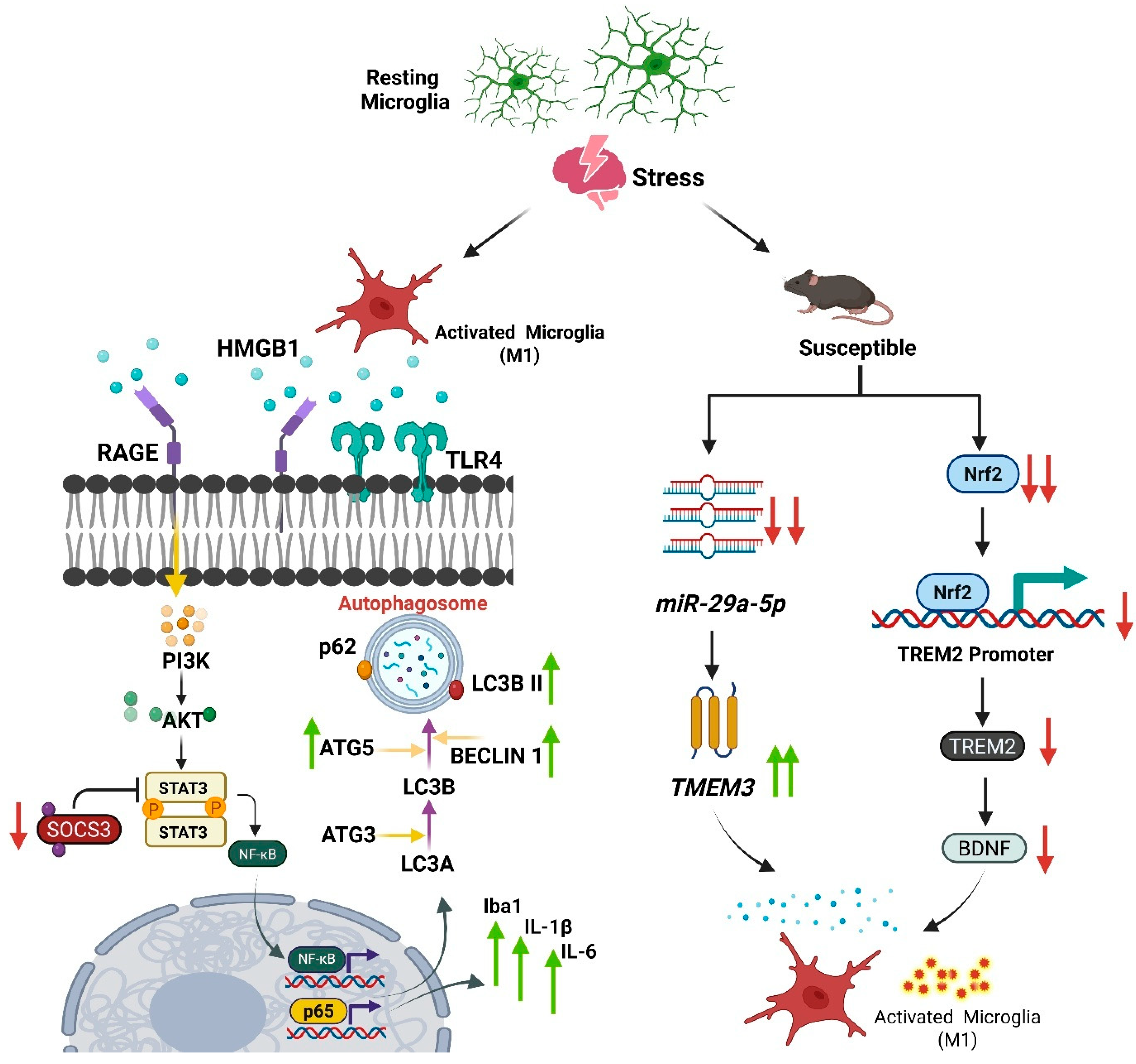
Accounting for approximately 10% of all brain cells, microglia are the resident immune cells of the CNS, acting as sentinels that constantly survey the brain microenvironment for signs of injury, infection, or homeostatic disturbance []. In response to such challenges, they undergo a rapid transformation from a resting, ramified state to an activated, amoeboid phenotype, capable of phagocytosis, antigen presentation, and the release of a vast array of signaling molecules, including cytokines, chemokines, and reactive oxygen species. While this response is essential for CNS protection, chronic and uncontrolled glial activation has been associated with aspects of MDD in some studies []. Growing evidence from both preclinical models and human studies demonstrates that chronic stress triggers a shift in microglial state, often towards a pro-inflammatory and neurotoxic phenotype that contributes to the synaptic and neuronal deficits underlying depressive behaviors [] (Figure 2).
Figure 2. Key transcriptional pathways in mPFC microglia that regulate stress susceptibility and resilience. Stress induces pro-inflammatory, pro-depressive signaling through two distinct pathways: (1) The elevation of HMGB1, which signals via RAGE/TLR4 to activate the STAT3/p65 axis, and (2) the downregulation of miR-29a-5p, which increases its target TMEM33 to promote M1 polarization. In contrast, the Nrf2-TREM2 axis promotes a resilient, anti-inflammatory phenotype. The activation of Nrf2 drives TREM2 transcription, leading to an anti-inflammatory state and antidepressant-like effects. Green upward arrows represent upregulation whereas red downward arrows represent down-regulation.
Definitive evidence establishing a causal role for these altered microglial states in driving depressive behaviors comes from depletion and repopulation experiments. Using a colony-stimulating factor 1 receptor (CSF1R) inhibitor (PLX5622) to selectively eliminate microglia, researchers have demonstrated that microglia are necessary for the development of stress-induced behavioral deficits []. One study showed that depleting microglia before repeated social defeat stress (RSDS) prevented the development of social withdrawal and cognitive impairment []. Early-life stress (ELS) induces enduring transcriptional reprogramming in hippocampal microglia that persists into adulthood and contributes to depressive vulnerability. While minimal changes are observed immediately after ELS (P9), adult mice (P200) exhibit 186 differentially expressed genes under basal conditions. Upregulated transcripts included inflammatory mediators such as Ripk and Gas6, Akirin1 and C1qbp, and genes linked to Akt signaling. Conversely, transcripts associated with cytoskeletal plasticity (Map, Stmn2, Stmn3) and gliogenesis (Cdh2, Metrn) were downregulated. Weighted gene co-expression network analysis identified an ELS-linked module enriched for protein polyubiquitination and proteasome-mediated degradation, indicating a lasting pro-inflammatory and catabolic microglial phenotype. Importantly, Gas6 upregulation was validated in post-mortem hippocampal tissue of depressed individuals with childhood abuse, where TMEM119+ microglia displayed elevated GAS6 [].
Stress engages the High Mobility Group Box 1 (HMGB1) pathway to drive transcriptional plasticity in microglia during depression. In a CSDS model, stress-susceptible mice showed elevated HMGB1 expression in mPFC microglia. This triggered signaling via the HMGB1/STAT3/p65 axis, with RAGE and TLR4 receptors promoting STAT3 and p65 phosphorylation, alongside reduced expression of SOCS3, a negative STAT3 regulator. Functionally, this reprogramming induced a pro-inflammatory microglial phenotype, reflected by hyper-ramified morphology, increased Iba1 expression, and upregulation of cytokines IL-1β and IL-6. In parallel, the pathway enhanced autophagy, marked by elevated Atg5, Beclin-1, and LC3B-II levels. Viral-mediated knockdown of HMGB1 in the mPFC abolished these molecular and cellular changes, rescuing depressive-like behaviors [] (see Figure 2).
In response to acute traumatic stress, such as electric foot-shocks, microglia exhibit a distinct pro-inflammatory transcriptional profile. Studies on mouse models of post-traumatic stress disorder (PTSD) have revealed that these cells increase in number and undergo morphological activation in the hippocampus. This is accompanied by a significant transcriptional upregulation of pro-inflammatory cytokines, including Interleukin-1β (IL-1β), IL-6, and Tumor Necrosis Factor-α (TNF-α). Mass cytometry has further shown increased expression of functional markers like iNOS and CD38. Pharmacological inhibition of this activation state alleviates the associated anxiety and fear behaviors []. Delving into the upstream molecular machinery, CSDS study revealed profound intracellular stress response centered on the interface between the endoplasmic reticulum (ER) and mitochondria. Stress signals, mediated by extracellular ATP, promote the formation of a tripartite tethering complex consisting of IP3R3-GRP75-VDAC1 at mitochondria-associated membranes (MAMs). This structural change facilitates excessive calcium (Ca2+) transfer from the ER into mitochondria. The resulting mitochondrial damage and reactive oxygen species (ROS) production serves as a direct trigger for the assembly and activation of the NLRP3 inflammasome, a key driver of microglial-mediated neuroinflammation in depression [].

4.1. Stress-Induced Changes in Microglial Density, Morphology, and State

Chronic stress does not simply cause a transient activation of microglia; it is a potent remodeling agent that induces durable phenotypic shifts, a process known as “priming”. Primed microglia is one that has been altered by a prior stimulus (stress) such that its response to a subsequent challenge is exaggerated or otherwise modified. A recent study using the chronic unpredictable mild stress (CUMS) paradigm in mice found a non-linear pattern of microglial activation in the hippocampal dentate gyrus. Microglial density decreased after two weeks, increased at four weeks, and declined again at six weeks, suggesting an initial adaptation, followed by reactive proliferation and eventual exhaustion under prolonged stress []. Similarly the RSDS model, which recapitulates aspects of psychosocial stress in humans, induces a rapid and sustained increase in microglial density and proliferation in the mPFC of mice that are susceptible to the stressor []. Morphologically, these microglia transition from their normal, highly ramified “surveying” state to a more amoeboid phenotype characterized by a larger cell body, shorter processes, and reduced process complexity [,]. In a rat model of CUMS, this activation was confirmed using multimodal approaches; in vivo positron emission tomography (PET) imaging with the translocator protein (TSPO) ligand [18F] DPA-714 showed significantly increased signal in the hippocampus of stressed rats, which was corroborated by post-mortem immunohistochemistry showing elevated protein levels of the microglial markers Iba1 and CD11b. Together, these studies establish that chronic stress is a powerful driver of microglial proliferation and morphological transformation in key mood-regulating circuits [].

4.2. Divergent Microglial Phenotypes in Stress Susceptibility vs. Resilience

An important development in depression research is the recognition that not all individuals exposed to stress develop the disorder, and this difference is reflected in microglial responses. The response of microglia to stress is not uniform; instead, these cells exhibit remarkable plasticity, adopting distinct phenotypes that can either confer vulnerability or promote resilience. Morphological analysis in a social defeat stress model reveals that this phenotypic divergence is structurally evident within the CA1 region of the hippocampus. In this model, mice resilient to stress are characterized by the predominance of a “hyper-ramified” microglial morphology, distinguished by longer and more complex processes. Functionally, these hyper-ramified microglia in resilient animals show significantly increased physical contact with both GABAergic and glutamatergic synaptic puncta compared to vulnerable counterparts. Conversely, vulnerable mice exhibit a shift toward a “de-ramified” phenotype with shorter, less complex processes and increased microglial density, indicating that distinct morphological states of microglia are tightly correlated with behavioral outcomes of stress [].
Beyond morphology, specific microRNA-mediated transcriptional regulation plays a crucial role in directing microglial polarization and determining stress outcomes. Research using a CUMS model in mice demonstrates that the expression of miR-29a-5p is significantly downregulated in the mPFC of susceptible animals. This downregulation leads to an increase in its target gene, transmembrane protein 33 (TMEM33). The resulting elevation in TMEM33 promotes the polarization of microglia toward a pro-inflammatory M1 state, characterized by increased levels of cytokines like IL-1β and TNF-α, while suppressing the anti-inflammatory M2 phenotype. Crucially, restoring miR-29a-5p levels in the mPFC rescues these effects; it suppresses TMEM33, facilitates M2 polarization, alleviates neuroinflammation, and ameliorates depressive-like behaviors, highlighting the miR-29a-5p/TMEM33 axis as a key transcriptional switch in microglial-mediated stress responses [] (Figure 2). Another critical transcriptional axis governing the microglial phenotype in depression involves the transcription factor NRF2 and its target TREM2. NRF2 directly regulates TREM2 transcription by binding to its promoter region. In response to stress, susceptible mice show reduced NRF2 expression and TREM2 transcription in the mPFC. The activation of NRF2 via the administration of sulforaphane (SFN) initiates TREM2 transcription, which in turn promotes an anti-inflammatory, arginase 1+ microglial phenotype. This NRF2-TREM2 pathway is associated with increased BDNF, and is essential for the antidepressant-like effects observed, implicating its importance in promoting a resilient microglial state [].
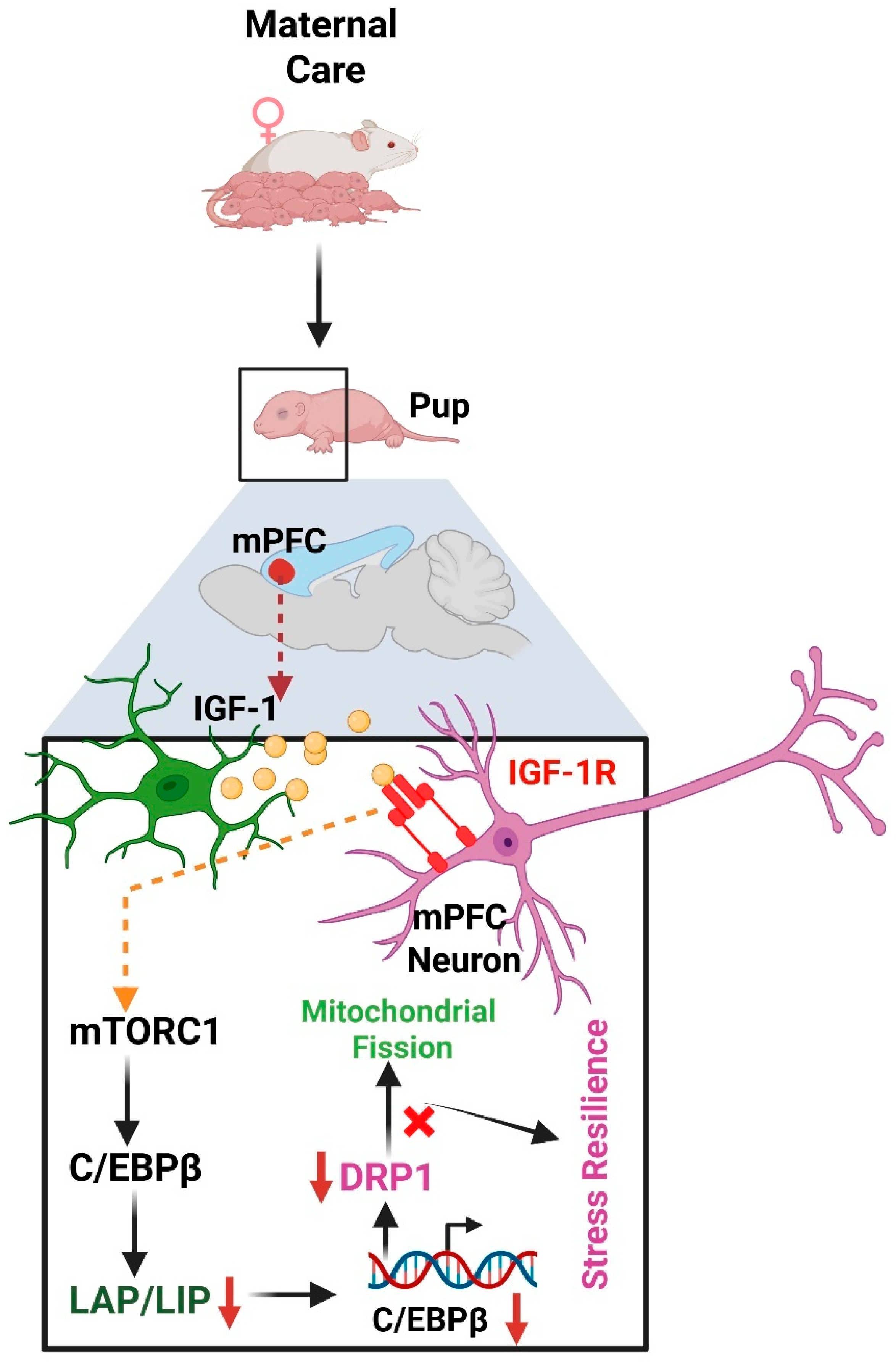
Microglial transcriptional plasticity not only alters their own inflammatory state but also directs the production of secreted factors that directly impact neuronal health and contribute to depressive phenotypes. For instance, in CUMS-susceptible mice, microglia in the prelimbic cortex (PrL) exhibit significantly increased transcription and expression of Vitamin D Binding Protein (VDBP). This microglia-derived VDBP acts on the neuronal receptor megalin, triggering the downstream SRC signaling pathway. The activation of this pathway precipitates neuronal apoptosis and synaptic damage, including reduced dendritic spine density and impaired synaptic transmission. Targeted overexpression of microglial VDBP is sufficient to induce depression-like behaviors and aggravate CUMS-induced synaptic pathology, identifying it as a key pathogenic mediator in microglia-neuron communication during stress []. Conversely to pathogenic signaling, microglial transcriptional activity can also be harnessed to promote resilience, particularly through pathways shaped by early-life experiences. Maternal care, for example, stimulates the release of insulin-like growth factor-1 (IGF-1) from microglia in the mPFC of pups. This microglial IGF-1 acts on neuronal IGF-1 receptors, activating mTORC1 signaling, which in turn alters the translational ratio of the transcription factor C/EBPβ toward its inhibitory isoform, LIP. This reduction in C/EBPβ transcriptional activity leads to the downregulation of its target gene Dnm1l (encoding DRP1), a key mediator of mitochondrial fission (Figure 3). By preventing stress-induced mitochondrial dysfunction, this microglia-neuron axis established by positive early-life experience confers lasting resilience to stress in adolescence [].
Figure 3. The microglia-to-neuron signaling axis mediating stress resilience. High maternal care stimulates microglia in the medial prefrontal cortex (mPFC) to release insulin-like growth factor-1 (IGF-1). This growth factor acts on neuronal receptors to activate mTORC1 signaling, which in turn increases the ratio of the inhibitory C/EBPβ-LIP isoform. The subsequent reduction in C/EBPβ transcriptional activity downregulates its target DRP1, preventing mitochondrial dysfunction and promoting resilience to stress.
Social factors, such as social hierarchy, can also powerfully modulate microglial transcriptional programs to influence susceptibility to stress-induced disorders like PTSD. In CSDS model, social rank was found to predict PTSD vulnerability, with dominant mice exhibiting greater susceptibility and more severe initial symptoms. At the molecular level, susceptible individuals show overactivation of microglia in the mPFC, which is associated with a specific transcriptional signature involving the activation of NF-κB, p65 and STAT1 pathways. This activation drives increased expression of the ubiquitin-like modifier activating enzyme 7 (Uba7) gene and subsequent production of the pro-inflammatory cytokine TNF-α, linking a defined social context to a distinct, pathogenic transcriptional state in microglia []. In a study using RSDS model, in the mPFC, stress-susceptible mice displayed a pro-inflammatory microglial profile, with elevated CD86 expression and upregulation of TNF-α, CXCL10, and IL-1β. By contrast, resilient mice, despite undergoing the same stress, showed a pro-resolution phenotype with increased CD206 and IL-10 expression [].

4.3. The Concept of Microglial “Priming” as an Epigenetic Memory

After the primary stressor is removed, microglia can return to a seemingly quiescent state, but they retain epigenetic marks on their histone tails that constitute a long-lasting memory of the event. Direct evidence for this epigenetic memory comes from studies of specific molecular modulators. In one primary study, priming microglia with an inflammatory trigger was shown to establish an “immune memory” that augmented the response to a second environmental stressor. The molecular underpinnings of this memory were identified as the enhanced deposition of active histone marks, specifically H3K27ac, H3K4me3, and H3K4me1 at gene regulatory regions []. Pharmacologically inhibiting the deposition of the key enhancer mark H3K27ac successfully prevented the formation of this immune memory and blocked the exaggerated secondary inflammatory response []. At a molecular level, priming involves the binding of transcription factors to enhancer regions of the genome, leading to the acquisition of these activating histone modifications []. DNA methylation provides another mechanism for this long-term cellular memory. A study investigating the lasting impact of ELS found that mice subjected to early social isolation displayed a drastic decrease in global DNA methylation levels specifically within their microglial cells. This stable alteration to the microglial methylome is one of the key mechanisms through which microglia “hold memories” of the early adverse experience, programming them to be more reactive to challenges later in life [].
A study examining a model of post-stroke anxiety found that the epigenetic regulator HDAC3 was specifically upregulated in microglia, not astrocytes. In this model, which combined photothrombotic stroke with restraint stress, it was microglial HDAC3 that drove anxiety-like behavior. The molecular mechanism involved HDAC3 deacetylating the p65 subunit of NF-κB. This post-translational modification altered the transcription of genes involved in prostaglandin synthesis, such as COX1, leading to an overproduction of prostaglandin E2 (PGE2) in the damaged cortex. This PGE2 then diffused to the amygdala, where it acted on neuronal EP2 receptors to elicit anxiety susceptibility, highlighting a distinct, microglia-specific epigenetic response to this particular combined stressor [].

4.4. DNA Methylation as a Molecular Imprint of Stress in Microglia

Among epigenetic mechanisms, DNA methylation- the covalent addition of a methyl group to cytosine residues, most commonly within CpG dinucleotides represents a particularly stable modification that can induce persistent alterations in gene expression potential. Owing to its stability, DNA methylation is considered a key mechanism through which early-life or chronic stress may imprint enduring molecular signatures within the microglial genome, thereby establishing a long-lasting predisposition to depressive pathology []. Progress in understanding the human microglial epigenome has been hampered by the difficulty of isolating these cells from the complex milieu of the brain. Genome-scale methylation analysis on pure microglia acutely isolated from post-mortem human brain tissue from 22 donors, including individuals with mood disorders and non-psychiatric controls revealed that microglia possess a DNA methylation profile that is distinct from other CNS cells. A key finding was that interindividual variation, rather than brain region, was the dominant source of methylation differences, suggesting that genetic background and life experiences including stress, profoundly shape the microglial epigenome [].

4.5. Microglial Regulation via Histone Modifications and Non-Coding RNAs in Stress

A recent study uncovered a novel and highly specific mechanism of communication between microglia and neurons in depression that is mediated by miRNAs. Using the CUMS rat model, tit was shown that stressed microglia significantly increase their secretion of extracellular vesicles known as exosomes. Critically, these exosomes were found to be enriched with a specific microRNA, miR-146a-5p []. These exosomes are taken up by neurons in the hippocampal dentate gyrus, a key site for adult neurogenesis that is implicated in depression. Once inside the neurons, miR-146a-5p exerts its regulatory function by directly suppressing the mRNA of Krüppel-like factor 4 (KLF4), a transcription factor essential for the differentiation and survival of new neurons.
A peripheral immune challenge with Bacille Calmette-Guerin (BCG) has been shown to induce persistent depression-like behaviors, providing a model to investigate lasting molecular changes in microglia. Proteomic profiling of isolated microglia after symptom onset revealed broad alterations in protein abundance and post-translational modifications, including histone acetylation. This was accompanied by reduced abundance of histone clusters 1-3 and increased H2A variants, suggesting disrupted chromatin architecture [].

4.5.1. The H3K27me3 Axis: EZH2 vs. Jmjd3

The trimethylation of histone H3 at lysine 27 (H3K27me3) is a canonical repressive mark that silences gene expression. The dynamic regulation of this mark is a central for the control of microglial activation. The “writers” of this mark are the histone methyltransferases of the Polycomb Repressive Complex 2 (PRC2), whose catalytic subunit is EZH2. Under homeostatic conditions, EZH2 deposits H3K27me3 at the promoters of inflammatory genes, keeping them silenced. However, in a mouse model of adolescent depression, the expression of Polycomb group factor 1 (PCGF1), a component of the related PRC1 complex that helps recruit PRC2 to its targets, was found to be significantly decreased in microglia. This reduction in PCGF1 function led to diminished deposition of both H2AK119ub (a mark placed by PRC1) and H3K27me3 at the promoter of the pro-inflammatory gene Matrix Metalloproteinase 10 (Mmp10). The resulting transcriptional de-repression of Mmp10 unleashed a neuroinflammatory cascade that drove neuronal damage and depressive-like behaviors [].
In direct opposition to EZH2 are the “erasers” of this mark, the H3K27 demethylases. The most prominent of these in microglia is JMJD3, also known as KDM6B []. Jmjd3 expression is induced by inflammatory stimuli, such as lipopolysaccharide (LPS) or chronic stress, via activation of the NF-κB signaling pathway. Once expressed, Jmjd3 is recruited to the promoters of pro-inflammatory cytokines, where it actively removes the repressive H3K27me3 mark. This epigenetic switch from a repressed to an active chromatin state allows for robust transcription of genes like Il-6 and Tnf-α, driving the inflammatory response [].

4.5.2. The H3K9me2 Axis: The G9a/GLP Complex

Another critical repressive mark is the dimethylation of histone H3 at lysine 9 (H3K9me2), deposited by a complex of the methyltransferases G9a and GLP. Inhibition of this complex in adult mice produces anxiolytic effects, suggesting that reducing H3K9me2-mediated repression is beneficial. However, the same inhibition during embryonic development increases anxiety-like behaviors in adult offspring, highlighting a critical developmental window for this epigenetic mechanism [].

4.6. The Heterogeneous Microglial Transcriptome in Depression

While preclinical models often point towards a pro-inflammatory microglial state, research in human post-mortem tissue has presented a more complex perspective. A key study performing RNA-sequencing on pure microglia isolated from the occipital cortex grey matter of MDD donors found a predominantly immune-suppressed profile. Strikingly, transcriptional alterations were restricted to GM microglia, defining a phenotype termed Depressive Disease-Associated Microglia (DepDAM), characterized by immune suppression rather than classical activation. A total of 81 genes were significantly downregulated, including key players in immune activation and phagocytosis such as the scavenger receptor. CD163, the opsonin SPP1 (osteopontin), and critical components of the classical complement pathway (C1QA, C1QB, C1QC). This signature suggests a state of functional inhibition. Interestingly, the upregulation of neuronal regulatory genes CD200 and CD47 was also observed in the microglia of MDD patients, suggesting this immunosuppressive phenotype may be actively maintained by signals from neurons []. However, few studies point to different molecular signatures, highlighting the heterogeneity of the microglial response. For instance, by combining spatial and single-cell transcriptomics, one study identified a specific microglial subgroup, designated Mic03, that was strongly correlated with depression. This subgroup was characterized by elevated expression of genes involved in controlling microglial activation and neuroinflammation, including IQGAP2, FYN, PDE7A, and ARHGEF3. These divergent findings may reflect differences in brain region, disease chronicity, or patient-specific factors [].
In models of prolonged, chronic mild stress, microglia can adopt an entirely different state characterized by widespread transcriptional repression. This response involves the significant downregulation of numerous interferon-regulated genes (IRGs), which correlates with decreased chromatin accessibility at Interferon-Stimulated Response Element (ISRE) motifs. This immunosuppressive phenotype appears to be actively mediated, driven not by a change in interferon levels but by the upregulation of the transcriptional repressor Activating Transcription Factor 3 (ATF3), demonstrating the remarkable and context-dependent plasticity of the microglial transcriptome in stress-related disorders [].

4.7. Therapeutic Implications of Microglial Epigenetic Regulation in Stress

Collectively, findings from diverse preclinical models and human studies strongly indicate that the microglial epigenome is a druggable target for novel antidepressant development. The studies summarized in Table 2 reveal that pharmacological interventions aimed at specific epigenetic regulators can successfully reverse stress-induced microglial activation and ameliorate depressive-like behaviors. A particularly robust strategy involves targeting the H3K27me3 demethylase JMJD3, as both the selective inhibitor GSK-J4 and the natural compound Ginsenoside Rd have been shown to be effective in distinct stress paradigms by restoring this repressive histone mark at inflammatory gene loci [,]. Furthermore, the field is advancing toward greater therapeutic precision, exemplified by the efficacy of a selective HDAC11 inhibitor, which suppresses microglial activation in an LPS-induced depression model []. The natural compound Nerolidol has been shown to exert antidepressant effects by downregulating DNA methyltransferase 1 (DNMT1) []. Finally, emerging evidence points to the therapeutic potential of modulating non-coding RNA networks (ncRNA), such as targeting the circular RNA circ-UBE2K, to rescue depressive phenotypes []. These studies establish that precision epigenetic modulators, designed to reprogram rather than simply suppress microglial function, represent a promising future class of therapeutics for stress-related disorders.
Table 2. Epigenetic Mechanisms and Therapeutic Targeting of Microglia in Stress-Related Depression.
Stress Epigenetic MechanismTarget Genes/PathwaysObserved Microglial ResponseBehavioral/Functional OutcomeTherapeutic Implications/InterventionsStudy Reference
Chronic Unpredictable Mild Stress (CUMS) in adolescent ratsHistone Demethylation (via KDM6B/JMJD3)Increased Jmjd3 expression, decreased H3K27me3 levels. Upregulation of pro-inflammatory cytokines (IL-1β, TNF-α).Microglial activation in the prefrontal cortex (PFC) and hippocampus (HIP).Depressive-like behaviors (anhedonia, despair). Increased susceptibility to stress in adulthood.Minocycline (microglial inhibitor) reversed microglial activation, normalized Jmjd3/H3K27me3 levels, and alleviated depressive-like behaviors.[]
Chronic Unpredictable Mild Stress (CUMS) in C57BL/6 and obese (ob/ob) miceHistone Demethylation (via JMJD3)Increased JMJD3 and NF-κB expression; decreased H3K27me3 levels. Increased pro-inflammatory cytokines. Downregulation of adiponectin (APN).Microglial activation in PFC and HIP; excessive activation in ob/ob mice.Depressive-like behaviors and memory impairment. Obese mice showed worse behavioral outcomes, suggesting increased susceptibility.GSK-J4 (selective JMJD3 inhibitor) relieved depressive-like behaviors, memory impairment, microglial activation, and normalized epigenetic/inflammatory markers.[]
Chronic Unpredictable Mild Stress (CUMS) in miceDNA Methylation (via DNMT1)Downregulation of DNA methyltransferase 1 (DNMT1).Suppression of CUMS-induced microglial activation and inflammatory response.Reduction in depressive-like behavior (assessed by OFT, SPT, TST).Nerolidol (a natural sesquiterpene) reduced DNMT1 levels and suppressed microglial activation.[]
Lipopolysaccharide (LPS)-induced depression model in miceHistone Deacetylation (via HDAC11)Inhibition of HDAC11 deacetylase function.Suppression of LPS-induced microglial activation. Initiation of autophagy and inhibition of nitric oxide (NO) production.Alleviation of depression-like behavior (reduced immobility, increased sucrose preference).Novel selective HDAC11 inhibitor (Compound 5) alleviated depressive-like behavior by inhibiting microglial activation.[]
Human Post-Mortem Brain Tissue (Dorsolateral Prefrontal Cortex) from individuals with Major Depressive Disorder (MDD)Chromatin Remodeling (Single-nucleus Assay for Transposase-Accessible Chromatin sequencing—snATAC-seq)Decreased accessibility at binding sites for TFs known to regulate immune homeostasis. Disruption of TF binding sites linked to genes affecting synaptic communication. Key TF mentioned: NR4A2 (activity-dependent, stress-reactive).A specific gray matter microglia cluster (termed Mic2) exhibited significantly decreased chromatin accessibility at key regulatory regions.The study directly links these cell type specific epigenetic alterations to the clinical diagnosis of MDD in humans, providing strong evidence for real, measurable brain changes underlying the disorder.Identification of a specific microglial subtype and regulatory mechanism opens pathways for developing highly targeted therapies[]
Gut microbiota-dysbiosis mouse model (germ-free mice colonized with microbiota from MDD patients vs. healthy controls).Non-coding RNAs (lncRNA-miRNA-mRNA networks)ceRNA networks involving lncRNAs (4930417H01Rik), miRNAs (mmu-miR-883b-3p), and mRNAs (Adcy1, Nr4a2). Axonal guidance and synaptogenesis pathways. Dysregulation of microglial function via altered inflammatory signaling (IFNG pathway), contributing to decreased neuronal activity (Fos downregulation)Dysregulated inflammatory response and neurodevelopment in the hippocampusIdentifies specific ncRNA networks in the gut–brain–epigenome axis as potential biomarkers and therapeutic targets for microbiota-based antidepressant strategies.[]
Chronic unpredictable mild stress (CUMS)circRNA-mediated regulationUBE2K (ubiquitin conjugating enzyme) downstream enrichment of TNF, IL-17, cytokine signaling.Microglia show morphological activation (shorter branches, increased soma size, elevated Iba1 intensity), increased iNOS/CD68 and pro-inflammatory cytokinesWorsened depression-like behaviours; increased neuroinflammation and neuronal/synaptic damage.Targeting circ-UBE2K (knockdown) or disrupting circ-UBE2K/HNRNPU axis reduced microglial activation and rescued depressive-like phenotypes suggests circRNA or HNRNPU as therapeutic targets/biomarkers[]
Lipopolysaccharide (LPS) systemic inflammation model in mice.Histone demethylation: activation of JMJD3 (H3K27me3 demethylase).TLR4, PI3K, AKT, NF-κB signaling; JMJD3-regulated pro-inflammatory gene transcription.Microglial overactivation with elevated IL-1β, IL-6, TNF-α; reduced by Rd treatment.LPS-induced depressive-like behavior with decreased sucrose preference and increased immobility; Rd rescues behavior and synaptic protein expression (PSD-95, SYP).Ginsenoside Rd reduces JMJD3 expression and suppresses TLR4-PI3K-AKT-NF-κB signaling, leading to decreased microglial activation and behavioral improvement.[]
Postmortem brain tissue (4 regions) from donors with mood disorders (n = 13) vs. control (n = 8) donors.DNA Methylation (Genome-wide methylation array)Differentially Methylated Regions (DMRs) associated with mood disorder status, linked to genes involved in myeloid cell function and neuropsychiatric disorders.Altered DNA methylation profiles in isolated microglia. Interindividual factors and age showed a larger effect on the methylome than brain regions.Findings suggest the microglial methylome is highly responsive to individual-specific factors, contributing to cellular heterogeneity.Identifies mood disorder-associated DMRs in microglia that could serve as biomarkers or targets for epigenetic drugs.[]

5. The Oligodendrocytes: Myelin Plasticity and Vulnerability in Depression

Oligodendrocytes are the myelinating cells of the CNS, responsible for ensheathing axons with lipid-rich myelin, which is essential for rapid saltatory nerve conduction and provides vital metabolic support to axons. For a long time, myelin was considered a static structural element, but this view is being rapidly overturned. There is now a wealth of evidence indicating that myelination is a dynamic and plastic process that continues throughout life and is crucial for learning, memory, and cognitive function [,]. Correspondingly, disruptions in oligodendrocyte function and myelin integrity have been increasingly identified as a core pathological feature of MDD. The lifelong process of generating new oligodendrocytes from a resident pool of oligodendrocyte precursor cells (OPCs), known as oligodendrogenesis, is critical for brain plasticity, learning, and repair. Chronic stress appears to directly affect this fundamental process. A study utilizing the RSDS mouse model, demonstrated that chronic psychosocial stress inflicts profound damage on the OPC population in the mPFC [].
The timing of stress exposure is also a critical variable. Adolescence is a period of dynamic white matter development and circuit refinement, rendering the brain particularly vulnerable to environmental insults. A recent study on adolescent mice uncovered a significant, region-specific impairment of oligodendrogenesis. In the mPFC and the lateral habenula (LHb), two brain regions heavily implicated in mood regulation and depression, stress caused a marked decrease in the number of proliferative cells and, consequently, a reduction in newly formed OPCs and mature oligodendrocytes. Notably, this effect was not observed in the amygdala, highlighting a circuit-specific vulnerability [].

5.1. Transcriptional Control of Myelination and Its Disruption by Stress

Numerous studies using animal models of chronic stress and depression have documented significant myelin deficits, demyelination, and reductions in the number and function of oligodendrocytes and their precursor cells (OPCs) in mood-related brain circuits. This pathology is mirrored in humans. Post-mortem studies of individuals with MDD have revealed altered expression of a suite of key myelin and oligodendrocyte related genes, including Proteolipid Protein 1 (PLP1), 2′,3′-Cyclic-Nucleotide 3′-Phosphodiesterase (CNP), Myelin Oligodendrocyte Glycoprotein (MOG), and the transcription factor OLIG1 []. Furthermore, a recent study using cell-type-specific nuclear isolation from the post-mortem basolateral amygdala found that expression of Myelin-Associated Oligodendrocyte Basic Protein (MOBP) was significantly decreased specifically in the oligodendrocytes of depressed individuals who had a history of childhood abuse, directly linking early life trauma to specific molecular deficits in this cell type []. A summary of key studies describing the modulation of oligodendrocytes in stress-induced depression is presented in Table 3.
These myelin deficits are not simply a passive consequence of neuronal damage but represent an active, pathological process driven by the epigenetic suppression of the myelinogenic program within oligodendrocytes. This concept of “epigenetic silencing” is well-established in neurological diseases, where mature oligodendrocytes present in lesions fail to produce myelin []. A clear molecular mechanism for this process was recently elucidated in a model of Parkinson’s disease, another disorder with myelin pathology []. In this model, the DNA methyltransferase (DNMT3A) was upregulated, leading to hypermethylation and silencing of the promoter for the transcription factor STAT5B. This reframes myelin integrity as a form of epigenetically regulated structural plasticity that is highly vulnerable to stress. Antidepressant treatments appear to counteract this pathological silencing. For instance, the immunomodulatory drug teriflunomide protects oligodendrocytes from stress-induced apoptosis and damage [], while the SSRI paroxetine promotes the proliferation and differentiation of OPCs, restoring myelin integrity in a corticosterone-induced depression model []. Furthermore, perinatal exposure to fluoxetine has been shown to directly alter the expression of myelin genes like Mag and Mbp through changes in DNA methylation, underscoring the sensitivity of the oligodendrocyte epigenome to pharmacological intervention during critical developmental windows [].
In models such CUMS and RSDS, animals exhibit depressive-like behaviors that are accompanied by significant pathology in the PFC, MYRF, a key region for mood regulation. Single-nucleus RNA sequencing of the PFC in these models reveals that chronic stress induces profound structural and transcriptional alterations in oligodendrocytes, with particularly strong effects observed in male mice, leading to disrupted neuro-glial interactions []. Pathway analyses of the widespread transcriptomic changes in the PFC of chronically stressed mice consistently point to a core set of upstream regulators that directly link the stress response to the fundamental machinery of myelination and its epigenetic control. Among the top predicted regulators are Myelin Regulatory Factor (Myrf), a master transcription factor essential for oligodendrocyte differentiation, and Methyl-CpG binding protein 2 (Mecp2), a key epigenetic “reader” protein that translates DNA methylation patterns into changes in gene expression [].
Stress induces a profound dysregulation of the oligodendrocyte transcriptome, shifting the cell’s genetic program away from myelination and maintenance toward a state of dysfunction. A comprehensive 2018 study using a chronic social stress (CSS) model in mice found a general downregulation of oligodendrocyte gene expression, including genes essential for myelin formation and axonal support. By using mice genetically engineered to be heterozygous for the oligodendrocyte gene Cnp1 (encoding 2′,3′-cyclic-nucleotide 3′-phosphodiesterase), thereby having constitutively reduced expression of a key myelin-related protein, the researchers demonstrated that these mice were more vulnerable to the behavioral effects of stress. This provides translational evidence that a reduction in oligodendrocyte gene expression is not merely a correlate of stress but is a significant contributor to the development of stress-induced aversion and anxiety phenotypes []. The robustness of this finding has been solidified by a large-scale congruent analysis performed in 2023. This work moved beyond a single stress paradigm by integrating and re-analyzing publicly available transcriptomic datasets from multiple, distinct mouse models of adversity, including social defeat, chronic unpredictable stress, early life stress, and adolescent social isolation. The analysis revealed a conserved transcriptional signature of oligodendrocyte dysregulation across all these models. Critically, the study extended this finding to humans, identifying a similar oligodendrocyte transcriptome change in post-mortem prefrontal cortex tissue from both male and female patients with MDD, establishing a clear translational bridge between preclinical models and the human condition [].

5.2. Early-Life Stress Impairs Oligodendrocyte Developmental Trajectories

The impact of ELS is particularly detrimental because it occurs during a critical period of oligodendrogenesis and myelination, creating a developmental vulnerability that can last a lifetime. Studies using the maternal separation model in rodents, a well-established paradigm for ELS, have uncovered a paradoxical mechanism of damage []. Unbiased mRNA profiling in the mPFC of maternally separated pups revealed that ELS induces a precocious differentiation of oligodendrocytes. While this leads to a transient increase in the expression of myelin-related genes during the postnatal period, it comes at a steep long-term cost: the premature exhaustion and subsequent depletion of the renewable OPC pool in the adult brain []. This ELS-induced disruption does not occur in isolation. The initial insult to the OPC population radiates outward, affecting the development and function of the entire glial network. A 2021 study by Wang and colleagues demonstrated that the ELS-induced reduction in the OPC population secondarily hinders the development of astrocytes in the hippocampus. This occurs because OPCs provide essential paracrine signals, specifically Wnt ligands, that are required for the proper formation of astrocytic networks. The loss of these signals due to OPC depletion leads to a secondary astrocytopathy, compounding the damage to the brain’s supportive cellular architecture []. The integrity of white matter tracts, which is critical for the rapid communication and synchronization of distributed neural networks requires healthy oligodendrocytes. Clinical studies have shown that prenatal maternal depression, a human correlate of ELS, is associated with significant alterations in the white matter microstructure of infants, a link that appears to be mediated by epigenetic modifications detectable at birth in umbilical cord blood [].

5.3. Linking Stress to Oligodendrocyte Transcriptional Dysfunction

The balance between histone acetylation and deacetylation is fundamental to oligodendrocyte lineage progression. This balance is maintained by the opposing actions of HATs and HDACs. A substantial body of work on affective disorders have established that HDAC activity is essential for OPCs to differentiate into mature, myelinating oligodendrocytes []. HDAC11, the sole member of the Class IV HDAC family, plays a complex and essential role. Its expression increases as oligodendrocytes mature, and it is required for the proper expression of the major myelin genes MBP and PLP. Disrupting HDAC11 function impairs both gene expression and the morphological development of oligodendrocytes []. Exposure to stress, particularly early-life adversity, induces widespread and persistent changes in DNA methylation patterns across the genome []. Methyl-CpG binding protein 2 (MeCP2) is the canonical reader of the DNA methylome in the brain, and its function is indispensable for normal oligodendrocyte development and myelination. MeCP2 deficiency in oligodendrocytes leads to profound dysregulation of myelin gene expression and impaired myelin formation. Intriguingly, studies in cultured rat oligodendrocytes have revealed that MeCP2 normally functions as a negative regulator, or a brake, on the expression of key myelin genes, including myelin basic protein (MBP), proteolipid protein (PLP), and myelin oligodendrocyte glycoprotein (MOG), as well as the neurotrophin (BDNF) [].
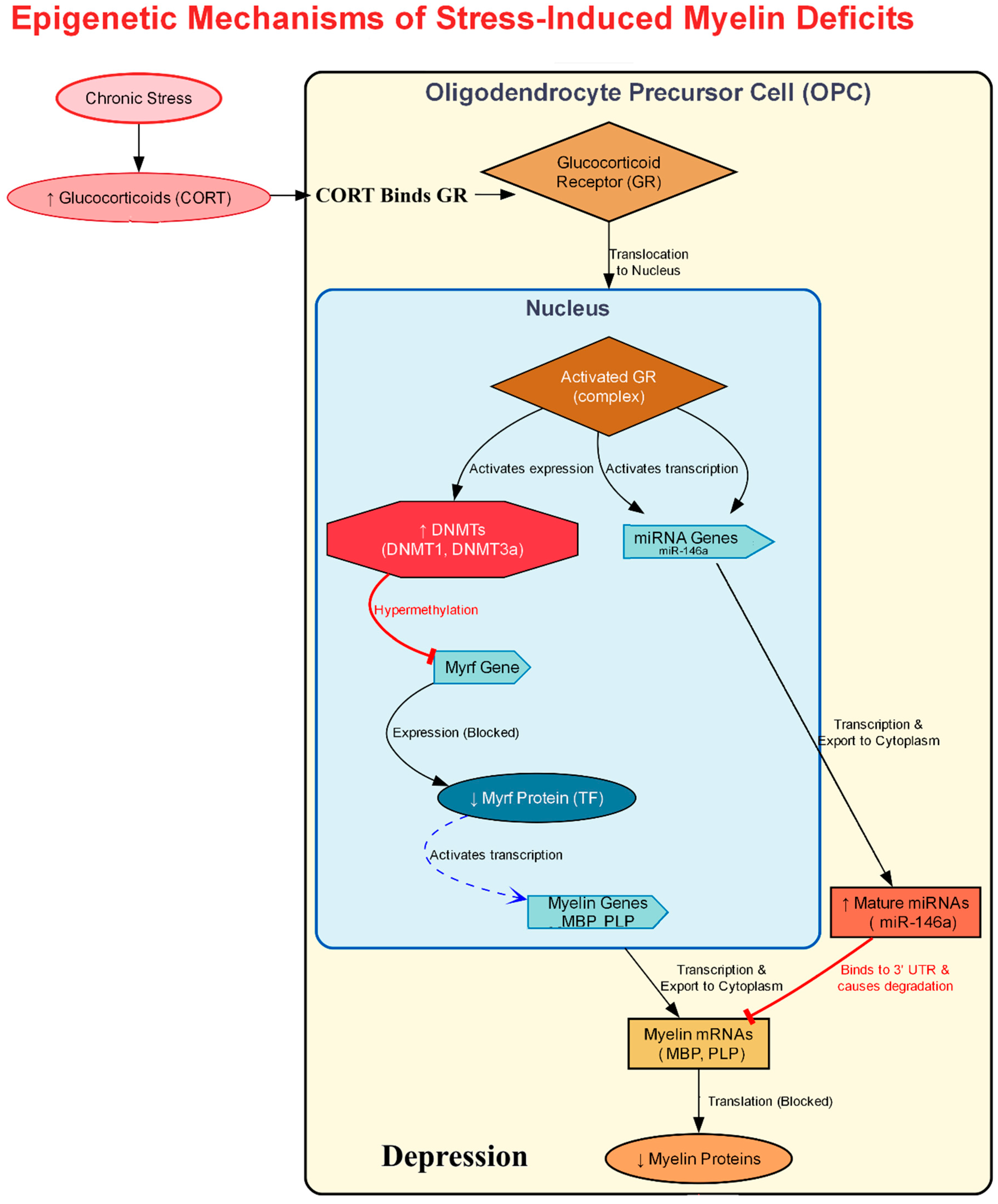
The diverse epigenetic insults triggered by stress likely converge on a select few master regulatory genes that orchestrate the complex process of oligodendrocyte differentiation. MYRF is one such master transcription factor; its timely activation is an absolute requirement for OPCs to differentiate and begin producing myelin []. The expression of Myrf is itself under tight control by upstream factors and is associated with specific promoter and enhancer elements []. The finding that Myrf is a top predicted upstream regulator of the entire transcriptomic response to chronic stress in the PFC strongly suggests that its regulatory regions are a key target of stress-induced epigenetic changes []. A plausible mechanism is that chronic stress, through the activation of DNMTs induces repressive hypermethylation at a critical enhancer element of the Myrf gene (Figure 4).
Figure 4. Epigenetic Mechanisms of Stress-Induced Myelin Deficits in Oligodendrocyte Precursor Cells (OPCs). Chronic stress contributes to myelin deficits and depression by initiating two parallel epigenetic pathways in oligodendrocyte precursor cells (OPCs) through increased glucocorticoids (CORT). When CORT binds to its receptor (GR), it triggers both DNA methylation and microRNA regulation. In the first pathway, the activated GR increases the expression of DNA methyltransferases (DNMTs), which hypermethylate and silence the Myelin Regulatory Factor (Myrf) gene. Since Myrf is a master regulator of differentiation, this silencing blocks the transcription of key myelin genes like MBP and PLP. Simultaneously, in the second pathway, the activated GR complex promotes the transcription of microRNAs (miRNAs) like miR-146a. These miRNAs bind to the MBP and PLP mRNAs in the cytoplasm, targeting them for degradation and blocking their translation into protein. Both of these epigenetic mechanisms; transcriptional repression via Myrf hypermethylation and post-transcriptional degradation via miRNAs converge to cause a significant decrease in myelin proteins, leading to the myelin deficits observed in depressive disorders.

5.4. MicroRNAs and Long Non-Coding RNAs in Oligodendrocyte Function

MicroRNAs are small (~22 nucleotide) RNA molecules that typically function by binding to messenger RNA (mRNA) targets, leading to their degradation or translational repression. A single miRNA can regulate hundreds of different mRNA targets, allowing it to act as a master regulator of entire cellular pathways. Specific miRNAs are known to be essential for normal oligodendrocyte development. For example, miR-219 is a critical factor for promoting the transition of OPCs into mature, myelinating oligodendrocytes; its absence can halt this developmental progression []. Recent research has directly implicated miRNA dysregulation in the pathophysiology of MDD. A 2024 study analyzing postmortem brain tissue from individuals with MDD identified a significant decrease in the expression of two specific miRNAs, miR-92a-3p and miR-129-5p []. This decrease was found specifically within extracellular vesicles (EVs), which are small membrane-bound particles released by cells for intercellular communication. Oligodendrocytes are a major source of brain-derived EVs, suggesting they may be the origin of this pathological signal.
Using a mouse model of CUMS, high-throughput sequencing of both miRNA and mRNA from the mPFC revealed a clear inverse relationship: the expression of mRNAs encoding proteins crucial for myelination, GABAergic and dopaminergic synapses, and neuronal growth was significantly downregulated in the stressed mice. Concurrently, the expression of a set of miRNAs predicted to target these very mRNAs was significantly upregulated. This finding demonstrates that stress activates a miRNA-driven program that actively dismantles the transcriptional machinery required for maintaining white matter integrity []. Complementing this work, a 2025 study analyzed human brain tissue from patients with a range of psychiatric and neurodegenerative disorders and identified 49 unique miRNAs whose expression levels were associated with conditions including MDD and PTSD, suggesting that specific miRNA candidates may represent points of convergent pathology, contributing to shared disease mechanisms across different stress-related disorders and providing potential targets for future therapeutic intervention [].
Long non-coding RNAs are a large and diverse class of RNA molecules greater than 200 nucleotides in length that do not code for proteins. Recent studies have identified lncRNAs that are specifically and critically involved in oligodendrocyte biology. One such example is MYRACL (MYelination RegulAting oligodendroCyte associated LncRNA), which is highly enriched in mature oligodendrocytes relative to their progenitor cells. Experimental manipulation in human cell culture models demonstrated that knocking down MYRACL expression disrupts oligodendrocyte maturation, whereas overexpressing it promotes differentiation and enhances myelin formation in vitro []. This identifies MYRACL as a potent pro-myelinating lncRNA whose expression could be a key target for disruption by chronic stress. Indeed, transcriptomic analyses from both human MDD brains and animal stress models reveal widespread dysregulation of the lncRNA landscape. A single-nucleus transcriptomic study of postmortem MDD brain tissue identified the lncRNA MALAT1 as one of the top-ranked genes whose expression within the oligodendrocyte lineage could distinguish MDD cases from healthy controls []. In parallel, studies in mice have shown that another lncRNA, NEAT1, is highly expressed in glial cells and is essential for regulating the adaptive behavioral response to stress. Loss of NEAT1 function results in neuronal hyperexcitability and panic-like behaviors, demonstrating its crucial role in maintaining emotional homeostasis []. The complex patterns of ncRNA dysregulation suggest they play two key roles in the oligodendrocyte response to stress. First, they act as “signal integrators.” A single environmental stressor triggers a coordinated change in the expression of hundreds of genes related to cell survival, metabolism, and differentiation.
Table 3. Summary of studies on Modulation of Oligodendrocytes in Stress-Induced Depression.
Stress Paradigm/ModelKey Genes/Pathways AffectedObserved Oligodendrocyte ResponseBehavioral/Functional OutcomeStudy Reference
Repeated Social Defeat Stress (RSDS) Mouse ModelOligodendrocyte Precursor Cells (OPCs)Profound damage and reduction in the OPC population in the medial prefrontal cortex (mPFC).Disruption of oligodendrogenesis, which is critical for brain plasticity, learning, and repair.[]
Major Depressive Disorder (MDD) (Post-mortem human prefrontal cortex)Altered expression of long non-coding RNA MALAT1Greatest transcriptional dysregulation found in immature OPCs; MALAT1 expression in early OPCs (OPC2 stage) has high predictive power for distinguishing MDD cases.Associated with the clinical diagnosis of MDD.[]
Human Post-Mortem MDD TissuePLP1, CNP, MOG, OLIG1, MOBPDecreased expression of key genes related to myelin and oligodendrocyte function. MOBP decrease linked to childhood abuse.Myelin deficits and impaired integrity of white matter tracts are considered core pathological features of MDD.[,]
Early-Life Stress (ELS)/Maternal Separation ModelWnt signaling pathwayAn initial, premature differentiation of oligodendrocytes leads to the long-term depletion and exhaustion of the renewable OPC pool in the adult brain.Impairs secondary development of astrocytes and leads to lasting white matter vulnerability.[,]
Chronic Unpredictable Mild Stress (CUMS)/RSDS ModelsMyelin Regulatory Factor (Myrf), Methyl-CpG binding protein 2 (Mecp2)Widespread transcriptomic changes, with Myrf and Mecp2 identified as top upstream regulators linking the stress response to myelination machinery.Shifts the genetic program away from myelination, contributing to stress-induced aversion and anxiety.[]
Chronic Unpredictable Mild Stress (CUMS) Mouse ModelUpregulation of miRNAs that target myelination-related mRNAsA significant downregulation of mRNAs for myelination, GABAergic/dopaminergic synapses, and neuronal growth, inversely correlated with an upregulation of their targeting miRNAs.Active dismantling of the transcriptional machinery required to maintain white matter integrity in the mPFC.[]

6. Limitations

While the synthesis of preclinical and human data presented in this review points toward a powerful role for glial transcriptional plasticity in depression, a critical perspective is required. There are often notable discrepancies between findings from controlled animal stress paradigms and observational studies of human post-mortem brain tissue. Human post-mortem brain tissue is an invaluable resource, providing the only direct window into the molecular pathology of MDD. However, data derived from this tissue must be interpreted with significant caution due to a host of uncontrollable confounding variables and technical challenges. Analysis provides only static, end-of-life information of a dynamic disease []. Gene expression is heavily influenced by confounding variables, including the agonal state, cause of death (suicide), post-mortem interval (PMI), and tissue quality metrics like pH and RNA Integrity Number (RIN). Furthermore, it is difficult to separate disease-specific signals from the cumulative effects of a lifetime of medication use, comorbidities, and substance use, all of which can alter the epigenome. Significant discrepancies also arise from fundamental species differences in glial biology. Human astrocytes are not merely larger but are profoundly more complex than their rodent counterparts. They are up to 2.6 times larger in diameter, with 10-fold more primary processes, allowing a single human astrocyte to contact up to 2 million synapses, far exceeding the capacity of mouse astrocytes []. The human brain also contains unique astrocyte subtypes, like interlaminar astrocytes, which are absent in rodents and are involved in higher cognitive circuits []. These structural differences are matched by functional divergence.
The path to clinical translation for these epigenetic modulators faces significant challenges that must be critically assessed. The foremost hurdle is specificity. Most epigenetic enzymes, such as HDACs or DNMTs, are ubiquitously expressed and regulate thousands of genes across all cell types. Systemic administration of a broad-spectrum inhibitor (say an HDAC inhibitor) could lead to severe, unpredictable off-target effects in peripheral organs and, within the brain, could disrupt essential homeostatic functions in healthy neurons and glia. The second major challenge is delivery. The BBB remains a major hinderance for most small-molecule drugs and virtually all biologic-based therapies like non-coding RNAs. Finally, the epigenetic landscape is highly dynamic; achieving a durable therapeutic effect without causing permanent, deleterious changes to the ‘epigenome’ is a delicate balance. Therefore, future strategies must move beyond broad-spectrum inhibitors and focus on developing novel systems for cell-type-specific delivery or designing drugs that target the specific protein–protein interactions of epigenetic complexes, rather than just the active catalytic site.

7. Conclusions

The paradigm for understanding major depression is shifting from a purely neuron-centric model to one that recognizes glial cells as one of the critical drivers of its pathophysiology. The evidence reviewed here establishes that chronic stress induces profound, cell-type-specific transcriptional and epigenetic reprogramming in astrocytes, microglia, and oligodendrocytes, which collectively orchestrate the neuroinflammation, synaptic deficits, and white matter abnormalities seen in depression. Stress compromises astrocytic control over glutamate homeostasis through histone modifications, epigenetically “primes” microglia for a chronic pro-inflammatory state, and impairs oligodendrocyte maturation and myelin integrity by silencing essential genes. These are not independent pathologies but interconnected consequences of maladaptive glial plasticity. This glial-centric approach along with neuron–glial interactions is critical for therapeutic development that moves beyond monoamine-based approaches. Future strategies should prioritize the development of precision epigenetic modulators capable of reversing these pathological changes. Targeting specific histone-modifying enzymes like HDACs or demethylases such as JMJD3, modulating DNA methylation, and harnessing ncRNAs offer tangible pathways to reprogram glia back to a homeostatic state.

Author Contributions

S.P.: Conceptualization, Data Curation, Investigation, Writing—Original Draft, Visualization, Writing—Review and Editing; R.K.: Data Curation, Writing—Original Draft, Visualization, Writing—Review and Editing; D.S.: Data Curation, Writing—Original Draft; A.K.: Supervision, Writing—Review and Editing; S.C.: Conceptualization, Supervision, Project administration, Funding acquisition, Writing—Review and Editing. All authors have read and agreed to the published version of the manuscript.

Funding

This work was supported by SERB-POWER Fellowship to S.C. [SPF/2021/000045].

Institutional Review Board Statement

Not applicable.

Data Availability Statement

No new data were created or analyzed in this study. Data sharing is not applicable to this article.

Acknowledgments

S.P. and R.K. acknowledge CSIR for providing doctoral fellowship and to Biorender.com for providing image creation platform. DS acknowledges UGC for doctoral fellowship. KIM Department of CSIR-IICT is acknowledged for generating institutional publication number IICT/Pubs./2025/316.

Conflicts of Interest

The authors declare no conflicts of interest.

References

  1. Jesulola, E.; Micalos, P.; Baguley, I.J. Understanding the Pathophysiology of Depression: From Monoamines to the Neurogenesis Hypothesis Model-Are We There Yet? Behav. Brain Res. 2018, 341, 79–90. [Google Scholar] [CrossRef] [PubMed]
  2. Lin, L.; Guo, Z.; Ren, Z.; Feng, Y.; Fang, P.; Wang, T.; Chen, M. Bibliometric Insights into Astrocytic Roles in Depression and Treatment. Front. Cell. Neurosci. 2025, 18, 1521398. [Google Scholar] [CrossRef]
  3. Rajkowska, G.; Miguel-Hidalgo, J. Gliogenesis and Glial Pathology in Depression. CNS Neurol. Disord. Drug Targets 2008, 6, 219–233. [Google Scholar] [CrossRef]
  4. Cui, L.; Li, S.; Wang, S.; Wu, X.; Liu, Y.; Yu, W.; Wang, Y.; Tang, Y.; Xia, M.; Li, B. Major Depressive Disorder: Hypothesis, Mechanism, Prevention and Treatment. Signal. Transduct. Target. Ther. 2024, 9, 30. [Google Scholar] [CrossRef]
  5. Rajkowska, G.; Stockmeier, C. Astrocyte Pathology in Major Depressive Disorder: Insights from Human Postmortem Brain Tissue. Curr. Drug Targets 2013, 14, 1225–1236. [Google Scholar] [CrossRef]
  6. O’Leary, L.A.; Belliveau, C.; Davoli, M.A.; Ma, J.C.; Tanti, A.; Turecki, G.; Mechawar, N. Widespread Decrease of Cerebral Vimentin-Immunoreactive Astrocytes in Depressed Suicides. Front. Psychiatry 2021, 12, 640963. [Google Scholar] [CrossRef]
  7. Sreeja, V.; Jose, A.; Patel, S.; Menon, B.; Athira, K.V.; Chakravarty, S. Pharmacogenetics of Selective Serotonin Reuptake Inhibitors (SSRI): A Serotonin Reuptake Transporter (SERT)-Based Approach. Neurochem. Int. 2024, 173, 105672. [Google Scholar] [CrossRef] [PubMed]
  8. Yang, X.-Y.; Wang, H.-Q.; Wang, Z.-Z.; Chen, N.-H. Linking Depression and Neuroinflammation: Crosstalk between Glial Cells. Eur. J. Pharmacol. 2025, 995, 177408. [Google Scholar] [CrossRef]
  9. Kushwaha, R.; Patel, S.; Yuvaraj, K.S.; Sharma, P.; Kumar, A.; Chakravarty, S. Investigating Molecular Mechanisms in Ischemic Preconditioning-Induced Resiliency to Severe Acute Global Cerebral Ischemia Using a Mouse Model of Chronic Cerebral Hypoperfusion. Cell. Mol. Neurobiol. 2025, 45, 27. [Google Scholar] [CrossRef]
  10. Villa, R.F.; Ferrari, F.; Moretti, A. Post-Stroke Depression: Mechanisms and Pharmacological Treatment. Pharmacol. Ther. 2018, 184, 131–144. [Google Scholar] [CrossRef] [PubMed]
  11. Labonté, B.; Turecki, G. Epigenetic Effects of Childhood Adversity in the Brain and Suicide Risk; CRC Press: Boca Raton, FL, USA, 2012. [Google Scholar]
  12. Wang, R.; Wang, W.; Xu, J.; Liu, D.; Wu, H.; Qin, X.; Jiang, H.; Pan, F. Jmjd3 Is Involved in the Susceptibility to Depression Induced by Maternal Separation via Enhancing the Neuroinflammation in the Prefrontal Cortex and Hippocampus of Male Rats. Exp. Neurol. 2020, 328, 113254. [Google Scholar] [CrossRef]
  13. Wolf, S.A.; Boddeke, H.W.G.M.; Kettenmann, H. Microglia in Physiology and Disease. Annu. Rev. Physiol. 2017, 79, 619–643. [Google Scholar] [CrossRef]
  14. Li, N.; Du, J.; Yang, Y.; Zhao, T.; Wu, D.; Peng, F.; Wang, D.; Kong, L.; Zhou, W.; Hao, A. Microglial PCGF1 Alleviates Neuroinflammation Associated Depressive Behavior in Adolescent Mice. Mol. Psychiatry 2025, 30, 914–926. [Google Scholar] [CrossRef]
  15. Patel, S.; Kushwaha, R.; Chakravarty, S. Neurobiology of Depression-Associated Disability: Gender-Specific Perspectives. In The Palgrave Encyclopedia of Disability; Bennett, G., Goodall, E., Eds.; Springer Nature: Cham, Switzerland, 2025; pp. 1–18. ISBN 978-3-031-40858-8. [Google Scholar]
  16. Sierra, A.; Beccari, S.; Diaz-Aparicio, I.; Encinas, J.M.; Comeau, S.; Tremblay, M.-È. Surveillance, Phagocytosis, and Inflammation: How Never-resting Microglia Influence Adult Hippocampal Neurogenesis. Neural Plast. 2014, 2014, 610343. [Google Scholar] [CrossRef]
  17. Teter, B.; Rozovsky, I.; Krohn, K.; Anderson, C.; Osterburg, H.; Finch, C. Methylation of the Glial Fibrillary Acidic Protein Gene Shows Novel Biphasic Changes during Brain Development. Glia 1996, 17, 195–205. [Google Scholar] [CrossRef]
  18. Kremer, L.P.M.; Cerrizuela, S.; El-Sammak, H.; Al Shukairi, M.E.; Ellinger, T.; Straub, J.; Korkmaz, A.; Volk, K.; Brunken, J.; Kleber, S. DNA Methylation Controls Stemness of Astrocytes in Health and Ischaemia. Nature 2024, 634, 415–423. [Google Scholar] [CrossRef]
  19. Neal, M.; Richardson, J.R. Epigenetic Regulation of Astrocyte Function in Neuroinflammation and Neurodegeneration. Biochim. Biophys. Acta Mol. Basis Dis. 2018, 1864, 432–443. [Google Scholar] [CrossRef] [PubMed]
  20. Yeh, H.; Ikezu, T. Transcriptional and Epigenetic Regulation of Microglia in Health and Disease. Trends Mol. Med. 2019, 25, 96–111. [Google Scholar] [CrossRef] [PubMed]
  21. Cheray, M.; Joseph, B. Epigenetics Control Microglia Plasticity. Front. Cell. Neurosci. 2018, 12, 243. [Google Scholar] [CrossRef]
  22. Pietrowski, M.J.; Gabr, A.A.; Kozlov, S.; Blum, D.; Halle, A.; Carvalho, K. Glial Purinergic Signaling in Neurodegeneration. Front. Neurol. 2021, 12, 654850. [Google Scholar] [CrossRef] [PubMed]
  23. Illes, P.; Rubini, P.; Ulrich, H.; Zhao, Y.; Tang, Y. Regulation of Microglial Functions by Purinergic Mechanisms in the Healthy and Diseased CNS. Cells 2020, 9, 1108. [Google Scholar] [CrossRef]
  24. Copray, S.; Huynh, J.L.; Sher, F.; Casaccia-Bonnefil, P.; Boddeke, E. Epigenetic Mechanisms Facilitating Oligodendrocyte Development, Maturation, and Aging. Glia 2009, 57, 1579–1587. [Google Scholar] [CrossRef]
  25. Shen, S.; Li, J.; Casaccia-Bonnefil, P. Histone Modifications Affect Timing of Oligodendrocyte Progenitor Differentiation in the Developing Rat Brain. J. Cell Biol. 2005, 169, 577–589. [Google Scholar] [CrossRef]
  26. Li, J.F.; Hu, W.Y.; Chang, H.X.; Bao, J.H.; Kong, X.X.; Ma, H.; Li, Y.F. Astrocytes Underlie a Faster-Onset Antidepressant Effect of Hypidone Hydrochloride (YL-0919). Front. Pharmacol. 2023, 14, 1175938. [Google Scholar] [CrossRef]
  27. Verkhratsky, A.; Nedergaard, M. Physiology of Astroglia. Physiol. Rev. 2018, 98, 239–389. [Google Scholar] [CrossRef]
  28. Varghese, S.M.; Patel, S.; Nandan, A.; Jose, A.; Ghosh, S.; Sah, R.K.; Menon, B.; KV, A.; Chakravarty, S. Unraveling the Role of the Blood-Brain Barrier in the Pathophysiology of Depression: Recent Advances and Future Perspectives. Mol. Neurobiol. 2024, 61, 10398–10447. [Google Scholar] [CrossRef] [PubMed]
  29. Dalbeyler, S.; Herud, A.; Zglinicki, B.; Ziuzia, P.; Piechota, M.; Hoinkis, D.; Bergauer, L.; Pando, C.M.; Soltys, Z.; Hanus, P. Glucocorticoid Receptors Mediate Reprogramming of Astrocytes in Depression. bioRxiv 2025. [Google Scholar] [CrossRef]
  30. Puentes-Orozco, M.; Albarracin, S.L.; Velásquez, M.M. Neuroinflammation and Major Depressive Disorder: Astrocytes at the Crossroads. Front. Cell. Neurosci. 2024, 18, 1504555. [Google Scholar] [CrossRef] [PubMed]
  31. Liddelow, S.A.; Barres, B.A. Reactive Astrocytes: Production, Function, and Therapeutic Potential. Immunity 2017, 46, 957–967. [Google Scholar] [CrossRef]
  32. Patel, S.; Kushwaha, R.; Holkar, A.; Dhaygude, S.S.; Kumar, A.; Chakravarty, S. Modeling Vicarious Social Defeat Stress in Rodents for Investigating Emotional Stress-Induced Depression. J. Psychiatr. Res. 2025, 190, 52–68. [Google Scholar] [CrossRef]
  33. Hao, T.; Du, X.; Yang, S.; Zhang, Y.; Liang, F. Astrocytes-Induced Neuronal Inhibition Contributes to Depressive-like Behaviors during Chronic Stress. Life Sci. 2020, 258, 118099. [Google Scholar] [CrossRef]
  34. Bansal, Y.; Codeluppi, S.A.; Banasr, M. Astroglial Dysfunctions in Mood Disorders and Rodent Stress Models: Consequences on Behavior and Potential as Treatment Target. Int. J. Mol. Sci. 2024, 25, 6357. [Google Scholar] [CrossRef]
  35. Lin, S.-S.; Zhou, B.; Liu, S.-L.; Ren, X.-Y.; Guo, J.; Tong, J.-L.; Chen, B.-J.; Jiang, R.-T.; Semyanov, A.; Yi, C. Astrocyte Ezrin Defines Resilience to Stress-Induced Depressive Behaviours in Mice. bioRxiv 2024. [Google Scholar] [CrossRef]
  36. Chi, D.; Zhang, K.; Zhang, J.; He, Z.; Zhou, H.; Huang, W.; Liu, Y.; Huang, J.; Zeng, W.; Bai, X. Astrocytic Pleiotrophin Deficiency in the Prefrontal Cortex Contributes to Stress-Induced Depressive-like Responses in Male Mice. Nat. Commun. 2025, 16, 2528. [Google Scholar] [CrossRef] [PubMed]
  37. Tao, K.; Cai, Q.; Zhang, X.; Zhu, L.; Liu, Z.; Li, F.; Wang, Q.; Liu, L.; Feng, D. Astrocytic Histone Deacetylase 2 Facilitates Delayed Depression and Memory Impairment after Subarachnoid Hemorrhage by Negatively Regulating Glutamate Transporter-1. Ann. Transl. Med. 2020, 8, 691. [Google Scholar] [CrossRef]
  38. Miguel-Hidalgo, J.J.; Waltzer, R.; Whittom, A.A.; Austin, M.C.; Rajkowska, G.; Stockmeier, C.A. Glial and Glutamatergic Markers in Depression, Alcoholism, and Their Comorbidity. J. Affect. Disord. 2010, 127, 230–240. [Google Scholar] [CrossRef]
  39. Ye, J.; Zhong, S.; Deng, Y.; Yao, X.; Liu, Q.; Wang, J.Z.; Xiao, S. HDAC7 Activates IKK/NF-ΚB Signaling to Regulate Astrocyte-Mediated Inflammation. Mol. Neurobiol. 2022, 59, 6141–6157. [Google Scholar] [CrossRef] [PubMed]
  40. Dang, R.; Wang, M.; Li, X.; Wang, H.; Liu, L.; Wu, Q.; Zhao, J.; Ji, P.; Zhong, L.; Licinio, J.; et al. Edaravone Ameliorates Depressive and Anxiety-like Behaviors via Sirt1/Nrf2/HO-1/Gpx4 Pathway. J. Neuroinflamm. 2022, 19, 41. [Google Scholar] [CrossRef]
  41. Sivalingam, K.; Doke, M.; Khan, M.A.; Samikkannu, T. Influence of Psychostimulants and Opioids on Epigenetic Modification of Class III Histone Deacetylase (HDAC)-Sirtuins in Glial Cells. Sci. Rep. 2021, 11, 21335. [Google Scholar] [CrossRef]
  42. Wu, X.; Chen, P.S.; Dallas, S.; Wilson, B.; Block, M.L.; Wang, C.-C.; Kinyamu, H.; Lu, N.; Gao, X.; Leng, Y. Histone Deacetylase Inhibitors Up-Regulate Astrocyte GDNF and BDNF Gene Transcription and Protect Dopaminergic Neurons. Int. J. Neuropsychopharmacol. 2008, 11, 1123–1134. [Google Scholar] [CrossRef]
  43. Wang, D.; Yu, Z.; Yao, R.; Zhang, J.; Cui, W.; Dai, J.; Li, J.; Qian, H.; Zhao, X. Quercetin Alleviates Depressive-like Behavior by Modulating Acetyl-H3K9 Mediated Ferroptosis Pathway in Hypothalamus of Perimenopausal Depression Rat Model. Biomed. Pharmacother. 2024, 179, 117369. [Google Scholar] [CrossRef]
  44. Fulton, S.L.; Bendl, J.; Di Salvo, G.; Fullard, J.F.; Al-Kachak, A.; Lepack, A.E.; Stewart, A.F.; Singh, S.; Poller, W.F.; Bastle, R.M. Major-Depressive-Disorder-Associated Dysregulation of ZBTB7A in Orbitofrontal Cortex Promotes Astrocyte-Mediated Stress Susceptibility. Neuron 2025, 113, 2656–2672.e13. [Google Scholar] [CrossRef]
  45. Doke, M.; Pendyala, G.; Samikkannu, T. Psychostimulants and Opioids Differentially Influence the Epigenetic Modification of Histone Acetyltransferase and Histone Deacetylase in Astrocytes. PLoS ONE 2021, 16, e0252895. [Google Scholar] [CrossRef]
  46. Xiu, J.; Li, J.; Liu, Z.; Wei, H.; Zhu, C.; Han, R.; Liu, Z.; Zhu, W.; Shen, Y.; Xu, Q. Elevated BICD2 DNA Methylation in Blood of Major Depressive Disorder Patients and Reduction of Depressive-like Behaviors in Hippocampal Bicd2-Knockdown Mice. Proc. Natl. Acad. Sci. USA 2022, 119, e2201967119. [Google Scholar] [CrossRef] [PubMed]
  47. Zhu, L.-L.; Li, L.-D.; Lin, X.-Y.; Hu, J.; Wang, C.; Wang, Y.-J.; Zhou, Q.-G.; Zhang, J. Plasma-Derived Small Extracellular Vesicles MiR-182-5p Is a Potential Biomarker for Diagnosing Major Depressive Disorder. Mol. Neurobiol. 2025, 62, 11099–11111. [Google Scholar] [CrossRef]
  48. Wang, Q.; Wang, H.; Dwivedi, Y. Integrated Long Noncoding RNA and Messenger RNA Expression Analysis Identifies Molecules Specifically Associated With Resiliency and Susceptibility to Depression and Antidepressant Response. Biol. Psychiatry Glob. Open Sci. 2024, 4, 100365. [Google Scholar] [CrossRef] [PubMed]
  49. Schröder, S.; Fuchs, U.; Gisa, V.; Pena, T.; Krüger, D.M.; Hempel, N.; Burkhardt, S.; Salinas, G.; Schütz, A.-L.; Delalle, I. PRDM16-DT Is a Novel LncRNA That Regulates Astrocyte Function in Alzheimer’s Disease. Acta Neuropathol. 2024, 148, 32. [Google Scholar] [CrossRef]
  50. Hu, N.; Zheng, Y.; Liu, X.; Jia, J.; Feng, J.; Zhang, C.; Liu, L.; Wang, X. CircKat6b Mediates the Antidepressant Effect of Esketamine by Regulating Astrocyte Function. Mol. Neurobiol. 2025, 62, 2587–2600. [Google Scholar] [CrossRef]
  51. Zhang, Y.; Tang, C. Navigating the Complex Terrain of Dysregulated Microglial Function in Depressive Disorders: Insights, Challenges and Future Directions. Aging Dis. 2024, 16, 1023. [Google Scholar] [CrossRef]
  52. Beurel, E.; Toups, M.; Nemeroff, C.B. The Bidirectional Relationship of Depression and Inflammation: Double Trouble. Neuron 2020, 107, 234–256. [Google Scholar] [CrossRef] [PubMed]
  53. González Ibáñez, F.; VanderZwaag, J.; Deslauriers, J.; Tremblay, M.-È. Ultrastructural Features of Psychological Stress Resilience in the Brain: A Microglial Perspective. Open Biol. 2024, 14, 240079. [Google Scholar] [CrossRef]
  54. Kokkosis, A.G.; Madeira, M.M.; Hage, Z.; Valais, K.; Koliatsis, D.; Resutov, E.; Tsirka, S.E. Chronic Psychosocial Stress Triggers Microglial-/Macrophage-induced Inflammatory Responses Leading to Neuronal Dysfunction and Depressive-related Behavior. Glia 2024, 72, 111–132. [Google Scholar] [CrossRef]
  55. Goodman, E.J.; DiSabato, D.J.; Sheridan, J.F.; Godbout, J.P. Novel Microglial Transcriptional Signatures Promote Social and Cognitive Deficits Following Repeated Social Defeat. Commun. Biol. 2024, 7, 1199. [Google Scholar] [CrossRef]
  56. Reemst, K.; Kracht, L.; Kotah, J.M.; Rahimian, R.; van Irsen, A.A.S.; Congrains Sotomayor, G.; Verboon, L.N.; Brouwer, N.; Simard, S.; Turecki, G.; et al. Early-Life Stress Lastingly Impacts Microglial Transcriptome and Function under Basal and Immune-Challenged Conditions. Transl. Psychiatry 2022, 12, 507. [Google Scholar] [CrossRef]
  57. Xu, K.; Wang, M.; Wang, H.; Zhao, S.; Tu, D.; Gong, X.; Li, W.; Liu, X.; Zhong, L.; Chen, J. HMGB1/STAT3/P65 Axis Drives Microglial Activation and Autophagy Exert a Crucial Role in Chronic Stress-Induced Major Depressive Disorder. J. Adv. Res. 2024, 59, 79–96. [Google Scholar] [CrossRef]
  58. Li, S.; Liao, Y.; Dong, Y.; Li, X.; Li, J.; Cheng, Y.; Cheng, J.; Yuan, Z. Microglial Deletion and Inhibition Alleviate Behavior of Post-Traumatic Stress Disorder in Mice. J. Neuroinflamm. 2021, 18, 7. [Google Scholar] [CrossRef]
  59. Zhang, J.-R.; Shen, S.-Y.; Zhai, M.-Y.; Shen, Z.-Q.; Li, W.; Liang, L.-F.; Yin, S.-Y.; Han, Q.-Q.; Li, B.; Zhang, Y.-Q. Augmented Microglial Endoplasmic Reticulum-Mitochondria Contacts Mediate Depression-like Behavior in Mice Induced by Chronic Social Defeat Stress. Nat. Commun. 2024, 15, 5199. [Google Scholar] [CrossRef]
  60. Chen, J.; Huang, J.; Han, T.; Kojima, N. Chronic Stress Modulates Microglial Activation Dynamics, Shaping Priming Responses to Subsequent Stress. Brain Sci. 2025, 15, 534. [Google Scholar] [CrossRef] [PubMed]
  61. Vidal-Itriago, A.; Radford, R.A.W.; Aramideh, J.A.; Maurel, C.; Scherer, N.M.; Don, E.K.; Lee, A.; Chung, R.S.; Graeber, M.B.; Morsch, M. Microglia Morphophysiological Diversity and Its Implications for the CNS. Front. Immunol. 2022, 13, 997786. [Google Scholar] [CrossRef] [PubMed]
  62. Wang, Y.L.; Han, Q.Q.; Gong, W.Q.; Pan, D.H.; Wang, L.Z.; Hu, W.; Yang, M.; Li, B.; Yu, J.; Liu, Q. Microglial Activation Mediates Chronic Mild Stress-Induced Depressive- and Anxiety-like Behavior in Adult Rats. J. Neuroinflamm. 2018, 15, 21. [Google Scholar] [CrossRef] [PubMed]
  63. Fujikawa, R.; Jinno, S. Identification of Hyper-Ramified Microglia in the CA1 Region of the Mouse Hippocampus Potentially Associated with Stress Resilience. Eur. J. Neurosci. 2022, 56, 5137–5153. [Google Scholar] [CrossRef]
  64. Yang, J.-C.; Zhao, J.; Chen, Y.-H.; Wang, R.; Rong, Z.; Wang, S.-Y.; Wu, Y.-M.; Wang, H.-N.; Yang, L.; Liu, R. MiR-29a-5p Rescues Depressive-like Behaviors in a CUMS-Induced Mouse Model by Facilitating Microglia M2-Polarization in the Prefrontal Cortex via TMEM33 Suppression. J. Affect. Disord. 2024, 360, 188–197. [Google Scholar] [CrossRef]
  65. He, L.; Zheng, Y.; Huang, L.; Ye, J.; Ye, Y.; Luo, H.; Chen, X.; Yao, W.; Chen, J.; Zhang, J. Nrf2 Regulates the Arginase 1+ Microglia Phenotype through the Initiation of TREM2 Transcription, Ameliorating Depression-like Behavior in Mice. Transl. Psychiatry 2022, 12, 459. [Google Scholar] [CrossRef]
  66. Kong, Y.; Zhang, X.; Li, L.; Zhao, T.; Huang, Z.; Zhang, A.; Sun, Y.; Jiao, J.; Zhang, G.; Liu, M. Microglia-Derived Vitamin D Binding Protein Mediates Synaptic Damage and Induces Depression by Binding to the Neuronal Receptor Megalin. Adv. Sci. 2025, 12, 2410273. [Google Scholar] [CrossRef]
  67. Chen, H.; Xu, R.; Wang, J.; Gao, F.; Lv, Y.; Li, X.; Li, F.; Zhao, J.; Zhang, X.; Wang, J. Maternal Behavior Promotes Resilience to Adolescent Stress in Mice through a Microglia-Neuron Axis. Nat. Commun. 2025, 16, 2333. [Google Scholar] [CrossRef] [PubMed]
  68. Gou, L.; Zheng, H.; Chen, J.; Gao, Y.; Ma, J.; Chen, D.; Li, Z.; Wu, C.; Lian, B.; Zhang, X. Social Hierarchy and Resilience Affect Stress-Induced PTSD via Uba7 Gene Expression and Subsequent Inflammation in Microglia of the MPFC. Mol. Psychiatry 2025, 1–14. [Google Scholar] [CrossRef] [PubMed]
  69. Huang, M.; Malovic, E.; Ealy, A.; Jin, H.; Anantharam, V.; Kanthasamy, A.; Kanthasamy, A.G. Microglial Immune Regulation by Epigenetic Reprogramming through Histone H3K27 Acetylation in Neuroinflammation. Front. Immunol. 2023, 14, 1052925. [Google Scholar] [CrossRef]
  70. Dantzer, R. From Stress Sensitization to Microglial Priming and Vice Versa: A New Era of Research in Biological Psychiatry. Biol. Psychiatry 2019, 85, 619–620. [Google Scholar] [CrossRef]
  71. Catale, C.; Bussone, S.; Lo Iacono, L.; Viscomi, M.T.; Palacios, D.; Troisi, A.; Carola, V. Exposure to Different Early-Life Stress Experiences Results in Differentially Altered DNA Methylation in the Brain and Immune System. Neurobiol. Stress. 2020, 13, 100249. [Google Scholar] [CrossRef] [PubMed]
  72. Zhu, H.; Guo, Y.; Huang, A.; Shen, H.; Chen, Y.; Song, J.; Guan, A.; Wu, L.; Wang, H.; Deng, B. HDAC3-Regulated PGE2 Production by Microglia Induces Phobic Anxiety Susceptibility After Stroke and Pointedly Exploiting a Signal-Targeted Gamma Visual Stimulation New Therapy. Front. Immunol. 2022, 13, 845678. [Google Scholar] [CrossRef] [PubMed]
  73. Rasmi, Y.; Shokati, A.; Hassan, A.; Aziz, S.G.G.; Bastani, S.; Jalali, L.; Moradi, F.; Alipour, S. The Role of DNA Methylation in Progression of Neurological Disorders and Neurodegenerative Diseases as Well as the Prospect of Using DNA Methylation Inhibitors as Therapeutic Agents for Such Disorders. IBRO Neurosci. Rep. 2023, 14, 28–37. [Google Scholar] [CrossRef] [PubMed]
  74. de Witte, L.D.; Wang, Z.; Snijders, G.L.J.L.; Mendelev, N.; Liu, Q.; Sneeboer, M.A.M.; Boks, M.P.M.; Ge, Y.; Haghighi, F. Contribution of Age, Brain Region, Mood Disorder Pathology, and Interindividual Factors on the Methylome of Human Microglia. Biol. Psychiatry 2022, 91, 572–581. [Google Scholar] [CrossRef]
  75. Fan, C.; Li, Y.; Lan, T.; Wang, W.; Long, Y.; Yu, S.Y. Microglia Secrete MiR-146a-5p-Containing Exosomes to Regulate Neurogenesis in Depression. Mol. Ther. 2022, 30, 572–581. [Google Scholar] [CrossRef]
  76. Rodriguez-Zas, S.L.; Wu, C.; Southey, B.R.; O’Connor, J.C.; Nixon, S.E.; Garcia, R.; Zavala, C.; Lawson, M.; McCusker, R.H.; Romanova, E.V.; et al. Disruption of Microglia Histone Acetylation and Protein Pathways in Mice Exhibiting Inflammation-Associated Depression-like Symptoms. Psychoneuroendocrinology 2018, 97, 47–58. [Google Scholar] [CrossRef]
  77. Wang, D.; Kosowan, J.; Samsom, J.; Leung, L.; Zhang, K.; Li, Y.; Xiong, Y.; Jin, J.; Petronis, A.; Oh, G. Inhibition of the G9a/GLP Histone Methyltransferase Complex Modulates Anxiety-Related Behavior in Mice. Acta Pharmacol. Sin. 2018, 39, 866–874. [Google Scholar] [CrossRef] [PubMed]
  78. Scheepstra, K.W.F.; Mizee, M.R.; van Scheppingen, J.; Adelia, A.; Wever, D.D.; Mason, M.R.J.; Dubbelaar, M.L.; Hsiao, C.-C.; Eggen, B.J.L.; Hamann, J. Microglia Transcriptional Profiling in Major Depressive Disorder Shows Inhibition of Cortical Gray Matter Microglia. Biol. Psychiatry 2023, 94, 619–629. [Google Scholar] [CrossRef]
  79. Wu, J.; Li, Y.; Huang, Y.; Liu, L.; Zhang, H.; Nagy, C.; Tan, X.; Cheng, K.; Liu, Y.; Pu, J. Integrating Spatial and Single-Nucleus Transcriptomic Data Elucidates Microglial-Specific Responses in Female Cynomolgus Macaques with Depressive-like Behaviors. Nat. Neurosci. 2023, 26, 1352–1364. [Google Scholar] [CrossRef] [PubMed]
  80. Zhang, Y.; Dong, Y.; Zhu, Y.; Sun, D.; Wang, S.; Weng, J.; Zhu, Y.; Peng, W.; Yu, B.; Jiang, Y. Microglia-Specific Transcriptional Repression of Interferon-Regulated Genes after Prolonged Stress in Mice. Neurobiol. Stress 2022, 21, 100495. [Google Scholar] [CrossRef]
  81. Chen, H.; Piao, J.; Geng, Z.; Han, X.; Li, R.; Cui, R.; Li, B. Ginsenoside Rd Alleviates LPS-Induced Neuroinflammation and Depressive-like Behaviors via Regulating TLR4-PI3K-NF-ΚB-JMJD3 Signaling. Int. Immunopharmacol. 2025, 162, 115071. [Google Scholar] [CrossRef]
  82. Wu, H.; Wang, R.; Qin, X.; Liu, D.; Wang, W.; Xu, J.; Jiang, H.; Pan, F. Effects of Chronic Stress on Depressive-like Behaviors and JMJD3 Expression in the Prefrontal Cortex and Hippocampus of C57BL/6 and Ob/Ob Mice. J. Psychiatr. Res. 2021, 133, 142–155. [Google Scholar] [CrossRef]
  83. Baek, S.Y.; Lee, J.; Kim, T.; Lee, H.; Choi, H.-S.; Park, H.; Koh, M.; Kim, E.; Jung, M.E.; Iliopoulos, D. Development of a Novel Histone Deacetylase Inhibitor Unveils the Role of HDAC11 in Alleviating Depression by Inhibition of Microglial Activation. Biomed. Pharmacother. 2023, 166, 115312. [Google Scholar] [CrossRef]
  84. Zhang, G.; Zhou, X.; Feng, Q.; Ke, W.; Pan, J.; Zhang, H.; Luan, Y.; Lei, B. Nerolidol Reduces Depression-like Behavior in Mice and Suppresses Microglia Activation by down-Regulating DNA Methyltransferase 1. Neuroreport 2024, 35, 457–465. [Google Scholar] [CrossRef]
  85. Cai, Y.; Ji, Y.; Liu, Y.; Zhang, D.; Gong, Z.; Li, L.; Chen, X.; Liang, C.; Feng, S.; Lu, J. Microglial Circ-UBE2K Exacerbates Depression by Regulating Parental Gene UBE2K via Targeting HNRNPU. Theranostics 2024, 14, 4058. [Google Scholar] [CrossRef]
  86. Wang, R.; Wang, W.; Xu, J.; Liu, D.; Jiang, H.; Pan, F. Dynamic Effects of Early Adolescent Stress on Depressive-like Behaviors and Expression of Cytokines and JMJD3 in the Prefrontal Cortex and Hippocampus of Rats. Front. Psychiatry 2018, 9, 471. [Google Scholar] [CrossRef] [PubMed]
  87. Chawla, A.; Cakmakci, D.; Fiori, L.M.; Zang, W.; Maitra, M.; Yang, J.; Żurawek, D.; Frosi, G.; Rahimian, R.; Mitsuhashi, H. Single-Nucleus Chromatin Accessibility Profiling Identifies Cell Types and Functional Variants Contributing to Major Depression. Nat. Genet. 2025, 57, 1890–1904. [Google Scholar] [CrossRef] [PubMed]
  88. Liu, L.; Wang, H.; Chen, X.; Zhang, Y.; Li, W.; Rao, X.; Liu, Y.; Zhao, L.; Pu, J.; Gui, S.; et al. Integrative Analysis of Long Non-Coding RNAs, Messenger RNAs, and MicroRNAs Indicates the Neurodevelopmental Dysfunction in the Hippocampus of Gut Microbiota-Dysbiosis Mice. Front. Mol. Neurosci. 2022, 14, 745437. [Google Scholar] [CrossRef]
  89. Liu, J.; Dietz, K.; Hodes, G.E.; Russo, S.J.; Casaccia, P. Widespread Transcriptional Alternations in Oligodendrocytes in the Adult Mouse Brain Following Chronic Stress. Dev. Neurobiol. 2018, 78, 152–162. [Google Scholar] [CrossRef]
  90. Fields, R.D. A New Mechanism of Nervous System Plasticity: Activity-Dependent Myelination. Nat. Rev. Neurosci. 2015, 16, 756–767. [Google Scholar] [CrossRef] [PubMed]
  91. Kokkosis, A.G.; Madeira, M.M.; Mullahy, M.R.; Tsirka, S.E. Chronic Stress Disrupts the Homeostasis and Progeny Progression of Oligodendroglial Lineage Cells, Associating Immune Oligodendrocytes with Prefrontal Cortex Hypomyelination. Mol. Psychiatry 2022, 27, 2833–2848. [Google Scholar] [CrossRef]
  92. Chen, H.; Kang, Z.; Liu, X.; Zhao, Y.; Fang, Z.; Zhang, J.; Zhang, H. Chronic Social Defeat Stress Caused Region-Specific Oligodendrogenesis Impairment in Adolescent Mice. Front. Neurosci. 2023, 16, 1074631. [Google Scholar] [CrossRef]
  93. Rajkowska, G.; Mahajan, G.; Maciag, D.; Sathyanesan, M.; Iyo, A.H.; Moulana, M.; Kyle, P.B.; Woolverton, W.L.; Miguel-Hidalgo, J.J.; Stockmeier, C.A. Oligodendrocyte Morphometry and Expression of Myelin–Related MRNA in Ventral Prefrontal White Matter in Major Depressive Disorder. J. Psychiatr. Res. 2015, 65, 53–62. [Google Scholar] [CrossRef] [PubMed]
  94. Perlman, K.; Curto, E.; Barnett-Burns, S.; Davoli, M.-A.; Yerko, V.; Merza, R.; Papadakis, A.I.; Spatz, A.; Pryce, C.R.; Turecki, G. Characterizing Oligodendrocyte-Lineage Cells and Myelination in the Basolateral Amygdala: Insights from a Novel Methodology in Postmortem Human Brain. bioRxiv 2025. [Google Scholar] [CrossRef] [PubMed]
  95. Liu, X.; Xin, D.E.; Zhong, X.; Zhao, C.; Li, Z.; Zhang, L.; Dourson, A.J.; Lee, L.; Mishra, S.; Bayat, A.E. Small-Molecule-Induced Epigenetic Rejuvenation Promotes SREBP Condensation and Overcomes Barriers to CNS Myelin Regeneration. Cell 2024, 187, 2465–2484. [Google Scholar] [CrossRef]
  96. Li, Y.; Su, Z.; Zhai, J.; Liu, Q.; Wang, H.; Hao, J.; Tian, X.; Gao, J.; Geng, D.; Wang, L. Oligodendrocyte-Specific STAT5B Overexpression Ameliorates Myelin Impairment in Experimental Models of Parkinson’s Disease. Cells 2025, 14, 1145. [Google Scholar] [CrossRef] [PubMed]
  97. Luo, S.; Wu, F.; Fang, Q.; Hu, Y.; Zhang, H.; Yuan, S.; Yang, C.; Shi, Y.; Luo, Y. Antidepressant Effect of Teriflunomide via Oligodendrocyte Protection in a Mouse Model. Heliyon 2024, 10, e29481. [Google Scholar] [CrossRef]
  98. Bu, J.; Liu, Y.; Zhao, Y.; Liu, L.; Shen, J.; Li, Y. Paroxetine Ameliorates Corticosterone-Induced Myelin Damage by Promoting the Proliferation and Differentiation of Oligodendrocyte Precursor Cells. Neuroscience 2025, 573, 344–354. [Google Scholar] [CrossRef]
  99. Ramsteijn, A.S.; Verkaik-Schakel, R.N.; Houwing, D.J.; Plösch, T.; Olivier, J.D.A. Perinatal Exposure to Fluoxetine and Maternal Adversity Affect Myelin-Related Gene Expression and Epigenetic Regulation in the Corticolimbic Circuit of Juvenile Rats. Neuropsychopharmacology 2022, 47, 1620–1632. [Google Scholar] [CrossRef]
  100. Brivio, E.; Kos, A.; Ulivi, A.F.; Karamihalev, S.; Ressle, A.; Stoffel, R.; Hirsch, D.; Stelzer, G.; Schmidt, M.V.; Lopez, J.P. Sex Shapes Cell-Type-Specific Transcriptional Signatures of Stress Exposure in the Mouse Hypothalamus. Cell Rep. 2023, 42, 112874. [Google Scholar] [CrossRef]
  101. Musaelyan, K.; Yildizoglu, S.; Bozeman, J.; Du Preez, A.; Egeland, M.; Zunszain, P.A.; Pariante, C.M.; Fernandes, C.; Thuret, S. Chronic Stress Induces Significant Gene Expression Changes in the Prefrontal Cortex alongside Alterations in Adult Hippocampal Neurogenesis. Brain Commun. 2020, 2, fcaa153. [Google Scholar] [CrossRef]
  102. Cathomas, F.; Azzinnari, D.; Bergamini, G.; Sigrist, H.; Buerge, M.; Hoop, V.; Wicki, B.; Goetze, L.; Soares, S.; Kukelova, D.; et al. Oligodendrocyte Gene Expression Is Reduced by and Influences Effects of Chronic Social Stress in Mice. Genes Brain Behav. 2019, 18, e12475. [Google Scholar] [CrossRef]
  103. Sharma, S.; Ma, W.; Ressler, K.J.; Anderson, T.; Li, D.C.; Jin, P.; Gourley, S.L.; Qin, Z. Dysregulation of Prefrontal Oligodendrocyte Lineage Cells Across Mouse Models of Adversity and Human Major Depressive Disorder Oligodendrocyte Dysregulation in Mouse Models of Stress and MDD. bioRxiv 2023. [Google Scholar] [CrossRef]
  104. Abraham, M.; Peterburs, J.; Mundorf, A. Oligodendrocytes Matter: A Review of Animal Studies on Early Adversity. J. Neural Transm. 2023, 130, 1177–1185. [Google Scholar] [CrossRef]
  105. Teissier, A.; Le Magueresse, C.; Olusakin, J.; Andrade da Costa, B.L.S.; De Stasi, A.M.; Bacci, A.; Imamura Kawasawa, Y.; Vaidya, V.A.; Gaspar, P. Early-Life Stress Impairs Postnatal Oligodendrogenesis and Adult Emotional Behaviour through Activity-Dependent Mechanisms. Mol. Psychiatry 2020, 25, 1159–1174. [Google Scholar] [CrossRef] [PubMed]
  106. Wang, Y.; Su, Y.; Yu, G.; Wang, X.; Chen, X.; Yu, B.; Cheng, Y.; Li, R.; Sáez, J.C.; Yi, C. Reduced Oligodendrocyte Precursor Cell Impairs Astrocytic Development in Early Life Stress. Adv. Sci. 2021, 8, 2101181. [Google Scholar] [CrossRef] [PubMed]
  107. Dean, D.C., III; Madrid, A.; Planalp, E.M.; Moody, J.F.; Papale, L.A.; Knobel, K.M.; Wood, E.K.; McAdams, R.M.; Coe, C.L.; Hill Goldsmith, H. Cord Blood DNA Methylation Modifications in Infants Are Associated with White Matter Microstructure in the Context of Prenatal Maternal Depression and Anxiety. Sci. Rep. 2021, 11, 12181. [Google Scholar] [CrossRef]
  108. Li, M.; Xiao, L.; Chen, X. Histone Acetylation and Methylation Underlie Oligodendroglial and Myelin Susceptibility in Schizophrenia. Front. Cell. Neurosci. 2022, 16, 823708. [Google Scholar] [CrossRef]
  109. Liu, H.; Hu, Q.; D’ercole, A.J.; Ye, P. Histone Deacetylase 11 Regulates Oligodendrocyte-specific Gene Expression and Cell Development in OL-1 Oligodendroglia Cells. Glia 2009, 57, 1–12. [Google Scholar] [CrossRef]
  110. Peng, H.; Zhu, Y.; Strachan, E.; Fowler, E.; Bacus, T.; Roy-Byrne, P.; Goldberg, J.; Vaccarino, V.; Zhao, J. Childhood Trauma, DNA Methylation of Stress-Related Genes, and Depression: Findings from Two Monozygotic Twin Studies. Psychosom. Med. 2018, 80, 599–608. [Google Scholar] [CrossRef]
  111. Sharma, K.; Singh, J.; Pillai, P.P.; Frost, E.E. Involvement of MeCP2 in Regulation of Myelin-Related Gene Expression in Cultured Rat Oligodendrocytes. J. Mol. Neurosci. 2015, 57, 176–184. [Google Scholar] [CrossRef]
  112. Huang, H.; Zhou, F.; Zhou, S.; Qiu, M. MYRF: A Mysterious Membrane-Bound Transcription Factor Involved in Myelin Development and Human Diseases. Neurosci. Bull. 2021, 37, 263–277. [Google Scholar] [CrossRef] [PubMed]
  113. Elbaz, B.; Popko, B. Molecular Control of Oligodendrocyte Development. Trends Neurosci. 2019, 42, 263–277. [Google Scholar] [CrossRef]
  114. Dugas, J.C.; Cuellar, T.L.; Scholze, A.; Ason, B.; Ibrahim, A.; Emery, B.; Zamanian, J.L.; Foo, L.C.; McManus, M.T.; Barres, B.A. Dicer1 and MiR-219 Are Required for Normal Oligodendrocyte Differentiation and Myelination. Neuron 2010, 65, 597–611. [Google Scholar] [CrossRef]
  115. Ibrahim, P.; Denniston, R.; Mitsuhashi, H.; Yang, J.; Fiori, L.M.; Żurawek, D.; Mechawar, N.; Nagy, C.; Turecki, G. Profiling Small RNA From Brain Extracellular Vesicles in Individuals With Depression. Int. J. Neuropsychopharmacol. 2024, 27, pyae013. [Google Scholar] [CrossRef]
  116. Ma, K.; Guo, L.; Xu, A.; Cui, S.; Wang, J.H. Molecular Mechanism for Stress-Induced Depression Assessed by Sequencing MiRNA and MRNA in Medial Prefrontal Cortex. PLoS ONE 2016, 11, e0159093. [Google Scholar] [CrossRef]
  117. Vattathil, S.M.; Gerasimov, E.S.; Canon, S.M.; Lori, A.; Tan, S.S.M.; Kim, P.J.; Liu, Y.; Lai, E.C.; Bennett, D.A.; Wingo, T.S. Mapping the MicroRNA Landscape in the Older Adult Brain and Its Genetic Contribution to Neuropsychiatric Conditions. Nat. Aging 2025, 5, 306–319. [Google Scholar] [CrossRef]
  118. Tsarouchas, T.; Vacante, F.; Kazakou, N.-L.; Wagstaff, L.; Bennett, M.; Zoupi, L.; Gibson, E.M.; Baker, A.H.; Williams, A.C. The LncRNA MYRACL Regulates Human Oligodendrocyte Maturation and Myelination. Mol. Ther. 2025. [Google Scholar] [CrossRef]
  119. Xie, Y.; Chen, L.; Wang, L.; Liu, T.; Zheng, Y.; Si, L.; Ge, H.; Xu, H.; Xiao, L.; Wang, G. Single-Nucleus Transcriptomic Analysis Reveals the Relationship between Gene Expression in Oligodendrocyte Lineage and Major Depressive Disorder. J. Transl. Med. 2024, 22, 109. [Google Scholar] [CrossRef] [PubMed]
  120. Kukharsky, M.S.; Ninkina, N.N.; An, H.; Telezhkin, V.; Wei, W.; de Meritens, C.R.; Cooper-Knock, J.; Nakagawa, S.; Hirose, T.; Buchman, V.L.; et al. Long Non-Coding RNA Neat1 Regulates Adaptive Behavioural Response to Stress in Mice. Transl. Psychiatry 2020, 10, 171. [Google Scholar] [CrossRef] [PubMed]
  121. Javier Meana, J.; Callado, L.F.; Morentin, B. Do Post-Mortem Brain Studies Provide Useful Information for Psychiatry? Rev. Psiquiatr. Salud Ment. 2014, 7, 101–103. [Google Scholar] [CrossRef]
  122. Oberheim, N.A.; Takano, T.; Han, X.; He, W.; Lin, J.H.C.; Wang, F.; Xu, Q.; Wyatt, J.D.; Pilcher, W.; Ojemann, J.G.; et al. Uniquely Hominid Features of Adult Human Astrocytes. J. Neurosci. 2009, 29, 3276–3287. [Google Scholar] [CrossRef] [PubMed]
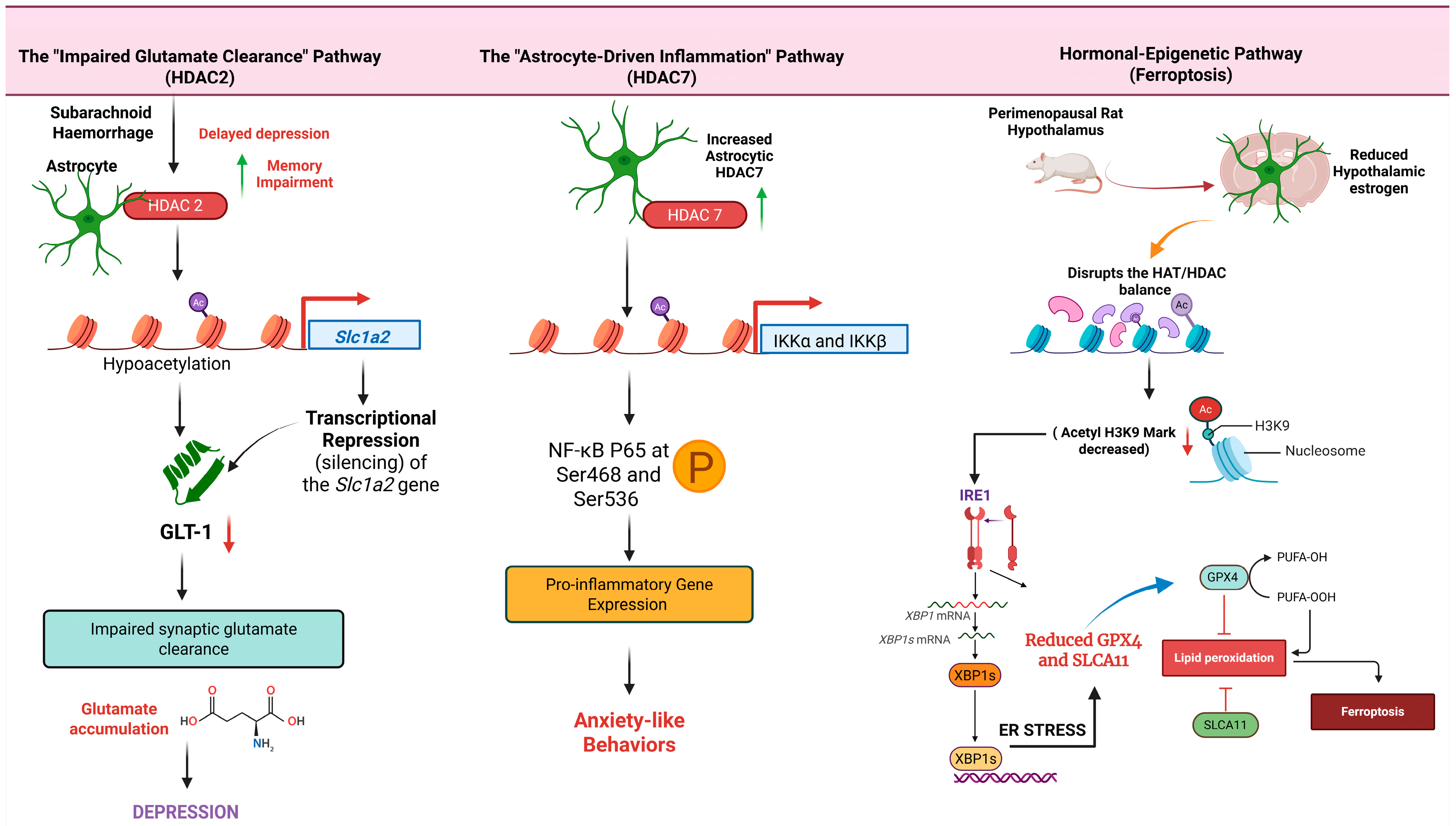
Disclaimer/Publisher’s Note: The statements, opinions and data contained in all publications are solely those of the individual author(s) and contributor(s) and not of MDPI and/or the editor(s). MDPI and/or the editor(s) disclaim responsibility for any injury to people or property resulting from any ideas, methods, instructions or products referred to in the content.

Article Metrics

Citations

Article Access Statistics

Multiple requests from the same IP address are counted as one view.