Article

Reelin Signaling by the Prime Neurogenic Niche of the Adult Brain

by
Francisco Javier Pérez-Martínez
1,2,*,†,
Manuel Cifuentes
2 and
Juan M. Luque
1,*
1
Instituto de Neurociencias, Universidad Miguel Hernández-Consejo Superior de Investigaciones Científicas, Campus de San Juan, 03550 San Juan de Alicante, Spain
2
Departamento de Biología Celular, Genética y Fisiología, Facultad de Ciencias, Universidad de Málaga, Campus de Teatinos, 29071 Málaga, Spain
*
Authors to whom correspondence should be addressed.
Current address: National Police Directorate, Valencia-Tránsitos Police Station, 7 Roncesvalles Square, 46009 Valencia, Spain.
Neuroglia 2025, 6(4), 43; https://doi.org/10.3390/neuroglia6040043
Submission received: 18 August 2025 / Revised: 9 October 2025 / Accepted: 31 October 2025 / Published: 6 November 2025

Abstract

Background: During development, reelin sets the pace of neocortical neurogenesis, enabling newborn neurons to migrate. However, whether—and, if so, how—reelin signaling affects the adult neurogenic niches remains uncertain. Methods: In the present study, we use both loss- and gain-of-function genetic approaches, along with in vivo and ex vivo assays, to investigate this question. Results: We show that reelin signaling, resulting in Dab1 phosphorylation, occurs in the ependymal-subependymal zone (EZ/SEZ) of the lateral ventricles, where, along with its associated rostral migratory stream (RMS), the highest density of functional ApoER2 accumulates. Mice deficient in Reelin, ApoER2, or Dab1 exhibit enlarged ventricles and a dysplastic RMS. Moreover, while the conditional ablation of Dab1 in neural progenitor cells (NPCs) enlarges the ventricles and impairs neuroblast clearance from the SEZ, the transgenic misexpression of Reelin in NPCs of Reelin-deficient mice normalizes the ventricular lumen and the density of ependymal cilia, thereby ameliorating neuroblast migration. Consistently, intraventricular infusion of reelin reroutes neuroblasts. Conclusions: These results demonstrate that reelin signaling persists, sustaining the germinal niche of the lateral ventricles and influencing neuroblast migration in the adult brain.

1. Introduction

Neurogenesis in the embryonic and adult stages emerges as a continuous event, as the lineage of neural progenitor cells (NPCs) and the conservation of many intrinsic signaling pathways are being revealed [1,2]. However, the origin and characteristics of extrinsic signals might still challenge this notion. Radial glia during development and a subpopulation of radial glia-derived astrocyte-like cells in the adult brain are regarded as the founder cells for most, if not all, neural lineages in the central nervous system (CNS). These neural stem cells undergo distinct modes of cell division, giving rise to the diversity of macroglial and neuronal cell types in the CNS [1]. In particular, neural cells do not function at their birth sites but instead undergo more or less extensive migrations. Active adult neurogenesis largely occurs in two restricted regions of the forebrain: the ependymal-subependymal zone (EZ/SEZ) of the lateral ventricles and the subgranular zone (SGZ) in the dentate gyrus of the hippocampus. The EZ/SEZ in the wall of the lateral ventricles constitutes the largest neurogenic niche of the adult mammalian brain [3]. It contains multiple cell populations, including ependymocytes lining the ventricle and SEZ astrocytes that are thought to behave as neural stem cells (so-called type B cells). Type B cells give rise to actively proliferating C cells that function as intermediate progenitor cells. C cells, in turn, generate immature neuroblasts (type A cells), which migrate in chains through the rostral migratory stream (RMS) to the olfactory bulb (OB), where they differentiate into granular and periglomerular interneurons [4,5,6,7]. Type B and C cells are also closely associated with blood vessels and with a nearby large extracellular matrix [8,9,10]. Type B cells retain important properties of radial glial cells, considered neural stem cells during brain development. For example, their cell bodies are generally located just under the EZ cell layer but have short processes that extend through the ependyma, with small apical endings on the ventricle contacting the cerebrospinal fluid (CSF) [9,11]. Thus, the SEZ can be regarded as a displaced neuroepithelium. Interestingly, neuroblast migration parallels CSF flow, and beating of ependymal cilia is required for normal CSF flow, the formation of CSF concentration gradients, and the directional migration of neuroblasts [12].
The conservation of many intrinsic signaling pathways underlies the remarkable similarity between embryonic and adult neurogenesis, although the origin and nature of extrinsic signals might differ [2]. A range of morphogens, including Notch, various Ephrins and Eph receptors, growth factors, neurotrophins, cytokines, neurotransmitters, and hormones, have been identified as major extracellular players in adult neurogenesis [2,13,14]. Knowledge of extracellular cues controlling targeted neuronal migration during adult neurogenesis, or of the molecular links connecting neurogenesis and migration, remains rather limited. A few adhesion molecules (such as β1-integrin, PSA-NCAM, and tenascin-R) and extracellular cues (such as neuregulins and slits) are known to regulate the stability, motility, or directionality of neuronal migration during EZ/SEZ neurogenesis [15,16].
The secreted glycoprotein reelin and its signaling machinery are well known to be required for proper brain development, affecting neuronal migration and the positioning of several neuronal lineages. It is well established that reelin binding to Apolipoprotein E Receptor 2 (ApoER2) or Very-Low-Density Lipoprotein Receptor (VLDLR) induces phosphorylation of the cytoplasmic adaptor protein Disabled 1 (Dab1), activating a tyrosine kinase signal transduction cascade and regulating Dab1 turnover. Indeed, reeler mice (deficient in Reelin) and mutant null mice for Dab1 or for both ApoER2 and Vldlr show comparable positioning defects [17,18,19,20]. However, reelin does not function simply as a positional signal. Rather, it appears to participate in multiple events critical for neuronal migration and cell positioning [21].
Recently, we have shown that during forebrain development, reelin, acting upstream of Notch signaling, is both required and sufficient to determine the rate of neocortical neurogenesis [22]. Such interaction is also required for neuronal migration and positioning [23]. Thus, the early function of reelin in the proliferative compartment might underlie its post-proliferative requirement, raising the possibility that it acts by coupling neurogenesis and neuronal migration, perhaps akin to a permissive rather than an instructive signal for cellular migration. Indeed, a reelin-dependent ApoER2 downregulation mechanism uncouples neocortical newborn neurons from NPCs, thereby enabling neurons to migrate [24]. Highlighting the difficulty in establishing a coherent model of reelin function, a number of studies have provided exceedingly contradictory evidence as to whether reelin plays a role during adult neurogenesis and cell migration [25,26,27,28,29,30,31,32,33,34,35,36,37].
In the present study, we use both loss- and gain-of-function genetic approaches, along with in vivo and ex vivo assays, to investigate reelin signaling in the adult brain. We seek to reveal whether it persists in the germinal niche of the lateral ventricles. Our results show that reelin signaling remains active within, and is necessary and sufficient to modulate, the EZ/SEZ neurogenic niche. They further suggest that cerebrospinal fluid reelin, acting upstream of ApoER2 and Dab1, regulates the integrity and functionality of the ventricular ependyma, the nearby NPCs, and eventually the differentiation and migratory behavior of neuroblasts through the RMS.

2. Materials and Methods

2.1. Mice

Heterozygous reeler mice were purchased from Jackson Laboratory (Bar Harbor, ME, USA). ApoER2-null and Vldlr-null mice were a gift from Johannes Nimpf (University of Vienna, Austria) and were genotyped as previously described [38]. Nestin-Reelin mice were a gift from Tom Curran (The Children’s Hospital of Philadelphia, PA, USA) and were genotyped as previously described [21]. Nestin-CreERT2 mice were a gift from Gordon Fishell (New York University, NY, USA) and were genotyped as previously described [39]. Dab1-null mice and conditional (floxed) Dab1 (Dab1c/c) mice were obtained and genotyped as described elsewhere [40,41]. Mutant mice (Nestin-CreER/+; Dab1c/c) were generated by crossing homozygous conditional Dab1 mice (Dab1c/c) with mice homozygous for the conditional Dab1 allele and heterozygous for the Nestin-CreER allele (Nestin-CreER/+; Dab1c/c). All primer sequences, PCR conditions, and the expected sizes of PCR products are available upon request. Animals were handled according to protocols approved by the European Union, NIH guidelines, and the Instituto de Neurociencias Animal Care and Use Committees.

2.2. Tamoxifen and Bromodeoxyuridine Administration

Tamoxifen (Sigma, St. Louis, MO, USA) was prepared as a 25 mg/mL stock solution in corn oil (C-8267; Sigma, St. Louis, MO, USA). For Cre induction and analysis of P60 Nestin-CreER/+; Dab1c/c mice, tamoxifen was administered by intraperitoneal injection (5 mg/35 g of body weight) every other day for a total of eight injections. After a 1-week break, another round of eight injections was administered. The recombination effectiveness of this dosage in Nestin-CreERT2 mice, and the lack of appreciable short- or long-term effects of tamoxifen exposure on proliferation in the EZ/SEZ, have been previously shown [39]. Bromodeoxyuridine (BrdU; Sigma) was administered intraperitoneally at 100 mg/g of body weight.

2.3. Alkaline Phosphatase-Reelin in Situ Staining

An alkaline phosphatase (AP)-fusion probe of the receptor-binding region of reelin (repeats 3 to 6, AP-RR36) was generated as previously described [42]. Validation of the probe, binding conditions, and alternative staining procedures have also been described [24,42]. AP-RR36 in situ binding was detected using AP substrates or by double immunofluorescence labeling of AP together with several cell markers.

2.4. Immunohistochemistry

Immunostaining was performed as previously described [22]. To ensure homogeneous sampling and reliable comparison across genotypes, we developed a gelatin-block embedding method that allows the simultaneous inclusion and sectioning of up to seven adult mouse brains in a single block. Each block was fixed in 4% paraformaldehyde and 30% sucrose, then sectioned at 50 μm using a freezing microtome, following standard neuroanatomical procedures [43]. This approach ensured equivalent rostro-caudal levels among specimens and identical histological and immunohistochemical conditions, thereby minimizing variability due to processing or anatomical level. Quantifications of ventricular area were performed from these serial, evenly spaced sections covering the full rostro-caudal extent of the lateral ventricles. Primary and secondary antibodies were acquired and diluted as indicated.
Primary antibodies: rabbit anti-human placental alkaline phosphatase (Abcam, Cambridge, UK, 1:100), mouse anti-human placental alkaline phosphatase (Abcam, 1:50), goat anti-doublecortin (Santa Cruz Biotechnology, Dallas, TX, USA, 1:100), rabbit anti-GFAP (Dako, Glostrup, Denmark, 1:500), rabbit anti-tyrosine hydroxylase (Pel-Freez Biologicals, Rogers, AR, USA, 1:1000), rabbit anti-calbindin (Swant AG, Marly, Switzerland, 1:10,000), rat anti-BrdU (Serotec, Kidlington, UK, 1:100), mouse anti-reelin (G10, Abcam, 1:1000), rabbit anti-S100b (Dako, 1:400), rabbit anti-Dab1 (B3, 1:500) [44,45], mouse anti-CRE (Novagen, Madison, WI, USA, 1:500), mouse anti-Mash1 (BD Pharmingen, San Diego, CA, USA, 1:500), mouse anti-phosphotyrosine (Sigma, St. Louis, MO, USA, 1:2000). Secondary antibodies: biotinylated donkey anti-rabbit, biotinylated goat anti-rat, biotinylated donkey anti-goat (Jackson ImmunoResearch, West Grove, PA, USA, 1:500); Cy3-donkey anti-rabbit, Cy3-goat anti-rat, Cy3-donkey anti-goat, Cy3-donkey anti-mouse, Cy2-donkey anti-rabbit, Cy2-donkey anti-mouse (Jackson ImmunoResearch, 1:200); HRP-goat anti-rabbit, HRP-goat anti-mouse (Jackson ImmunoResearch, 1:5000).
Images were captured on a Leica TCS SP2 AOBS inverted laser-scanning confocal microscope. ImageJ (NIH, http://rsb.info.nih.gov/ij/, version 1.54, accessed on 16 June 2024) and Adobe Photoshop software (version 24.2.1) were used for image processing and analysis.

2.5. Production of Reelin

Recombinant reelin was produced using the mammalian expression construct pCrl, which encodes mouse Reelin cDNA [46]. Cell culture and transfection procedures have been previously described [47].

2.6. Dab1 Phosphorylation Assay

Brains were harvested from P60 reeler mice. Two-millimeter-thick coronal slices were obtained, and the EZ/SEZ at the striatal side of the lateral ventricles was microdissected under a low-power dissecting microscope in ice-cold DMEM. Tissues were homogenized in cell lysis buffer and precleared by brief centrifugation. Anti-Dab1 immunoprecipitation was carried out from equal amounts of protein, and Dab1 protein levels and phosphorylation status upon reelin stimulation were determined as previously described [48].

2.7. Ultrastructural Analysis

For scanning electron microscopy, P60 wild-type, reeler, and reeler ne-Reelin mice were used. Briefly, the entire wall of the lateral ventricles was dissected as a whole mount and fixed with 2.5% glutaraldehyde/2% paraformaldehyde overnight. The tissues were incubated in 2% OsO4 for 1 h, dehydrated with ethanol, and dried with a critical-point drier. After sputter-coating with platinum, the mounted samples were examined with a Hitachi S-4100 scanning electron microscope.

2.8. Intraventricular Infusion of Reelin

Recombinant reelin was produced and concentrated as previously described [47]. The concentrated protein (or mock) was infused using a mini-osmotic pump (Alzet model 2001, Campbell, CA, USA) into the caudal region of the lateral ventricle of P60 wild-type and reeler mice at the coordinates −0.7 mm, 1.2 mm, and 2.0 mm (anterior, lateral, and depth coordinates relative to bregma). Five days later, the brains were processed for immunohistochemistry using anti-doublecortin antibodies.

3. Results

3.1. The EZ/SEZ and RMS Contain the Highest Density of Functional Reelin Receptors in the Adult Mouse Brain

To test whether the reelin pathway continues to be active in postnatal ages, it was essential to determine whether there is continuity in the expression of its functional receptors in adult animals. In fact, we found that during embryonic development the lateral ganglionic eminence (LGE)—but not the medial ganglionic eminence (MGE), the main origin of the EZ/SVZ/RMS system of the adult brain [49,50]—expresses FRR (Supplementary Figure S1). For this reason, we analyzed its expression in the adult brain using the fusion protein AP-RR36, which we previously developed and extensively validated in isolated cells and embryonic tissues [24,42]. The excellent resolution offered by the enzymatic development of this technique made it possible to elaborate a rostral–caudal cartography of FRR expression, from the olfactory bulb to the brainstem, including the cerebellum (Supplementary Figure S2).
Thus, we clearly observed the highest density of FRR in the adult brain within the two known adult neurogenic niches: the ependymal/subependymal zone (EZ/SVZ) of the lateral ventricles, including the rostral migratory stream (RMS), and the subgranular zone (SGZ) of the dentate gyrus in the hippocampus. Diffuse staining was also observed in the glomerular layer of the olfactory bulb, the medial habenular nucleus, and the molecular layer of the cerebellum. Moreover, the strongest contribution to this signal in the brain’s neurogenic niches is due to ApoER2, as shown by null mutants of this receptor, which display a near-complete lack of staining except for a diffuse signal in the glomerular layer of the olfactory bulb.
Consistent with these results are those obtained with null mutants for the other canonical reelin receptor, VLDLR: wild-type-like FRR staining is observed throughout the brain but is minimal in the cerebellum (an expression pattern practically complementary to that of the ApoER2 mutant), confirming that VLDLR is the main receptor present in this structure. On the other hand, staining with the AP-reelin probe shows a gradual increase as the genetic dosage of reelin decreases. Thus, the signal observed in the reeler brain is stronger than in the heterozygous brain for Reelin, which in turn is stronger than in the wild-type brain. This supports the conclusion that reelin causes a downregulation of the receptor. Details of the localization of functional reelin receptors in the adult brain’s neurogenic niches can be observed in Figure 1.

3.2. Functional ApoER2 Is Present in the Main Cellular Compartments of the EZ/SEZ/RMS Neurogenic Axis

By examining the specific regions in which ApoER2 is expressed in the brain, our next objective was to determine which cell types of the neurogenic niche of the lateral ventricles are competent to respond to a signal triggered by reelin, as well as to examine the localization of ApoER2 in the cellular components constituting the RMS. The combination of the in situ binding assay with the AP-RR36 probe and detection by immunofluorescence shows that, to a greater or lesser extent, all cellular subpopulations of the EZ/SEZ are competent to receive reelin as a ligand, as demonstrated by the colocalization of functional ApoER2 with specific markers for ependymocytes (E-type cells) [S100β], subventricular astrocytes (including B-type cells) [GFAP], transient amplifying cells (type C cells) [Mash1, also known as Ascl1], neuroblasts (type A cells) [DCX], as well as cells in S phase of the cell cycle [pulse and harvest 1 h after BrdU injection] (Figure 2). It is noteworthy that the highest colocalization of ApoER2 occurs in neuroblasts. Thus, an embryo-to-adult continuity is evident in the expression of ApoER2 across the different cell lineages of the neurogenic niche.
Regarding the components of the rostral migratory stream, neuroblasts (positive for the doublecortin marker, DCX) consistently express ApoER2, together with astrocytes (GFAP+) that form the “tubes” through which they migrate, facilitating their movement through the adult environment. As previously described, neuroblasts also undergo divisions along the RMS, from their birth in the EZ/SEZ until they reach their destination in the different layers of the olfactory bulb [51]. It is equally significant that these dividing elements are also competent to respond to reelin (Supplementary Figure S3).

3.3. Reelin Signaling Remains Active in the EZ/SEZ

Although ApoER2 is considered a promiscuous receptor, the evidence we had so far suggesting that reelin-triggered signaling remains active in the adult brain was more than sufficient to justify the following experiment. Our group had observed that the adult reeler mutant shows exaggerated ventricular enlargement and a hyperplastic RMS. The cells of the EZ/SEZ/RMS are competent to receive the reelin signal, as they express one of the canonical elements of the signaling pathway, the ApoER2 receptor. In addition, the presence of the intracellular adaptor Dab1 has been described in the RMS, another of the classical components of the pathway [26].
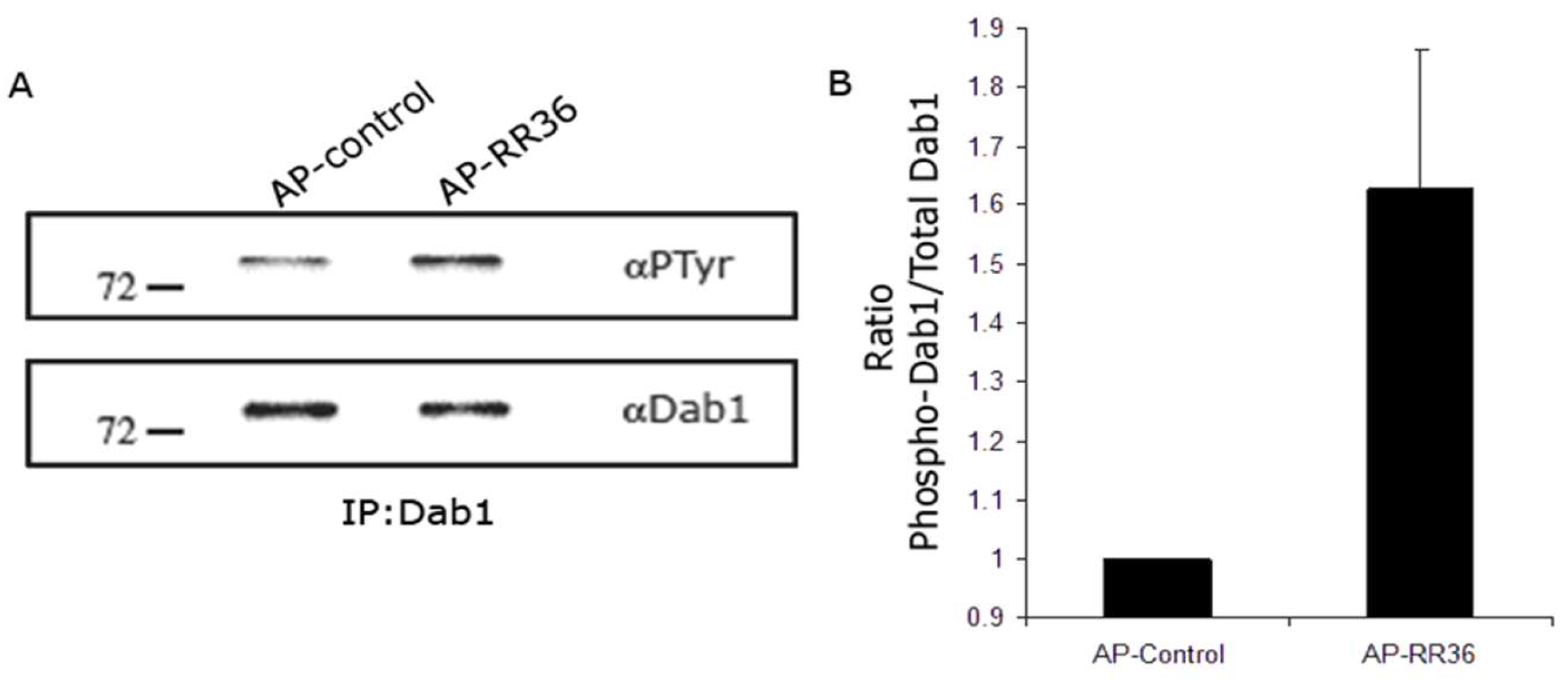
As is known, a requirement for reelin signaling through its receptor to be transduced is the phosphorylation of Dab1 [48]. During embryonic development, we have shown that reelin, through a mechanism of downregulation of ApoER2, promotes the dissociation of newborn neurons from their parent cells and is able to normalize Dab1 levels on a reeler genetic background [42]. To assess whether the reelin signaling pathway remains active in the adult brain, we performed an ex vivo Dab1 phosphorylation assay under reelin stimulation, using homogenates of cells extracted from the EZ/SEZ of adult reeler mice as a substrate.
The reelin fragment comprising repeats 3 to 6 (RR36) recapitulates the vast majority of the functions of the protein and is capable of triggering signaling [52]. To continue the line of experiments carried out thus far, we used the AP-RR36 fusion protein for this Dab1 phosphorylation assay (although previously, and with the same result, we had already performed it with recombinant reelin generated in our laboratory by transfection of HEK293T cells). After incubation, immunoprecipitation of Dab1 was necessary to enrich the sample with this protein, since an antibody that generically detects phosphorylated tyrosine residues was used.
The results of this assay show that Dab1 phosphorylation indeed occurs after incubation of EZ/SEZ cells with AP-RR36. The increase in the phosphotyrosine band, together with the decrease in the Dab1 signal—consistent with the onset of its degradation—supports the conclusion that reelin signaling persists in the adult EZ/SEZ. (Figure 3).

3.4. Lack of Function of Reelin, ApoER2 or Dab1, but Not of VLDLR, Causes Analogous Phenotypes in the Lateral Ventricles and RMS

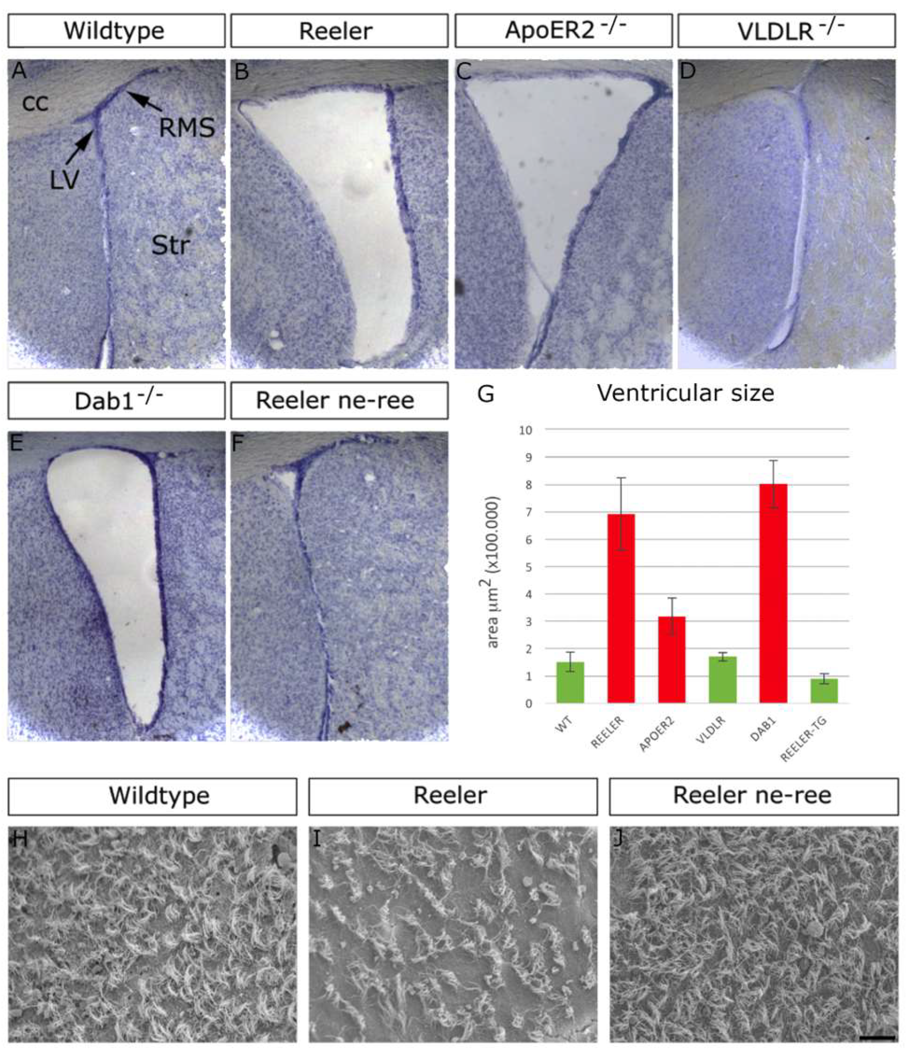
Loss of function of Reelin, ApoER2, or Dab1 in mutants lacking these proteins causes hyperplasia of the RMS [30]. Closely related to this structure are the lateral ventricles of the adult brain. When analyzing the neuroanatomy of these mutants, we observed evident ventriculomegaly (increased ventricular size; Figure 4B,C,E), but not in Vldlr mutants (Figure 4D), which do not exhibit phenotypic alterations in any of these structures. This supports the conclusion that the main reelin receptor in the neurogenic niches of the adult brain—if not the only one—is ApoER2.

3.5. Partial Rescue of Lateral Ventricle Integrity and Neuroblast Migration by Ectopic Expression of Reelin in NPCs of Reeler Mutants

To define the possible function of reelin and its signaling pathway in the adult EZ/SEZ system, we carried out a series of studies with genetically manipulated animals. As in our previous work [24], we used the ne-Reelin transgenic mouse to evaluate the effect of Reelin gain-of-function on a wild-type background and in a rescue paradigm on a reeler (reeler ne-Reelin) adult background. The study in which this transgenic mouse was first used [21] highlighted the rescue of cerebellar foliation in reeler ne-Reelin animals, with the consequent disappearance of the ataxic phenotype, although cortical lamination was apparently not restored. In fact, we demonstrated that cortical lamination is partially recovered above an ectopic subplate [24]. The analysis of ectopic Reelin expression in the adult now provides key information for deepening the understanding of reelin function.
The protein ectopically expressed by neural progenitors of the EZ/SEZ (under the Nestin promoter) on a reeler background causes normalization of ventricular size (Figure 4B,F) and, in fact, recovery of the density of ependymal cilia, analyzed by scanning electron microscopy (Figure 4H–J). Graphic representation of the values obtained with software analysis shows statistically significant differences between the ventricular size of reeler, Dab1, and ApoER2 mutants (Figure 4G, red columns).

3.6. Conditional Ablation Dab1 in NPCs Impairs Neuroblast Clearance from the SVZ

To address this issue from the opposite perspective, we carried out a conditional ablation of Dab1, since this protein is expressed in both embryonic radial glia [45] and the adult EZ/SEZ [26], which would provide data perfectly complementary to those obtained from gain-of-function experiments. For this purpose, we used the CRE-lox system induced by tamoxifen to interrupt reelin signaling in progenitors, as it allows the selection of both the site and the time at which the gene of interest—in this case Dab1—can be eliminated. By crossing Nestin-CreERT2 transgenic mice (expressing the Cre recombinase enzyme under the Nestin promoter [39]) with floxed-Dab1 mice (the Dab1 gene flanked by loxP sites [37,41]), we achieved the conditional ablation of reelin signaling in the progenitors of adult mice.
Effective ablation of Dab1 after tamoxifen administration was evident by immunodetection of the protein in the RMS at the level of the olfactory bulb, as well as in more caudal regions such as the anterior commissure, the lateral ventricles, and the subgranular zone of the hippocampus, the latter being neurogenic niches where Nestin is expressed more intensely (Supplementary Figure S4). We performed a 3-day BrdU pulse experiment to demonstrate that conditional ablation of Dab1 in progenitors (Figure 5A,B) increases the size of the lateral ventricles (Figure 5C,D) and impairs the migration of neuroblasts from the SEZ toward the RMS, causing their accumulation (Figure 5E,F). This confirms that the phenotype observed in mutants of the reelin signaling pathway is not a development-dependent event.
Thus, we observed that interruption of the reelin pathway by selective elimination of Dab1 results in an accumulation of cells in the more caudal regions of the RMS. Under normal conditions, a neuroblast born in the EZ/SEZ takes about 3 days to migrate to the olfactory bulb through the RMS. In this case, in Dab1flx/flx; Cre+ mice, a dramatic increase in BrdU+ profiles was observed in the EZ/SEZ, in contrast with Dab1+/flx; Cre+ mice, in which the majority of BrdU-incorporating cells had already left their neurogenic niche to begin their migration toward the olfactory bulb. Genetic ablation of Dab1 therefore causes a defect in neuroblast migration.
In a complementary approach, we found the opposite effect in transgenic reeler ne-Reelin brains. We first tested the function of the ne-Reelin transgene in the adult brain by immunofluorescence to detect the presence of reelin in the EZ/SEZ of the lateral ventricle, confirming its ectopic expression in this area (Figure 6A–I). We then demonstrated that the accumulation of neuroblasts in the RMS observed in reeler mutants, with the consequent migratory difficulty in reaching the olfactory bulb, tends to normalize in the presence of ectopic reelin, also restoring the morphology of the RMS at the level of the olfactory bulb (Figure 6J–L).

3.7. Intraventricular Infusion of Recombinant Reelin Alters the Migration of Neuroblasts

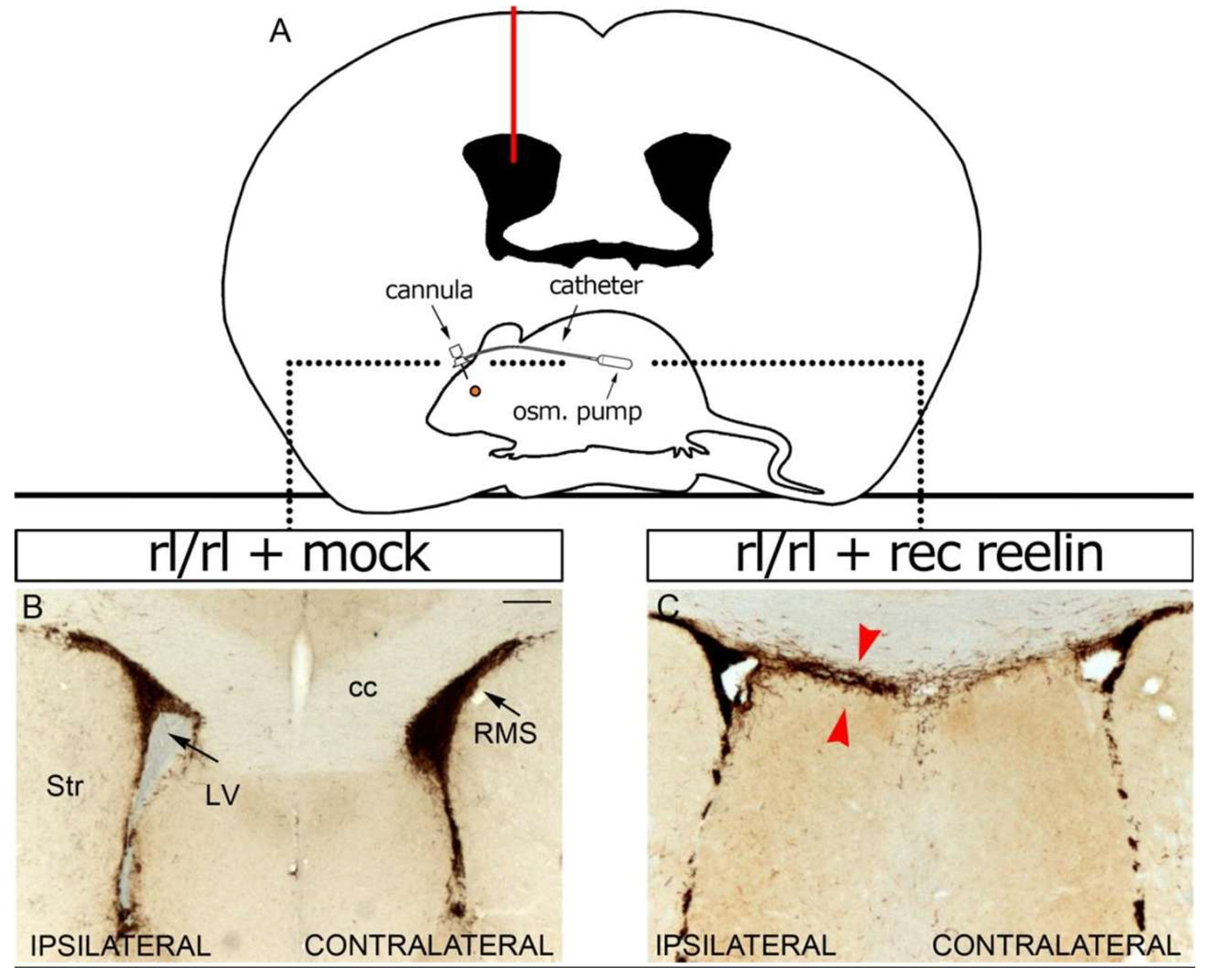
Taken together, the previous results point to a role for reelin in the maintenance of adult neurogenic niches. Interruption of the reelin signaling pathway causes defects in neuroblast migration, together with alterations in the anatomy of the lateral ventricles. In a complementary approach, in an environment with excess reelin, we used osmotic mini-pumps to administer recombinant reelin directly into the lateral ventricle of adult reeler mice (Figure 7A). The pumps were carefully implanted using stereotaxic surgery and left in place for 5 days, releasing recombinant reelin at a rate of 1 μL/hour. Subsequently, immunohistochemical techniques were performed to detect Doublecortin+ cells (neuroblasts). Infusion of control culture medium did not alter the distribution of neuroblasts (Figure 7B), whereas infusion of reelin affected their migratory behavior on a reeler genetic background, producing a remarkable alteration in the usual location of neuroblasts on the side ipsilateral to the site of cannula insertion of the reelin pump (Figure 7C, red arrowheads). Therefore, we conclude that reelin influences the migration of neuroblasts in the adult brain, probably acting from the CSF, which, as our group showed previously, naturally contains reelin [47].

4. Discussion

Here we show that in the adult brain, the reelin-activated biochemical signaling cascade, resulting in phosphorylation of the intracellular adaptor Dab1, occurs in the ependymal–subependymal zone (EZ/SVZ) of the lateral ventricles. We also show that the EZ/SEZ, which contains stem cells and other neural progenitors, together with its extensive rostral migratory stream (RMS) of neuroblasts, concentrates the highest density of functional ApoER2 in the adult brain. Mutant mice lacking Reelin (reeler), ApoER2, or Dab1 exhibit a prominent enlargement of ventricular volume and marked hypercellularity of the RMS, particularly in its proximal aspect. Conditional ablation of Dab1 in neural progenitor cells of wild-type mice increases ventricular volume and inhibits cell migration from the SVZ to the RMS. In contrast, ectopic transgene expression of Reelin in the neural progenitors of reeler mutants normalizes the ventricular lumen and the density of ependymal cilia, thereby improving neuroblast migration. Consistently, intraventricular infusion of recombinant reelin in reeler mutants also alters the migration of neuroblasts, indicating that reelin acts from the cerebrospinal fluid. These results show that reelin signaling not only remains active but also is necessary and sufficient to modulate the adult EZ/SEZ/RMS neurogenic axis.
The dynamics and molecular composition of cerebrospinal fluid (CSF) are the object of renewed interest, particularly as CSF emerges as a key component in the regulation of the EZ/SVZ neurogenic niche [53]. The presence of reelin in the CSF of the adult brain has been documented by our group [47] and later confirmed by others [54,55,56]. Various mechanisms are involved in the complex dynamics of CSF [57,58]. One of them, the coordinated movement of ependymal cilia lining the ventricular wall, generates a laminar supraependymal flow approximately 200 μm thick [59,60,61]. This movement has been shown to be necessary not only for the normal flow of CSF in the lateral ventricle but also for the formation of concentration gradients of molecules contained in the fluid and for the directional migration of neuroblasts, which runs parallel to this flow [12]. In addition, multiple lines of evidence link ciliary alterations and planar polarity of ependymocytes with ventricular distension and hydrocephalus [62,63]. Our results, demonstrating the activity of reelin in maintaining the integrity of the ciliated ependymal layer/ventricular volume and in regulating neurogenesis and neuroblast migration, are consistent with these observations. Moreover, the small apical surface of B cells is in direct contact with CSF, whose soluble factors can modulate the behavior of these stem cells [64,65].
Moreover, as radial glia and neuroepithelial cells do during embryonic development, the vast majority of B cells contact the ventricle through small apical processes containing a single primary cilium [9,11]. This primary cilium may integrate signals directly from molecules present in the CSF [66]. In another vein, the ependymal cells themselves—surrounding the apical processes of B cells and constituting the main interface between CSF and brain parenchyma—are interconnected by gap junctions, a type of intercellular connection that does not impose a strict restriction on the diffusion of molecules between both compartments [67]. Particularly intriguing is the detection of ApoER2 and VLDLR (as well as Notch and some of its ligands) within the CSF itself [55,56]. Their presence may be related to the release of extracellular membrane particles (prominin/CD133+) from neural progenitor cells and other epithelial cells into the CSF [68,69]. Where, then, does CSF reelin originate? Most likely, CSF reelin is not derived from blood but from the CNS [54,70]. On the other hand, although its expression is detectable during embryonic development [71,72,73], reelin was not found in the secretome of the choroid plexus of the lateral ventricle, classically considered a primary producer of CSF and more recently identified as a key component of the adult EZ/SVZ niche [74].
However, it is possible that reelin reaches the EZ/SVZ neurogenic niche after being secreted into the CSF from other circumventricular organs (CVOs). For example, the subfornical organ, located on the ventral surface of the fornix near the foramen of Monro that interconnects the lateral ventricles with the third ventricle, strongly expresses reelin [71]. Traditionally considered a sensory CVO, its secretory capacity has recently been demonstrated [75]. Reelin is also expressed in the subcommissural organ (SCO), located in the dorsocaudal region of the third ventricle at the entrance to the cerebral aqueduct [71]. The SCO is known to secrete SCO-spondin, transthyretin, and basic fibroblast growth factor (bFGF), proteins that participate in various aspects of neurogenesis such as stem cell proliferation, neuronal differentiation, and axonal guidance. Some unidentified soluble compounds secreted by the SCO have also been detected in the lateral ventricle [76]. Structurally, the main SCO secretion, SCO-spondin, belongs to the TSR superfamily (thrombospondin type 1 repeat), which includes F-spondin and thrombospondin-1 among other proteins [77]. Reelin contains a domain homologous to the N-terminus of F-spondin [78], and in turn, F-spondin can interact, through its TSR domains, with ApoER2 [79]. Thrombospondin-1 (TSP-1) can interact with ApoER2 and VLDLR, and it has been proposed that TSP-1 stabilizes chains of neural precursors derived from SVZ explants [33]; however, its in vivo function remains unclear. Interestingly, impaired secretory function of the SCO is a common feature in various animal models of hydrocephalus [80], classically defined as a distension of the ventricular system due to the active accumulation of CSF. Multiple lines of evidence associate hydrocephalus with abnormal neurogenesis, and more recent findings implicate neurogenic alterations rather than CSF accumulation as key factors in its pathogenesis [81]. In any case, regardless of the origin of adult brain CSF reelin, there is a suggestive parallel between this causal association and our own results, which show that inhibition of reelin activity in the EZ/SVZ niche leads to alterations compromising both neurogenesis and ventricular volume.
It is well established that the activation of Notch signaling promotes and maintains the neural progenitor state, while inhibiting neuronal differentiation. Conversely, the protein Numb antagonizes Notch function during neural precursor division [82,83]. Canonical Notch signaling is highly active in the EZ/SVZ neurogenic niche, where it regulates the maintenance of neural stem progenitors [84,85]. Although the precise mechanisms regulating Notch activity in the mouse EZ/SVZ are not fully understood, several Notch ligands are known to be expressed there, and both Delta1 and Jagged1 have been detected in IPCs as well as in neuroblasts [66,85,86]. Interestingly, postnatal ablation of Numb/Numblike proteins in the EZ/SVZ results in enlargement of the lateral ventricles and marked hypercellularity of the RMS [87], phenotypes similar to those described here for deficiencies of Reelin, ApoER2, or Dab1. Consistently, overexpression of the activated intracellular domain of Notch in the EZ/SVZ phenocopies the hypercellularity of the RMS; however, the integrity and size of the lateral ventricles remain unaffected, suggesting that Numb regulation of ependymal integrity may occur through a Notch-independent mechanism [87]. In contrast, a more recent study shows that ependymal cells are dependent on canonical Notch signaling, which actively maintains their quiescence, phenotype, and position. Inhibition of Notch signaling exclusively in the ventricular ependyma allows ependymocytes to re-enter the cell cycle and generate olfactory bulb neurons, whereas forced activation of Notch is sufficient to block the plastic response of ependymal cells to cerebral infarction [88].
Undoubtedly, Notch signaling exerts distinct functions across the different cellular populations of the lateral ventricle neurogenic niche. Although the interaction between reelin and Notch signaling is known to play an important role in the modulation of neurogenesis and cell migration during cortical development [22,23], the precise molecular mechanisms underlying this interaction remain unclear. In fact, hundreds of genes are directly regulated in vivo in cortical NSCs by the Notch intracellular domain (NICD) together with its cofactor RBPJ. Among them is Dab1, whose transcription, following Notch activation, is not induced but rather repressed [89]. Moreover, Numb and Dab1 function as endocytic accessory proteins that regulate endocytosis and the subsequent post-endocytic trafficking of their associated receptors, including Notch, TrkB, APP, VLDLR, and ApoER2 [90]. Since both proteins share a phosphotyrosine-binding (PTB/NPXY) domain, the possibility of cross-interaction depending on the cellular context cannot be ruled out. A recent study demonstrated that inhibition of Notch signaling in the adult neurogenic niche of the lateral ventricle accelerates the migration of cells from the SVZ to the RMS compared with controls. In contrast, Notch overexpression increases the proportion of cells retained in the SVZ for longer periods than in control brains [91], a phenotype strikingly similar to that observed after conditional ablation of Dab1 in this work. Furthermore, the high levels of Notch in PSA-NCAM+ neuroblasts located in the RMS, which are much lower in cells that have already reached the OB, suggest that Notch activity may inhibit premature differentiation [92]. This expression pattern strongly resembles that described here for ApoER2 localization. It would therefore be of particular interest to investigate whether the interaction between reelin and Notch signaling persists within the adult EZ/SVZ/RMS neurogenic axis.
The literature on the manipulation of the PSA-NCAM glycoconjugate is also of potential relevance in our context. Poly-α2,8-sialic acid (PSA), a unique post-translational modification of the neural cell adhesion molecule (NCAM), is strongly associated with neural development and plasticity. Elimination of PSA—through genetic ablation of its synthesizing enzymes—independently of NCAM, results in both an expansion of the RMS and ventricular enlargement [93], phenotypes comparable to those reported here for the absence of reelin, ApoER2, or Dab1. Moreover, intrathecal administration of endoneuraminidase (endo N), an enzyme that selectively removes PSA, has demonstrated that PSA not only promotes rostral tangential migration of neuroblasts but also suppresses NCAM-induced differentiation. This differentiation, which depends on cell–cell contact, involves activation of the mitogen-activated protein kinase (MAPK) pathway [94]. Notably, as we show here for ApoER2, wild-type mice display a downregulation of PSA once neuroblasts reach the OB and initiate radial migration and differentiation. Interestingly, reelin has been shown to induce the dissociation of neuroblasts migrating in chains from SVZ explants [26], and this effect also depends on MAPK pathway activation [32]. In addition, intraventricular administration of endo N (but not the genetic elimination of PSA) causes massive dispersion of neuroblasts into surrounding brain regions, including the striatum [95]—an effect comparable to the redirection of neuroblasts observed here after intraventricular infusion of recombinant reelin in reeler mutants. Although no direct interaction is known between reelin or ApoER2 and PSA or NCAM, PSA on NCAM is capable of directly binding bioactive molecules such as neurotrophins, neurotransmitters, and growth factors, thereby regulating their extracellular concentrations and signaling dynamics. For instance, PSA binds brain-derived neurotrophic factor (BDNF), which, when associated with the PSA chain, can migrate to TrkB and p75NTR receptors [96]. In turn, neurotrophins, including BDNF, can regulate the proteolytic processing of ApoER2 through activation of Trk signaling [97]. Furthermore, NCAM can interact heterophilically with other cell adhesion molecules such as L1 [98]. A recent study demonstrated proteolytic cleavage of L1 by reelin, further highlighting its importance for neuronal migration during cortical development [99]. It would be equally interesting to investigate whether these transitive molecular interactions persist in the adult EZ/SVZ/RMS neurogenic axis.
Recently, it has been shown that ependymal cells and adult neural stem cells share common embryonic progenitors. Although the number of cells in the lateral ventricle decreases with age, ventricular growth is accompanied by an expansion of the apical surface area of ependymal cells, which accommodates the larger ventricular size [100]. Impairment of this mechanism, due to defective reelin signaling, may underlie the ventriculomegaly observed here in Reelin, ApoER2, and Dab1 deficient mice. Moreover, reelin has been shown to signal through mTOR (mammalian target of rapamycin) to regulate dendritic growth [101]. Interestingly, both mTORC1 and primary cilia are required for brain ventricle morphogenesis [102], and the timing of mTOR activation is also critical for EZ/SVZ/RMS neurogenesis [103].
Although our findings strongly support a persistent and functional role of reelin signaling in the adult EZ/SEZ–RMS neurogenic axis, some limitations should be noted. Developmental compensations in reelin-deficient mice cannot be entirely excluded and might partially mask specific adult effects. However, the combined use of loss-of-function, gain-of-function, and conditional strategies helps to mitigate this concern and provides convergent support for our conclusions. Intraventricular infusion experiments may create non-physiological gradients, and functional analyses were based on static imaging rather than direct measurements of neuroblast migration dynamics. Moreover, ultrastructural and in vitro data were limited, and the final fate of migrating cells was not examined. Future studies addressing these aspects will help to refine and expand the model proposed here.
We propose that, acting through ApoER2 and Dab1, CSF-derived reelin regulates both the integrity and functionality of the ventricular ependyma, as well as the behavior of progenitor cells in the EZ/SVZ—ultimately controlling the production, differentiation, and migration of RMS neuroblasts at least up to the core of the OB. Although less evident, this model is not at odds with the one we previously proposed for embryonic cortical development [24]. Indeed, the requirement for a downregulation of ApoER2 in RMS neuroblasts of wild-type mice is clearly inferred from the higher intensity of functional ApoER2 observed in reeler mutants. Considering that neuroblasts migrating along the RMS have not yet exited the cell cycle—that is, they do not constitute an entirely post-mitotic population—the downregulation of ApoER2 would not reach the minimal levels observed in the embryonic cortical plate. However, once neuroblasts reach the OB core, a complete downregulation of ApoER2 may be required to permit, perhaps via cues other than reelin, the onset of radial migration of the olfactory interneurons generated in the adult neurogenic niche. Notably, the consistent detection of functional ApoER2 in the subgranular zone of the adult hippocampal dentate gyrus supports this view and warrants further investigation. We believe our findings open new avenues of research, suggesting that reelin signaling constitutes an integral component of a master mechanism coupling neurogenesis and neuronal migration throughout life.

Supplementary Materials

The following supporting information can be downloaded at: https://www.mdpi.com/article/10.3390/neuroglia6040043/s1, Figure S1: Functional reelin receptors are expressed in the LGE; Figure S2: The major reelin receptor in the neurogenic niches of the adult mouse brain is ApoER2; Figure S3: Functional reelin receptors are expressed in the RMS; Figure S4: Conditional ablation of Dab1.

Author Contributions

F.J.P.-M. and J.M.L. conceived the project. F.J.P.-M. contributed to all experiments. M.C. performed scanning electron microscopy of the lateral ventricle and the stereotaxic infusion of recombinant reelin. F.J.P.-M. and J.M.L. wrote the manuscript. J.M.L. supervised the project. All authors have read and agreed to the published version of the manuscript.

Funding

This work was supported by grants from the Spanish Ministry of Science and Innovation SAF2004-07685 and the Fundación Médica Mutua Madrileña [to J.M.L.].

Institutional Review Board Statement

Animals were handled according to protocols approved by the European Union, NIH guidelines and the Instituto de Neurociencias Animal Care and Use Committees, approval code UMH-CSIC 2007/VSC/PEA/0014 (19-07-2007).

Informed Consent Statement

Not applicable.

Data Availability Statement

The original data presented in the study are openly available in doi: https://doi.org/10.1101/2021.12.12.472284.

Acknowledgments

We deeply appreciate J. Nimpf for ApoER2- and Vldlr-null mice; B. Howell for Dab1-null and conditional (floxed) Dab1 (Dab1c/c) mice, and for Dab1 (B3) antibody; and M. Hattori for the AP-RR36 reelin fusion protein. We also thank T. Curran for the Nestin-Reelin allele and the Reelin pCrl plasmid, and G. Fishell for the Nestin-CreER allele. This work was presented in partial fulfilment of the requirements for a PhD degree in Biology (F. Javier Pérez-Martínez). The reviews and comments by E. Soriano, L. García-Alonso, M.C. Pedraza and three anonymous referees are gratefully acknowledged. To our fathers, in memoriam.

Conflicts of Interest

The authors declare that the research was conducted in the absence of any commercial or financial relationships that could be construed as a potential conflict of interest.

References

  1. Kriegstein, A.; Alvarez-Buylla, A. The glial nature of embryonic and adult neural stem cells. Annu. Rev. Neurosci. 2009, 32, 149–184. [Google Scholar] [CrossRef]
  2. Ming, G.L.; Song, H. Adult neurogenesis in the mammalian brain: Significant answers and significant questions. Neuron 2011, 70, 687–702. [Google Scholar] [CrossRef] [PubMed]
  3. Ihrie, R.A.; Alvarez-Buylla, A. Lake-front property: A unique germinal niche by the lateral ventricles of the adult brain. Neuron 2011, 70, 674–686. [Google Scholar] [CrossRef] [PubMed]
  4. Lois, C.; Alvarez-Buylla, A. Long-distance neuronal migration in the adult mammalian brain. Science 1994, 264, 1145–1148. [Google Scholar] [CrossRef] [PubMed]
  5. Doetsch, F.; Caillé, I.; Lim, D.A.; García-Verdugo, J.M.; Alvarez-Buylla, A. Subventricular zone astrocytes are neural stem cells in the adult mammalian brain. Cell 1999, 97, 703–716. [Google Scholar] [CrossRef]
  6. Carleton, A.; Petreanu, L.T.; Lansford, R.; Alvarez-Buylla, A.; Lledo, P.M. Becoming a new neuron in the adult olfactory bulb. Nat. Neurosci. 2003, 6, 507–518. [Google Scholar] [CrossRef]
  7. Brill, M.S.; Ninkovic, J.; Winpenny, E.; Hodge, R.D.; Ozen, I.; Yang, R.; Lepier, A.; Gascón, S.; Erdelyi, F.; Szabo, G.; et al. Adult generation of glutamatergic olfactory bulb interneurons. Nat. Neurosci. 2009, 12, 1524–1533. [Google Scholar] [CrossRef]
  8. Mercier, F.; Kitasako, J.T.; Hatton, G.I. Anatomy of the brain neurogenic zones revisited: Fractones and the fibroblast/macrophage network. J. Comp. Neurol. 2002, 451, 170–188. [Google Scholar] [CrossRef]
  9. Shen, Q.; Wang, Y.; Kokovay, E.; Lin, G.; Chuang, S.M.; Goderie, S.K.; Roysam, B.; Temple, S. Adult SVZ stem cells lie in a vascular niche: A quantitative analysis of niche cell-cell interactions. Cell Stem Cell 2008, 3, 289–300. [Google Scholar] [CrossRef]
  10. Tavazoie, M.; Van der Veken, L.; Silva-Vargas, V.; Louissaint, M.; Colonna, L.; Zaidi, B.; Garcia-Verdugo, J.M.; Doetsch, F. A specialized vascular niche for adult neural stem cells. Cell Stem Cell 2008, 3, 279–288. [Google Scholar] [CrossRef]
  11. Mirzadeh, Z.; Merkle, F.T.; Soriano-Navarro, M.; Garcia-Verdugo, J.M.; Alvarez-Buylla, A. Neural stem cells confer unique pinwheel architecture to the ventricular surface in neurogenic regions of the adult brain. Cell Stem Cell 2008, 3, 265–278. [Google Scholar] [CrossRef]
  12. Sawamoto, K.; Wichterle, H.; Gonzalez-Perez, O.; Cholfin, J.A.; Yamada, M.; Spassky, N.; Murcia, N.S.; Garcia-Verdugo, J.M.; Marin, O.; Rubenstein, J.L.; et al. New neurons follow the flow of cerebrospinal fluid in the adult brain. Science 2006, 311, 629–632. [Google Scholar] [CrossRef] [PubMed]
  13. Zhao, C.; Deng, W.; Gage, F.H. Mechanisms and functional implications of adult neurogenesis. Cell 2008, 132, 645–660. [Google Scholar] [CrossRef] [PubMed]
  14. Jang, M.H.; Song, H.; Ming, G.L. Regulation of Adult Neurogenesis by Neurotransmitters. In Adult Neurogenesis; Gage, F.H., Kempermann, G., Song, H., Eds.; CSHL Press: Cold Spring Harbor, NY, USA, 2008; pp. 397–423. [Google Scholar]
  15. Lledo, P.M.; Alonso, M.; Grubb, M.S. Adult neurogenesis and functional plasticity in neuronal circuits. Nat. Rev. Neurosci. 2006, 7, 179–193. [Google Scholar] [CrossRef] [PubMed]
  16. Ming, G.L.; Song, H. Adult neurogenesis in the mammalian nervous system. Annu. Rev. Neurosci. 2005, 28, 223–250. [Google Scholar] [CrossRef]
  17. Rice, D.S.; Curran, T. Role of the reelin signaling pathway in central nervous system development. Annu. Rev. Neurosci. 2001, 24, 1005–1039. [Google Scholar] [CrossRef]
  18. Tissir, F.; Goffinet, A.M. Reelin and brain development. Nat. Rev. Neurosci. 2003, 4, 496–505. [Google Scholar] [CrossRef]
  19. Luque, J.M. Puzzling out the reeler brainteaser: Does reelin signal to unique neural lineages? Brain Res. 2007, 1140, 41–50. [Google Scholar] [CrossRef]
  20. Cooper, J.A. A mechanism for inside-out lamination in the neocortex. Trends Neurosci. 2008, 31, 113–119. [Google Scholar] [CrossRef]
  21. Magdaleno, S.; Keshvara, L.; Curran, T. Rescue of ataxia and preplate splitting by ectopic expression of Reelin in reeler mice. Neuron 2002, 33, 573–586. [Google Scholar] [CrossRef]
  22. Lakomá, J.; Garcia-Alonso, L.; Luque, J.M. Reelin sets the pace of neocortical neurogenesis. Development 2011, 138, 5223–5234. [Google Scholar] [CrossRef]
  23. Hashimoto-Torii, K.; Torii, M.; Sarkisian, M.R.; Bartley, C.M.; Shen, J.; Radtke, F.; Gridley, T.; Sestan, N.; Rakic, P. Interaction between Reelin and Notch signaling regulates neuronal migration in the cerebral cortex. Neuron 2008, 60, 273–284. [Google Scholar] [CrossRef]
  24. Pérez-Martínez, F.J.; Luque-Río, A.; Sakakibara, A.; Hattori, M.; Miyata, T.; Luque, J.M. Reelin-dependent ApoER2 downregulation uncouples newborn neurons from progenitor cells. Biol. Open. 2012, 1, 1258–1263. [Google Scholar] [CrossRef]
  25. Kim, H.M.; Qu, T.; Kriho, V.; Lacor, P.; Smalheiser, N.; Pappas, G.D.; Guidotti, A.; Costa, E.; Sugaya, K. Reelin function in neural stem cell biology. Proc. Natl. Acad. Sci. USA 2002, 99, 4020–4025. [Google Scholar] [CrossRef]
  26. Hack, I.; Bancila, M.; Loulier, K.; Carroll, P.; Cremer, H. Reelin is a detachment signal in tangential chain-migration during postnatal neurogenesis. Nat. Neurosci. 2002, 5, 939–945. [Google Scholar] [CrossRef]
  27. Won, S.J.; Kim, S.H.; Xie, L.; Wang, Y.; Mao, X.O.; Jin, K.; Greenberg, D.A. Reelin-deficient mice show impaired neurogenesis and increased stroke size. Exp. Neurol. 2006, 198, 250–259. [Google Scholar] [CrossRef] [PubMed]
  28. Zhao, S.; Chai, X.; Frotscher, M. Balance between neurogenesis and gliogenesis in the adult hippocampus: Role for reelin. Dev. Neurosci. 2007, 29, 84–90. [Google Scholar] [CrossRef] [PubMed]
  29. Heinrich, C.; Nitta, N.; Flubacher, A.; Müller, M.; Fahrner, A.; Kirsch, M.; Freiman, T.; Suzuki, F.; Depaulis, A.; Frotscher, M.; et al. Reelin deficiency and displacement of mature neurons, but not neurogenesis, underlie the formation of granule cell dispersion in the epileptic hippocampus. J. Neurosci. 2006, 26, 4701–4713. [Google Scholar] [CrossRef] [PubMed]
  30. Andrade, N.; Komnenovic, V.; Blake, S.M.; Jossin, Y.; Howell, B.; Goffinet, A.; Schneider, W.J.; Nimpf, J. ApoER2/VLDL receptor and Dab1 in the rostral migratory stream function in postnatal neuronal migration independently of Reelin. Proc. Natl. Acad. Sci. USA 2007, 104, 8508–8513. [Google Scholar] [CrossRef]
  31. Gong, C.; Wang, T.W.; Huang, H.S.; Parent, J.M. Reelin regulates neuronal progenitor migration in intact and epileptic hippocampus. J. Neurosci. 2007, 27, 1803–1811. [Google Scholar] [CrossRef]
  32. Simó, S.; Pujadas, L.; Segura, M.F.; La Torre, A.; Del Río, J.A.; Ureña, J.M.; Comella, J.X.; Soriano, E. Reelin induces the detachment of postnatal subventricular zone cells and the expression of the Egr-1 through Erk1/2 activation. Cereb. Cortex 2007, 17, 294–303. [Google Scholar] [CrossRef]
  33. Blake, S.M.; Strasser, V.; Andrade, N.; Duit, S.; Hofbauer, R.; Schneider, W.J.; Nimpf, J. Thrombospondin-1 binds to ApoER2 and VLDL receptor and functions in postnatal neuronal migration. EMBO J. 2008, 27, 3069–3080. [Google Scholar] [CrossRef]
  34. Massalini, S.; Pellegatta, S.; Pisati, F.; Finocchiaro, G.; Farace, M.G.; Ciafrè, S.A. Reelin affects chain-migration and differentiation of neural precursor cells. Mol. Cell Neurosci. 2009, 42, 341–349. [Google Scholar] [CrossRef]
  35. Pujadas, L.; Gruart, A.; Bosch, C.; Delgado, L.; Teixeira, C.M.; Rossi, D.; de Lecea, L.; Martínez, A.; Delgado-García, J.M.; Soriano, E. Reelin regulates postnatal neurogenesis and enhances spine hypertrophy and long-term potentiation. J. Neurosci. 2010, 30, 4636–4649. [Google Scholar] [CrossRef] [PubMed]
  36. Courtès, S.; Vernerey, J.; Pujadas, L.; Magalon, K.; Cremer, H.; Soriano, E.; Durbec, P.; Cayre, M. Reelin controls progenitor cell migration in the healthy and pathological adult mouse brain. PLoS ONE 2011, 6, e20430. [Google Scholar] [CrossRef] [PubMed]
  37. Teixeira, C.M.; Kron, M.M.; Masachs, N.; Zhang, H.; Lagace, D.C.; Martinez, A.; Reillo, I.; Duan, X.; Bosch, C.; Pujadas, L.; et al. Cell-Autonomous Inactivation of the Reelin Pathway Impairs Adult Neurogenesis in the Hippocampus. J. Neurosci. 2012, 32, 12051–12065. [Google Scholar] [CrossRef]
  38. Trommsdorff, M.; Gotthardt, M.; Hiesberger, T.; Shelton, J.; Stockinger, W.; Nimpf, J.; Hammer, R.E.; Richardson, J.A.; Herz, J. Reeler/Disabled-like disruption of neuronal migration in knockout mice lacking the VLDL receptor and ApoE receptor. Cell 1999, 97, 689–701. [Google Scholar] [CrossRef] [PubMed]
  39. Balordi, F.; Fishell, G. Mosaic removal of hedgehog signaling in the adult SVZ reveals that the residual wild-type stem cells have a limited capacity for self-renewal. J. Neurosci. 2007, 27, 14248–14259. [Google Scholar] [CrossRef]
  40. Howell, B.W.; Hawkes, R.; Soriano, P.; Cooper, J.A. Neuronal position in the developing brain is regulated by mouse disabled. Nature 1997, 389, 733–737. [Google Scholar] [CrossRef]
  41. Pramatarova, A.; Chen, K.; Howell, B.W. A genetic interaction between the APP and Dab1 genes influences brain development. Mol. Cell. Neurosci. 2008, 37, 178–186. [Google Scholar] [CrossRef]
  42. Uchida, T.; Baba, A.; Pérez-Martínez, F.J.; Hibi, T.; Miyata, T.; Luque, J.M.; Nakajima, K.; Hattori, M. Downregulation of functional Reelin receptors in projection neurons implies that primary Reelin action occurs at early/premigratory stages. J. Neurosci. 2009, 29, 10653–10662. [Google Scholar] [CrossRef]
  43. Heimer, L.; Robards, M.J. Neuroanatomical Tract-Tracing Methods; Springer: Boston, MA, USA, 1981. [Google Scholar]
  44. Howell, B.W.; Gertler, F.B.; Cooper, J.A. Mouse disabled (mDab1): A Src binding protein implicated in neuronal development. EMBO J. 1997, 16, 121–132. [Google Scholar] [CrossRef]
  45. Luque, J.M.; Morante-Oria, J.; Fairén, A. Localization of ApoER2, VLDLR and Dab1 in radial glia: Groundwork for a new model of reelin action during cortical development. Dev. Brain Res. 2003, 140, 195–203. [Google Scholar] [CrossRef] [PubMed]
  46. D’Arcangelo, G.; Nakajima, K.; Miyata, T.; Ogawa, M.; Mikoshiba, K.; Curran, T. Reelin is a secreted glycoprotein recognized by the CR-50 monoclonal antibody. J. Neurosci. 1997, 17, 23–31. [Google Scholar] [CrossRef] [PubMed]
  47. Sáez-Valero, J.; Costell, M.; Sjögren, M.; Andreasen, N.; Blennow, K.; Luque, J.M. Altered levels of cerebrospinal fluid reelin in frontotemporal dementia and Alzheimer’s disease. J. Neurosci. Res. 2003, 72, 132–136. [Google Scholar] [CrossRef] [PubMed]
  48. Howell, B.W.; Herrick, T.M.; Cooper, J.A. Reelin-induced tyrosine phosphorylation of disabled 1 during neuronal positioning. Genes Dev. 1999, 13, 643–648. [Google Scholar] [CrossRef]
  49. Wichterle, H.; Garcia-Verdugo, J.M.; Herrera, D.G.; Alvarez-Buylla, A. Young neurons from medial ganglionic eminence disperse in adult and embryonic brain. Nat. Neurosci. 1999, 2, 461–466. [Google Scholar] [CrossRef] [PubMed]
  50. Wichterle, H.; Turnbull, D.H.; Nery, S.; Fishell, G.; Alvarez-Buylla, A. In utero fate mapping reveals distinct migratory pathways and fates of neurons born in the mammalian basal forebrain. Development 2001, 128, 3759–3771. [Google Scholar] [CrossRef] [PubMed]
  51. Menezes, J.R.; Smith, C.M.; Nelson, K.C.; Luskin, M.B. The division of neuronal progenitor cells during migration in the neonatal mammalian forebrain. Mol. Cell. Neurosci. 1995, 6, 496–508. [Google Scholar] [CrossRef]
  52. Jossin, Y.; Ignatova, N.; Hiesberger, T.; Herz, J.; Lambert de Rouvroit, C.; Goffinet, A.M. The central fragment of Reelin, generated by proteolytic processing in vivo, is critical to its function during cortical plate development. J. Neurosci. 2004, 24, 514–521. [Google Scholar] [CrossRef] [PubMed] [PubMed Central]
  53. Lun, M.P.; Monuki, E.S.; Lehtinen, M.K. Development and functions of the choroid plexus-cerebrospinal fluid system. Nat. Rev. Neurosci. 2015, 16, 445–457. [Google Scholar] [CrossRef]
  54. Ignatova, N.; Sindic, C.J.; Goffinet, A.M. Characterization of the various forms of the Reelin protein in the cerebrospinal fluid of normal subjects and in neurological diseases. Neurobiol. Dis. 2004, 15, 326–330. [Google Scholar] [CrossRef]
  55. Guldbrandsen, A.; Vethe, H.; Farag, Y.; Oveland, E.; Garberg, H.; Berle, M.; Myhr, K.M.; Opsahl, J.A.; Barsnes, H.; Berven, F.S. In-depth characterization of the cerebrospinal fluid (CSF) proteome displayed through the CSF proteome resource (CSF-PR). Mol. Cell Proteomics 2014, 13, 3152–3163. [Google Scholar] [CrossRef] [PubMed]
  56. Macron, C.; Lane, L.; Núñez Galindo, A.; Dayon, L. Identification of Missing Proteins in Normal Human Cerebrospinal Fluid. J. Proteome Res. 2018, 17, 4315–4319. [Google Scholar] [CrossRef] [PubMed]
  57. Iliff, J.J.; Wang, M.; Zeppenfeld, D.M.; Venkataraman, A.; Plog, B.A.; Liao, Y.; Deane, R.; Nedergaard, M. Cerebral arterial pulsation drives paravascular CSF-interstitial fluid exchange in the murine brain. J. Neurosci. 2013, 33, 18190–18199. [Google Scholar] [CrossRef] [PubMed]
  58. Hladky, S.B.; Barrand, M.A. Mechanisms of fluid movement into, through and out of the brain: Evaluation of the evidence. Fluids Barriers CNS 2014, 11, 26. [Google Scholar] [CrossRef]
  59. Worthington, W.C., Jr.; Cathcart, R.S., 3rd. Ciliary currents on ependymal surfaces. Ann. N. Y. Acad. Sci. 1966, 130, 944. [Google Scholar] [CrossRef]
  60. Cifuentes, M.; Rodríguez, S.; Pérez, J.; Grondona, J.M.; Rodríguez, E.M.; Fernández-Llebrez, P. Decreased cerebrospinal fluid flow through the central canal of the spinal cord of rats immunologically deprived of Reissner’s fibre. Exp. Brain Res. 1994, 98, 431–440. [Google Scholar] [CrossRef]
  61. Siyahhan, B.; Knobloch, V.; de Zélicourt, D.; Asgari, M.; Schmid Daners, M.; Poulikakos, D.; Kurtcuoglu, V. Flow induced by ependymal cilia dominates near-wall cerebrospinal fluid dynamics in the lateral ventricles. J. R. Soc. Interface 2014, 11, 20131189. [Google Scholar] [CrossRef]
  62. Jiménez, A.J.; Domínguez-Pinos, M.D.; Guerra, M.M.; Fernández-Llebrez, P.; Pérez-Fígares, J.M. Structure and function of the ependymal barrier and diseases associated with ependyma disruption. Tissue Barriers 2014, 2, e28426. [Google Scholar] [CrossRef]
  63. Ohata, S.; Alvarez-Buylla, A. Planar Organization of Multiciliated Ependymal (E1) Cells in the Brain Ventricular Epithelium. Trends Neurosci. 2016, 39, 543–551. [Google Scholar] [CrossRef] [PubMed]
  64. Lehtinen, M.K.; Walsh, C.A. Neurogenesis at the brain-cerebrospinal fluid interface. Annu. Rev. Cell Dev. Biol. 2011, 27, 653–679. [Google Scholar] [CrossRef] [PubMed]
  65. Zappaterra, M.D.; Lisgo, S.N.; Lindsay, S.; Gygi, S.P.; Walsh, C.A.; Ballif, B.A. A comparative proteomic analysis of human and rat embryonic cerebrospinal fluid. J. Proteome Res. 2007, 6, 3537–3548. [Google Scholar] [CrossRef] [PubMed]
  66. Fuentealba, L.C.; Obernier, K.; Alvarez-Buylla, A. Adult neural stem cells bridge their niche. Cell Stem Cell 2012, 10, 698–708. [Google Scholar] [CrossRef]
  67. Whish, S.; Dziegielewska, K.M.; Møllgård, K.; Noor, N.M.; Liddelow, S.A.; Habgood, M.D.; Richardson, S.J.; Saunders, N.R. The inner CSF-brain barrier: Developmentally controlled access to the brain via intercellular junctions. Front. Neurosci. 2015, 9, 16. [Google Scholar] [CrossRef]
  68. Marzesco, A.M.; Janich, P.; Wilsch-Bräuninger, M.; Dubreuil, V.; Langenfeld, K.; Corbeil, D.; Huttner, W.B. Release of extracellular membrane particles carrying the stem cell marker prominin-1 (CD133) from neural progenitors and other epithelial cells. J. Cell Sci. 2005, 118, 2849–2858. [Google Scholar] [CrossRef]
  69. Huttner, H.B.; Janich, P.; Köhrmann, M.; Jászai, J.; Siebzehnrubl, F.; Blümcke, I.; Suttorp, M.; Gahr, M.; Kuhnt, D.; Nimsky, C.; et al. The stem cell marker prominin-1/CD133 on membrane particles in human cerebrospinal fluid offers novel approaches for studying central nervous system disease. Stem Cells 2008, 26, 698. [Google Scholar] [CrossRef]
  70. Aasebø, E.; Opsahl, J.A.; Bjørlykke, Y.; Myhr, K.M.; Kroksveen, A.C.; Berven, F.S. Effects of blood contamination and the rostro-caudal gradient on the human cerebrospinal fluid proteome. PLoS ONE 2014, 9, e90429. [Google Scholar] [CrossRef]
  71. Lein, E.S.; Hawrylycz, M.J.; Ao, N.; Ayres, M.; Bensinger, A.; Bernard, A.; Boe, A.F.; Boguski, M.S.; Brockway, K.S.; Byrnes, E.J.; et al. Genome-wide atlas of gene expression in the adult mouse brain. Nature 2007, 4457124, 168. [Google Scholar] [CrossRef]
  72. Johansson, P.A.; Irmler, M.; Acampora, D.; Beckers, J.; Simeone, A.; Götz, M. The transcription factor Otx2 regulates choroid plexus development and function. Development 2013, 140, 1055–1066. [Google Scholar] [CrossRef]
  73. Johansson, P.A. The choroid plexuses and their impact on developmental neurogenesis. Front. Neurosci. 2014, 8, 340. [Google Scholar] [CrossRef] [PubMed]
  74. Silva-Vargas, V.; Maldonado-Soto, A.R.; Mizrak, D.; Codega, P.; Doetsch, F. Age-Dependent Niche Signals from the Choroid Plexus Regulate Adult Neural Stem Cells. Cell Stem Cell 2016, 19, 643–652. [Google Scholar] [CrossRef] [PubMed]
  75. Agassandian, K.; Grobe, J.L.; Liu, X.; Agassandian, M.; Thompson, A.P.; Sigmund, C.D.; Cassell, M.D. Evidence for intraventricular secretion of angiotensinogen and angiotensin by the subfornical organ using transgenic mice. Am. J. Physiol. Regul. Integr. Comp. Physiol. 2017, 312, R973–R981. [Google Scholar] [CrossRef] [PubMed]
  76. Guerra, M.M.; González, C.; Caprile, T.; Jara, M.; Vío, K.; Muñoz, R.I.; Rodríguez, S.; Rodríguez, E.M. Understanding How the Subcommissural Organ and Other Periventricular Secretory Structures Contribute via the Cerebrospinal Fluid to Neurogenesis. Front. Cell Neurosci. 2015, 9, 480. [Google Scholar] [CrossRef]
  77. Adams, J.C.; Tucker, R.P. The thrombospondin type 1 repeat (TSR) superfamily: Diverse proteins with related roles in neuronal development. Dev. Dyn. 2000, 218, 280–299. [Google Scholar] [CrossRef]
  78. Ranaivoson, F.M.; von Daake, S.; Comoletti, D. Structural Insights into Reelin Function: Present and Future. Front. Cell Neurosci. 2016, 10, 137. [Google Scholar] [CrossRef]
  79. Hoe, H.S.; Wessner, D.; Beffert, U.; Becker, A.G.; Matsuoka, Y.; Rebeck, G.W. F-spondin interaction with the apolipoprotein E receptor ApoEr2 affects processing of amyloid precursor protein. Mol. Cell. Biol. 2005, 25, 9259–9268. [Google Scholar] [CrossRef]
  80. Meiniel, A. The secretory ependymal cells of the subcommissural organ: Which role in hydrocephalus? Int. J. Biochem. Cell Biol. 2007, 39, 463. [Google Scholar] [CrossRef]
  81. Furey, C.G.; Choi, J.; Jin, S.C.; Zeng, X.; Timberlake, A.T.; Nelson-Williams, C.; Mansuri, M.S.; Lu, Q.; Duran, D.; Panchagnula, S.; et al. De Novo Mutation in Genes Regulating Neural Stem Cell Fate in Human Congenital Hydrocephalus. Neuron 2018, 99, 302–314.e4. [Google Scholar] [CrossRef]
  82. Roegiers, F.; Jan, Y.N. Asymmetric cell division. Curr. Opin. Cell Biol. 2004, 16, 195–205. [Google Scholar] [CrossRef]
  83. Engler, A.; Rolando, C.; Giachino, C.; Saotome, I.; Erni, A.; Brien, C.; Zhang, R.; Zimber-Strobl, U.; Radtke, F.; Artavanis-Tsakonas, S.; et al. Notch2 Signaling Maintains NSC Quiescence in the Murine Ventricular- Subventricular Zone. Cell Rep. 2018, 22, 992–1002. [Google Scholar] [CrossRef]
  84. Imayoshi, I.; Sakamoto, M.; Yamaguchi, M.; Mori, K.; Kageyama, R. Essential roles of Notch signaling in maintenance of neural stem cells in developing and adult brains. J. Neurosci. 2010, 30, 3489–3498. [Google Scholar] [CrossRef] [PubMed]
  85. Aguirre, A.; Rubio, M.E.; Gallo, V. Notch and EGFR pathway interaction regulates neural stem cell number and self-renewal. Nature 2010, 4677313, 323–327. [Google Scholar] [CrossRef] [PubMed]
  86. Irvin, D.K.; Nakano, I.; Paucar, A.; Kornblum, H.I. Patterns of Jagged1, Jagged2, Delta-like 1 and Delta-like 3 expression during late embryonic and postnatal brain development suggest multiple functional roles in progenitors and differentiated cells. J. Neurosci. Res. 2004, 75, 330–343. [Google Scholar] [CrossRef] [PubMed]
  87. Kuo, C.T.; Mirzadeh, Z.; Soriano-Navarro, M.; Rasin, M.; Wang, D.; Shen, J.; Sestan, N.; Garcia-Verdugo, J.; Alvarez-Buylla, A.; Jan, L.Y.; et al. Postnatal deletion of Numb/Numblike reveals repair and remodeling capacity in the subventricular neurogenic niche. Cell 2006, 127, 1253–1264. [Google Scholar] [CrossRef] [PubMed] [PubMed Central]
  88. Carlén, M.; Meletis, K.; Göritz, C.; Darsalia, V.; Evergren, E.; Tanigaki, K.; Amendola, M.; Barnabé-Heider, F.; Yeung, M.S.; Naldini, L.; et al. Forebrain ependymal cells are Notch-dependent and generate neuroblasts and astrocytes after stroke. Nat. Neurosci. 2009, 12, 259–267. [Google Scholar] [CrossRef]
  89. Li, Y.; Hibbs, M.A.; Gard, A.L.; Shylo, N.A.; Yun, K. Genome-wide analysis of N1ICD/RBPJ targets in vivo reveals direct transcriptional regulation of Wnt, SHH, and hippo pathway effectors by NotchStem. Cells 2012, 30, 741–752. [Google Scholar] [CrossRef]
  90. Yap, C.C.; Winckler, B. Adapting for endocytosis: Roles for endocytic sorting adaptors in directing neural development. Front. Cell Neurosci. 2015, 9, 119. [Google Scholar] [CrossRef]
  91. Piccin, D.; Yu, F.; Morshead, C.M. Notch signaling imparts and preserves neural stem characteristics in the adult brain. Stem. Cells Dev. 2013, 22, 1541–1550. [Google Scholar] [CrossRef]
  92. Givogri, M.I.; de Planell, M.; Galbiati, F.; Superchi, D.; Gritti, A.; Vescovi, A.; de Vellis, J.; Bongarzone, E.R. Notch signaling in astrocytes and neuroblasts of the adult subventricular zone in health and after cortical injury. Dev. Neurosci. 2006, 28, 81–91. [Google Scholar] [CrossRef]
  93. Weinhold, B.; Seidenfaden, R.; Röckle, I.; Mühlenhoff, M.; Schertzinger, F.; Conzelmann, S.; Marth, J.D.; Gerardy-Schahn, R.; Hildebrandt, H. Genetic ablation of polysialic acid causes severe neurodevelopmental defects rescued by deletion of the neural cell adhesion molecule. J. Biol. Chem. 2005, 280, 42971. [Google Scholar] [CrossRef]
  94. Petridis, A.K.; El-Maarouf, A.; Rutishauser, U. Polysialic acid regulates cell contact-dependent neuronal differentiation of progenitor cells from the subventricular zone. Dev. Dyn. 2004, 230, 675–684. [Google Scholar] [CrossRef]
  95. Battista, D.; Rutishauser, U. Removal of polysialic acid triggers dispersion of subventricularly derived neuroblasts into surrounding CNS tissues. J. Neurosci. 2010, 30, 3995–4003. [Google Scholar] [CrossRef]
  96. Sato, Y.; Mii, A.; Hamazaki, Y.; Fujita, H.; Nakata, H.; Masuda, K.; Nishiyama, S.; Shibuya, S.; Haga, H.; Ogawa, O.; et al. Heterogeneous fibroblasts underlie age-dependent tertiary lymphoid tissues in the kidney. JCI Insight 2016, 1, e87680. [Google Scholar] [CrossRef] [PubMed] [PubMed Central]
  97. Larios, J.A.; Jausoro, I.; Benitez, M.L.; Bronfman, F.C.; Marzolo, M.P. Neurotrophins regulate ApoER2 proteolysis through activation of the Trk signaling pathway. BMC Neurosci. 2014, 15, 108. [Google Scholar] [CrossRef] [PubMed]
  98. Horstkorte, R.; Schachner, M.; Magyar, J.P.; Vorherr, T.; Schmitz, B. The fourth immunoglobulin-like domain of NCAM contains a carbohydrate recognition domain for oligomannosidic glycans implicated in association with L1 and neurite outgrowth. J. Cell Biol. 1993, 121, 1409–1421. [Google Scholar] [CrossRef] [PubMed]
  99. Lutz, D.; Sharaf, A.; Drexler, D.; Kataria, H.; Wolters-Eisfeld, G.; Brunne, B.; Kleene, R.; Loers, G.; Frotscher, M.; Schachner, M. Proteolytic cleavage of transmembrane cell adhesion molecule L1 by extracellular matrix molecule Reelin is important for mouse brain development. Sci. Rep. 2017, 7, 15268. [Google Scholar] [CrossRef]
  100. Redmond, S.A.; Figueres-Oñate, M.; Obernier, K.; Nascimento, M.A.; Parraguez, J.I.; López-Mascaraque, L.; Fuentealba, L.C.; Alvarez-Buylla, A. Development of Ependymal and Postnatal Neural Stem Cells and Their Origin from a Common Embryonic Progenitor. Cell Rep. 2019, 27, 429–441. [Google Scholar] [CrossRef]
  101. Jossin, Y.; Goffinet, A.M. Reelin signals through phosphatidylinositol 3- kinase and Akt to control cortical development and through mTor to regulate dendritic growth. Mol. Cell Biol. 2007, 27, 7113–7124. [Google Scholar] [CrossRef]
  102. Foerster, P.; Daclin, M.; Asm, S.; Faucourt, M.; Boletta, A.; Genovesio, A.; Spassky, N. mTORC1 signaling and primary cilia are required for brain ventricle morphogenesis. Development 2017, 144, 201–210. [Google Scholar] [CrossRef]
  103. Magri, L.; Cominelli, M.; Cambiaghi, M.; Cursi, M.; Leocani, L.; Minicucci, F.; Poliani, P.L.; Galli, R. Timing of mTOR activation affects tuberous sclerosis complex neuropathology in mouse models. Dis. Model Mech. 2013, 6, 1185–1197. [Google Scholar] [CrossRef]
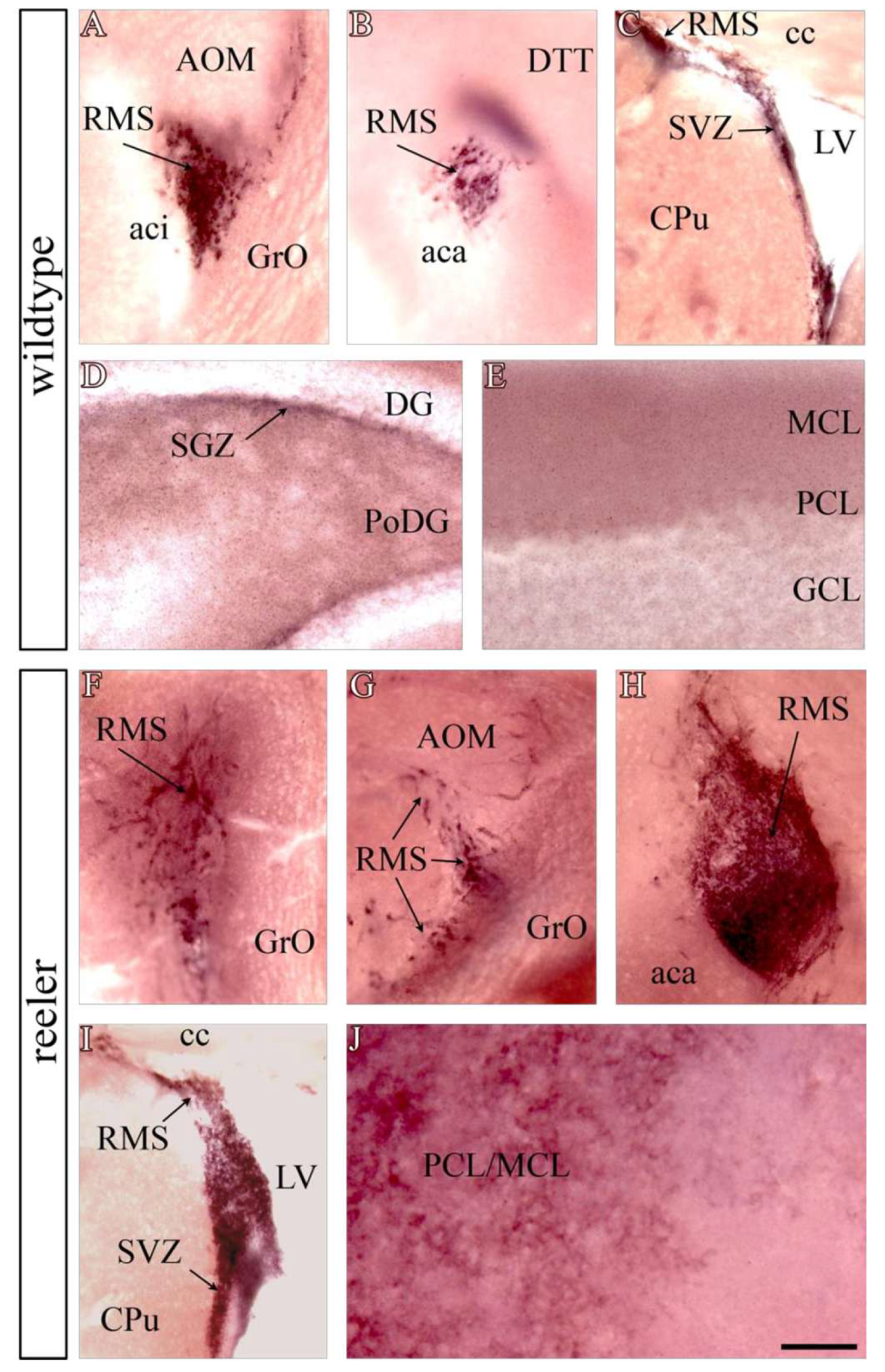
Figure 1. Localization of expression sites of functional reelin receptors in the adult brain. AP-reelin staining in wild-type (AE) and reeler (FJ) mice reveals the presence of functional reelin receptors along the SEZ/RMS in both wild-type (AC) and reeler (FI) brains. The RMS of reeler mutants appears very thin (hypocellular) in its rostral portion (FG), with no signs of neuroblast accumulation in the nucleus of the olfactory bulb, but considerably thicker (extremely hypercellular) in its caudal part (HI). FRRs are also present in the subgranular zone of the hippocampus (D). Note the weak staining of the molecular layer of the cerebellum in wild-type mice (E), which is markedly increased in reeler (J). Abbreviations: aca, anterior part of the anterior commissure; aci, intrabulbar part of the anterior commissure; AOM, medial part of the anterior olfactory nucleus; cc, corpus callosum; CPu, caudate–putamen nucleus; DTT, dorsal tenia tecta; DG, dentate gyrus; GCL, cerebellar granular layer; GrO, granular layer of the olfactory bulb; LV, lateral ventricle; MCL, cerebellar molecular layer; PCL, Purkinje cell layer; PoDG, polymorphic layer of the dentate gyrus; RMS, rostral migratory stream; SVZ, subventricular zone. Scale bar: 200 μm.
Neuroglia 06 00043 g001
Figure 2. Reelin binds to the main cell types of the major adult neurogenic niche. All EZ/SEZ cell subpopulations are competent to receive reelin as a ligand. Colocalization of functional ApoER2 with specific markers is shown for S100β+ cells, corresponding to ependymal type E cells (AC). Subventricular astrocytes, known as type B cells and expressing GFAP, also exhibit some degree of reelin receptor expression (DF). The strongest colocalization is observed between ApoER2 and doublecortin, a marker of neuroblasts or type A cells (GI). Cells in S phase of the cell cycle (1 h after BrdU injection, (JL)) and transient amplifying cells, or type C cells, which express Mash1 (also known as Ascl1), are also competent for reelin binding. Scale bar: (AC); (GI): 50 μm. (DE): 30 μm; (MO): 40 μm.
Neuroglia 06 00043 g002
Figure 3. Reelin signaling remains active in the adult SVZ. Ex vivo Dab1 phosphorylation assay after stimulation of a dissected tissue extract from the EZ/SEZ of reeler brains with the AP-RR36 fusion protein. Because of the lack of specific antibodies against phosphorylated Dab1 tyrosine residues, immunoprecipitation of this protein was required to enrich the sample for detection of phosphorylated tyrosine residues by Western blot (A). The addition of the central fragment of reelin resulted in a 60% increase in the ratio between phosphorylated Dab1 and total Dab1, in parallel with a 20% decrease in total Dab1 present in the sample (B).
Neuroglia 06 00043 g003
Figure 4. Increased ventricular volume in mutants related to reelin and its signaling pathway and recovery of ependymal cilia density due to ectopic expression of Reelin. Nissl staining showing the size of the lateral ventricles in different mutants compared with the wild-type genotype (A). Null mutants for Reelin, ApoER2, and Dab1 (B,C,E), as well as conditional ablation of Dab1 in progenitors (G,H), show an increase in ventricular size, whereas gain of Reelin function normalizes this defect (F). The Vldlr mutant exhibits a normal ventricular phenotype (D). Photomicrograph obtained by scanning electron microscopy of ependymal cilia in the wall of the lateral ventricle (H). Cilia density and morphology are severely affected in reeler mutants (I). Expression of Reelin by neural progenitors in the adult subependymal zone results in normalization of this phenotypic defect (J). Scale bar: (AF), 100 μm; (HJ), 10 μm.
Neuroglia 06 00043 g004
Figure 5. Effects of Dab1 ablation after tamoxifen treatment in adult mice. Immunohistochemical detection of Dab1. The signal is present in neuroblasts of the subependymal zone and RMS (arrows, (A)) and disappears in cells expressing the Cre recombinase enzyme (arrowheads, (B)). Nissl staining shows enlargement of the lateral ventricle in Dab1flx/flx; Cre+ mice after tamoxifen exposure (D), compared to control (C). Three-day BrdU pulse and chase (E,F). In mice with both Dab1 floxed alleles (F), an accumulation of BrdU+ cells (neuroblasts) is observed along the subventricular zone and in the corticostriatal sulcus, the onset of the RMS, compared to control (E), where neuroblasts have already left the zone to migrate into the olfactory bulb. Scale bar: 100 μm. [CPu, caudate–putamen nucleus; LV, lateral ventricle; SEZ, subependymal zone; RMS, rostral migratory stream].
Neuroglia 06 00043 g005
Figure 6. Ectopic Reelin expression in the adult Nestin-Reelin transgenic mouse and rescue of RMS morphology in the core of the olfactory bulb. Immunofluorescence for the detection of reelin using the monoclonal antibody G10. No immunoreactivity to reelin is observed in wild-type animals (AC) or in reeler mutants (DF). However, the Nestin promoter directs ectopic Reelin expression in the subependymal zone of reeler ne-Reelin transgenic mice (GI). Immunohistochemical detection of BrdU after a 3-day pulse-and-chase experiment in wild-type mice (J). In reeler mutants, cells appear scattered and reduced in number compared to controls (K). The presence of ectopic reelin on a reeler background rescues this phenotype, both in morphology and in the number of cells reaching the center of the olfactory bulb (L). [cc, corpus callosum; LV, lateral ventricle; Str, striate nucleus]. Scale bar: (AI), 150 μm; (JL), 200 μm.
Neuroglia 06 00043 g006
Figure 7. Reelin modifies neuroblast migration in vivo. Infusion of recombinant reelin or control medium for 5 days into the right ventricle of adult reeler mice by implantation of osmotic mini-pumps (A). Infusion of control medium produces no change in neuroblast distribution, as shown by immunohistochemistry for doublecortin detection (B). In contrast, the presence of recombinant reelin dramatically alters the normal localization of neuroblasts around the ventricle ipsilateral to the infusion site ((C), arrowheads). [cc, corpus callosum; LV, lateral ventricle; RMS, rostral migratory stream; Str, striatal nucleus]. Scale bar: 200 μm.
Neuroglia 06 00043 g007
Disclaimer/Publisher’s Note: The statements, opinions and data contained in all publications are solely those of the individual author(s) and contributor(s) and not of MDPI and/or the editor(s). MDPI and/or the editor(s) disclaim responsibility for any injury to people or property resulting from any ideas, methods, instructions or products referred to in the content.