Review

A Mini-Review on Enhancing Solubility in Topical Hydrogel Formulations Using Solid Dispersion Technology for Poorly Water-Soluble Drugs

by
Zaid Dahma
1,
Covadonga Álvarez-Álvarez
1,2,* and
Paloma Marina de la Torre-Iglesias
1,2,*
1
Department of Pharmaceutics and Food Technology, Faculty of Pharmacy, Complutense University of Madrid, Plaza Ramón y Cajal s/n, 28040 Madrid, Spain
2
Instituto Universitario de Farmacia Industrial, Complutense University of Madrid, Plaza Ramón y Cajal s/n, 28040 Madrid, Spain
*
Authors to whom correspondence should be addressed.
Colloids Interfaces 2025, 9(2), 17; https://doi.org/10.3390/colloids9020017
Submission received: 21 January 2025 / Revised: 12 March 2025 / Accepted: 18 March 2025 / Published: 21 March 2025
(This article belongs to the Special Issue Biocolloids and Biointerfaces: 2nd Edition)

Abstract

:
The solubility behavior of drugs is a critical factor in formulation development. Approximately 40–45% of new drugs face market entry challenges due to low water solubility. Enhancing drug bioavailability is thus essential in developing pharmaceutical dosage forms. Many biopharmaceutical class II and IV drugs are commonly prescribed to treat inflammations, infections, and pain from various pathologies. Their oral administration has several drawbacks, including significant first-pass liver effects, low bioavailability, and adverse gastrointestinal effects. Topical application has gained relevance due to its advantages in delivering drugs directly to the target site, avoiding gastrointestinal irritation, and increasing their effectiveness. However, topical hydrogel formulations with poorly water-soluble drugs face challenges related to the skin’s permeability. Therefore, preparing topical hydrogels using solid dispersions (SDs) is an effective strategy to enhance the dissolution rate of poorly soluble drugs, thereby improving their topical bioavailability. In this review, the concepts of SDs, topical delivery systems, and topical hydrogel formulations incorporating SDs, as well as their preparation methods, characterization, and applications, will be discussed.

Graphical Abstract

1. Introduction

The solubility of a drug is a physicochemical property that influences its absorption and therapeutic efficacy. Drugs having poor aqueous solubility can lead to unsuccessful formulation development. Improving the solubility and dissolution rates of hydrophobic drugs remains a significant challenge in pharmaceutical development. Several strategies have been developed to enhance the solubility and bioavailability of poorly water-soluble drugs, including solid dispersions (SDs) [1,2,3], complexation [3], and micronization [4,5]. Lipid-based systems, such as self-emulsifying drug delivery systems (SEDDSs), which form fine emulsions in the gastrointestinal tract, have also shown promise in improving drugs’ solubility and absorption [6,7]. Other techniques like co-crystals [8,9] and nanonization [10,11] have been widely explored for their ability to enhance drug dissolution rates. Furthermore, nanostructured lipid carriers (NLCs) and solid lipid nanoparticles (SLNs) are emerging as effective carriers that offer improved drug stability, controlled release, and enhanced solubility [12]. Thiomers, a class of thiolated polymers, also contribute to improving bioavailability by prolonging the retention of drugs at the mucosal sites through bioadhesion mechanisms [13]. These diverse approaches are critical to overcoming the challenges associated with delivering poorly soluble drugs.
SDs have been shown to be an effective method for increasing the dissolution rate and bioavailability of a variety of poorly water-soluble active pharmaceutical ingredients (APIs). SDs refer to a class of solid products that have at least two separate components, in general a hydrophobic drug and a hydrophilic matrix. This matrix can either be amorphous or crystalline. There are several ways to prepare SDs, including spray-drying, gel entrapment, solvent evaporation, kneading, melting, a melting solvent method, co-precipitation, using modified solvents, co-precipitation with supercritical fluid and co-grinding, and evaporation [14].
The Biopharmaceutical Classification System (BCS) is a scientific framework that classifies drugs into four categories based on their aqueous solubility and intestinal permeability. This framework helps with predicting and deciding whether there is a need for in vivo bioequivalence studies. Drugs are divided into class I (high solubility, high permeability), class II (low solubility, high permeability), class III (high solubility, low permeability), and class IV (low solubility, low permeability). Both the World Health Organization (WHO) [15] and the U.S. Food and Drug Administration (FDA) [16] apply the BCS to support biowaivers, particularly for immediate-release oral dosage forms, streamlining the approval of generic medicines when certain criteria are met. Biopharmaceutical class II drugs, which have high permeability but low solubility, along with class IV drugs, which exhibit both poor solubility and permeability, are commonly prescribed to treat inflammations, infections, and pains caused by a variety of symptoms. However, their oral administration has many adverse effects, with the majority being related to the gastrointestinal tract [17], and since a large number of inflammatory infections occur locally and near the body’s surface [18], topical application has become more important, with the advantage of delivering drugs directly to the site of inflammation, avoiding gastrointestinal irritation and decreasing adverse systemic effects. Moreover, cutaneous inflammatory illnesses impact a high number of people worldwide. While these conditions generally have lower mortality rates compared to those for other diseases, they significantly affect patients’ quality of life. According to the Institute of Health Metrics and Dermatology Experts, dermatitis, cellulitis, psoriasis, acne vulgaris, seborrheic dermatitis, and pyoderma are among the most common skin diseases [19]. Hydrogels have emerged as versatile carriers for drug delivery due to their ability to retain large amounts of water while maintaining their structural integrity. Their biocompatibility and tunable properties make them particularly valuable for biomedical applications, including controlled drug release. Although traditionally used for hydrophilic drugs, recent advances have enabled the incorporation of hydrophobic drugs into hydrogel matrices, significantly enhancing their solubility, bioavailability, and controlled-release profiles. Several approaches have been developed to achieve this, including micelle formation, nanostructured lipid carriers, cyclodextrin inclusion complexes, and amphiphilic copolymers that create hydrophobic domains within the hydrogel network. Thermosensitive hydrogels, such as those based on a poly(lactic-co-glycolic acid)-poly(ethylene glycol)-poly(lactic-co-glycolic acid) (PLGA-PEG-PLGA) triblock copolymer, are particularly promising, as they transition from a liquid into a gel at body temperature, enabling localized and sustained drug delivery. This feature is particularly relevant in cancer therapy, where hydrogels loaded with hydrophobic chemotherapeutic agents have shown improved therapeutic efficacy while minimizing systemic toxicity. Furthermore, pH-sensitive hydrogels have been designed to provide controlled drug release in response to environmental pH variations, making them suitable for targeted delivery to specific tissues or diseased areas. This ability to improve the solubility and controlled release of hydrophobic drugs is highly relevant in the context of topical drug delivery, where achieving adequate solubility and skin permeability remains a major challenge [20].
In this work, the combination of SDs with hydrogels for topical drug delivery, solid dispersion hydrogels (SDHs), has been reviewed. Several relevant studies on SDHs for topical applications were selected, spanning from 2014 to 2024. The selection of this limited number of studies was due to the specificity of the topical application domain. However, through a detailed exploration of this topic, the need to emphasize the advantages and potential of SDHs has been identified. Therefore, the focus of this mini-review is to provide a concise overview of SDs, topical delivery systems, and SDHs as effective approaches to enhancing the dissolution rate of poorly soluble drugs in topical preparations. This review includes an examination of the different types of SDs, their preparation methods, and topical delivery systems, before delving into hydrogels that incorporate SDs. It highlights the challenges associated with the low water solubility of APIs and their permeability through the skin. Additionally, the concepts of SDHs, their methods of preparation, their characterization, and their applications in boosting drugs’ topical bioavailability are discussed.

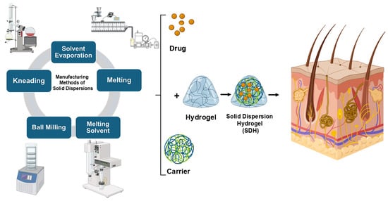
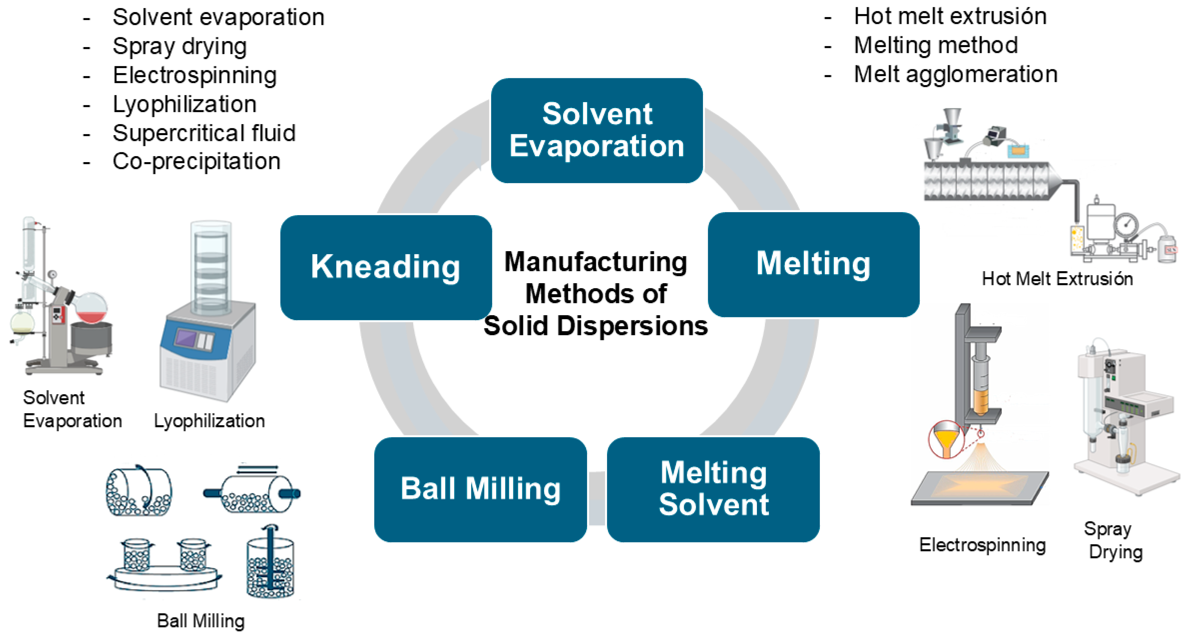
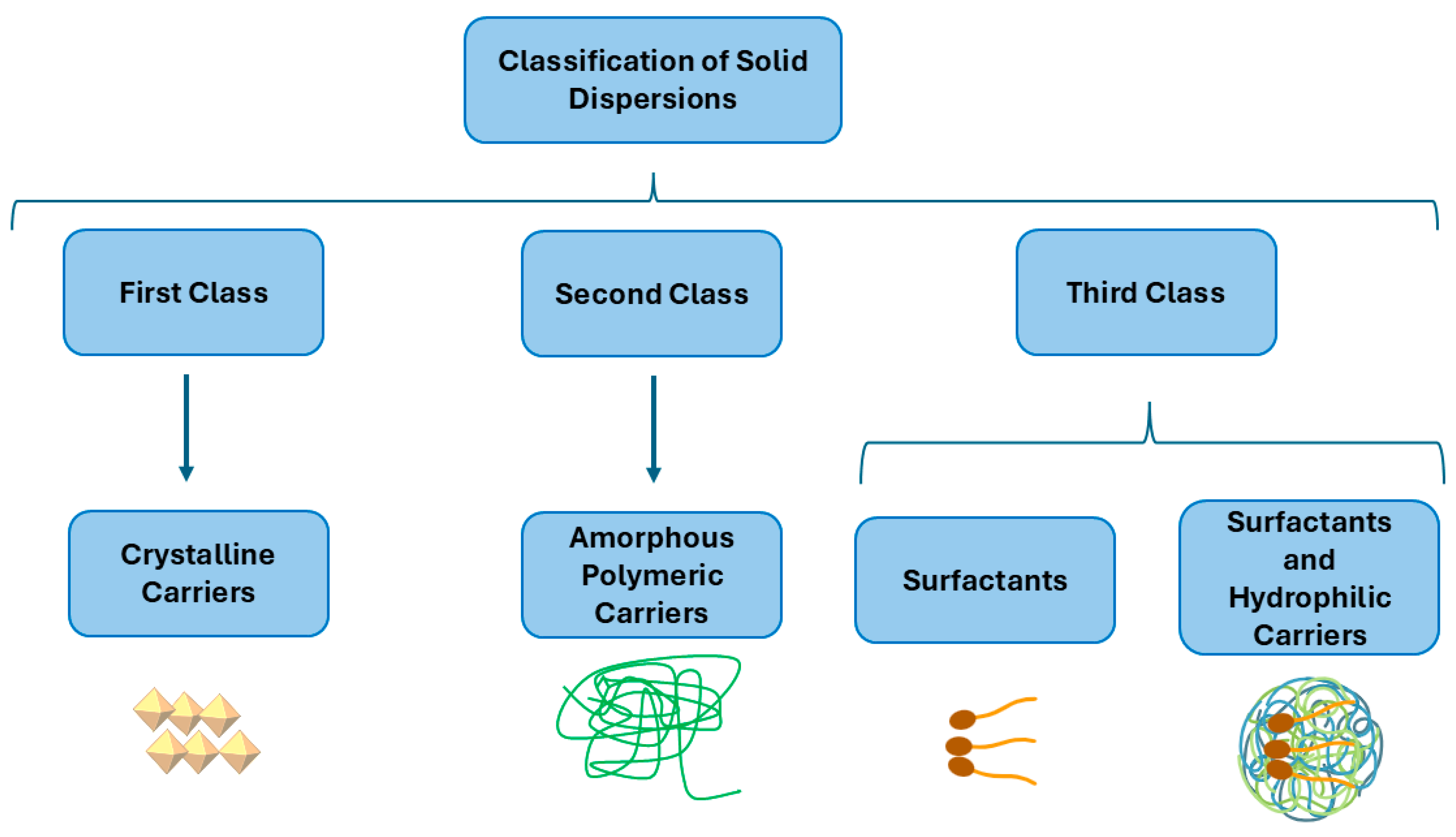
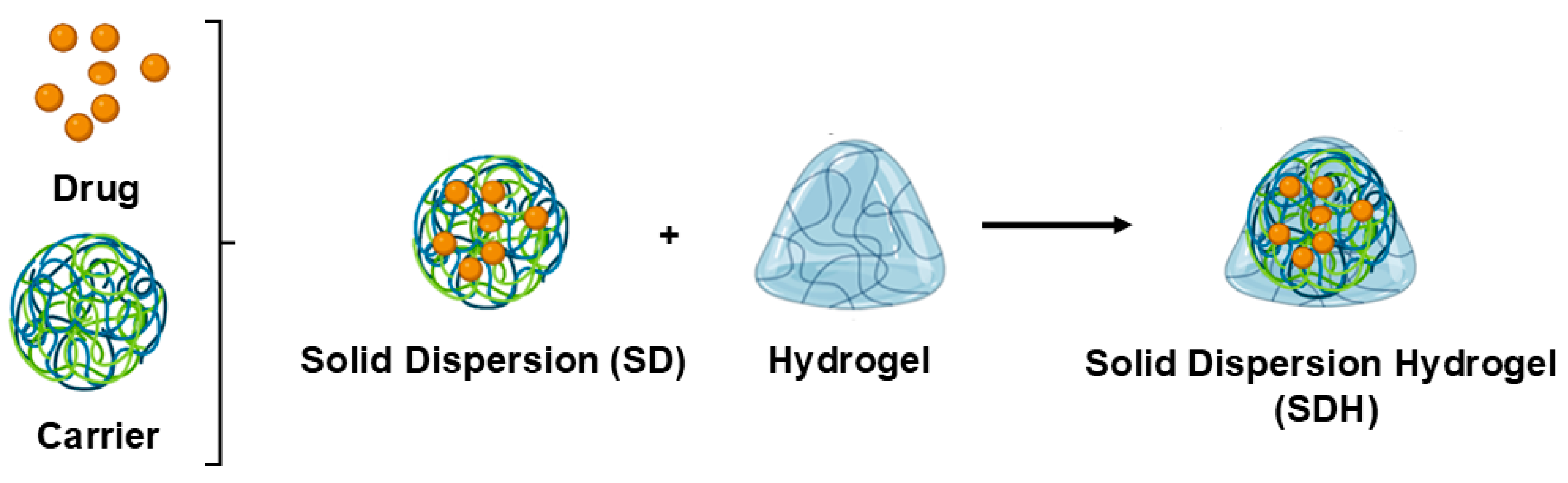
2. Solid Dispersions (SDs)

Gibbs free energy is a thermodynamic measure that indicates the maximum amount of useful work a system can perform at a constant temperature and pressure. It depends on enthalpy, which is related to the total heat content of the system, and entropy, which measures the disorder or randomness of the particles. A change in the Gibbs free energy determines whether a process will occur spontaneously. If this change is negative, the process is spontaneous; if it is positive, the process is non-spontaneous; and if it is zero, the system is in equilibrium. In the context of SD formulations, which are solid products containing a hydrophobic drug dispersed in hydrophilic carriers, this concept is particularly relevant. SD formulations increase a drug’s solubility, surface area, and dissolution rate by altering its crystalline structure, thus reducing its Gibbs free energy. This reduction in its Gibbs energy contributes to the enhancement of the drug’s solubility and dissolution rate, making the process more thermodynamically favorable [21].
SDs are divided into three classes depending on the carriers that are used and on the fabrication method. The first class of SDs was created by Sekiguchi and Obi in 1961 [22]. In this class, an SD is produced utilizing crystalline carriers such as urea and sugars. However, these formulations were thermodynamically unstable, which resulted in slower drug release. Due to this thermodynamic instability of the first class of SDs [23], a second class of SDs was introduced that utilized amorphous polymeric carriers [24] instead of urea or sugars. These carriers are synthetic polymers such as polyethylene glycol (PEG) [25], polyvinylpyrrolidone (PVP) [26,27,28], polymethacrylates [29], natural polymers including ethyl cellulose [30], hydroxypropylmethylcellulose (HPMC) [31,32,33], and starch derivatives such as cyclodextrins (CDs) [34]. As a result, the drug particle size becomes smaller, improving its wettability and increasing its solubility.
Recently, a third class of SDs was created. A surfactant alone or in a combination with other hydrophilic carriers is used in the preparation of this class of SDs (Figure 1). This class provides a significant advantage in terms of its solubility, stability, and formulation flexibility, making these SDs a valuable tool in pharmaceutical development. Surfactants are commonly used to enhance the solubility of poorly water-soluble drugs and play a significant role in the pharmaceutical industry. Additionally, they serve as wetting agents, emulsifiers, detergents, dispersants, and foaming agents. Many surfactants are used to prepare SDs, such as inulin lauryl carbamate (Inutec®) [29], lauroyl polyoxyl-32 glycerides (Gelucire® 44/14 [35], glyceryl dibehenate (Compritol® 888 ATO) [36], a polyoxyethylene-polyoxypropylene block copolymer (Poloxamer 407) [37], and sodium dodecyl sulfate (SDS) [38].
Soluplus®-based solid dispersions have emerged as a promising strategy to enhance the solubility and bioavailability of poorly water-soluble drugs. Soluplus®, an amphiphilic polymer, facilitates the formation of stable amorphous solid dispersions, thereby improving drug dissolution rates. For instance, a recent study demonstrated that a Soluplus®-mediated amorphous solid dispersion of diosgenin (BCS class II) significantly increased its solubility and stability compared to these properties in the pure drug [39]. Additionally, third-generation solid dispersions combining Soluplus® and poloxamer 407 have been shown to enhance the oral bioavailability of resveratrol (BCS class II), increasing its solubility and reducing intestinal efflux and metabolism mechanisms [37]. These advantages underscore the potential of Soluplus®-based solid dispersions in improving the therapeutic performance of hydrophobic drugs.
SD formulations can be prepared using various methods, each with distinct advantages and limitations. These methods include solvent evaporation, kneading, melting, melting solvent methods, and ball milling (Figure 2).
The melting method, introduced by Sekiguchi and Obi in 1961 [40], involves heating a physical mixture of a drug and a hydrophilic carrier until both melt at a temperature slightly above their eutectic point. The resulting mixture is then rapidly cooled and solidified in an ice bath while being stirred. This approach is favored for its simplicity, cost-effectiveness, and ease of scalability. However, it poses a risk of thermal degradation for heat-sensitive drugs. Various fabrication techniques, such as hot melt extrusion and melt agglomeration, have been developed based on this principle, with continued use in modern research [41]. A notable variation of the melting method is the spray-congealing technique, which, although similar, introduces an additional atomization step. Instead of simply cooling the melted mixture, the spray-congealing method involves spraying it into a cold environment, causing the droplets to solidify into spherical particles. This modification allows for more controlled particle shapes and sizes, offering the potential to improve the solubility and bioavailability of poorly soluble drugs by enhancing their surface area. Both methods aim to enhance the solubility and bioavailability of poorly soluble drugs, but spray-congealing adds the step of atomization to create specific particle shapes and sizes [42].
The solvent evaporation method, which has been widely employed in the pharmaceutical industry since its introduction by Tachibana and Nakamura in 1965 [43], is especially useful for heat-sensitive compounds. In this process, both the drug and carrier are dissolved in a volatile solvent, eliminating the need for heat, which is a key advantage over the melting method. The solvent is then evaporated under constant stirring, leading to the formation of an SD. This technique is versatile and can be adapted through various fabrication processes, including spray-drying, electrospinning [44], lyophilization, supercritical fluid technology, and co-precipitation, which offer additional control over the final product’s properties [45].
The kneading method, studied by Dhandapani and El-Gied [29], involves dispersing the carrier in water to form a paste, into which the drug is incorporated and kneaded thoroughly. After kneading, the mixture is dried, and, if necessary, sieved. This method is advantageous because it is simple and scalable for industrial production and allows for the optimization of key process variables, such as the drug-to-polymer ratio and kneading time. Furthermore, it is compatible with a wide range of polymers, including surfactant-based ones like Poloxamer 188, which enhances the drug solubility and is generally safe for oral use [46]. However, it may not be as effective for drugs with extremely low solubility or for those that require fine-tuned particle size control.
The melting solvent method, as explored by Goldberg et al. [47], combines elements of both melting and solvent evaporation techniques. In this approach, the drug is dissolved in an appropriate solvent and then mixed with the melted carrier. The mixture is then evaporated to dryness, effectively creating an SD. This method is particularly beneficial for drugs with high melting points, although it requires careful control over the evaporation and mixing processes to avoid solvent residue or product inconsistency.
Lastly, the ball milling method involves grinding the drug and the carrier together in a ball mill to produce a uniform, fine mixture [48]. This technique is highly effective in enhancing the solubility and dissolution rate of poorly soluble drugs by increasing their surface area and reducing their particle size. While it offers clear benefits in terms of solubility enhancement, the ball milling method requires specialized equipment and may pose a risk of contamination from the milling media, which can impact the purity of the final product [49,50].
Each of these methods brings its own strengths and challenges, and the choice of method depends on the properties of the drug and the carrier and the desired characteristics of the final SD.
A recent study highlights the use of Fused Deposition Modeling (FDM) 3D printing technology as a novel approach to the fabrication of solid dispersions. This method offers precise control over the composition, structure, and release profile of drug delivery systems, making it a promising alternative to traditional fabrication techniques. FDM 3D printing enables the creation of solid dispersions with tailored physicochemical properties, such as a uniform drug distribution and enhanced dissolution rates. These properties are particularly beneficial for improving the solubility and bioavailability of poorly water-soluble drugs. This technology also provides advantages such as scalability, reproducibility, and the ability to design complex dosage forms, positioning it as an innovative and valuable tool for pharmaceutical development [51].
Figure 2. Conventional manufacturing methods for SDs. This figure was created by the authors using BioRender.com (accessed on 5 March 2025) and Microsoft PowerPoint. The electrospinning illustration was adapted with modifications from Ref. [43], and the ball milling illustration was adapted with permission from Ref. [48]. Copyright 2021, Springer.
Colloids 09 00017 g002
During the fabrication of SDs, many excipients have been used as carriers (shown in Table 1), like PEG 4000 and Gelucire® 50/13 [52], PVP K25 and PVP K30 [53], Gelucire® 44/14 and Gelucire® 50/13 [54], amino methacrylate copolymer (Eudragit® EPO) and HPMC [55], Gelucire® 50/13: Gelucire® 48/16 [42], polyvinylpyrrolidone-vinyl acetate (Kollidon® VA 64) [56], low-substituted hydroxypropyl cellulose (LHPC) [38,57], maltodextrin [58], kaolin [49], lactose [50], Poloxamer 407, Poloxamer 188, and Gelucire® 44/14 [59]. These carriers offer several advantages: they enhance the solubility and dissolution rate of poorly soluble drugs, improve their bioavailability, and provide stability to drug formulations. Additionally, they allow for the customization of drug release profiles and are compatible with various manufacturing processes, making them versatile and effective in pharmaceutical applications [60].
While SDs offer numerous advantages for enhancing the bioavailability of drugs through an increase in their solubility, they also come with certain disadvantages, such as changes in crystallinity and a decline in the dissolution rate over time. Due to their thermodynamic instability, they are particularly sensitive to temperature and humidity during storage. These conditions can induce phase separation and crystallization by increasing the molecular mobility, lowering the glass transition temperature (Tg), or disrupting the interactions between the drug and its carrier, ultimately leading to the reduced solubility and dissolution rate of the drug [21,61]. To overcome this challenge, over the past few decades, various SD systems have been developed by incorporating poorly water-soluble drugs into different water-soluble carrier polymers. Numerous comprehensive reviews have summarized the methods for preparing, characterizing, and stabilizing these amorphous SDs [62,63]. To improve SDs and prevent the recrystallization of the API, carriers and surfactants can be optimized in several ways. Using polymers with high glass transition temperatures (Tg) helps maintain the API in an amorphous state, reducing the recrystallization risk. For example, PVP and HPMC are excellent carriers because they can form strong hydrogen bonds with the API, stabilizing the amorphous form. Eudragit® EPO, an amino methacrylate copolymer, is another effective carrier known for its ability to form stable hydrogen bonds with various drugs, further inhibiting recrystallization. Several factors, including reduced molecular mobility and drug–polymer interactions, have been found to prevent drugs’ crystallization from the amorphous state in polymer carriers. With the availability of a wide variety of polymers, a major challenge in SD design is the polymer selection. Incorporating surfactants can enhance the miscibility of the drug and the carrier, stabilizing the amorphous form and increasing its solubility. Poloxamers (e.g., Poloxamer 188 and 407) are block copolymers that improve the solubility and dissolution rate of drugs by forming micelles and hydrogen bonds with the drug molecules. Gelucire® surfactants, such as Gelucire® 50/13 and 44/14, are lipid-based and help maintain the amorphous state of the drug by enhancing its miscibility with the carrier. SDS, an anionic surfactant, reduces surface tension and forms micelles, which can further stabilize the drug in its amorphous form. By carefully choosing and optimizing these carriers and surfactants, the stability and bioavailability of a drug can be significantly improved. The formation of hydrogen bonds between the drug and the carrier or surfactant is crucial, as these bonds reduce the mobility of the drug molecules, preventing them from rearranging into a crystalline structure and thus maintaining the drug in a more soluble and bioavailable amorphous state [21,64].

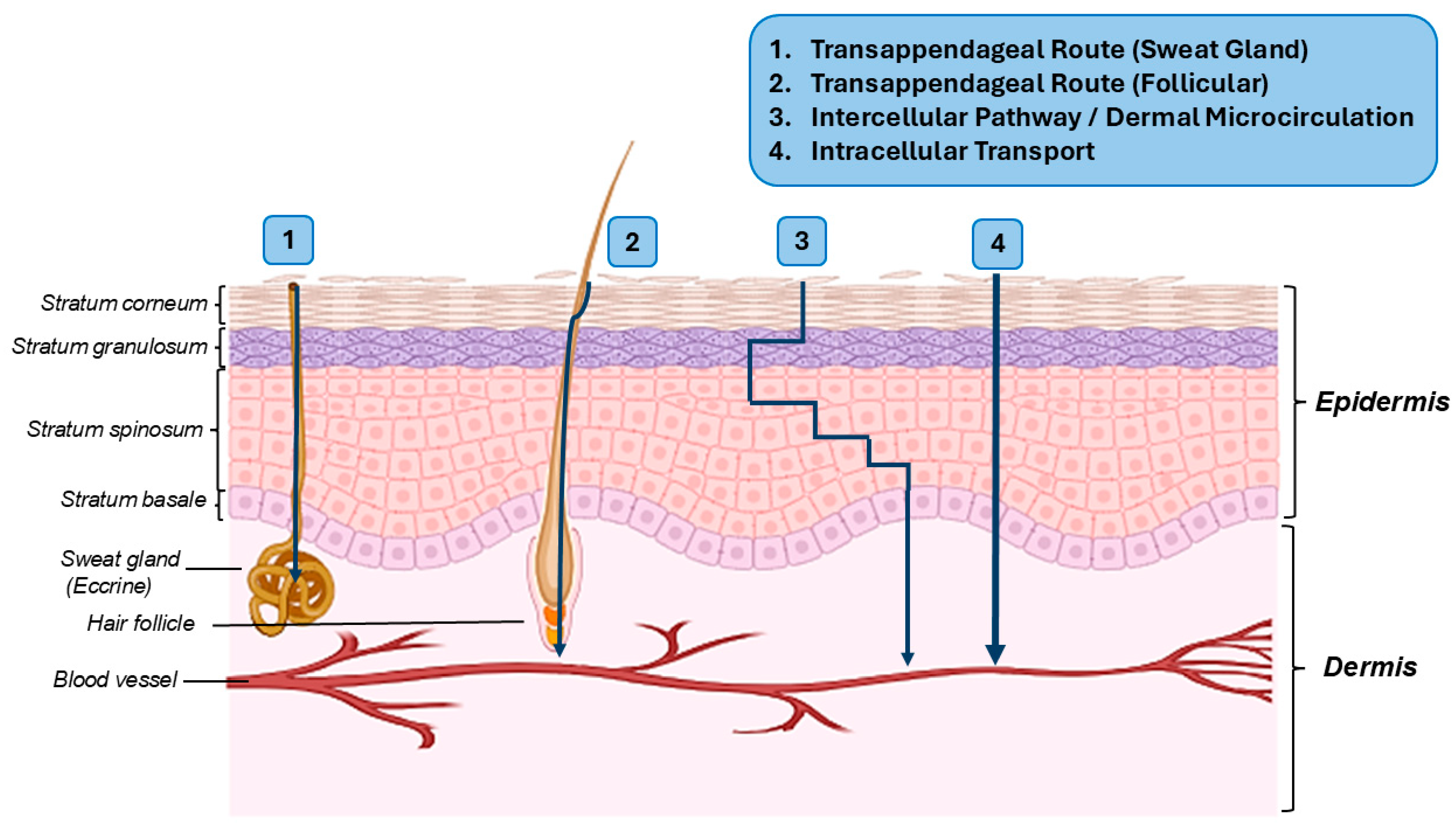
3. Topical Drug Delivery Systems and Their Pathways to the Target Tissues

Topical drug delivery is the application of a drug directly to the skin in order to treat or cure skin-related diseases. These topical drug delivery methods are typically utilized for local skin infections, such as fungal infections, or local inflammation caused by many pathologies. A topical delivery system is referred to as an element that brings a specific drug into contact with and through the skin. This delivery system consists of two essential types of products, external and internal. External formulations are the preparations that are spread or dispersed onto the cutaneous tissues to cover the targeted area. Meanwhile, internal topical formulations are used for local activity on anorectal tissues, vaginally, or on the mucous membranes of the mouth. Topical drug applications offer numerous advantages by delivering drugs more selectively to the target site. This not only avoids gastrointestinal irritation and reduces adverse systemic effects but also bypasses the gastrointestinal metabolism. Additionally, topical applications can enhance the treatment efficacy and provide faster symptom relief [65]. The main types of topical formulations include creams, ointments, hydrogels, lotions, and pastes, each designed to deliver active ingredients to the skin for various therapeutic effects [66].
Topical hydrogels, which are essential in modern medicine due to their wide range of applications, are formulations designed to deliver APIs through the skin. These hydrophilic polymeric networks can absorb large amounts of water while maintaining their structural integrity, making them highly effective carriers for both hydrophilic and hydrophobic drugs. Their unique properties, including their biocompatibility, tunable mechanical strength, and responsiveness to physiological stimuli, allow for controlled drug release, improved bioavailability, and enhanced skin permeability. Beyond topical applications, hydrogels have proven invaluable in various biomedical fields. In drug delivery, they enable sustained and targeted therapeutic release, minimizing systemic side effects. Their ability to maintain a moist environment and promote tissue regeneration makes them particularly useful in wound healing. Additionally, in tissue engineering, hydrogels act as scaffolds that mimic the extracellular matrix, supporting cell adhesion, proliferation, and differentiation. Their role in transdermal and ocular delivery systems further underscores their versatility in overcoming key challenges in localized and systemic treatments [67]. These hydrogels typically consist of a three-dimensional network of hydrophilic polymers, such as polyvinyl alcohol (PVA) or polyacrylic acid (Carbopol®), which can absorb and retain significant amounts of water. PVA gels through a process called physical crosslinking, where the polymer chains form crystalline regions that act as junction points, creating a network structure. Carbopol®, on the other hand, gels through a process of neutralization, where the acidic polymer is neutralized with a base such as sodium or potassium hydroxide or triethanolamine, causing it to swell and form a gel. Other common gelling agents include alginate, which gels through ionic crosslinking with calcium ions; gelatin, which gels through thermal gelation as it cools; and xanthan gum, which forms gels through hydrogen bonding and the entanglement of its polymer chains. This unique structure allows for the sustained and controlled release of the APIs, enhancing their therapeutic efficacy as stabilizers [68].
The integration of 3D printing technologies into hydrogel fabrication has emerged as an innovative method for designing structures with specific mechanical and functional properties. For instance, a 3D printing method based on the use of carbomer as a rheological modifier has been developed, enabling the direct printing of various multifunctional hydrogel inks. This approach allows for the creation of hydrogels with double networks, magnetic hydrogels, thermoresponsive hydrogels, and biogels, offering excellent printability and biocompatibility. These advancements hold significant potential for medical applications, including tissue engineering, wound healing, and controlled drug delivery [69].
Additionally, specialized hydrogels, such as mucoadhesive hydrogels, are designed to adhere to the mucosal tissues, prolonging drugs’ residence time and improving their bioavailability in applications such as oral, ocular, and vaginal drug delivery. These formulations rely on mucoadhesive polymers like chitosan, Carbopol®, and sodium carboxymethylcellulose, which interact with mucins through electrostatic forces and hydrogen bonding to enhance the adhesion and drug retention at the site of application [70]. A further advancement in mucoadhesive technology involves thiolated polymers (thiomers), which form covalent disulfide bonds with the cysteine-rich domains of mucins, significantly enhancing the adhesion strength and resistance to mucosal turnover. Examples include thiolated chitosan and poly(acrylic acid)-cysteine conjugates, which have been shown to improve drugs’ residence time and permeability, making them promising carriers for mucosal drug delivery [71].
The importance of hydrogels lies in their versatility and biocompatibility. They can be used to treat a variety of skin conditions, from minor irritations to chronic wounds, due to their ability to maintain a moist environment that promotes healing. Additionally, hydrogels can be formulated to provide cooling and soothing effects, making them ideal for burns and other inflammatory conditions. Common excipients in hydrogel formulations include (1) gelling agents, which provide the structural matrix of the hydrogel, ensuring it maintains its form and consistency; (2) preservatives, which prevent microbial growth, ensuring the hydrogel remains safe and effective over time; and (3) stabilizers, which help maintain the physical and chemical stability of the hydrogel, preventing the degradation of active ingredients. Hydrogels may also contain neutralizing (alkalinizing) agents, such as Carbopol®, as well as antioxidants like butylated hydroxyanisole (BHA), butylated hydroxytoluene (BHT), sodium benzoate, benzoic acid, and others. The advantages of hydrogels include moisture retention, which helps maintain a moist environment beneficial for wound healing; controlled release, which allows for the sustained and controlled release of active ingredients; cooling effects, which provide a soothing and cooling sensation ideal for burns and inflammatory conditions; and biocompatibility, which generally ensures they are well tolerated by the skin, reducing the risk of irritation. However, hydrogels also have disadvantages, such as a limited drug load, which may make them unsuitable for delivering large amounts of active ingredients; stability issues, as they can be prone to dehydration or microbial contamination if not properly formulated and stored; cost, as the production of hydrogels can be more expensive compared to that of other topical formulations; and application limitations, as they may not adhere well to certain areas of the body or under certain conditions [72]. Overall, topical hydrogels represent a versatile and effective solution for various dermatological and medical applications, offering improved patient outcomes through enhanced drug delivery and wound care [73]. Hydrogels, thanks to their biocompatibility, high water retention capacity, and tunable controlled-release properties, act as versatile carriers for other advanced drug delivery systems, such as nanoparticles, effectively combining the structural benefits of hydrogels with the enhanced solubility, targeting, and cellular uptake capabilities of nanoparticles. Incorporating nanoparticles into hydrogels enhances drug solubility, targeting efficiency, and controlled release, making these hybrid systems highly versatile. Examples include polyacrylamide hydrogel nanoparticles functionalized with F3 peptides for tumor-targeted cisplatin delivery, showing tumor reductions and improved safety. Similarly, chitosan-based hydrogel nanoparticles have improved the nasal delivery of piperine for Alzheimer’s treatment, enhancing brain targeting while reducing nasal irritation. In vaccines, cholesteryl-pullulan hydrogel nanoparticles have been used to deliver pneumococcal antigens, inducing protective immune responses. These hydrogel–nanoparticle systems also enable stimuli-responsive release, such as electroresponsive phenytoin delivery for epilepsy or salt-triggered protein release, demonstrating their potential for personalized and controlled therapies. Overall, these hybrid systems offer improved solubility, targeting, and controlled release, making them promising tools for next-generation drug delivery in oncology, neurology, and immunotherapy [74].
The stratum corneum, the skin’s outermost layer, serves as a protective barrier, making passive penetration through it the rate-limiting step for epidermal drug transport [75]. Topically applied drugs can accumulate in the stratum corneum, epidermis, dermis, and fatty tissue, creating a reservoir for sustained release to the surrounding tissues (Figure 3) [75,76,77].
The reservoir’s efficiency is influenced by factors such as APIs, the protein-binding capacity, clearance, application parameters, compound concentration, lipid/water solubility, and percutaneous absorption [75]. The drug’s physical state—solubilized or suspended—significantly impacts its permeation, with solubilized drugs achieving a higher flux due to their increased thermodynamic activity and improved partitioning. This enhances their efficacy at lower concentrations, minimizes irritation risks, and reduces costs [77]. Moreover, formulation characteristics like the type of system (monophasic vs. multiphasic), viscosity, and pH play a crucial role in the drug transport across the skin. Developing effective topical drug products requires careful consideration of the factors that influence drug penetration and permeation. To reach the dermal level, substances may pass through the sweat ducts or hair follicles [78,79] and penetrate nearby blood vessels for distribution to deeper tissues (Figure 3) [80]. A drug’s ability to penetrate the stratum corneum and the underlying tissues depends on its molecular properties (Table 2), including size, water solubility, and lipophilicity (its ability to dissolve in fats is also crucial for penetrating the lipid-rich stratum corneum). Drugs with a molecular weight below 500 g/mol, such as ibuprofen (354.27 g/mol), meloxicam (351.403 g/mol) [57], indomethacin (357.787 g/mol) [38], and diclofenac (296 g/mol), traverse the skin barrier more easily [81]. A high water solubility enhances absorption, while sufficient lipophilicity aids passage through the lipid-rich stratum corneum [82,83,84]. Enhancers like dimethyl sulfoxide (DMSO), ethanol, and propylene glycol improve penetration by disrupting the stratum corneum barrier [85]. Additionally, formulations with a low pKa and high acidity promote the presence of the drug in its protonated form, facilitating its interaction with the membrane’s lipid components and improving the absorption efficiency. The balance of ionized and unionized forms, governed by a drug’s pKa and environmental pH, further determines its membrane permeability [86,87,88,89].

4. The Current State of SDHs

SDHs provide numerous advantages over conventional hydrogels, especially in the fields of drug delivery and biomedical applications. One key benefit is their improved solubility and bioavailability for drugs that are poorly water-soluble. By incorporating a drug into a solid matrix, these hydrogels can enhance its dissolution rate and offer more consistent and controlled release of the API. A particularly promising type of SDH is crosslinked hydrogels, such as poly(2-hydroxyethyl methacrylate) (PHEMA), which have gained attention as innovative carriers for solid dispersions. Unlike traditional water-soluble polymers, PHEMA-based hydrogels provide a sustained supersaturation effect through a feedback-controlled diffusion mechanism. This allows for gradual and controlled release of the drug, helping to prevent rapid crystallization and ensuring that the drug remains in an amorphous, soluble form over an extended period. This feature is particularly beneficial for improving the bioavailability of poorly water-soluble drugs that typically struggle with achieving the necessary therapeutic concentrations in the body [90]. In addition to crosslinked PHEMA hydrogels, there are other types of hydrogels that are effective carriers for solid dispersions, such as non-crosslinked polymers like Carbopol, Poloxamer, HPMC, and hydroxypropyl guar gum (HPG). These non-crosslinked hydrogels offer significant advantages, especially since they do not require the complex modifications needed for crosslinking. For instance, Carbopol (also known as Carbomer) is a widely used polymer known for its ability to form gel matrices that provide controlled drug release. Similarly, Poloxamer is a triblock copolymer with thermoresponsive properties that can be used to create gels at body temperature, offering flexibility in drug delivery formulations. HPMC is another versatile polymer used in both oral and topical formulations, where it can control the viscosity and modulate the release rate of drugs. Likewise, HPG is a natural polymer that forms gels with water and provides a simple yet effective means to enhance drug solubility and bioavailability without requiring crosslinking. These non-crosslinked hydrogels provide flexibility in drug delivery systems, particularly in oral and topical formulations. They can be easily modified to suit a range of therapeutic needs by adjusting the polymer concentration, molecular weight, and other formulation factors. Both crosslinked and non-crosslinked hydrogels, such as PHEMA, Carbopol, Poloxamer, HPMC, and HPG, offer a variety of benefits, including improved physical stability, tailored release profiles for immediate- or controlled-release formulations, and a wide range of therapeutic applications. These hydrogels are appropriate for topical uses such as wound healing, localized pain relief, and the treatment of skin conditions. The versatility of SDHs, including both crosslinked and non-crosslinked hydrogels, extends to their preparation methods, which can be customized to incorporate a wide variety of APIs and excipients, optimizing the formulation for specific topical applications (Figure 4). Overall, the unique properties of SDHs make them a promising platform for advancing topical drug delivery systems and improving patient outcomes [91].

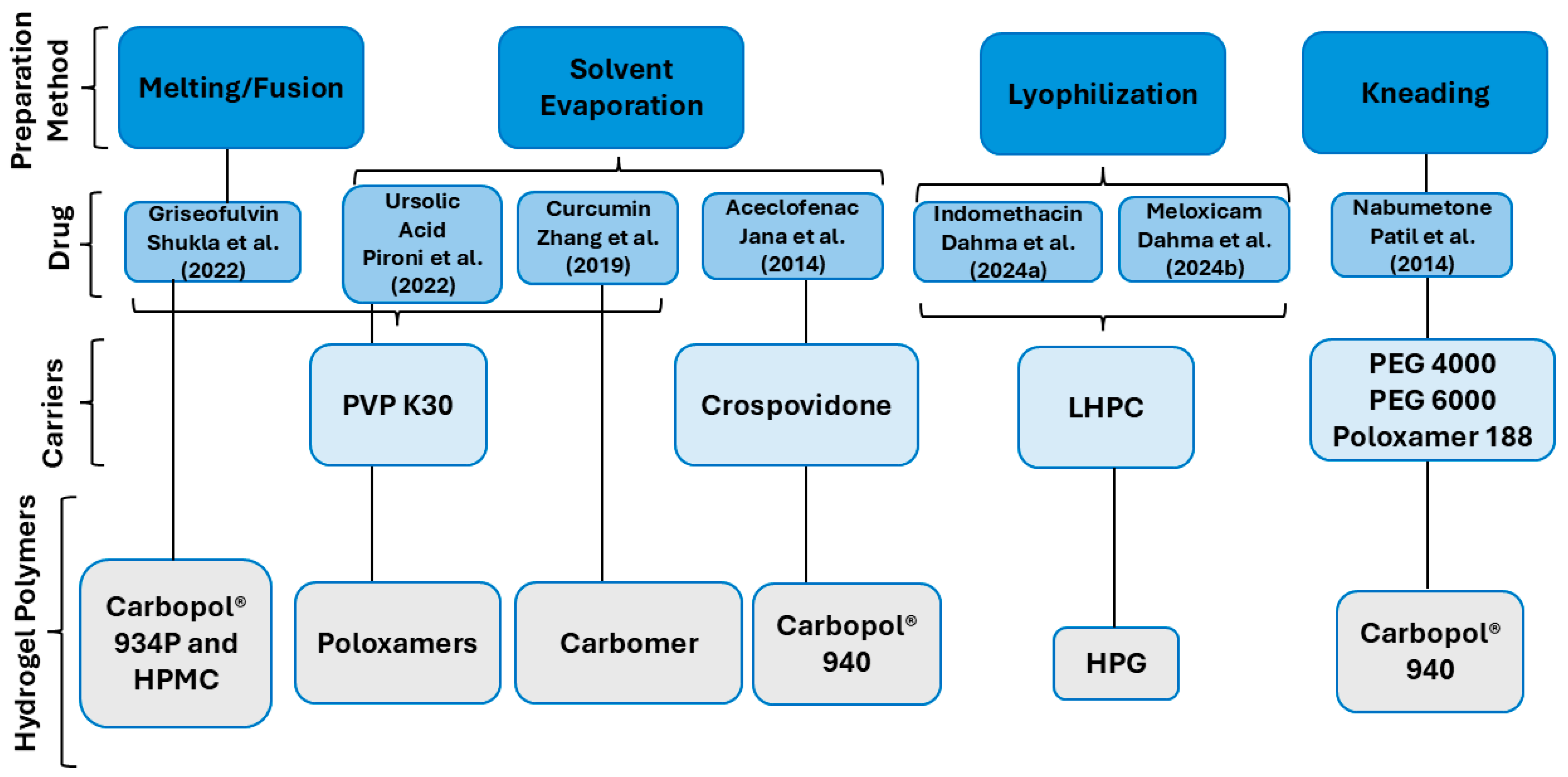
4.1. The Preparation of SDHs

To prepare SDHs, SDs are initially formulated using various methods and materials. The SDs are then incorporated into a hydrogel matrix with different gelling agents, with each contributing to the unique properties of the final SDH (Scheme 1). For instance, an aceclofenac–crospovidone SDH was prepared using Carbopol® 940, a crosslinked polyacrylate polymer known for its high viscosity control, which is beneficial for forming stable gels and suspensions. The Carbopol® 940 gels containing the aceclofenac–crospovidone SD were prepared as follows: the required amount of aceclofenac–crospovidone SD (equivalent to 150 mg of aceclofenac) was dissolved in ethanol and deionized water, respectively, and mixed thoroughly. Next, 100 mg of Carbopol 940, soaked in 6.50 mL of deionized water overnight, was added to the mixture and stirred at 500 rpm for 1 h. Finally, triethanolamine was added to form a clear gel [92].
Similarly, curcumin and PVP K30 SDHs were formulated using components such as ethacridine lactate, carbomer, glycerin, polysorbate 80 (Tween® 80), ethanol, and triethanolamine. Temperature-sensitive in situ hydrogels containing curcumin SDs were prepared as follows: Poloxamer 407 and Poloxamer 188 (6:1, w/w) were dissolved in water. Milled curcumin SDs (180-mesh powders) were added to the solution and agitated to formcurcumin SDHs containing 20 mg/mL of curcumin. These hydrogels were flowing liquids and stored at 4 °C in darkness until use [93].
Additionally, gels loaded with an SD of griseofulvin, utilizing mannitol and PVP K30, were formulated with two gel-forming polymers: Carbopol® 934P and HPMC. Both polymers provide advantages such as a favorable in vitro diffusion profile, good viscosity, and excellent spreadability. SD gels of griseofulvin were prepared using Carbopol® 934P or HPMC. The SD was dispersed in purified water and heated to 50 °C with constant stirring. For the Carbopol gels, Carbopol was added to the solution under continuous stirring, maintaining the temperature at 50 °C to avoid the entrapment of air. The gels were neutralized using triethanolamine to achieve a neutral pH. In the HPMC gels, HPMC was added in a similar manner, and the dispersions were neutralized with 10% sodium hydroxide (NaOH) to form the gels. Both types of gels were stirred to ensure their uniformity and clarity [93].
In other studies, SDHs were prepared using indomethacin and meloxicam in combination with LHPC and HPG, a hydrophilic compound that aids in maintaining skin hydration. Different control hydrogels of HPG were prepared by dispersing 85 mg of HPG polymer in 5 mL of pH 6.0 phosphate buffer and stirring it at 600 rpm until a homogeneous hydrogel was obtained. For hydrogels containing various SDs, 5 g of hydrogel blank was weighed, and an amount equivalent to 65 mg of the drug was added to each SD. The mixture was stirred at 600 rpm for 2 min [38,57].
Furthermore, SDs containing ursolic acid (UA), PVP K30, and poloxamer 407 were formulated into poloxamer hydrogels, known for their thermoreversible gelation properties. The UA-SD-loaded poloxamer hydrogels were prepared using the “cold method”. The UA-SDs (equivalent to 0.3% (w/v) of the drug) were dispersed in a 0.9% sodium chloride solution. Poloxamer 407 was then added to this solution, which was stirred at 100 rpm in an ice bath for 30 min. The hydrogels were stored in a refrigerator at 4 °C for 24 h to allow for the complete dissolution of the polymer [91].
Lastly, nabumetone (N) was formulated into PEG 4000/PEG 6000/Poloxamer 188 SD topical hydrogels using Carbopol® 940 at different concentrations. Carbopol® 940 was accurately weighed and soaked in 100 mL of water for 10 h. The SD equivalent to 1 g of N was then dispersed into the Carbopol® dispersion with continuous stirring. Triethanolamine was added to neutralize the polymer, followed by the addition of methyl paraben. Stirring was continued for approximately 30 min to obtain a clear gel [65].
Each of these formulations highlights the versatility and adaptability of the preparation of SDHs.

4.2. Characterization of SDHs

To ensure the quality, stability, and effectiveness of topical SDHs, various properties must be evaluated, including their morphology, using scanning electron microscopy (SEM); degree of crystallinity, via differential scanning calorimetry (DSC) and X-ray diffraction (XRD); molecular interactions, through Fourier-transform infrared spectroscopy (FTIR); solubility; dissolution rate; rheological behavior; cytotoxicity; penetration characteristics; and in vivo performance. Each characterization method mentioned above is summarized below.

4.2.1. SEM

SEM is an ideal technique for characterizing particle morphology and size in SDHs. SEM enables detailed imaging of the surface structures and dispersed particles within the hydrogel, allowing for observation of the interaction between the hydrogel matrix and solid particles. For analysis, the hydrogel typically requires specific preparation steps. Generally, the hydrogel is dehydrated to prevent interference from a high moisture content during imaging. If the material is non-conductive, it is coated with a thin layer of metal (such as gold or platinum) to prevent charging effects under the electron beam. The information obtained from SEM provides valuable insights into the distribution and adhesion of solid particles within the hydrogel matrix, which is crucial for understanding controlled release and formulation stability. This technique is particularly useful for developing optimized SDHs for pharmaceutical and biomedical applications, as it reveals both the size of the dispersed particles and the structural details of the hydrogel’s surface [57,65,92,93].

4.2.2. The Degree of Crystallinity

The crystallinity of SDHs significantly influences the mobility and release profile of the drug. A lower degree of crystallinity can lead to a faster drug release due to increased drug mobility. To evaluate the crystallinity behavior of SDs in SDHs, DSC and XRD are commonly used. In a DSC analysis, the heat uptake of an SDH sample is compared with that of a reference sample to monitor the phase transitions. This technique provides insight into the thermal behavior of the SD within the hydrogel matrix, helping to identify any crystalline or amorphous states present [95]. An XRD analysis involves irradiating the SDH sample with X-rays and analyzing the scattered radiation at different angles. XRD offers structural information on the phases, crystal orientations, crystallinity levels, and crystal defects within the SD of the hydrogel. This characterization is essential for understanding structural properties that influence the release and stability of the drug in SDHs, optimizing their use in controlled drug delivery applications [96].

4.2.3. FTIR

FTIR is a valuable technique for analyzing the molecular interactions and chemical composition of SDHs. FTIR can detect functional groups and identify possible interactions between the hydrogel matrix and the dispersed drug or solid particles, such as hydrogen bonding or van der Waals interactions, which can influence drug release and stability. During an FTIR analysis, an infrared beam is directed through the SDH sample, and the absorbed wavelengths are recorded, providing a spectrum that represents the molecular vibrations of different bonds within the material. Shifts in characteristic peaks or changes in intensity can indicate interactions between the components of the SDH, as well as any changes in the chemical environment of the drug when it is incorporated into the hydrogel. This information is essential for understanding the compatibility of the drug with the hydrogel matrix and the stability of the formulation. An FTIR analysis allows for a detailed evaluation of the molecular structure of SDHs, supporting the optimization of drug delivery and the overall performance of the hydrogel system [97].

4.2.4. Solubility

A solubility analysis is a crucial method for evaluating the performance and bioavailability of SDHs. Improving the solubility of poorly water-soluble drugs is a key goal in the development of SDHs, as enhanced solubility can significantly improve drug release and absorption. In this analysis, SDH samples are dispersed in an appropriate solvent, typically water or a physiological buffer, and stirred until equilibrium is reached. Afterward, the solution is filtered to remove undissolved particles, and the concentration of the dissolved drug is measured using spectroscopic techniques, such as UV–Visible spectrophotometry or High-Performance Liquid Chromatography (HPLC). This process helps determine the enhancement in solubility provided by the SD within the hydrogel matrix compared to that of the pure drug alone. Solubility studies can reveal how well the drug is incorporated and stabilized within the SDH, providing insights into the formulation’s effectiveness for improving drug release and ensuring consistent therapeutic levels. This method is essential for optimizing SDH formulations for improved bioavailability and drug delivery performance [98].

4.2.5. In Vitro Drug Release

An in vitro release rate analysis is an essential method for assessing the drug release profile of SDHs and predicting their performance in drug delivery applications. This method helps determine how efficiently a drug is released from the hydrogel matrix and the potential release mechanism, such as diffusion or matrix erosion. To conduct this analysis, the SDH sample is placed in a release medium (typically a buffer solution that mimics physiological conditions) and maintained at a controlled temperature and agitation to simulate in vivo conditions. Samples of the release medium are collected at specific time intervals and replaced with fresh medium to maintain sink conditions. The concentration of the drug released in each sample is then measured using techniques like UV–Visible spectrophotometry or HPLC. By plotting the cumulative drug release over time, this method provides a release profile that can be analyzed to understand the kinetics and mechanism of drug release. Mathematical models such as zero-order, first-order, or Higuchi models can be applied to the data to further characterize the release behavior. This information is critical for optimizing SDH formulations to achieve controlled and sustained drug release, ultimately enhancing the therapeutic efficacy and patient compliance [38,57].

4.2.6. Rheological Studies

Rheological analyses are a valuable method for studying the mechanical and flow properties of SDHs. Understanding the rheology of SDHs is essential for optimizing their application in drug delivery, as properties like viscosity, elasticity, and yield stress can affect both the stability and release profile of the encapsulated drug. In a typical rheological study, a sample of the SDH is subjected to various shear rates or oscillatory stresses using a rheometer to evaluate parameters such as its viscosity, shear-thinning behavior, and viscoelastic properties. These measurements are often performed at controlled temperatures to mimic physiological conditions, such as 37 °C. By analyzing the changes in viscosity and elasticity with an increasing shear rate or oscillatory stress, this study reveals how the hydrogel responds to forces similar to those encountered during administration or application. Rheological data help characterize the internal structure of the SDH, including interactions between the hydrogel matrix and the dispersed particles, which may influence the consistency and stability of the formulation. For instance, a hydrogel with the appropriate viscoelastic properties can provide sustained and controlled drug release by resisting deformation and maintaining its structure under applied forces. This method is crucial for developing SDHs with tailored rheological properties, ensuring their ease of application, enhanced stability, and optimal drug release performance [38,57,65,93,99].

4.2.7. Cytotoxicity

This type of study aims to evaluate the potential harmful effects of substances, particularly SD systems incorporated into hydrogels, on cellular health and viability. SDHs are a novel class of biomaterials that involve the incorporation of poorly soluble drugs or bioactive compounds into a SD matrix within a hydrogel network. While these SDHs offer enhanced drug release profiles and bioavailability, their safety profile, particularly their cytotoxicity, is critical to assess. These investigations focus on determining whether the SDs within the hydrogel negatively affect cell growth or proliferation or induce apoptosis. Various cell lines may be exposed to SDHs to identify possible cellular responses such as oxidative stress, membrane damage, or inflammatory reactions. Dahma et al. [38] conducted a cytotoxicity study using the fluorescent dye resazurin. They examined four types of mammalian cells: monkey epithelial cells (Vero CCL-81), human ovarian cells (HeLa), murine macrophages (J774), and murine fibroblasts (L929) [38,100,101]. Meanwhile, Marena et al. utilized an agar overlay qualitative assay to evaluate the cytotoxicity of a UA-loaded hydrogel on L-929 cells [92,102].

4.2.8. Permeation Studies

Research on the permeation of SDHs for topical drug delivery investigates how these advanced materials interact with and permeate through both biological and synthetic membranes. SDHs are designed to enhance the solubility and bioavailability of poorly soluble drugs, offering controlled release and improving therapeutic outcomes. In topical applications, the permeation of SDHs through the skin barrier is critical for achieving effective drug concentrations at the target site. To evaluate this, studies often employ in vitro models, such as Franz diffusion cells, using excised animal skin membranes (e.g., from pigs, rats, or rabbits), which are chosen due to their structural similarities to human skin. Additionally, synthetic membranes, such as polymeric films or silicone, are increasingly used in permeation studies due to their reproducibility, consistency, and ability to mimic the specific barrier properties of human skin [103]. These models allow researchers to assess the rate and extent of drug permeation and to investigate how factors such as the hydrogel’s composition, the molecular dispersion of the drug, the physicochemical properties of the drug, and the hydrogel’s swelling behavior influence drug release and skin permeation. Physiologically, the Franz cells indicate the initial penetration of the active ingredient through the skin, which is the first step towards reaching the site of action. However, further evaluation is needed to assess its permeation through deeper tissue layers to the target sites [104].
In the context of topical hydrogel systems, cellular permeation studies are pivotal for assessing the ability of APIs to cross cellular barriers and effectively reach the target site of action. In vitro models, such as Transwell assays and 3D microvessel systems, are highly valuable for evaluating the permeability of APIs through cellular layers. For instance, Transwell assays use endothelial cell monolayers to mimic the skin or mucosal tissues, shedding light on how well the drugs incorporated into hydrogels diffuse through these layers. These systems are essential for optimizing hydrogel formulations, improving drug release, and ensuring effective absorption at the site of application. By tracking molecule diffusion, coupled with fluorescence-based detection or chromatography, the permeation behavior can be quantified, fine-tuned, and adjusted for diverse therapeutic needs. In addition, 3D microvessel models offer a more advanced representation of in vivo conditions, wherein hydrogel-loaded active ingredients undergo permeability and efficacy testing in a more complex and dynamic environment. This combination of in vitro techniques contributes to improving hydrogel formulations, ultimately enhancing the bioavailability and therapeutic outcomes of drugs in topical treatments. However, it is important to note that alongside these in vitro models, in vivo and in silico methodologies also play a significant role. While in vivo studies simulate more complex biological environments, offering insights into the drug behavior in living organisms, in silico approaches such as molecular dynamics simulations help explore the molecular transport mechanisms that govern API permeation. By integrating in vitro, in vivo, and in silico methods, researchers are better equipped to design and refine drug delivery systems, ensuring more precise and effective treatments [105].

4.2.9. In Vivo Study

In vivo methods are essential for evaluating the efficacy of SDHs in topical drug delivery, providing valuable insights into their therapeutic potential and safety profile. Animal models are commonly used to assess the effectiveness of SDHs for localized drug delivery to the skin or mucosal surfaces. After applying the SDH formulation to the targeted area, a drug’s permeation, bioavailability, and release rates are measured over time. Techniques such as skin sampling, HPLC, and mass spectrometry (MS) are used to quantify the drug’s concentration in the tissues and assess its absorption across the skin barrier. Additionally, therapeutic effects such as wound healing, the anti-inflammatory response, or pain relief are monitored through clinical evaluations and histological analyses. These assessments include examining the tissue at the application site for signs of irritation, inflammation, or necrosis, as well as evaluating the overall healing process or therapeutic improvement. In vivo studies also help assess the safety and biocompatibility of SDHs, ensuring that a formulation does not cause significant adverse reactions or toxicity. Overall, these in vivo methods are crucial for confirming the performance of SDHs in topical drug delivery, optimizing their design for clinical applications, and ensuring their effectiveness in treating various skin-related conditions [91,92,94].

4.3. SDHs as a Promising Carrier System for the Topical Delivery of Poorly Water-Soluble Drugs

SDHs have emerged as promising carrier systems for the topical delivery of poorly water-soluble drugs, offering enhanced drug solubility, controlled release, and improved therapeutic outcomes. This innovative approach has been the focus of several studies exploring their potential to optimize the drug delivery, improve bioavailability, and provide targeted therapeutic effects. Through a range of formulations and in vitro and in vivo evaluations, SDHs are being investigated for their ability to overcome the challenges of drug solubility and absorption, paving the way for more effective topical treatments.
Pironi et al. [91] developed an SD made of UA in poloxamer hydrogels with the aim of improving the rheological properties for topical applications and enhancing its in vivo anti-inflammatory effect. The results indicated that the SDHs showed a lower resistance to flow and a higher sol–gel transition temperature compared to these properties in hydrogels made with only sodium chloride (NaCl) solution. The increased viscosity of these hydrogels helped regulate the drug diffusion process. A diffusion study of the SDHs using Franz diffusion cells showed an enhancement in UA’s skin permeability. The developed formulations were found to be biocompatible with the L929 cell line. In vivo tests indicated that all of the formulations were effective and offered improved properties for the local treatment of inflammatory skin disorders compared to those of the free drug.
Shukla et al. [93] conducted a study aimed at improving the topical and systemic delivery of griseofulvin. An SD containing this drug was formulated to enhance its aqueous solubility and then incorporated into a water-based gel for the optimal topical application. The findings indicated that the hydrogel formulation of the SDH (prepared with 1% Carbopol® as the gelling agent and triethanolamine as the crosslinking agent) loaded with the SD (using a 1:3 ratio of griseofulvin to mannitol) was likely the most effective formulation, demonstrating an excellent in vitro diffusion profile, viscosity, and spreadability.
Zhang et al. [94] investigated the use of curcumin SDHs to treat bacterial infections. This study found that these hydrogels had high therapeutic efficacy when they were applied topically, reducing inflammation and enhancing wound healing more effectively than the marketed Lincomycin/Lidocaine gel. Despite their weak in vitro antibacterial effect, the curcumin SDs exhibited strong in vivo antibacterial activity by promoting the growth of intravaginal Lactobacilli.
Jana et al. [92] explored the development of an aceclofenac–crospovidone SD using a “Quality by Design (QbD)” approach. Their findings revealed that a Carbopol® 940 topical gel containing the aceclofenac–crospovidone SD demonstrated sustained aceclofenac permeation over 10 h in an ex vivo skin permeation study using excised mouse skin. The gels were evaluated for their pH, viscosity, and gel strength. The FTIR analysis confirmed no significant interactions between aceclofenac and the other excipients in the formulation. In vivo anti-inflammatory tests on male Sprague-Dawley rats with carrageenan-induced paw edema showed that the optimized gel was as effective as a marketed gel and did not cause skin irritation.
Patil et al. [65] conducted an in vitro diffusion study and found that gels incorporating nabumetone SDs showed significantly improved diffusion compared to that of gels with pure nabumetone. These findings suggest that gels with SDs are superior in enhancing the dissolution and diffusion of nabumetone while also helping mitigate the drug’s gastric side effects.
Dahma et al. [38] investigated the characterization and cellular toxicity of SD-loaded hydrogels based on indomethacin (IND). They found that the solubility coefficient for the SD increased significantly (p < 0.05), showing a five-fold improvement compared to that of the raw IND material. However, further increases in LHPC did not enhance IND’s solubility. The in vitro release of the drug from the indomethacin hydrogel exhibited a gradual release pattern, typical for drugs with low solubility. For the SD hydrogel, factors such as hydrogen bonding in the SD, the inclusion of hydrophilic carriers like LHPC, and drug amorphization likely contributed to the improved dissolution of IND, as evidenced through DSC, XRD, SEM, viscosity, and FTIR studies. Additionally, no cytotoxic effects were observed in various mammalian cell types, indicating that the SD hydrogel was biocompatible and non-toxic.
Dahma et al. [57] also formulated SD formulations to enhance the solubility and permeability of meloxicam (MX) hydrogels, which have low water solubility. The reduction in the drug’s crystallinity and the increase in hydrogen bonds in various SDs have been linked to the better dissolution of meloxicam from HPG hydrogels. Using low LHPC ratios as a carrier significantly improved the solubility by 4.61 to 5.64 times, respectively. DSC studies of SDs demonstrated that increasing the LHPC ratios significantly lowered the MX crystallinity percentages to 18.12%. These findings were corroborated by XRPD studies. However, DSC studies indicated that higher LHPC ratios in the SDs did not achieve further reductions in crystallinity. The FTIR analysis revealed the presence of hydrogen bonds characteristic of drug/polymer and polymer/polymer interactions in the various SDs. The hydrogels formulated with SDs showed significant enhancements in the drug release profiles.

4.4. Future Perspectives and Challenges of SDHs

SDHs have demonstrated remarkable potential in topical drug delivery due to their ability to enhance the solubility and bioavailability of poorly water-soluble drugs. The versatility of SDHs, driven by advancements in the preparation methods and the inclusion of various polymers and excipients, paves the way for innovations in drug formulations tailored to specific therapeutic applications. Despite these promising attributes, significant challenges remain in fully realizing the potential of SDHs in clinical practice.
One of the primary challenges involves achieving and maintaining the stability of the SD within the hydrogel matrix. The degree of crystallinity plays a crucial role in the release kinetics of drugs, with a lower crystallinity favoring faster and more consistent drug release. Advanced characterization techniques like DSC and XRD are essential for optimizing the crystalline properties of SDHs. Furthermore, the compatibility of the drug with the hydrogel matrix must be meticulously evaluated using tools like FTIR to ensure the stability and efficacy of the formulation.
The rheological properties of SDHs present another critical area for improvement. Optimizing their viscosity, elasticity, and shear-thinning behavior is essential to enhancing their ease of application, stability, and controlled drug release. Innovations in the polymer selection and crosslinking strategies are expected to address these challenges, enabling the formulation of hydrogels with superior mechanical and flow properties.
From a therapeutic perspective, the permeation and efficacy of SDHs in vivo need to be validated further. Although in vitro permeation studies using Franz diffusion cells and synthetic membranes provide valuable insights, in vivo studies are essential to evaluate the actual drug absorption and therapeutic outcomes under physiological conditions. These studies must also evaluate the long-term biocompatibility and potential toxicity of SDHs to ensure patient safety.
Emerging trends in SDH research include the incorporation of multifunctional excipients and polymers that offer additional benefits, such as anti-inflammatory or antimicrobial properties. Thermoresponsive and pH-sensitive hydrogels are gaining attention for their ability to provide targeted and stimuli-responsive drug release, which can enhance the therapeutic efficacy of SDHs in topical applications. These hydrogels can offer a controlled release in response to local changes in temperature or pH, improving the drug delivery to affected areas. Moreover, integrating SDHs with nanotechnology, such as the inclusion of nanoparticles or liposomes, holds promise for further improving the drug solubility, stability, and controlled release.
Additionally, recent advancements in 3D printing technologies offer a novel approach to fabricating SDHs with precise control over their structure and drug release profiles. This technique allows for the design of customized hydrogels with unique mechanical and functional properties, providing new opportunities for the fabrication of hydrogels tailored to specific therapeutic needs. By using 3D printing, it is possible to create hydrogels with complex geometries and controlled porosity, enhancing the drug delivery efficiency and improving patient outcomes.

5. Conclusions

In conclusion, the studies reviewed here underscore the considerable potential of SDHs in enhancing the topical delivery of poorly water-soluble drugs. These SDHs offer several key advantages, including improvements in drug solubility, controlled and sustained release profiles, better skin penetration, and enhanced rheological properties, which contribute to their ease of application and improved stability. In vivo studies demonstrate that SDHs not only improve therapeutic outcomes but are also biocompatible and non-toxic, making them suitable for long-term use. Furthermore, they facilitate the regulation of drug diffusion, ensuring the optimal dosing and effectiveness. While SDHs represent a groundbreaking approach to topical drug delivery, addressing challenges related to their stability and clinical validation will be critical for their successful translation into routine clinical use. Future research efforts should focus on refining the formulation strategies, exploring novel polymers and excipients, and conducting comprehensive in vivo studies to unlock the full potential of SDHs as a versatile and effective drug delivery platform, offering an alternative that improves the bioavailability and enhances the drug delivery for the treatment of various dermal conditions and inflammatory diseases.

Author Contributions

Writing—review and editing and supervision: P.M.d.l.T.-I.; writing—original draft preparation: Z.D.; editing and supervision: C.Á.-Á. All authors have read and agreed to the published version of the manuscript.

Funding

This work was supported by the Spanish Ministry of Science and Innovation (PID2022-136438OB-I00) and the Complutense University of Madrid (Research Group 910939).

Data Availability Statement

No new data were created or analyzed in this study. Data sharing is not applicable to this article.

Acknowledgments

The authors gratefully acknowledge the support from Department of Pharmaceutics and Food Technology, the Faculty of Pharmacy, Complutense University of Madrid and Instituto Universitario de Farmacia Industrial, Complutense University of Madrid.

Conflicts of Interest

The authors declare no conflicts of interest.

Abbreviations

The following abbreviations are used in this manuscript:
APIActive Pharmaceutical Ingredient
BCSBiopharmaceutical Classification System
BHAButylated Hydroxyanisole
BHTButylated Hydroxytoluene
CDCyclodextrin
DMSODimethyl Sulfoxide
DSCDifferential Scanning Calorimetry
FDAFood and Drug Administration
FTIRFourier-Transform Infrared Spectroscopy
FDMFused Deposition Modeling
HeLaHuman Ovarian Cells
HPGHydroxypropyl Guar Gum
HPLCHigh-Performance Liquid Chromatography
HPMCHydroxypropylmethylcellulose
INDIndomethacin
INDNa-PVPVASodium IND–Copovidone
IND-PVPVAIND–Copovidone
J774Murine Macrophages
L929Murine Fibroblasts
LHPCLow-Substituted Hydroxypropyl Cellulose
MSMass Spectrometry
NNabumetone
NaClSodium Chloride
NaOHSodium Hydroxide
NLCNanostructured Lipid Carrier
PEGPolyethylene Glycol
PHEMAPoly(2-hydroxyethyl methacrylate)
PLGA-PEG-PLGAPoly(lactic-co-glycolic acid)-poly(ethylene glycol)-poly(lactic-co-glycolic acid)
PVAPolyvinyl Alcohol
PVPPolyvinylpyrrolidone
QbDQuality by Design
SDSolid Dispersion
SDHSolid Dispersion Hydrogel
SDSSodium Dodecyl Sulfate
SEDDSSelf-Emulsifying Drug Delivery System
SEMScanning Electron Microscopy
SLNSolid Lipid Nanoparticle
TgGlass Transition Temperature
UAUrsolic Acid
Vero CCL-81Monkey Epithelial Cells
WHOThe World Health Organization
XRDX-Ray Diffraction

References

  1. Choi, J.-S.; Lee, S.-E.; Jang, W.S.; Byeon, J.C.; Park, J.-S. Solid dispersion of dutasteride using the solvent evaporation method: Approaches to improve dissolution rate and oral bioavailability in rats. Mater. Sci. Eng. C 2018, 90, 387–396. [Google Scholar] [CrossRef]
  2. Xu, W.; Sun, Y.; Du, L.; Chistyachenko, Y.S.; Dushkin, A.V.; Su, W. Investigations on solid dispersions of valsartan with alkalizing agents: Preparation, characterization and physicochemical properties. J. Drug Deliv. Sci. Technol. 2018, 44, 399–405. [Google Scholar] [CrossRef]
  3. Choi, J.S.; Kwon, S.H.; Lee, S.E.; Jang, W.S.; Byeon, J.C.; Jeong, H.M.; Park, J.S. Use of acidifier and solubilizer in tadalafil solid dispersion to enhance the in vitro dissolution and oral bioavailability in rats. Int. J. Pharm. 2017, 526, 77–87. [Google Scholar] [CrossRef] [PubMed]
  4. Karashima, M.; Sano, N.; Yamamoto, S.; Arai, Y.; Yamamoto, K.; Amano, N.; Ikeda, Y. Enhanced pulmonary absorption of poorly soluble itraconazole by micronized cocrystal dry powder formulations. Eur. J. Pharm. Biopharm. 2017, 115, 65–72. [Google Scholar] [CrossRef] [PubMed]
  5. Seo, B.; Kim, T.; Park, H.J.; Kim, J.-Y.; Lee, K.D.; Lee, J.M.; Lee, Y.-W. Extension of the hansen solubility parameter concept to the micronization of cyclotrimethylenetrinitramine crystals by supercritical anti-solvent process. J. Supercrit. Fluids 2016, 111, 112–120. [Google Scholar] [CrossRef]
  6. Chen, X.; McClements, D.J.; Zhu, Y.; Chen, Y.; Zou, L.; Liu, W.; Cheng, C.; Fu, D.; Liu, C. Enhancement of the solubility, stability and bioaccessibility of quercetin using protein-based excipient emulsions. Food. Res. Int. 2018, 114, 30–37. [Google Scholar] [CrossRef]
  7. Kim, C.H.; Lee, S.G.; Kang, M.J.; Lee, S.; Choi, Y.W. Surface modification of lipid-based nanocarriers for cancer cell-specific drug targeting. J. Pharm. Investig. 2017, 47, 203–227. [Google Scholar] [CrossRef]
  8. Reggane, M.; Wiest, J.; Saedtler, M.; Harlacher, C.; Gutmann, M.; Zottnick, S.H.; Piechon, P.; Dix, I.; Müller-Buschbaum, K.; Holzgrabe, U.; et al. Bioinspired co-crystals of imatinib providing enhanced kinetic solubility. Eur. J. Pharm. Biopharm. 2018, 128, 290–299. [Google Scholar] [CrossRef]
  9. Wong, J.J.L.; Yu, H.; Lim, L.M.; Hadinoto, K. A trade-off between solubility enhancement and physical stability upon simultaneous amorphization and nanonization of curcumin in comparison to amorphization alone. Eur. J. Pharm. Sci. 2018, 114, 356–363. [Google Scholar] [CrossRef]
  10. Park, J.B.; Park, C.; Piao, Z.Z.; Amin, H.H.; Meghani, N.M.; Tran, P.H.L.; Tran, T.T.D.; Cui, J.-H.; Cao, Q.-R.; Oh, E. pH-independent controlled release tablets containing nanonizing valsartan solid dispersions for less variable bioavailability in humans. J. Drug Deliv. Sci. Technol. 2018, 46, 365–377. [Google Scholar] [CrossRef]
  11. Chen, B.Q.; Kankala, R.K.; Wang, S.-B.; Chen, A.Z. Continuous nanonization of lonidamine by modified-rapid expansion of supercritical solution process. J. Supercrit. Fluids 2018, 133, 486–493. [Google Scholar] [CrossRef]
  12. Elmowafy, M.; Al-Sanea, M.M. Nanostructured Lipid Carriers (NLCs) as Drug Delivery Platform: Advances in Formulation and Delivery Strategies. Saudi Pharm. J. 2021, 29, 999–1012. [Google Scholar] [CrossRef]
  13. Mfoafo, K.; Mittal, R.; Eshraghi, A.A.; Omidian, H. Thiolated Polymers: An Overview of Mucoadhesive Properties and Their Potential in Drug Delivery via Mucosal Tissues. J. Drug Deliv. Sci. Technol. 2023, 85, 104596. [Google Scholar] [CrossRef]
  14. Schittny, A.; Huwyler, J.; Puchkov, M. Mechanisms of increased bioavailability through amorphous solid dispersions: A review. Drug. Deliv. 2020, 27, 110–127. [Google Scholar] [CrossRef] [PubMed]
  15. World Health Organization (WHO). Multisource (Generic) Pharmaceutical Products: Guidelines on Registration Requirements to Establish Interchangeability. 2018. Available online: https://www.who.int/publications/m/item/annex-6-trs-1003 (accessed on 7 March 2025).
  16. U.S. Food and Drug Administration (FDA). Waiver of In Vivo Bioavailability and Bioequivalence Studies for Immediate-Release Solid Oral Dosage Forms Based on a Biopharmaceutics Classification System Guidance for Industry. 2017. Available online: https://www.fda.gov/files/drugs/published/Bioavailability-and-Bioequivalence-Studies-Submitted-in-NDAs-or-INDs-%E2%80%94-General-Considerations.pdf (accessed on 7 March 2025).
  17. Bashiardes, S.; Christodoulou, C. Orally Administered Drugs and Their Complicated Relationship with Our Gastrointestinal Tract. Microorganisms 2024, 12, 242. [Google Scholar] [CrossRef] [PubMed]
  18. Wang, W.; Sun, C.; Jing, L.; Zhou, M.; Gao, J.; Shi, Y.; Zhang, S. Multifunctional nanocomposite hydrogel dressing with low temperature photothermal and controlled antibiotics release for combating bacterial infection. Mater. Des. 2024, 239, 112812. [Google Scholar] [CrossRef]
  19. Karimkhani, C.; Dellavalle, R.P.; Coffeng, L.E.; Flohr, C.; Hay, R.J.; Langan, S.M.; Nsoesie, E.O.; Ferrari, A.J.; Erskine, H.E.; Silverberg, J.I.; et al. Global skin disease morbidity and mortality an update from the global burden of disease study 2013. JAMA Dermatol. 2017, 153, 406–412. [Google Scholar] [CrossRef]
  20. McKenzie, M.; Betts, D.; Suh, A.; Bui, K.; Kim, L.D.; Cho, H. Hydrogel-Based Drug Delivery Systems for Poorly Water-Soluble Drugs. Molecules 2015, 20, 20397–20408. [Google Scholar] [CrossRef]
  21. Bhujbal, S.V.; Mitra, B.; Jain, U.; Gong, Y.; Agrawal, A.; Karki, S.; Taylor, L.S.; Kumar, S.; Zhou, Q.T. Pharmaceutical amorphous solid dispersion: A review of manufacturing strategies. J. Pharm. Sci B 2021, 11, 2505–2536. [Google Scholar] [CrossRef]
  22. Khan, K.U.; Minhas, M.U.; Badshah, S.F.; Suhail, M.; Ahmad, A.; Ijaz, S. Overview of nanoparticulate strategies for solubility enhancement of poorly soluble drugs. Life Sci. 2022, 15, 120301. [Google Scholar] [CrossRef]
  23. Bazzo, G.C.; Pezzini, B.R.; Stulzer, H.K. Eutectic mixtures as an approach to enhance solubility, dissolution rate and oral bioavailability of poorly water-soluble drugs. Int. J. Pharm. 2020, 588, 119741. [Google Scholar] [CrossRef] [PubMed]
  24. Kumar, A.; Kumar, J. Solid dispersion techniques: A review. Int. J. Res. Eng. Sci. Manag. 2021, 4, 104–111. [Google Scholar]
  25. Jiménez-Santos, C.J.; Pérez-Martínez, J.I.; Gómez-Pantoja, M.E.; Moyano, J.R. Enhancement of albendazole dissolution properties using solid dispersions with Gelucire 50/13 and PEG 15000. J. Drug. Deliv. Sci. Technol. 2017, 42, 261–272. [Google Scholar] [CrossRef]
  26. Haser, A.; Cao, T.; Lubach, J.; Listro, T.; Acquarulo, L.; Zhang, F. Melt extrusion vs. spray drying: The effect of processing methods on crystalline content of naproxen-povidone formulations. Eur. J. Pharm. Sci. 2017, 102, 115–1252. [Google Scholar] [CrossRef]
  27. Motallae, S.; Taheri, A.; Homayouni, A. Preparation and characterization of solid dispersions of celecoxib obtained by spray-drying ethanolic suspensions containing PVP-K30 or isomalt. J. Drug Deliv. Sci. Technol. 2018, 46, 188–196. [Google Scholar] [CrossRef]
  28. Choi, J.S.; Lee, S.E.; Jang, W.S.; Byeon, J.C.; Park, J.S. Tadalafil solid dispersion formulations based on PVP/VA S-630: Improving oral bioavailability in rats. Eur. J. Pharm. Sci. 2017, 106, 152–158. [Google Scholar] [CrossRef] [PubMed]
  29. Tran, P.; Pyo, Y.C.; Kim, D.H.; Lee, S.E.; Kim, J.K.; Park, J.S. Overview of the manufacturing methods of solid dispersion technology for improving the solubility of poorly water-soluble drugs and application to anticancer drugs. Pharmaceutics 2019, 11, 132. [Google Scholar] [CrossRef]
  30. Zhang, X.; Xing, H.; Zhao, Y.; Ma, Z. Pharmaceutical dispersion techniques for dissolution and bioavailability enhancement of poorly water-soluble drugs. Pharmaceutics 2018, 10, 74. [Google Scholar] [CrossRef]
  31. Fan, N.; He, Z.; Ma, P.; Wang, X.; Li, C.; Sun, J.; Sun, Y.; Li, J. Impact of HPMC on inhibiting crystallization and improving permeability of curcumin amorphous solid dispersions. Carbohydr. Polym. 2018, 181, 543–550. [Google Scholar] [CrossRef]
  32. Fan, N.; Ma, P.; Wang, X.; Li, C.; Zhang, X.; Zhang, K.; Li, J.; He, Z. Storage stability and solubilization ability of HPMC in curcumin amorphous solid dispersions formulated by Eudragit E100. Carbohydr. Polym. 2018, 199, 492–498. [Google Scholar] [CrossRef]
  33. Wang, S.; Liu, C.; Chen, Y.; Zhang, Z.; Zhu, A.; Qian, F. A high-sensitivity HPLC-ELSD method for HPMC-AS quantification and its application in elucidating the release mechanism of HPMC-AS based amorphous solid dispersions. Eur. J. Pharm. Sci. 2018, 122, 303–310. [Google Scholar] [CrossRef] [PubMed]
  34. Kashani, E.; Maghsoudi, S.; Rezania, H.; Yarazavi, M.; Hajiabbas, M.; Benkovics, G.; Bilensoy, E.; Lacík, I.; Heydari, A. Cyclodextrin polymers containing ionizable and ionic groups: A comprehensive review from classifications and synthesis methods to applications. Mater. Today Chem. 2024, 39, 102186. [Google Scholar] [CrossRef]
  35. Almeida, H.; Ferreira, B.; Fernandes-Lopes, C.; Araújo, F.; Bonifácio, M.J.; Vasconcelos, T.; Sarmento, B. Third-Generation Solid Dispersion Through Lyophilization Enhanced Oral Bioavailability of Resveratrol. ACS Pharmacol. Transl. Sci. 2024, 7, 888–898. [Google Scholar] [CrossRef] [PubMed]
  36. Biedrzycka, K.; Marcinkowska, A. The use of hot melt extrusion to prepare a solid dispersion of ibuprofen in a polymer matrix. Polymers 2023, 15, 2912. [Google Scholar] [CrossRef] [PubMed]
  37. Vasconcelos, T.; Prezotti, F.; Araújo, F.; Lopes, C.; Loureiro, A.; Marques, S.; Sarmento, B. Third-generation solid dispersion combining Soluplus and poloxamer 407 enhances the oral bioavailability of resveratrol. Int. J. Pharm. 2021, 595, 120245. [Google Scholar] [CrossRef]
  38. Dahma, Z.; Ibáñez-Escribano, A.; Fonseca-Berzal, C.; García-Rodríguez, J.J.; Álvarez-Álvarez, C.; Torrado-Salmerón, C.; Torrado-Santiago, S.; Torre-Iglesias, P.M. Development, Characterization, and Cellular Toxicity Evaluation of Solid Dispersion-Loaded Hydrogel Based on Indomethacin. Polymers 2024, 16, 2174. [Google Scholar] [CrossRef]
  39. Liu, P.; Zhou, J.Y.; Chang, J.H.; Liu, X.G.; Xue, H.F.; Wang, R.X.; Liu, C.Z. Soluplus-Mediated Diosgenin Amorphous Solid Dispersion with High Solubility and High Stability: Development, Characterization and Oral Bioavailability. Drug Des. Dev. Ther. 2020, 14, 2959–2975. [Google Scholar] [CrossRef]
  40. Sekiguchi, K.; Obi, N. Studies on absorption of eutectic mixture. I. A comparison of the behavior of eutectic mixture of sulfathiazole and that of ordinary sulfathiazole in man. Chem. Pharm. Bull. 1961, 9, 866–872. [Google Scholar]
  41. Ugwu, F.E.; Hyma, P. Enhancement of solubility of poorly water soluble drugs (Amlodipine Besylate) by using Solid Dispersion Technique. Int. J. Res. Anal. Rev. 2024, 11, 562–574. [Google Scholar]
  42. Bertoni, S.; Albertini, B.; Ferraro, L.; Beggiato, S.; Dalpiaz, A.; Passerini, N. Exploring the use of spray congealing to produce solid dispersions with enhanced indomethacin bioavailability: In vitro characterization and in vivo study. Eur. J. Pharm. BioPharm. 2019, 139, 132–141. [Google Scholar] [CrossRef]
  43. Nguyen, T.D.; Roh, S.; Nguyen, M.T.N.; Lee, J.S. Structural Control of Nanofibers According to Electrospinning Process Conditions and Their Applications. Micromachines 2023, 14, 2022. [Google Scholar] [CrossRef] [PubMed]
  44. Tachibana, T.; Nakamura, A. A method for preparing an aqueous colloidal dispersion of organic materials by using water-soluble polymers: Dispersion of B-carotene by polyvinylpyrrolidone Kolloid-Z. Z. Für Polym. 1965, 203, 130–133. [Google Scholar] [CrossRef]
  45. Visan, A.I.; Negut, I. Development and Applications of PLGA Hydrogels for Sustained Delivery of Therapeutic Agents. Gels 2024, 10, 497. [Google Scholar] [CrossRef]
  46. Kalyani, K.; Sravya, D. A Study on Enhancement of Solubility and Dissolution Properties of Voriconazole by Solid Dispersion Technique. J. Pharm. Insights Res. 2024, 2, 089–096. [Google Scholar] [CrossRef]
  47. Goldberg, A.H.; Gibaldi, M.; Kanig, J.L. Increasing dissolution rates and gastrointestinal absorption of drugs via solid solutions and eutectic mixtures III. J. Pharm. Sci. 1966, 55, 487–492. [Google Scholar] [CrossRef]
  48. Sitotaw, Y.W.; Habtu, N.G.; Gebreyohannes, A.Y.; Nunes, S.P.; Van Gerven, T. Ball Milling as an Important Pretreatment Technique in Lignocellulose Biorefineries: A Review. Biomass Convers. Biorefinery 2023, 13, 15593–15616. [Google Scholar] [CrossRef]
  49. Bejaoui, M.; Kalfat, R.; Galai, H. The effect of adding PVP to the binary solid dispersion (indomethacin: Kaolin) on the formation of physically stable amorphous drug. J. Pharm. Innov. 2021, 17, 736–746. [Google Scholar] [CrossRef]
  50. Rojas-Oviedo, I.; Retchkiman-Corona, B.; Quiri-no-Barreda, C.T.; Cárdenas, J.; Schabes-Retchkiman, P. Solubility enhancement of a poorly water soluble drug by forming solid dispersions using mechanochemical activation. Ind. J. Pharm. Sci. 2012, 74, 505. [Google Scholar] [CrossRef]
  51. Khalid, G.M.; Billa, N. Solid Dispersion Formulations by FDM 3D Printing—A Review. Pharmaceutics 2022, 14, 690. [Google Scholar] [CrossRef]
  52. El-Badry, M.; Fetih, G.; Fathy, M. Improvement of solubility and dissolution rate of indomethacin by solid dispersions in Gelucire 50/13 and PEG4000. Saudi Pharm. J. 2009, 17, 217–225. [Google Scholar] [CrossRef]
  53. Ji, Y.; Paus, R.; Prudic, A.; Lübbert, C.; Sadowski, G. A novel approach for analyzing the dissolution mechanism of solid dispersions. Pharm. Res. 2015, 32, 2559–2578. [Google Scholar] [CrossRef] [PubMed]
  54. Daravath, B.; Tadikonda, R.R.; Vemula, S.K. Formulation and pharmacokinetics of Gelucire solid dispersions of flurbiprofen. Drug Dev. Ind. Pharm. 2015, 41, 1254–1262. [Google Scholar] [CrossRef] [PubMed]
  55. Xie, T.; Gao, W.; Taylor, L.S. Impact of Eudragit EPO and hydroxypropyl methylcellulose on drug release rate, supersaturation, precipitation outcome and redissolution rate of indomethacin amorphous solid dispersions. Int. J. Pharm. 2017, 531, 313–323. [Google Scholar] [CrossRef]
  56. Chiang, C.W.; Tang, S.; Mao, C.; Chen, Y. Effect of Buffer pH and Concentration on the Dissolution Rates of Sodium Indomethacin–Copovidone and Indo-methacin–Copovidone Amorphous Solid Dispersions. Mol. Pharm. 2023, 12, 6451–6462. [Google Scholar] [CrossRef]
  57. Dahma, Z.; Torrado-Salmerón, C.; Álvarez-Álvarez, C.; Guarnizo-Herrero, V.; Martínez-Alonso, B.; Torrado, G.; Torrado-Santiago, S.; Torre-Iglesias, P.M. Topical Meloxicam Hydroxypropyl Guar Hydrogels Based on Low-Substituted Hydroxypropyl Cellulose Solid Dispersions. Gels 2024, 10, 207. [Google Scholar] [CrossRef] [PubMed]
  58. Benavent, C.; Torrado-Salmerón, C.; Torrado-Santiago, S. Development of a solid dispersion of nystatin with maltodextrin as a carrier agent: Improvements in antifungal efficacy against Candida spp. biofilm infections. Pharmaceuticals 2021, 14, 397. [Google Scholar] [CrossRef]
  59. Colombo, M.; Lima-Melchiades, G.; Michels, L.R.; Figueiró, F.; Bassani, V.L.; Teixeira, H.F.; Koester, L.S. Solid dispersion of kaempferol: Formulation development, characterization, and oral bioavailability assessment. AAPS PharmSciTech 2019, 20, 106. [Google Scholar] [CrossRef]
  60. Rusdin, A.; Mohd -Gazzali, A.; Thomas, N.; Megantara, S.; Aulifa, D.L.; Budiman, A.; Muchtaridi, M. Advancing drug delivery paradigms: Polyvinyl pyrolidone (PVP)-based amorphous solid dispersion for enhanced physicochemical properties and therapeutic efficacy. Polymers 2024, 16, 286. [Google Scholar] [CrossRef]
  61. Zhang, J.; Guo, M.; Luo, M.; Cai, T. Advances in the development of amorphous solid dispersions: The role of polymeric carriers. Asian J. Pharm. Sci. 2023, 18, 100834. [Google Scholar] [CrossRef]
  62. Liu, X.; Zhao, L.; Wu, B.; Chen, F. Improving solubility of poorly water-soluble drugs by protein-based strategy: A review. Int. J. Pharm. 2023, 634, 122704. [Google Scholar] [CrossRef]
  63. Ting, J.M.; Porter, I.W.; Mecca, J.M.; Bates, F.S.; Reineke, T.M. Advances in polymer design for enhancing oral drug solubility and delivery. Bioconjugate Chem. 2018, 29, 939–952. [Google Scholar] [CrossRef]
  64. Tian, Y.; Jacobs, E.; Jones, D.S.; Mc-Coy, C.P.; Wu, H.; Andrews, G.P. The design and development of high drug loading amorphous solid dispersion for hot-melt extrusion platform. Int. J. Pharm. 2020, 586, 119545. [Google Scholar] [CrossRef]
  65. Patil, S.S.; Mohite, S.K. Development and evaluation of solid dispersion incorporated topical gel of Nabumetone. Res. J. Pharm. Tech. 2014, 7, 1413–1419. [Google Scholar]
  66. Harrison, I.P.; Spada, F. Hydrogels for atopic dermatitis and wound management: A superior drug delivery vehicle. Pharmaceutics 2018, 10, 71. [Google Scholar] [CrossRef] [PubMed]
  67. Cascone, S.; Lamberti, G. Hydrogel-Based Commercial Products for Biomedical Applications: A Review. Int. J. Pharm. 2020, 573, 118803. [Google Scholar] [CrossRef]
  68. Croisfelt, F.M.; Tundisi, L.L.; Ataide, J.A.; Silveira, E.; Tambourgi, E.B.; Jozala, A.F.; Souto, E.M.; Mazzola, P.G. Modified-release topical hydrogels: A ten-year review. J. Mater. Sci. 2019, 54, 10963–10983. [Google Scholar] [CrossRef]
  69. Chen, Z.; Zhao, D.; Liu, B.; Nian, G.; Li, X.; Yin, J.; Qu, S.; Yang, W. 3D Printing of Multifunctional Hydrogels. Adv. Funct. Mater. 2019, 29, 1900971. [Google Scholar] [CrossRef]
  70. Chatterjee, S.; Mohanta, A.; De, A.; Mukherjee, A.; Hazra, A.; Niloy, P.P.; Tudu, M.; Chattopadhyay, K.; Samanta, A. Evaluation of Gum Odina/Carbopol Composite Mucoadhesive Hydrogel on Pharmaceutical Performance: Focusing on Potential Periodontal Treatment. Int. J. Biol. Macromol. 2025, 288, 138708. [Google Scholar] [CrossRef]
  71. Choi, H.; Choi, W.S.; Jeong, J.O. A Review of Advanced Hydrogel Applications for Tissue Engineering and Drug Delivery Systems as Biomaterials. Gels 2024, 10, 693. [Google Scholar] [CrossRef]
  72. Pires, P.C.; Mascarenhas-Melo, F.; Pedrosa, K.; Lopes, D.; Lopes, J.; Macário-Soares, A.; Peixoto, D.; Giram, P.S.; Veiga, F.; Paiva-Santos, A.C. Polymer-based biomaterials for pharmaceutical and biomedical applications: A focus on topical drug administration. Eur. Polym. J. 2023, 187, 111868. [Google Scholar] [CrossRef]
  73. Lai, W.-F.; He, Z.-D. Design and Fabrication of Hydrogel-Based Nanoparticulate Systems for In Vivo Drug Delivery. J. Control. Release 2016, 243, 269–282. [Google Scholar] [CrossRef] [PubMed]
  74. Phatale, V.; Vaiphei, K.K.; Jha, S.; Patil, D.; Agrawal, M.; Alexander, A. Overcoming skin barriers through advanced transdermal drug delivery approaches. J. Control. Release 2022, 351, 361–380. [Google Scholar] [CrossRef] [PubMed]
  75. Clijsen, R.; Baeyens, J.P.; Barel, A.O. In vivo determination of the diclofenac skin reservoir: Comparison between passive, occlusive, and iontophoretic application. Drug Des. Dev. Ther. 2015, 9, 835–840. [Google Scholar] [CrossRef]
  76. Zhu, J.; Tang, X.; Jia, Y.; Ho, C.T.; Huang, Q. Applications and delivery mechanisms of hyaluronic acid used for topical/transdermaldelivery–areview. Int. J. Pharm. 2020, 578, 119127. [Google Scholar] [CrossRef] [PubMed]
  77. Danby, S.G.; Draelos, Z.D.; Gold, L.F.; Cha, A.; Vlahos, B.; Aikman, L.; Sanders, P.; Wu-Linhares, D.; Cork, M.J. Vehicles for atopic dermatitis therapies: More than just a placebo. J. Dermatol. Treat. 2022, 2022 3, 685–698. [Google Scholar] [CrossRef]
  78. Anantrao, J.H.; Nath, P.A.; Nivrutti, P.R. Drug penetration enhancement techniques in transdermal drug delivery system: A review. J. Pharm. Res. Int. 2021, 33, 46–61. [Google Scholar] [CrossRef]
  79. Supe, S.; Takudage, P. Methods for evaluating penetration of drug into the skin: A re-view. Ski. Res. Technol. 2021, 27, 299–308. [Google Scholar] [CrossRef]
  80. Elmowafy, M. Skin penetration/permeation success determinants of nanocarriers: Pursuit of a perfect formulation. Colloids Surf. B Biointerfaces 2021, 203, 111748. [Google Scholar] [CrossRef]
  81. Ng, K.W. Penetration enhancement of topical formulations. Pharmaceutics 2018, 10, 51. [Google Scholar] [CrossRef]
  82. Kumari, L.; Choudhari, Y.; Patel, P.; Gupta, G.D.; Singh, D.; Rosenholm, J.M.; Bansal, K.K.; Kurmi, B.D. Advancement in Solubilization Approaches: A Step towards Bioavailability Enhancement of Poorly Soluble Drugs. Life 2023, 13, 1099. [Google Scholar] [CrossRef]
  83. Roberts, M.S.; Cheruvu, H.S.; Mangion, S.E.; Alinaghi, A.; Benson, H.A.; Mo-hammed, Y.; Holmes, A.; Hoek, J.; Pastore, M.; Grice, J.E. Topical drug delivery: History, percutaneous absorption, and product development. Adv. Drug Deliv. Rev. 2021, 177, 113929. [Google Scholar] [CrossRef] [PubMed]
  84. Praça, F.S.; Medina, W.S.; Eloy, J.O.; Petrilli, R.; Campos, P.M.; Ascenso, A.; Bentley, M.V. Evaluation of critical parameters for in vitro skin permeation and penetration studies using animal skin models. Eur. J. Pharm. Sci. 2018, 111, 121–132. [Google Scholar] [CrossRef] [PubMed]
  85. Raghav, R.S.; Verma, S.; Monika, A. Comprehensive Review on Potential Chemical and Herbal Permeation Enhancers Used in Transdermal Drug Delivery Systems. Recent Adv. Drug Deliv. Formul. Former. Recent Pat. Drug Deliv. Formul. 2024, 18, 21–34. [Google Scholar] [CrossRef]
  86. Suvarna, V.; Sawant, N.; Jadhav, P.; Desai, N. Bile Acid Nanoparticles-An Emerging Approach for Site Specific Drug Targeting. Curr. Nanomed. 2024, 14, 212–226. [Google Scholar] [CrossRef]
  87. Derry, S.; Conaghan, P.; Da- Silva, J.A.P. Topical NSAIDs for chronic musculoskeletal pain in adults. Cochrane Database Syst. Rev. 2016, 4, 7400. [Google Scholar] [CrossRef]
  88. Martina, H.; Baker, M. Skin penetration and tissue permeation after topical administration of diclofenac. Curr. Med. Res. Opin. 2017, 33, 1623–1634. [Google Scholar] [CrossRef]
  89. Bolla, P.K.; Clark, B.A.; Juluri, A.; Cheruvu, H.S.; Renukuntla, J. Evaluation of formulation parameters on permeation of ibuprofen from topical formulations using Strat-M® membrane. Pharmaceutics 2020, 12, 151. [Google Scholar] [CrossRef]
  90. Sun, D.D.; Lee, P.I. Crosslinked Hydrogels—A Promising Class of Insoluble Solid Molecular Dispersion Carriers for Enhancing the Delivery of Poorly Soluble Drugs. Acta Pharm. Sin. B 2014, 4, 26–36. [Google Scholar] [CrossRef]
  91. Pironi, A.M.; Melero, A.; Eloy, J.O.; Guillot, A.J.; Santos, K.P.; Chorilli, M. Solid dipersions included in poloxamer hydrogels have favorable rheological properties for topical application and enhance the in vivo antiinflammatory effect of ursolic acid. J. Drug Deliv. Sci. Technol. 2022, 74, 103602. [Google Scholar] [CrossRef]
  92. Jana, S.; Ali, S.A.; Nayak, A.K.; Sen, K.K.; Basu, S.K. Development of topical gel containing aceclofenac-crospovidone solid dispersion by “Quality by Design (QbD) approach. Chem. Eng. Res. Des. 2014, 92, 2095–2105. [Google Scholar] [CrossRef]
  93. Shukla, S.; Upadhyay, S.; Gupta, R.K. Formulation and Evaluation of Griseofulvin Solid Dispersion incorporated gel for topical application. Res. J. Pharm. Technol. 2022, 15, 4389–4394. [Google Scholar] [CrossRef]
  94. Zhang, M.; Zhuang, B.; Du, G.; Han, G.; Jin, Y. Curcumin solid dispersion-loaded in situ hydrogels for local treatment of injured vaginal bacterial infection and improvement of vaginal wound healing. J. Pharm. Pharmacol 2019, 71, 1044–1054. [Google Scholar] [CrossRef]
  95. Ahmadi-Khoshooei, M.; Fazlollahi, F.; Maham, Y. A review on the application of differential scanning calorimetry (DSC) to petroleum products: Characterization and kinetic study. J. Therm. Anal. Calorim. 2019, 138, 3455–3484. [Google Scholar] [CrossRef]
  96. Ameh, E.S. A review of basic crystallography and x-ray diffraction applications. Int. J. Adv. Manuf. Technol. 2019, 105, 3289–3302. [Google Scholar] [CrossRef]
  97. Hmingthansanga, V.; Singh, N.; Banerjee, S.; Manickam, S.; Velayutham, R.; Natesan, S. Improved topical drug delivery: Role of permeation enhancers and advanced approaches. Pharmaceutics 2022, 14, 2818. [Google Scholar] [CrossRef]
  98. Jagtap, S.; Magdum, C.; Jadge, D.; Jagtap, R. Solubility enhancement technique: A review. J. Pharm. Sci. Res. 2018, 10, 2205–2211. [Google Scholar]
  99. Bhuyan, C.; Saha, D.; Rabha, B. A brief review on topical gels as drug delivery system. J. Pharm. Res. Int. 2021, 33, 344–357. [Google Scholar] [CrossRef]
  100. Ibáñez-Escribano, A.; Fonseca-Berzal, C.; Martínez-Montiel, M.; Álvarez-Márquez, M.; Gómez-Núñez, M.; Lacueva-Arnedo, M.; Espinosa-Buitrago, T.; Martín-Pérez, T.; Escario, J.A.; Merino-Montiel, P. Thio-and selenosemicarbazones as antiprotozoal agents against Trypanosoma cruzi and Trichomonas vaginalis. J. Enzym. Inhib. Med. 2022, 37, 781–791. [Google Scholar] [CrossRef] [PubMed]
  101. Ibáñez-Escribano, A.; Meneses-Marcel, A.; Marrero-Ponce, Y.; Nogal-Ruiz, J.J.; Arán, V.J.; Gómez-Barrio, A.; Escario, J.A. A sequential procedure for rapid and accurate identification of putative trichomonacidal agents. J. Microbiol. Methods 2014, 105, 162–167. [Google Scholar] [CrossRef]
  102. Marena, G.D.; Fonseca-Santos, B.; Matheus Aparecido dos Santos, R.; dos-Santos, K.C.; Bauab, T.M.; Corilli, M. Incorporation of ursolic acid in liquid crystalline systems improves the antifungal activity against Candida sp. J. Pharm. Innov. 2021, 16, 576–586. [Google Scholar] [CrossRef]
  103. Patel, M.; Patel, A.; Desai, J.; Patel, S. Cutaneous pharmacokinetics of topically applied novel dermatological formulations. AAPS Pharm. Sci. Tech. 2024, 25, 46. [Google Scholar] [CrossRef] [PubMed]
  104. Rapalli, V.K.; Mahmood, A.; Waghule, T.; Gorantla, S.; Kumar-Dubey, S.; Alexander, A.; Singhvi, G. Revisiting techniques to evaluate drug permeation through skin. Expert Opin. Drug Deliv. 2021, 18, 1829–1842. [Google Scholar] [CrossRef] [PubMed]
  105. Jorgensen, C.; Linville, R.M.; Galea, I.; Lambden, E.; Vögele, M.; Chen, C.; Troendle, E.P.; Ruggiu, F.; Ulmschneider, M.B.; Schiøtt, B.; et al. Permeability Benchmarking: Guidelines for Comparing in Silico, in Vitro, and in Vivo Measurements. J. Chem. Inf. Model. 2025, 65, 1234–1249. [Google Scholar] [CrossRef] [PubMed]
Figure 1. Classification of SDs. This figure was created by the authors using BioRender.com (accessed on 5 March 2025) and Microsoft PowerPoint.
Colloids 09 00017 g001
Figure 3. Anatomy of the skin and the penetration of drugs from topical hydrogels through the skin. This figure was created by the authors using BioRender.com (accessed on 5 March 2025) and Microsoft PowerPoint.
Colloids 09 00017 g003
Figure 4. Schematic representation of SDH preparation. This figure was created by the authors using BioRender.com (accessed on 5 March 2025) and Microsoft PowerPoint.
Colloids 09 00017 g004
Scheme 1. The preparation methods, active ingredients, carriers, and hydrogel polymers of SDHs [38,57,65,91,92,93,94]. This scheme was created by the authors using Microsoft PowerPoint.
Colloids 09 00017 sch001
Table 1. Composition of SD formulations with drug-to-carrier ratios, their preparation methods, and solubility results.
Preparation MethodsDrug: CarriersSolubility (µg/mL or mg/mL)
(Increase in Drug Solubility)
Ref.
MeltingIndomethacin (IND):PEG 4000
IND:Gelucire® 50/13
Phosphate buffer, pH of 7.4El-Badry et al. [52]
1:4 (4-fold)
1:4 (3.5-fold)
Spray-dryingIND:PVP K25Phosphate bufferJi et al. [53]
pH of 4.98: 0.47 mg/mL
pH of 6.05: 4.27 mg/mL
pH of 7.25: 65.96 mg/mL
Solvent evaporation Flurbiprofen: Gelucire® 44/14Phosphate buffer solution, pH of 7.2,
0.618 ± 0.26 mg/mL (3.5-fold)
Daravath et al. [54]
Solvent evaporation IND:Eudragit® EPO (3:7)
IND:HPMC (5:5 and 1:9)
pH 2.2 Mcllvaine buffer > 80 µg/mL 36 µg/mL
35 µg/mL
>80 µg/mL
Xie et al. [55]
Spray
congealing
IND:Gelucire® 50/13:
Gelucire® 48/16
(1:9:0)
(1:4.5:4.5)
(1:2.7:6.3)
Phosphate buffer, pH of 5.8
0.194 ± 0.044 mg/mL (4-fold) 0.466 ± 0.045 mg/mL (19-fold) 0.775 ± 0.025 mg/mL (31-fold)
Bertoni et al. [42]
Solvent evaporation IND-Copovidone
(IND-PVPVA)
sodium IND–Copovidone
(INDNa-PVPVA)
Phosphate buffer, pH of 4.7–7.2
INDNa-PVPVA: 250 mg/mL
Chiang et al. [56]
LyophilizationIND:LHPCAcetate buffer, pH of 5.8
656.09 ± 6.28 µg/mL (4.9-fold)
Dahma et al. [38]
LyophilizationMeloxicam:LHPCAcetate buffer, pH of 5.8
709.17 ± 10.15 µg/mL (5.6-fold)
Dahma et al. [57]
LyophilizationNystatin:MaltodextrinPhosphate buffer, pH of 4.5 (1.3-fold)Benavent et al. [58]
Ball millingIND:Kaolin
+ 10% PVP

+ 25% PVP

+ 50% PVP

+ 75% PVP
Water
16.66 ± 0.1 µg/mL
(1.8-fold)
29.05 ± 0.1 µg/mL
(3.1-fold)
43.44 ± 0.1 µg/mL
(4.5-fold)
44.44 ± 0.1 µg/mL (4.8-fold)
Bejaoui et al. [49]
Ball millingIND:lactose (1:1)Water
39.18 ± 0.004 µg/mL (2.7-fold)
Rojas-Oviedo et al. [50]
Solvent and meltingKaempferol:
Poloxamer 407 (1:5)
Water
670.16 ± 90.53 µg/mL (4000 fold)
Colombo et al. [59]
Table 2. Factors influencing the penetration of drugs from hydrogels through the skin.
FactorsMolecular
Size
AcidityWater
Solubility
Penetration
Enhancer
Protein
Binding
Site and Method of Application Considerations
DescriptionActive ingredients with a molecular size < 500 g/mol can easily pass through the stratum corneum [81].Hydrogels with high acidity can penetrate the cell membrane and reach the neutral intracellular space at higher concentrations [88,89].
Acidic formulations with low pKa values ionize easily and cross barrier membranes more effectively [84].
To pass through the stratum corneum and lipid matrix, the compound must be both water-soluble and lipophilic [84,87].Increases penetration through the stratum corneum [84,85].The drug concentration is higher in joints and tissues with high albumin concentrations (e.g., inflamed joints) [86].Topical hydrogels more easily reach the superficial joints (e.g., knee, fingers) compared to deeper ones (e.g., hip joints) [79]. The bioavailability of the drug can significantly increase with repetitive administration [80].
Disclaimer/Publisher’s Note: The statements, opinions and data contained in all publications are solely those of the individual author(s) and contributor(s) and not of MDPI and/or the editor(s). MDPI and/or the editor(s) disclaim responsibility for any injury to people or property resulting from any ideas, methods, instructions or products referred to in the content.

