Abstract
Background: The treatment of soft tissue vascular anomalies is a challenge in materials science, requiring injectable biomaterials that can conform to complex lesion architectures while providing controlled drug delivery. Conventional liquid sclerosants fail due to poor localization. This study reports on the formulation and clinical performance of an in situ-forming, drug-eluting composite foam designed to overcome these limitations. Methods: A multicomponent composite foam was formulated from a liquid phase containing bleomycin and polidocanol and a gaseous phase of room air using a standardized Tessari emulsification technique. The therapeutic performance of this composite was evaluated retrospectively in 14 patients with high-risk airway venous malformations (AVMs) by quantifying lesion volume reduction on magnetic resonance imaging (MRI) and assessing clinical outcomes. Biocompatibility was determined by monitoring adverse tissue reactions. Results: The injectable composite foam demonstrated superior clinical performance with a 100% therapeutic response rate. Full target lesion ablation, defined as a complete response, was achieved in 10 of 14 cases (71.4%), demonstrating the composite’s high efficacy. The material exhibited excellent biocompatibility, with adverse events limited to minor, localized mucosal necrosis (21.4%) that resolved without intervention, indicating predictable material-tissue interaction. Conclusions: The bleomycin-polidocanol composite foam is an effective, therapeutic biomaterial whose performance is directly linked to its unique physicochemical structure. This work validates a material-based strategy for treating complex vascular lesions and highlights the potential for further optimization of such injectable composites by enhancing their long-term stability.
Keywords:
foam; composite; biomaterial; bleomycin; drug delivery; polidocanol; venous malformation; sclerosant; sclerotherapy; pediatric 1. Introduction
Congenital vascular anomalies represent a diverse spectrum of disorders, ranging from complex systemic conditions, such as congenital portosystemic shunts [], to the localized, low-flow venous malformations (VMs) that are the focus of this study. Venous malformations (VMs) are congenital vascular anomalies caused by somatic mutations in the TEK gene (also known as TIE2), a key regulator of angiogenesis. Located on chromosome 9p21.2, the TEK gene encodes the TIE2 receptor, which is involved in signal transduction from angiopoietins 1 and 2. The most common mutation in VMs is c.2740C>T (p. Leu914Phe), which causes ligand-independent hyperactivation of TIE2. This, in turn, leads to the constitutive activation of the PI3K/AKT/mTOR signaling pathway and the formation of structurally defective venous channels [,].
The mosaic pattern of these mutations leads to the variation in clinical symptoms and makes diagnosis challenging. The lesion mainly affects small veins and venules that lack a normal smooth muscle layer. This causes the formation of underdeveloped venous channels. These abnormal vessels are prone to progressive dilation, blood stasis, and secondary thrombosis. Because angiogenesis and vascular remodeling continue postnatally, VMs may appear or become more obvious in childhood, adolescence, or adulthood [,].
Airway venous malformations (AVMs) represent a clinically significant subtype of vascular anomalies that can cause hemorrhage, pain, dysphagia, respiratory distress, and rhinolalia. While the overall prevalence of venous malformations is estimated at 1 in 5000 to 1 in 10,000, involvement of the airway is exceptionally rare. In a previous literature review, we identified only 14 patients who had undergone sclerotherapy for AVMs, described across a handful of case reports and small series [,,]. These publications vary significantly in study design, the sclerosing agents used, and outcome assessment methods, highlighting the extreme rarity of this condition and the scarcity of comprehensive clinical data. The variability across these reports in study design, therapeutic agents, and evaluation methods underscores the exceptional rarity of this clinical entity and the paucity of aggregated data.
Sclerotherapy is a first-line minimally invasive treatment for VMs []. When administered as a foam, bleomycin exerts its efficacy by inducing apoptosis, causing DNA strand breaks, and inhibiting the PI3K/AKT/mTOR signaling pathway, which in turn blocks pathological angiogenesis and promotes fibrosis of the vessel wall [].
The first systematic clinical application of polidocanol foam for treating vascular anomalies was described by Juan Cabrera et al. in 1997 []. They presented their experience with a cohort of 260 patients, demonstrating the method’s safety and efficacy.
In 2000, Tessari described a simplified method for preparing high-quality foam using two plastic syringes and a three-way stopcock, a technique that has since become the most widely adopted approach in clinical practice []. The rapid and widespread adoption of the Tessari method can be attributed to its simplicity, low cost, and the reproducible quality of the resulting foam composite. Numerous subsequent studies have validated the high clinical efficacy and safety profile of foam produced by this method, confirming the initial promising results [,]. Further investigations into material science have characterized the physicochemical properties of Tessari-prepared foam, analyzing critical parameters such as bubble size distribution and stability, which are directly linked to therapeutic outcomes []. This validation culminated in the method’s inclusion validation culminated in the method’s inclusion in major clinical practice guidelines, solidifying its role as the endorsed standard for foam preparation in modern sclerotherapy [].
It is important to establish clear criteria for classifying foams as composites. Composite polymer foams are defined as materials consisting of a polymer phase with voids and an additional solid or hollow phase distributed throughout the polymer []. Therefore, the foam used in our treatment, which is based on the compound described in [,], is classified as a composite foam.
A contemporary approach in sclerotherapy involves the use of foamed bleomycin, which enhances contact with the vascular endothelium and ensures a more uniform distribution within the lesion. However, a significant limitation is the instability of the foam, particularly during its transit through long delivery systems. This issue becomes even more critical when treating venous malformations located below the glottis, where extended catheters (such as infusion sets) are required. These long delivery systems can cause the foam to degrade, losing its homogeneity before it reaches the target site [,]. This degradation compromises therapeutic efficacy underscoring the urgent need for stabilized foam formulations—a critical area for future research.
Sclerosant foam is a gas-in-liquid dispersion, where sclerosant molecules are adsorbed onto the surface of gas bubbles. Such structures provide several key physicochemical advantages:
- The “Empty Vein” Effect: The foam completely displaces blood from the malformation’s lumen, which prevents dilution of the sclerosant [].
- Maximized Endothelial Contact: The large surface area of the foam maximizes the contact between the sclerosing agent and the vascular endothelium [].
- Prolonged Dwell Time: The foam is retained within the vascular channels four to six times longer than its liquid counterpart, prolonging the therapeutic effect [].
- Mechanical Displacement: By physically replacing the blood volume, the foam ensures direct and uniform contact between the sclerosant and the vessel wall [].
- Reduced Systemic Absorption: The lower liquid volume required for foam reduces the risk of systemic toxicity, a critical advantage when using agents like bleomycin [].
Composite biomaterials have emerged as a promising avenue for enhancing the treatment of venous malformations, particularly in the context of sclerotherapy. These materials, which combine biocompatible polymers with bioactive agents, can be engineered to stabilize sclerosant foams, improving their delivery and therapeutic efficacy [,]. For instance, composite scaffolds incorporating polidocanol or bleomycin can provide controlled release mechanisms, ensuring prolonged contact with the vascular endothelium and reducing foam degradation during transit through long catheters. Such advancements address critical limitations in current sclerotherapy practices and offer potential for targeted, minimally invasive interventions.
In the context of a rare pathology lacking clinical guidelines, our study makes a significant contribution by reporting the largest cohort to date (n = 14) of patients with AVMs managed with a consistent therapeutic strategy and uniform assessment criteria. Consequently, this research lays the groundwork for developing future standards of care for this challenging and potentially life-threatening condition.
2. Materials and Methods
2.1. Research Design
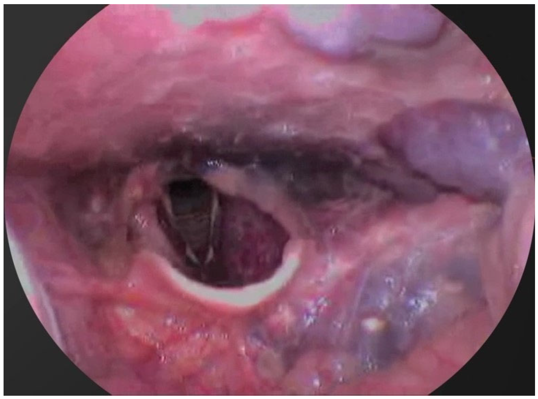
A multicenter, retrospective clinical study was conducted to assess the efficacy and safety of bleomycin-polidocanol foam sclerotherapy in patients with airway venous malformations (AVMs) affecting respiratory function (Figure 1). Patients were treated between January 2022 and April 2025 at clinical centers in Moscow and Makhachkala.
Figure 1.
Endoscopic view of airway venous malformation (AVM) causing respiratory compromise.
2.2. Inclusion Criteria
Patients were eligible for inclusion if they met the following criteria:
- A diagnosis of a venous malformation confirmed by radiological imaging (magnetic resonance imaging (MRI), CT, or ultrasound) and/or direct endoscopic visualization.
- Location of the venous malformation (VM) within the airway, including the soft palate, tongue, oropharynx, or subglottic space.
- The presence of clinical symptoms attributable to the VM, such as respiratory distress, nocturnal apnea, dysphagia, or dysphonia.
- Age between 4 and 67 years.
- Provision of signed informed consent by the patient or their legal guardian.
2.3. Patient Characteristics
The study included 14 patients (7 female and 7 male), consisting of 8 pediatric and 6 adult cases. The anatomical distribution of the lesions was as follows:
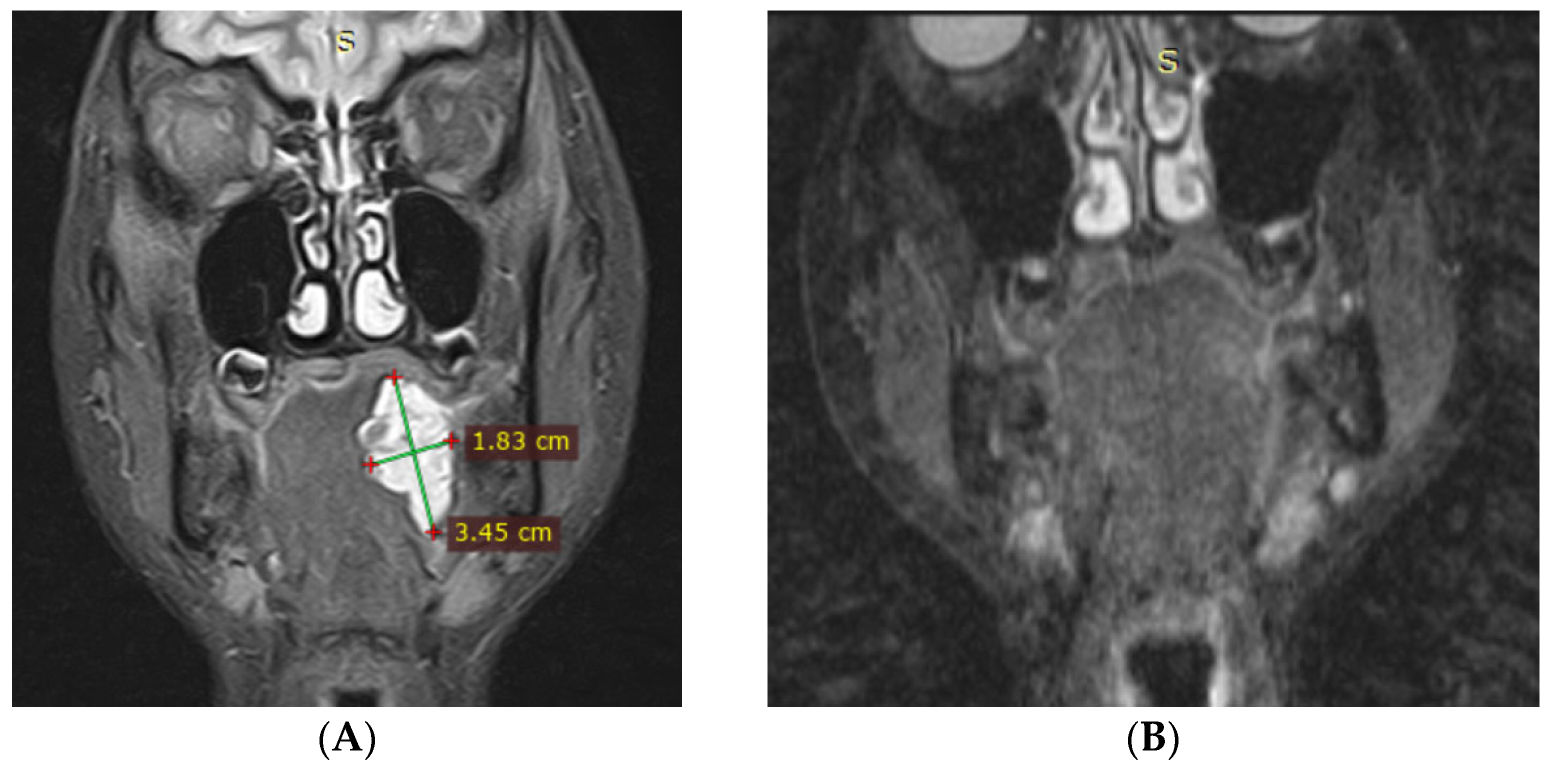
- The tongue (involving the body, base, and floor of the mouth) in 5 patients (Figure 2).
Figure 2. Magnetic resonance imaging (MRI) of a patient with a venous malformation of the tongue base. - The soft palate and palatopharyngeal arches in 6 patients.
- The oropharynx in 2 patients.
- The subglottic space with extension into the cervical soft tissues in 1 patient (Figure 3).
Figure 3. Magnetic resonance imaging (MRI) of a patient with a venous malformation involving the neck and airway.
The presenting symptoms and clinical signs included episodes of nocturnal apnea, stridor, impaired nasal and oral breathing, dysphonia, localized soft tissue enlargement, and dysphagia.
2.4. Treatment Protocol
All patients were treated with foam sclerotherapy using a composition prepared immediately before injection. The formulation consisted of:
- Bleomycin (Bleocin®) at a final concentration of 1.5 to 3 mg/mL
- 3% Polidocanol (Aethoxysklerol®)
- Room air
The foam was generated using the Tessari method, which involves a three-way stopcock and two syringes to combine the bleomycin solution, polidocanol, and room air in a 1:1:4 ratio.
The procedure was performed under either endoscopic or ultrasound guidance, depending on the anatomical site. The injected foam volume ranged from 2 to 20 mL. The choice of anesthesia (local or general) was based on the patient’s age, the location of the VM, and the level of patient cooperation.
In selected cases with severe airway stenosis, a tracheostomy was performed or a nasopharyngeal airway was placed prior to the procedure. Post-procedural monitoring included pulse oximetry, follow-up imaging with CT or MRI, and video endoscopic surveillance.
2.5. Efficacy and Safety Assessment
The primary endpoint for assessing therapeutic efficacy was the quantitative reduction in malformation volume, measured by magnetic resonance imaging (MRI). The percentage of volume reduction was calculated for each patient.
Based on this, the therapeutic response was categorized into three distinct groups:
- Complete response: 100% reduction in lesion volume.
- Significant reduction: volume reduction of more than 50% but less than 100%.
- Limited reduction: volume reduction of less than 50%.
Secondary efficacy outcomes included the resolution of clinical symptoms (e.g., dyspnea, apnea, dysphonia, dysphagia) and the total number of sclerotherapy sessions required to achieve a stable clinical result.
Safety was assessed by documenting the frequency and severity of all adverse events during and after the procedures.
2.6. Statistical Analysis
All statistical analyses were performed using Python (version 3.14) with the SciPy (version 1.16.3) and Pingouin (version 0.5.5) libraries.
Continuous variables, such as lesion volume, were tested for normality using the Shapiro–Wilk test. As the data distribution was found to be non-normal, results are presented as median and interquartile range (IQR).
To compare lesion volumes before and after treatment, the non-parametric Wilcoxon signed-rank test for paired samples was used. The magnitude of the treatment effect was quantified using the Rank-Biserial Correlation (RBC).
Categorical data, such as treatment outcomes and complication rates, are presented as frequencies and percentages (%). A p-value of less than 0.05 was considered statistically significant.
3. Results
All 14 patients in this study, diagnosed with airway venous malformations (AVMs) were treated using a single, standardized protocol for bleomycin-polidocanol foam sclerotherapy. Standardization was applied to the foam preparation technique, the final bleomycin concentration (1.5 to 3 mg/mL), the component ratio (1:1:4; bleomycin solution: polidocanol: air), the injection method, and the criteria for evaluating treatment efficacy.
3.1. Magnetic Resonance Imaging (MRI) Findings
MRI is the primary imaging modality used to diagnose airway venous malformations and assess treatment response. It is highly sensitive to the fluid content within venous lacunae, enabling precise delineation of the lesion’s extent, the degree of soft-tissue infiltration, and the potential for airway compromise. MRI is especially indispensable for evaluating lesions in the retropharyngeal space, the epiglottic region, or when deep or multilocular involvement is suspected.
The most informative sequences include:
T2-weighted images (T2WI): These reveals marked hyperintensity within the malformation due to the stagnant venous blood in the dilated channels.
Short-tau inversion recovery (STIR) sequences: These enhance the contrast between the lesion and surrounding soft tissues, crucial for evaluating the infiltrative margins.
Fat-suppressed T1-weighted images (T1WI): These are vital for precisely delineating the lesion’s topography.
A key diagnostic feature is the presence of phleboliths, which appear as multiple, small, ovoid foci of low signal intensity within the otherwise hyperintense malformation. This finding suggests the lesion’s chronicity and the associated stasis, leading to secondary thrombosis.
Contrast enhancement was not employed in this study, as the characteristic signal intensity of venous malformations on non-contrast sequences is typically sufficient to distinguish them from lymphatic malformations, arteriovenous malformations, and neoplastic processes.
3.2. Clinical Outcomes
The cohort’s baseline median lesion volume was 6782.0 mm3 (IQR: 4943.38857.0 mm3). Following the treatment course, the median volume decreased to 1545.5 mm3 (IQR: 0.04403.8 mm3). The Wilcoxon signed-rank test confirmed that this reduction was statistically highly significant (p < 0.001). The magnitude of this effect was large (Rank-Biserial Correlation = 1.0), indicating a strong and clinically meaningful therapeutic response across the cohort.
A positive clinical response was observed in all 14 patients (100%). Based on our predefined criteria, 8 patients (57.1%) achieved complete response, 4 patients (28.6%) achieved significant reduction in malformation, and 2 patients (14.3%) achieved limited reduction in malformation. A representative case of complete response after a single session is shown in Figure 4, which documents the complete radiological reduction in a left tongue root malformation.
Figure 4.
Magnetic resonance imaging (MRI) of a 13-year-old female patient (Patient #3) with a venous malformation of the left tongue root. (A) The pre-treatment axial T2-weighted scan reveals a hyperintense lesion (arrow). (B) The post-treatment scan, performed after a single session of sclerotherapy, demonstrates complete radiological resolution of the malformation with no residual signs of the lesion.
The detailed clinical characteristics, treatment parameters, and outcomes are summarized in Table 1. Treatment-related complications, limited to localized necrosis, were recorded in three patients (21.4%).
Table 1.
Clinical characteristics and treatment outcomes in 14 patients with airway venous malformations (AVMs).
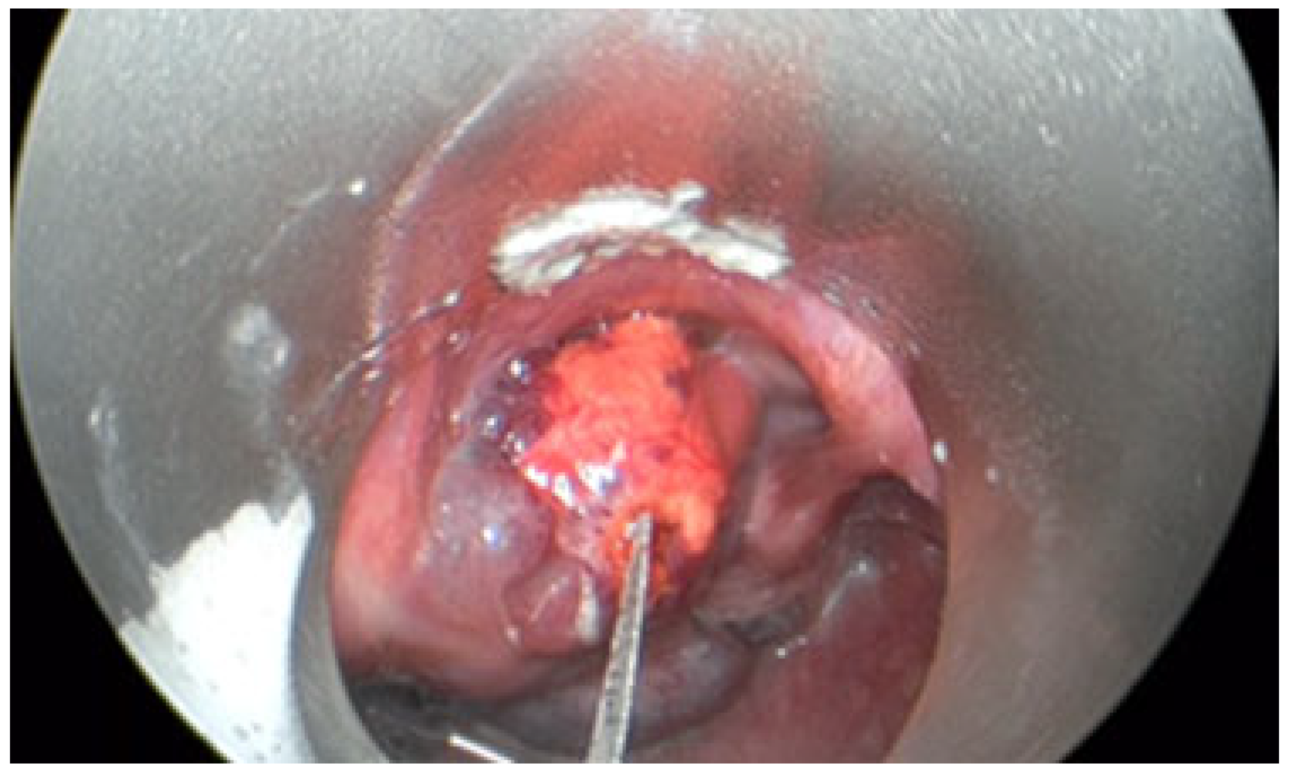
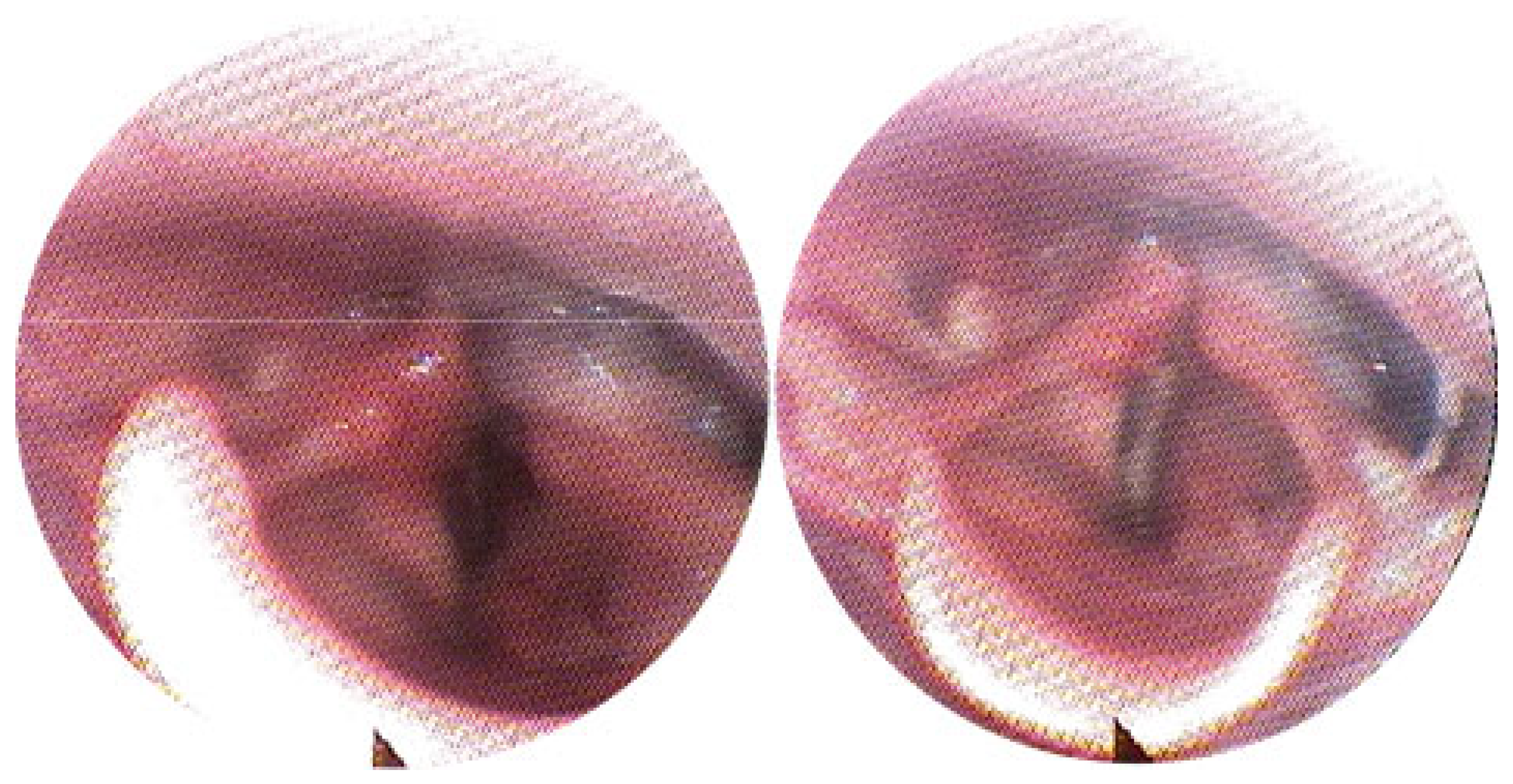
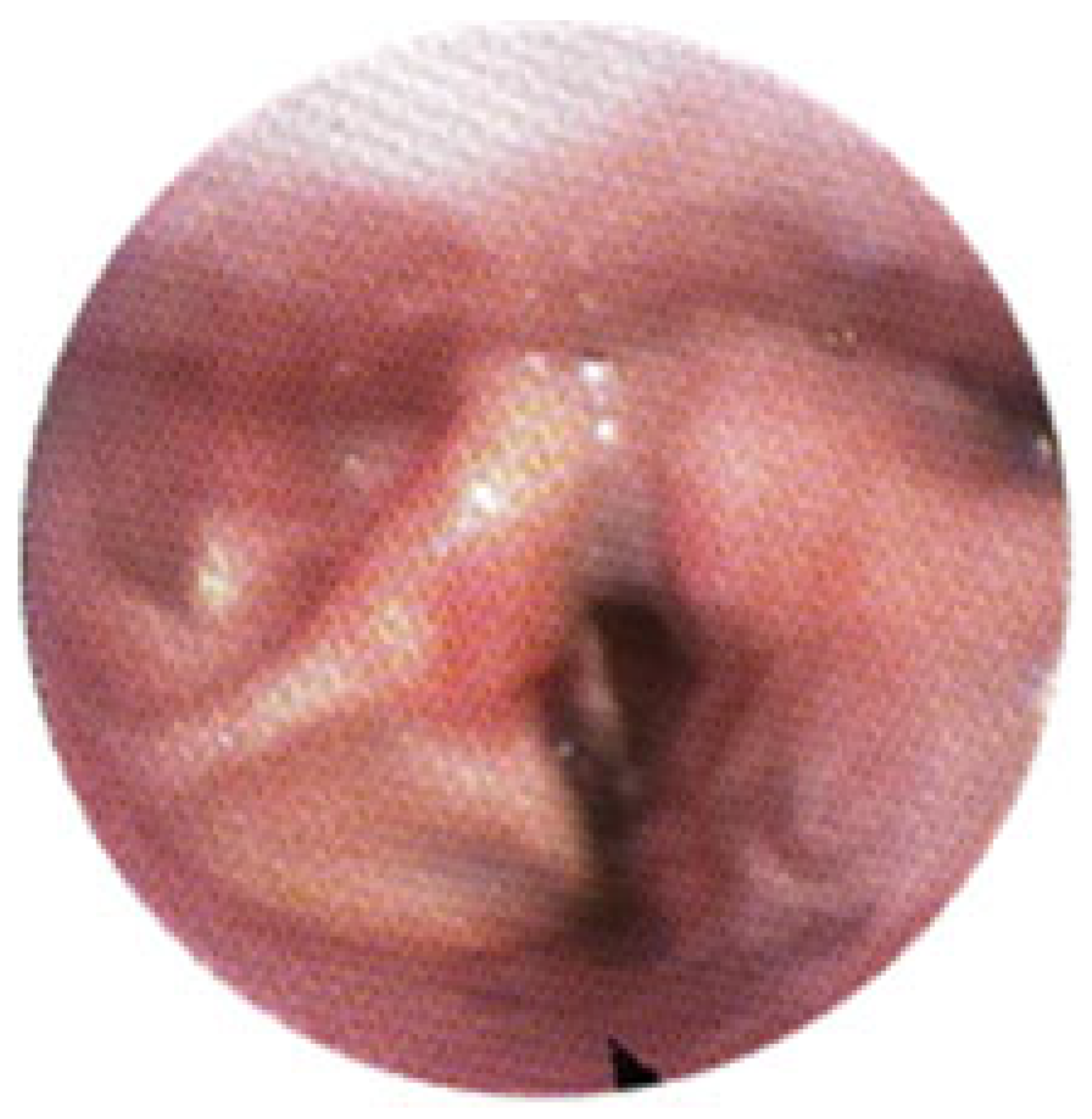
The patient with a subglottic lesion presented with severe luminal narrowing and tracheal deviation, necessitating a prophylactic tracheostomy prior to treatment. Following three sclerotherapy sessions, a substantial reduction in the malformation’s volume and restoration of airway patency were achieved. This clinical improvement facilitated successful decannulation (Figure 5, Figure 6, Figure 7 and Figure 8).
Figure 5.
Venous malformation (VM) of the arytenoid cartilage before sclerotherapy.
Figure 6.
Injection of the combined Bleomycin-Polidocanol foam composition into the venous malformation (VM) of the arytenoid cartilage.
Figure 7.
Endoscopic view after one session of sclerotherapy, demonstrating partial regression of the venous malformation.
Figure 8.
Endoscopic view of the arytenoid cartilage after three sessions of sclerotherapy.
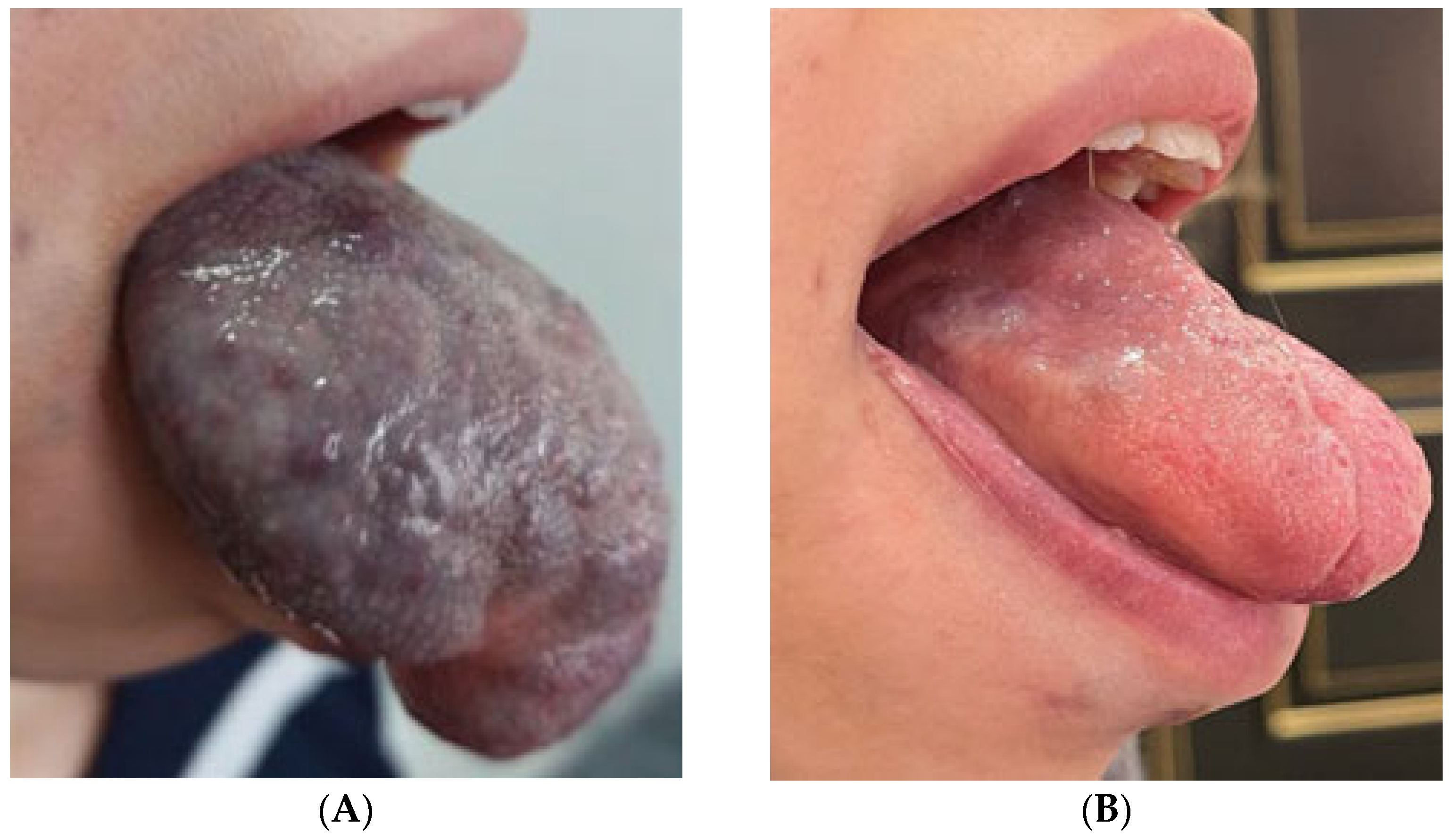
A notable case involved a pediatric female patient who presented with an extensive venous malformation encompassing the entire tongue, leading to severe macroglossia. She underwent a treatment course of 12 weekly sclerotherapy sessions (Figure 9), resulting in a durable complete response, marked by the normalization of tongue morphology and full resolution of symptoms. Follow-up MRI confirmed regression of the malformation. No adverse effects were observed throughout treatment course.
Figure 9.
Venous malformation (VM) of the tongue. (A)—pre-treatment appearance. (B)—post-treatment appearance after 12 sessions of combined bleomycin-polidocanol foam sclerotherapy, demonstrating a complete response.
Clinical efficacy was assessed based on changes in respiratory symptoms (including inspiratory dyspnea, stridor, and rhinolalia) as well as findings from follow-up endoscopic examinations and/or MRI.
A complete or significant partial response was observed in 12 of 14 patients (85.7%) after a treatment course from 1 to 12 sessions. The remaining two patients were also classified as having a partial response; although their malformations persisted, there was a notable reduction in lesion volume, accompanied by a marked improvement in clinical symptoms.
3.3. Complications
The recorded adverse events were infrequent and primarily limited to minor, localized complications. The most common event was mucosal necrosis of the soft palate, which occurred in three patients (21.4%). One of these cases was also associated with a local hematoma and tenderness.
- In two patients, the necrosis was subclinical, asymptomatic, and resolved spontaneously without intervention.
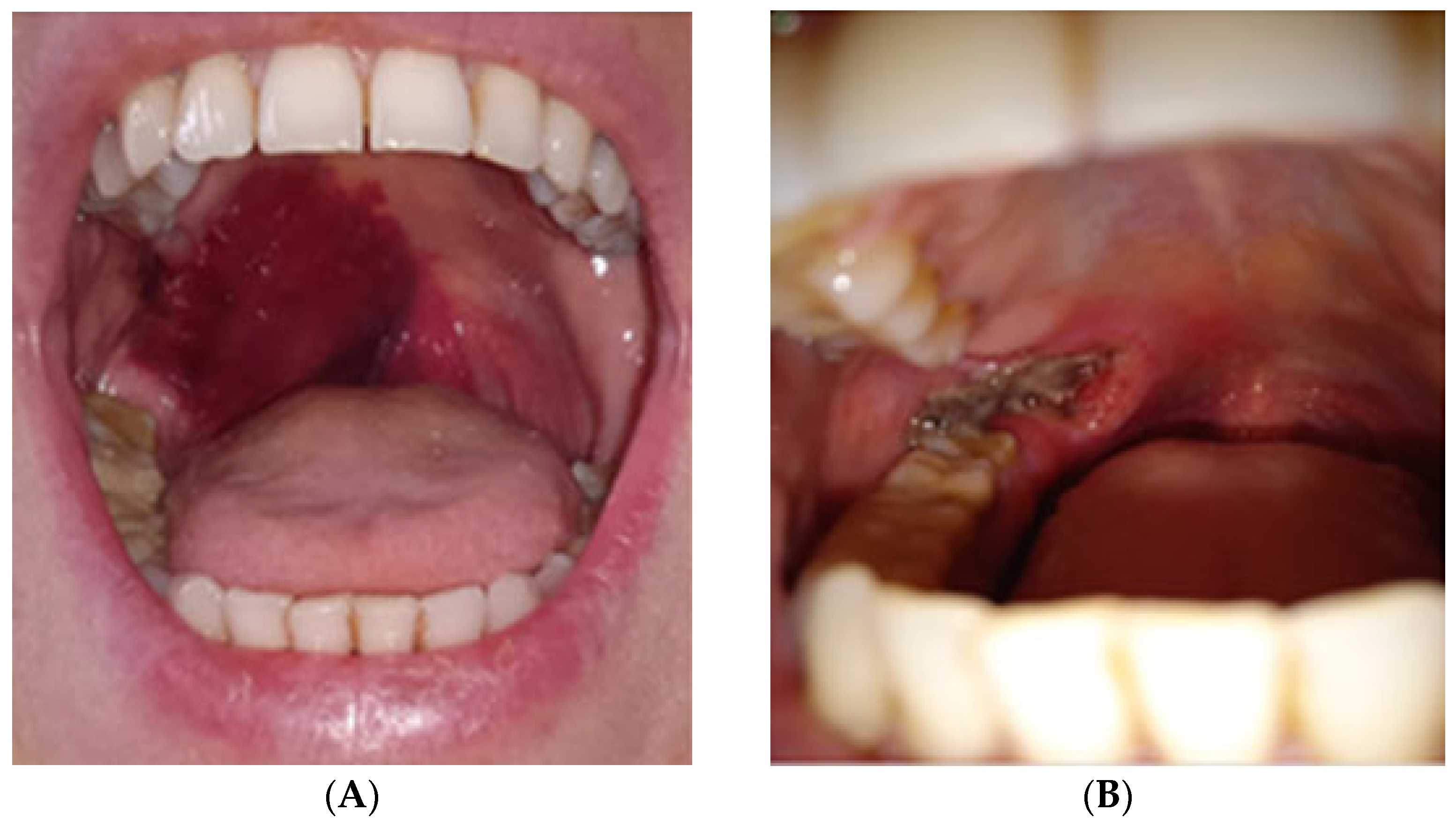
- The third case involved a 47-year-old female who developed localized mucosal necrosis secondary to a hematoma, which was associated with significant tenderness. The patient managed the condition with local wound care, and the lesion healed completely within 11 days without the need for systemic therapy (Figure 10).
Figure 10. Adverse mucosal events following combined bleomycin-polidocanol foam sclerotherapy. (A)—Mucosal ecchymosis observed on day 2 post-treatment. (B)—Progression to focal necrosis by day 7.
No major or systemic complications were observed, including stenosis, laryngeal edema, dysphonia, or the necessity for hospitalization or surgical intervention.
4. Discussion
The results of this study demonstrate the high clinical efficacy and favorable safety profile of our bleomycin-polidocanol foam composition for treating airway venous malformations (AVMs). The method proved effective for both superficial and deep-seated lesions across multiple anatomical sites, including the tongue, soft palate, oropharynx, and subglottic space.
- Complete response (based on both clinical and MRI criteria) was achieved in 8 of 14 patients (57.1%).
- ○
- Of these, 6 patients (42.9% of the total cohort) achieved a complete response after a single sclerotherapy session—two with tongue base venous malformations (VMs) and two with soft palate VMs.
- A partial response, characterized by significant lesion regression, was observed in the remaining 4 patients (28.6%).
- ○
- Significant reduction, with >50% volume reduction, in 4 patients (28.6%).
- ○
- Limited reduction, with <50% volume reduction, in 2 patients (14.3%).
- No patients failed to respond to treatment, resulting in a 0% non-response rate.
One illustrative case involved a patient from Iran who had a diffuse malformation of the entire tongue. She achieved a durable, complete clinical and radiological response after undergoing 12 weekly procedures. In the patient with the subglottic VM and associated tracheal deviation, a prophylactic tracheostomy was performed. Successful decannulation occurred after just three sclerotherapy sessions.
Complications were reported in 3 of 14 patients (21.4%), all of which involved focal mucosal necrosis of the soft palate, primarily affecting the palatopharyngeal arch. Two of these cases were subclinical, while one was associated with tenderness and resolved spontaneously within 11 days.
The occurrence of mucosal necrosis (21.4%), though minor and self-limiting in all cases, warrants a discussion of strategies to minimize its incidence. Our experience indicates that a multi-faceted approach—emphasizing dosage, foam preparation, and injection technique is essential for enhancing patient safety.
First, strict adherence to weight-based dosing represents a fundamental safety principle. In our practice, we did not exceed the maximum recommended doses of 1 mg/kg for bleomycin and 2 mg/kg for polidocanol per session, which helps mitigate both local and systemic toxicity.
Second, the physicochemical properties of the foam are crucial. The standardized Tessari method, employing a 1:1:4 ratio of bleomycin solution, polidocanol, and air, ensures the production of a consistent and stable foam. Reducing the proportion of air increases the concentration of the sclerosing agents, thereby elevating the risk of excessive tissue reaction and necrosis.
Finally, the injection technique itself is likely the most critical modifiable factor. We observed that necrosis was associated with superficial, submucosal deposition of the foam. Therefore, we advocate for deeper, intramural injections into the bulk of the malformation whenever possible. In cases involving superficial phlebectasias where this is unavoidable, we recommend a gentle digital massage of the injection site for approximately two minutes post-procedure. This maneuver helps disperse the foam, preventing localized pooling and reducing the direct cytotoxic effect on the overlying mucosa. Regarding management, all observed complications were treated conservatively with local wound care, confirming their manageable nature.
A key observation during this study was the issue of foam degradation during transit through long infusion systems. In the patient with the subglottic VM, the sclerosant was delivered via an extended infusion set, and the foam was observing to lose some of its structural integrity before reaching the target lesion. This finding highlights the need for future research aimed at enhancing foam stability, particularly for treating lesions in deep or complex anatomical locations.
In contrast to previously published reports, which were limited to small case series (e.g., Kamijo et al., 2013, n = 5; Oomen et al., 2015, n = 6; Azene et al., 2016, n = 1) [,,], the present study represents the largest and most homogeneous cohort of patients with airway VMs treated with a single, standardized protocol in the published literature.
The use of combined bleomycin-polidocanol foam offers several key procedural advantages:
- It is a minimally invasive procedure that can often be performed under local anesthesia.
- The treatment is scarless, resulting in superior cosmetic outcomes.
- The foam effectively fills complex, multilocular cavities, ensuring uniform distribution of the sclerosant.
- The injection and distribution of the foam can be monitored in real-time under image guidance (ultrasound, endoscopy, or fluoroscopy).
The superior clinical efficacy of sclerotherapy foam over liquid formulations is attributed to a complex interplay of synergistic biological and physiological effects. Upon injection, the foam initiates a rapid, multifactorial cascade that targets the vessel wall on multiple levels, resulting in efficient and permanent fibrotic obliteration. The key mechanisms underlying this process include:
- Sclerophlebitis: Foam induces a more pronounced and localized aseptic inflammation of the vessel wall compared to liquid agents, accelerating fibrosis process and vessel occlusion [,].
- Venospasm: The injection of foam triggers a reflex contraction of the vein’s smooth muscle layer, promoting luminal collapse, reducing blood outflow, and extending the sclerosant’s exposure time [].
- Thrombogenic Effect: The close apposition of foam to the endothelium accelerates both the formation and its subsequent organization into an occlusive thrombus [].
- Prolonged Retention: Due to its high viscosity, microfoam (bubbles < 250 μm) remains at the injection site longer than liquid, ensuring sustained contact [].
- Uniform Endothelial Destruction: The finely dispersed nature of the foam results in more uniform and profound endothelial damage at the molecular level compared to liquid sclerosants [].
The high efficacy of the combined foam is not solely attributable to its physical properties, but rather is fundamentally rooted in the powerful pharmacological synergy of its active components. The formulation utilizes two distinct yet complementary mechanisms of action: the immediate, ablative effect of a detergent sclerosant and the long-term, biological control of a cytostatic and anti-angiogenic agent. This dual-pronged approach to the malformation’s endothelium drives a durable clinical response through the following effects:
- Detergent Action of Polidocanol: As a detergent, polidocanol disrupts the cell membranes of the endothelium, initiating the localized inflammatory response necessary for fibrosis [].
- Cytostatic Effect of Bleomycin: Bleomycin induces DNA damage and apoptosis in endothelial cells. Critically, it also inhibits pathological angiogenesis by blocking the PI3K/AKT/mTOR signaling pathway [,,].
- Synergistic Effect of the Combination: The use of a combined bleomycin-polidocanol foam creates a synergistic effect, enhancing the depth of endothelial injury and thereby reducing the risk of lesion recurrence [,].
This study has several inherent limitations that must be acknowledged. The primary limitation is its retrospective, non-randomized design and the absence of a direct comparator or control group.
Our analysis of clinical outcomes such as “symptom resolution” or “successful decannulation” does not formally account for potential competing events (e.g., the need for alternative procedures, intercurrent illnesses affecting airway status, or other unrelated adverse events that could influence the outcome). Consequently, a formal cumulative incidence analysis was not performed.
While our results are promising, this study design precludes definitive conclusions about the superiority of this specific composite foam over other treatment modalities. The extreme rarity of airway venous malformations makes conducting prospective, randomized controlled trials ethically challenging and practically infeasible, necessitating reliance on well-documented case series such as this one.
To contextualize our findings, we compared them with historical controls from the limited available literature. Previous reports on sclerotherapy for AVMs have been restricted to very small cohorts and have employed varied agents and protocols, such as bleomycin alone or different sclerosants [,,]. Although a direct statistical comparison is impossible due to this heterogeneity, our observed complete response rate of 71.4% with a standardized protocol appears highly favorable and makes a meaningful contribution to the existing evidence base.
Furthermore, alternative treatment modalities for AVMs include surgical excision, laser therapy, and systemic medical management (e.g., with mTOR inhibitors like sirolimus). Surgical resection in the airway carries a high risk of morbidity, including nerve damage, scarring, and functional impairment. Laser ablation can be effective for superficial lesions but may require multiple sessions and carries a risk of collateral thermal injury. Systemic therapies are typically reserved for extensive, syndromic, or refractory cases and have their own profile of systemic side effects.
In this context, our material-based approach using an in situ forming composite foam offers a compelling, minimally invasive, scarless, and highly targeted alternative that directly addresses the localized nature of these lesions. Future prospective, multicenter registry studies will be essential to build upon these findings and further define the optimal treatment algorithm for this rare condition.
Foam stability is a critical technical challenge in sclerotherapy. The degradation of foam, particularly as it passes through long catheters or infusion systems, becomes especially problematic when treating lesions located below the glottis. The primary mechanisms of foam degradation include liquid drainage, bubble coalescence, and disproportionation [].
Future efforts to advance this technique should focus on enhancing foam stability, which can be achieved by modifying three key components. First, the gas phase can be modified by replacing room air with more soluble gases like CO2 or, with less soluble gases, like sulfur hexafluoride (SF6), which helps reduce disproportionation. Second, the liquid phase can be enhanced by incorporating viscosity-enhancing agents, such as glycerol or hyaluronic acid, to slow drainage, or by adding surfactants to strengthen the bubble structure. Finally, the preparation method itself can be refined through techniques such as multi-cycle emulsification or the use of micromesh devices, combined with immediate point-of-care administration to ensure the foam is delivered in its most stable state.
5. Conclusions
Combined bleomycin-polidocanol foam sclerotherapy is a highly effective treatment for airway venous malformations (AVMs), demonstrating a 100% response rate in our cohort, which includes a 71% complete response rate and a 29% partial response rate.
The treatment has a favorable safety profile, with a complication rate of 21.4%. All adverse events were localized, minor, and fully reversible.
Foam instability during delivery into the lower airway represents a significant technical challenge that warrants further investigation to optimize both the formulation and administration technique.
These findings provide a strong foundation for the standardization of care for airway venous malformations and significantly advance the clinical understanding of this rare but challenging pathology.
Further development of this method depends on improving foam stability.
Author Contributions
Conceptualization, A.M.; Methodology, L.V.; Software, E.A.; Validation, E.A. and A.A.V.; Formal analysis, E.A.; Investigation, L.V.; Resources, L.V.; Data curation, L.V. and E.A.; Writing—original draft, L.V. and E.A.; Writing—review & editing, E.A. and R.V.; Visualization, L.V. and E.A.; Supervision, A.M., A.A.V., R.V. and D.T.; Project administration, A.A.V., R.V. and D.T.; Funding acquisition, A.M. All authors have read and agreed to the published version of the manuscript.
Funding
This research was funded by the Ministry of Health of the Russian Federation within the framework of a state assignment (Theme No. 124031100087-1).
Institutional Review Board Statement
The study was conducted in accordance with the Declaration of Helsinki, and approved by the Inter-University Ethics Committee at the Russian University of Medicine (ROSUNIMED) (No. 05-19, approval date: 16 May 2019).
Informed Consent Statement
Informed consent was obtained from all subjects involved in the study. Written informed consent has been obtained from the patients to publish this paper.
Data Availability Statement
The data presented in this study are not publicly available due to ethical restrictions and patient privacy concerns. Data may be made available upon reasonable request to the corresponding author, subject to approval from the Institutional Review Board.
Acknowledgments
The authors would like to thank a native speaker Vasilisa Shunikhina for her assistance with the edition of the submission’s final version.
Conflicts of Interest
The authors declare no conflicts of interest.
Abbreviations
The following abbreviations are used in this manuscript:
| AVM | Airway Venous Malformation |
| CR | Complete Response |
| CT | Computed Tomography |
| IRB | Institutional Review Board |
| MRI | Magnetic Resonance Imaging |
| NR | No Response |
| STIR | Short-Tau Inversion Recovery |
| T1WI | T1-Weighted Image |
| T2WI | T2-Weighted Image |
| VM | Venous Malformation |
References
- Garbuzov, R.V.; Polyaev, Y.A.; Stepanov, A.E.; Mylnikov, A.A. Abernathy malformations in children. Experience in endovascular and surgical treatment. Russ. J. Pediatr. Surg. 2020, 24, 71–77. [Google Scholar] [CrossRef]
- Castel, P.; Carmona, F.J.; Grego-Bessa, J.; Berger, M.F.; Viale, A.; Anderson, K.V.; Bague, S.; Scaltriti, M.; Antonescu, C.R.; Baselga, E.; et al. Somatic PIK3CA mutations as a driver of sporadic venous malformations. Sci. Transl. Med. 2016, 8, 332ra42. [Google Scholar] [CrossRef]
- Uebelhoer, M.; Nätynki, M.; Kangas, J.; Mendola, A.; Nguyen, H.L.; Soblet, J.; Godfraind, C.; Boon, L.M.; Eklund, L.; Limaye, N.; et al. Venous malformation-causative TIE2 mutations mediate an AKT-dependent decrease in PDGFB. Hum. Mol. Genet. 2013, 22, 3438–3448. [Google Scholar] [CrossRef] [PubMed]
- Queisser, A.; Seront, E.; Boon, L.M.; Vikkula, M. Genetic Basis and Therapies for Vascular Anomalies. Circ. Res. 2021, 129, 155–173. [Google Scholar] [CrossRef]
- Wassef, M.; Blei, F.; Adams, D.; Alomari, A.; Baselga, E.; Berenstein, A.; Burrows, P.; Frieden, I.J.; Garzon, M.C.; Lopez-Gutierrez, J.C.; et al. Vascular Anomalies Classification: Recommendations From the International Society for the Study of Vascular Anomalies. Pediatrics 2015, 136, e203–e214. [Google Scholar] [CrossRef]
- Azene, E.; Mitchell, S.; Radvany, M.; Agrawal, N.; Eisele, D.; Weiss, C. Foamed bleomycin sclerosis of airway venous malformations: The role of interspecialty collaboration. Laryngoscope 2016, 126, 2726–2732. [Google Scholar] [CrossRef]
- Kamijo, A.; Hatsushika, K.; Kanemaru, S.; Moriyama, M.; Kase, Y.; Masuyama, K. Five adult laryngeal venous malformation cases treated effectively with sclerotherapy. Laryngoscope 2013, 123, 2766–2769. [Google Scholar] [CrossRef]
- Oomen, K.P.; Paramasivam, S.; Waner, M.; Niimi, Y.; Fifi, J.T.; Berenstein, A.; O, T.M. Endoscopic transmucosal direct puncture sclerotherapy for management of airway vascular malformations. Laryngoscope 2016, 126, 205–211. [Google Scholar] [CrossRef]
- Gurgacz, S.; Zamora, L.; Scott, N.A. Percutaneous sclerotherapy for vascular malformations: A systematic review. Ann. Vasc. Surg. 2014, 28, 1335–1349. [Google Scholar] [CrossRef] [PubMed]
- Zhang, W.; Chen, G.; Ren, J.G.; Zhao, Y.F. Bleomycin induces endothelial mesenchymal transition through activation of mTOR pathway: A possible mechanism contributing to the sclerotherapy of venous malformations. Br. J. Pharmacol. 2013, 170, 1210–1220. [Google Scholar] [CrossRef]
- Cabrera, J.; Cabrera García-Olmedo, J.R. Nuevo método de esclerosis en las varices tronculares. Patol. Vasc. 1995, 4, 55–73. [Google Scholar]
- Tessari, L.; Cavezzi, A.; Frullini, A. Preliminary experience with a new sclerosing foam in the treatment of varicose veins. Dermatol. Surg. 2001, 27, 58–60. [Google Scholar] [CrossRef]
- Cavezzi, A.; Mosti, G.; Campana, F.; Tessari, L.; Bastiani, L.; Urso, S.U. Catheter Foam Sclerotherapy of the Great Saphenous Vein, with Perisaphenous Tumescence Infiltration and Saphenous Irrigation. Eur. J. Vasc. Endovasc. Surg. 2017, 54, 629–635. [Google Scholar] [CrossRef]
- Azmoun, S.; Liu, Y.; Bian, K.; Chen, A.; Liu, S. The Effect of Pushing Rate on Foam Stability in the Tessari Method. Dermatol. Surg. 2024, 50, 542–545. [Google Scholar] [CrossRef]
- Shi, X.; Liu, Y.; Li, D.; Tursun, M.; Azmoun, S.; Liu, S. The Stability of Physician-Compounded Foam is Influenced by the Angle of Connector. Ann. Vasc. Surg. 2024, 99, 217–222. [Google Scholar] [CrossRef] [PubMed]
- Rabe, E.; Breu, F.X.; Cavezzi, A.; Coleridge Smith, P.; Frullini, A.; Gillet, J.L.; Guex, J.J.; Hamel-Desnos, C.; Kern, P.; Partsch, B.; et al. European guidelines for sclerotherapy in chronic venous disorders. Phlebology 2014, 29, 338354. [Google Scholar] [CrossRef] [PubMed]
- Chavla, K.K.; Gladysh, G. Composite Foams. In Encyclopedia of Polymer Science and Technology, 3rd ed.; Mark, H.F., Ed.; Wiley-Interscience: Hoboken, NJ, USA, 2003; Volume 9, pp. 267–281. [Google Scholar]
- Zhang, H.; Liu, S.; Chen, A. A Novel Compound Sclerosant: Polidocanol-Bleomycin Foam. Dermatol. Surg. 2020, 46, 1712–1714. [Google Scholar] [CrossRef] [PubMed]
- Sun, Y.; Gu, H.; Yang, X.; Cai, R.; Shang, Y.; Hu, L.; Wang, Y.; Chen, H.; Lin, X. Bleomycin Polidocanol Foam (BPF) Stability—In Vitro Evidence for the Effectiveness of a Novel Sclerosant for Venous Malformations. Eur. J. Vasc. Endovasc. Surg. 2020, 59, 1011–1018. [Google Scholar] [CrossRef]
- Yang, X.; Chen, H.; Gu, H.; Jin, Y.; Hu, L.; Hua, C.; Wang, Y.; Sun, Y.; Yu, W.; Lin, X. Interim results of bleomycin-polidocanol foam sclerotherapy as a highly efficient technique for venous malformations. J. Vasc. Surg. Venous Lymphat. Disord. 2020, 8, 1066–1073. [Google Scholar] [CrossRef]
- Frullini, A.; Cavezzi, A. Sclerosing foam in the treatment of varicose veins and telangiectases: History and analysis of safety and complications. Dermatol. Surg. 2002, 28, 11–15. [Google Scholar] [CrossRef]
- Wang, B.; Lin, Y.; Yue, H.J.; Guo, W.B.; Chen, L.; Lyu, K.X.; Huang, D.Y.; Lei, W.B. Comparison of pingyangmycin fibrin glue composite and pingyangmycin dexamethasone composite in the treatment of pharyngolaryngeal venous malformation. Chin. J. Otorhinolaryngol. Head Neck Surg. 2023, 58, 552–557. (In Chinese) [Google Scholar] [CrossRef]
- Jia, X.; Mowatt, G.; Burr, J.M.; Cassar, K.; Cook, J.; Fraser, C. Systematic review of foam sclerotherapy for varicose veins. Br. J. Surg. 2007, 94, 925–936. [Google Scholar] [CrossRef]
- Cavezzi, A.; Parsi, K. Complications of foam sclerotherapy. Phlebology 2012, 27 (Suppl. S1), 46–51. [Google Scholar] [CrossRef] [PubMed]
- Cavezzi, A.; Frullini, A.; Ricci, S.; Tessari, L. Treatment of Varicose Veins by Foam Sclerotherapy: Two Clinical Series. Phlebology 2002, 17, 13–18. [Google Scholar] [CrossRef]
- Bai, T.; Liu, Y.; Jiang, W.; Li, Y.; Liu, J.; Yu, C.; Fan, Y. A Review of Sclerosing Foam Stability in the Treatment of Varicose Veins. Dermatol. Surg. 2020, 46, 249–257. [Google Scholar] [CrossRef] [PubMed]
- Parsi, K. Interaction of detergent sclerosants with cell membranes. Phlebology 2015, 30, 306–315. [Google Scholar] [CrossRef]
- Chen, F.; Zhao, W.; Du, C.; Chen, Z.; Du, J.; Zhou, M. Bleomycin induces senescence and repression of DNA repair via downregulation of Rad51. Mol. Med. 2024, 30, 54. [Google Scholar] [CrossRef] [PubMed]
- Roy, T.; Boateng, S.T.; Uddin, M.B.; Banang-Mbeumi, S.; Yadav, R.K.; Bock, C.R.; Folahan, J.T.; Siwe-Noundou, X.; Walker, A.L.; King, J.A.; et al. The PI3K-Akt-mTOR and Associated Signaling Pathways as Molecular Drivers of Immune-Mediated Inflammatory Skin Diseases: Update on Therapeutic Strategy Using Natural and Synthetic Compounds. Cells 2023, 12, 1671. [Google Scholar] [CrossRef]
- Huang, T.T.; Lampert, E.J.; Coots, C.; Lee, J.M. Targeting the PI3K pathway and DNA damage response as a therapeutic strategy in ovarian cancer. Cancer Treat. Rev. 2020, 86, 102021. [Google Scholar] [CrossRef]
- Sun, B.; Sun, X.; Gao, Q.; Wang, Q. Advances in Sclerosants for Low-Flow Vascular Malformations. Ann. Vasc. Surg. 2025, 122, 511–523. [Google Scholar] [CrossRef] [PubMed]
- Fraissenon, A.; Fortin, F.; Durous, V.; Chauvel-Picard, J.; Gleizal, A.; Viremouneix, L.; Cabet, S.; Guibaud, L. Percutaneous Sclerotherapy of Large Venous Malformations Using Consecutive Polidocanol and Bleomycin Foam: MR Imaging Volumetric and Quality-of-Life Assessment. J. Vasc. Interv. Radiol. 2024, 35, 127–136.e1. [Google Scholar] [CrossRef] [PubMed]
Disclaimer/Publisher’s Note: The statements, opinions and data contained in all publications are solely those of the individual author(s) and contributor(s) and not of MDPI and/or the editor(s). MDPI and/or the editor(s) disclaim responsibility for any injury to people or property resulting from any ideas, methods, instructions or products referred to in the content. |
© 2025 by the authors. Licensee MDPI, Basel, Switzerland. This article is an open access article distributed under the terms and conditions of the Creative Commons Attribution (CC BY) license (https://creativecommons.org/licenses/by/4.0/).