Abstract
Blastocystis spp. are among the few enteric parasites with a prevalence that can reach up to approximately 80% in communities of developing countries. This systematic review updates and summarizes available literature on the molecular prevalence and subtype distribution of Blastocystis spp. in Latin American people. This work follows the PRISMA (Preferred Reporting Items for Systematic Reviews and Meta-Analyses) guidelines. The literature revised covers from 1 January 2015 to 6 October 2023 in seven different scientific databases, and the material was selected through inclusion and exclusion criteria. According to data found in the 36 selected articles, the prevalence of Blastocystis spp. in Latin America ranged between 5.8% (Bolivian rural communities) and 94.0% (Colombian general public). Generally, genomic DNA was extracted from approximately 200 mg fecal sediments using commercial kits, such as the QIAamp Stool Mini Kit (QIAGEN, Hilden, Germany) or the Norgen Stool DNA Isolation Kit (Norgen Biotek Corporation, Thorold, ON, Canada). Subtype-specific primers (such as the couple of primers BhRDr–RD5) developed from unique sequences of the SSU rRNA gene were applied to Blastocystis subtyping. Ten specific subtypes (STs) were found as well as various mixed infections, and the most circulating Blastocystis STs were in the order ST3, ST1, ST2, and ST4. The most recent data about Blastocystis spp. molecular epidemiology and the STs in communities of Latin America are limited to studies from specific countries. Novel scientific data from the other countries are required to obtain a complete picture and truly understand the distribution and prevalence of Blastocystis spp. and the STs.
1. Introduction
Blastocystis spp. are anaerobic, unicellular, intestinal parasitic protists distributed worldwide and able to colonize the large intestine of many vertebrate species, including humans [1,2,3,4]. Blastocystis spp. present multiple evolutionary stages or life cycles, i.e., vacuolar, granular, multi-vacuolar, a-vacuolar, ameboid, and cystic forms [5,6].
The main methods to detect Blastocystis spp. are microscopy, culturing, and molecular assays [7]. Two standard sets of primers, i.e., BhRDr/RD5 (~600 bp, Barcoding primers) and Blast 505–532/Blast 998–1017 (~500 bp, Santin primers), were frequently used for sequence regions of the small subunit ribosomal RNA (SSU rRNA) gene and typified Blastocystis spp. [8,9]. Molecular methods include: (1) restricted fragment length polymorphism (RFLP), (2) subtype-specific sequence-tagged site (STS), and (3) real-time polymerase chain reaction (PCR) [10,11]. Maloney, Molokin, and Santin [12], more recently, developed a novel method using Oxford Nanopore MinION long-read sequencing and universal eukaryotic primers to produce full-length (>1800 bp) SSU rRNA gene sequences for Blastocystis spp. The Public Databases for Molecular Typing and Microbial Genome Diversity (PubMLST) reports provenance and phenotype information linked to Blastocystis spp. molecular typing information [13].
Blastocystis spp., isolated from both human and animal hosts, are currently classified into 18S rRNA gene subtypes (STs), i.e., ST1–ST17, ST21, and ST23–ST32 [13,14]. Many STs, i.e., ST1–ST8, ST10, ST12, ST14, and ST16, are detected in different hosts (humans and domestic and wild animals) [13,15,16]. The frequency and variety of Blastocystis spp. STs in various species of animals strengthens the hypothesis of parasite zoonotic transmission [17,18]. Four STs, i.e., ST1, ST2, ST3, and ST4, are found frequently in human studies [19,20,21,22]; to date, ST9 has been identified only in humans [16,18]. However, complete SSU rRNA gene sequences are available for 17 STs (ST1–ST17) [23,24].
Blastocystis spp. are among the few enteric parasites with a prevalence that frequently exceeds 5% in citizens of industrialized countries [25] and can reach up to approximately 80% in developing countries [26,27]. The parasite transmission among humans generally occurs via the fecal–oral route, and may be either direct (i.e., person-to-person or zoonotic) or indirect (i.e., foodborne and waterborne) [28,29].
People living in rural contexts or developing countries, such as those located in the Caribbean and Latin America, with deficient water sanitary supply services, inadequate wastewater treatments, and close animal contact, are particularly exposed to health risks derived from a Blastocystis spp. infection [30]. Additionally, favorable climatic conditions, i.e., high humidity and warm temperature, increase the chance of transmission of Blastocystis spp. in tropical areas [31,32]. The Blastocystis spp.-infected patients are, in many cases, asymptomatic or generally experience mild symptoms, such as diarrhea, abdominal pain, flatulence, bloating, constipation, and skin lesions [33]. However, specific groups of individuals, i.e., children, elderly people, and patients with anemia or irritable bowel syndrome, could experiment serious Blastocystosis symptoms [16,34,35].
The occurrence of Blastocystis spp. has been observed in various water sources [36,37], including rivers [38], lakes [39], streams [40], sewage [41], surface water [42], stored water [43], tap water [44], and water tanks [45]. Outbreaks of Blastocystosis in poor communities have been linked to drinking water contaminated with fecal matter and poor sanitation services [40,46]. Scientific reports and evidence about the occurrence and prevalence of parasites in water sources have given the international community a boost to adopt safety strategies, such as the safe drinking water, sanitation, and hygiene programs (WASH) of the World Health Organization (WHO) [47].
Despite the recent efforts of the scientific community, governments, and international health organizations to detect and typify Blastocystis spp. in the Caribbean and Latin American countries, the epidemiological–molecular characterization and spatial distribution are not yet fully clarified. This systematic review updates and summarizes available literature on the molecular prevalence and subtype distribution of Blastocystis spp. in individuals of Latin America.
2. Materials and Methods
The systematic review was performed according to the guidelines set forth in the Preferred Reporting Items for Systematic Reviews and Meta-Analyses (PRISMA) and the checklist of Moher et al. [48] (Supplementary File S1).
2.1. Search Strategy
Searching the literature was carried out on 6–7 October 2023 by an author (Y.S.-G.). Full-text articles were searched in seven electronic scientific databases, including ISI Web of Science (Clarivate Analytics), EMBASE (Elsevier), Science Direct (Elsevier), Scopus (Elsevier), SciELO (São Paulo Research Foundation—FAPESP), PubMed (National Library of Medicine of USA—NLM), and EBSCOhost (EBSCO Industries), using the following Boolean equation: (“Blastocystis”) AND (“Argentina” OR “Belize” OR “Bolivia” OR “Brazil” OR “Chile” OR “Colombia” OR “Costa Rica” OR “Cuba” OR “Ecuador” OR “El Salvador” OR “Guatemala” OR “Guyana” OR “French Guyana” OR “Honduras” OR “Mexico” OR “Nicaragua” OR “Panama” OR “Paraguay” OR “Peru” OR “Puerto Rico” OR “Dominican Republic” OR “Suriname” OR “Uruguay” OR “Venezuela”).
2.2. Inclusion Criteria
The inclusion criteria, applied to full texts for assessing their eligibility, were: (a) original article focusing on molecular identification of Blastocystis spp. in Latin American humans, (b) article published from 1 January 2015 to 6 October 2023, (c) article written in English and/or Spanish, (d) study limited to humans, (e) cross-sectional study, and (f) article published in peer-reviewed journals inserted in the Scimago Journal Ranking (SJR) database.
2.3. Exclusion Criteria
The exclusion criteria, applied to full texts for assessing their eligibility, were: (a) abstract not associated with the full article, (b) article published in non-peer-reviewed source, (c) article not written in English or Spanish, (d) review papers of literature or meta-analyses, (e) retrospective studies, (f) short communication, (g) letters to the editor, and (h) studies with ≤3 points based on the Joanna Briggs Institute (JBI) tool [49].
2.4. Selection of Studies
The Mendeley Desktop Reference Management System 1.19.8 software was used to compile the identified articles and to remove the duplicates. Subsequently, articles were independently screened for title and abstract pertinence by two authors (Y.S.-G. and J.E.B.). Irrelevant titles were removed. Disagreements between the two researchers were resolved through consultation with a third author (C.F.). Inclusion and exclusion criteria were applied to full texts to assess their eligibility. Two authors (Y.S.-G. and J.E.B.) independently analyzed the full-text papers and only those that met all criteria were finally selected.
2.5. Data Extraction and Analysis
The information from each selected paper was extracted and organized in a matrix with the following subjects: (a) reference, (b) quartile, (c) rural/urban, (d) quality, (e) collection period, (f) demographic group studied, (g) age, (h) number of repeat samples, (i) concentration method, (j) DNA extraction method, (k) Blastocystis-specific SSU-rDNA primers, (l) product size, (m) amplification, (n) subtypes, (o) prevalence, and (p) 95% confidence intervals (CI).
2.6. Quality Assessment
The quality of the included studies was assessed with standardized critical appraisal instruments from the JBI for prevalence [49]; the tool, composed by nine questions with four answer options (yes, no, unclear, and not applicable), assists in assessing the trustworthiness, relevance, and results of published scientific articles.
Based on a quality score rating system, the papers were divided into two categories: high-quality study (score 7–9) and moderate-quality study (score 4–6).
The nine questions (Q) of the JBI instrument are: (Q1) Was the sample frame appropriate to address the target population? (Q2) Were study participants sampled in an appropriate way? (Q3) Was the sample size adequate? (Q4) Were the study subjects and the setting described in detail? (Q5) Was the data analysis conducted with sufficient coverage of the identified sample? (Q6) Were valid methods used for the identification of the condition? (Q7) Was the condition measured in a standard, reliable way for all participants? (Q8) Was there appropriate statistical analysis? (Q9) Was the response rate adequate and, if not, was the low response rate managed appropriately?
Two researchers (Y.S.-G. and J.E.B.) worked independently, and disagreements were resolved through consultation with a third author (C.F.) (Supplementary File S2).
3. Results
3.1. Literature Search
The PRISMA Statement flow diagram that indicates the four phases of the literature search (identification, screening, eligibility, and inclusion) is shown in Figure 1. During the identification phase, a total of 948 publications were recorded. Duplicated articles were automatically removed via a bibliographic management software and the remaining 374 papers were screened for title and abstract pertinence.
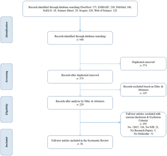
Figure 1.
PRISMA flow diagram.
The eligibility of 229 full-text articles that passed the title and abstract screening phase was assessed based on preset inclusion and exclusion criteria. Finally, 36 articles [50,51,52,53,54,55,56,57,58,59,60,61,62,63,64,65,66,67,68,69,70,71,72,73,74,75,76,77,78,79,80,81,82,83,84,85] were included in this systematic review.
3.2. Characteristics of Included Studies
Figure 2 shows the eighteen articles published in Q1 SJR journals [50,51,52,54,55,61,63,64,65,70,71,75,76,77,79,80,81,82], the fifteen articles in Q2 SJR journals [57,58,59,60,62,66,67,68,69,72,73,74,83,84,85], and the three articles found in Q3 SJR journals [53,56,78].
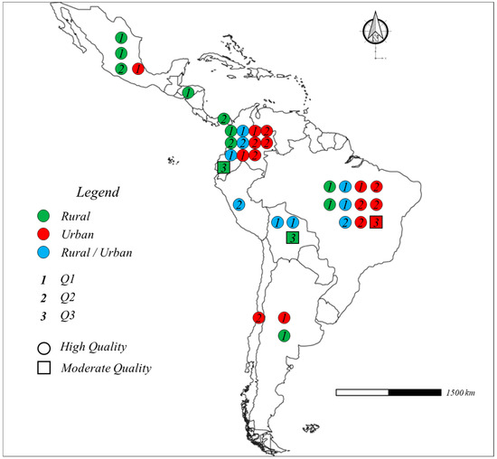
Figure 2.
General characteristics of the included studies [50,51,52,53,54,55,56,57,58,59,60,61,62,63,64,65,66,67,68,69,70,71,72,73,74,75,76,77,78,79,80,81,82,83,84,85].
Based on the JBI score rating system tool, thirty-three articles were considered as high-quality scientific papers [50,51,52,54,55,57,58,59,60,61,62,63,64,65,66,67,68,69,70,71,72,73,74,75,76,77,79,80,81,82,83,84,85] and only three were of moderate quality [53,56,78]. The studies were reported from Argentina [50,51], Bolivia [52,53,54], Brazil [55,56,57,58,59,60,61,62,63,64,65], Chile [66], Colombia [67,68,69,70,71,72,73,74,75,76,77], Ecuador [78], Honduras [79], Mexico [80,81,82,83], Panama [84], and Peru [85]. Fifteen studies were conducted on urban citizens [51,56,57,58,59,62,64,66,67,69,71,73,74,75,80], twelve were performed in rural settings [50,53,61,65,68,77,78,79,81,82,83,84], and nine studies focused on rural–urban mixed communities [52,54,55,60,63,70,72,76,85]. The papers, mainly cross-sectional studies, investigated the prevalence and typification of Blastocystis spp. in different Latin American social groups, i.e., aborigines, children, teenagers, pregnant women, farmers, urticaria patients, diabetes mellitus patients, other patients, and the elderly [50,51,52,53,54,55,56,57,58,59,60,61,62,63,64,65,66,67,68,69,70,71,72,73,74,75,76,77,78,79,80,81,82,83,84,85] (Table 1).

Table 1.
General characteristics of the selected studies published.
3.3. Molecular Characteristics of the Selected Articles
The main characteristics of the applied experimental methodologies are shown in Table 2.
Table 2.
Molecular characteristics of the selected studies in the systematic review.
The most common concentration methods, applied to separate protozoan cysts from excess fecal debris and to detect scanty microorganisms, were Ritchie methods [50,56,60,63,67,70,71,74,80], Kato–Katz [52,63,71,74,79], the zinc sulfate flotation technique [57,59,65,73], and formalin-ethyl acetate concentration techniques [51,75,84]. Other methods, such as spontaneous sedimentation [58,61,64] and centrifugal sedimentation [55], were routinely used. Eight studies did not report the concentration method used [62,72,76,77,78,81,82,83].
In general, genomic DNA was extracted from approximately 200 mg fecal sediments using commercial kits, such as the QIAamp Stool Mini Kit (QIAGEN, Hilden, Germany) [50,51,55,57,58,59,60,61,62,63,64,65,66,68,69,70,78,81,82,83,84] or the Norgen Stool DNA Isolation Kit (Norgen Biotek Corporation, Thorold, ON, Canada) [67,71,72,74,75,77,85], following the manufacturer’s instructions. Other commercial kit, such as the MP FastDNA soil kit [76,79], NucleoSpin Tissue kit [53,54], and Magnex DNA kit [56], were also used. Only Potes-Morales et al. [73] achieved rapid isolation and purification of genomic DNA through the phenol-chloroform isoamyl alcohol extraction method.
Subtype-specific primers developed from unique sequences of the SSU rRNA gene were applied to Blastocystis subtyping. For instance, the BhRDr primer was designed to be combined with the RD5 primer, and the expected size of the PCR product is 600 bp. This set of primers was used in 21 of the 36 selected studies [50,51,52,53,54,55,56,57,58,59,60,61,62,63,64,65,66,67,68,69,70,71,72,73,74,75,76,77,78,79,80,81,82,83,84,85]. Other sets of primers, such as F1/R1 (1100 bp SSU rRNA, barcoding region) [53,54,56,80] and Blast 505–532/Blast 998–1017 (500 bp SSU rRNA, barcoding region) [61,64,68,81], were used in just a few studies.
Different molecular approaches, i.e., PCR developed in 29 studies [50,51,52,53,54,55,57,58,59,60,61,62,63,64,65,66,67,68,70,71,72,73,74,78,80,81,83,84,85], qPCR [75,76,77,82], PCR-RFLP [56], multi-parallel qPCR [82], and semi/nested PCR [69], were used to amplify small segments of DNA.
3.4. Prevalence of Blastocystis spp. and Subtypes
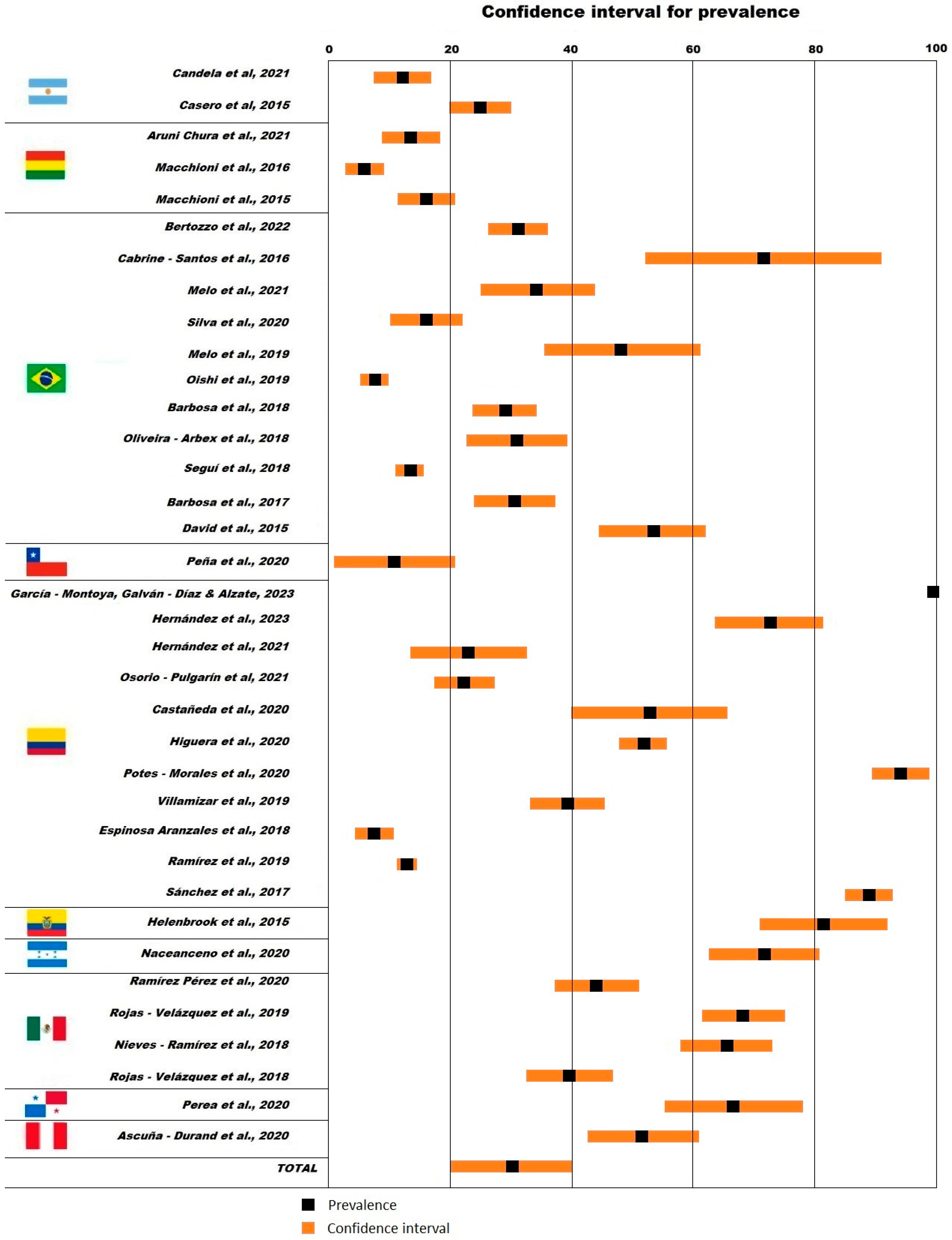
The size of the studied groups ranged between 21 [56] and 2026 individuals [76]. The prevalence of Blastocystis spp. (Figure 3) in the analyzed fecal samples based on molecular identification methods ranged between 5.8% (Bolivian rural communities, confidence interval (CI) 2.8–8.9%) [53] and 94.0% (Colombian general public, CI 89.3–98.7%) [73]. One-third of the selected studies reported a prevalence of Blastocystis spp. less than 20.0% in specific population groups of Argentina [50], Bolivia [52,53,54], Brazil [58,60,63], Chile [66], and Colombia [75,76]. Only three studies reported a prevalence of Blastocystis spp. higher than 80.0% in Ecuadorian and Colombian people [73,77,78].

Figure 3.
Reported prevalence of Blastocystis spp. in Latin America [50,51,52,53,54,55,56,57,58,59,60,61,62,63,64,65,66,67,68,69,70,71,72,73,74,75,76,77,78,79,80,81,82,83,84,85].
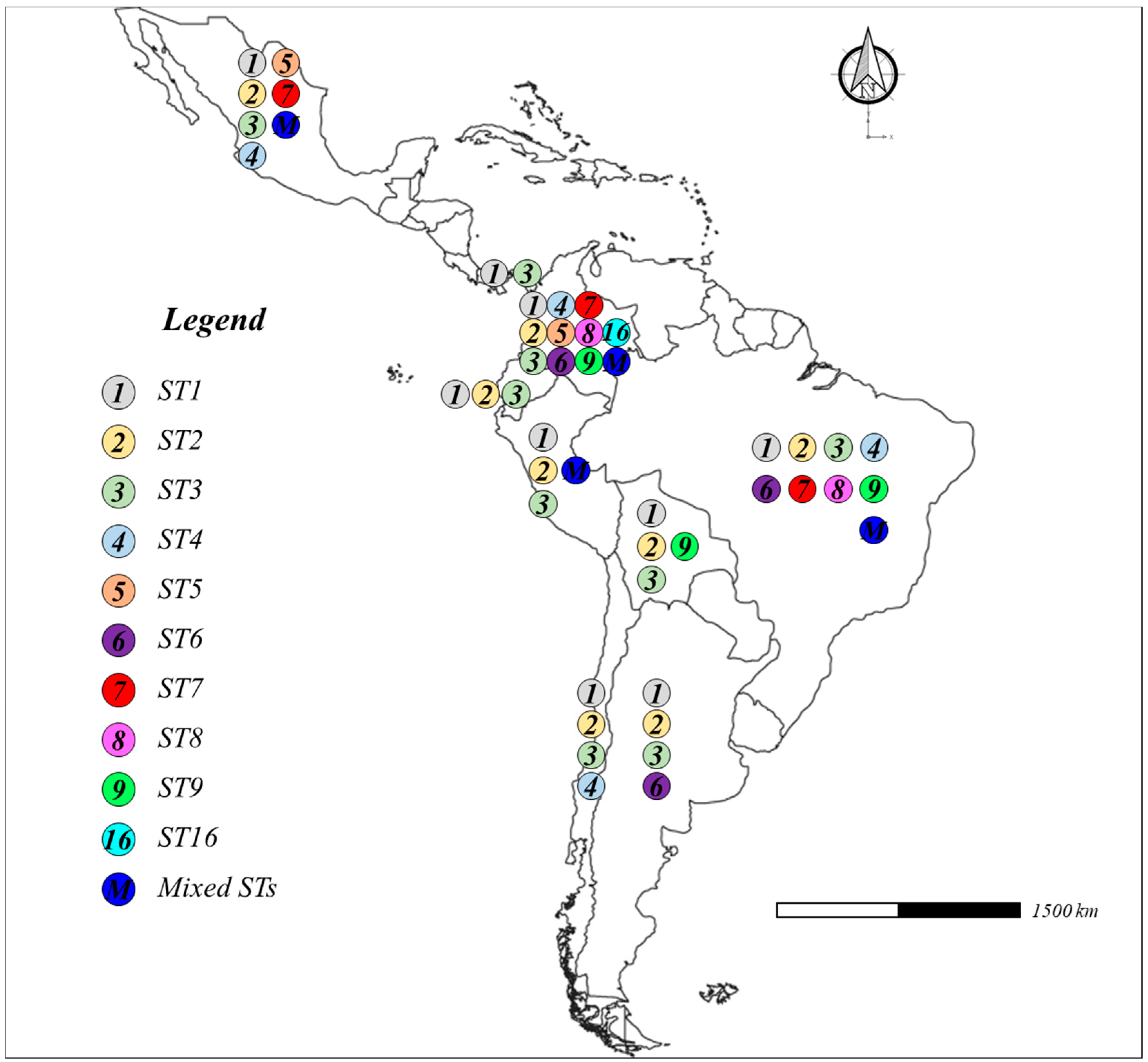
Ten STs were found, as follows: ST1 [50,51,52,53,55,56,57,58,60,61,62,63,64,65,66,67,68,69,70,72,73,74,76,77,78,80,81,84,85], ST2 [50,51,52,53,54,55,56,57,58,59,60,61,62,63,64,65,66,67,68,69,70,72,73,74,76,77,78,80,81,85], ST3 [50,51,52,53,54,55,56,57,58,59,60,61,62,63,64,65,66,67,68,69,70,72,73,74,76,77,78,80,81,82,83,84,85], ST4 [55,59,61,63,64,66,68,70,72,74,76,77,80], ST5 [68,80], ST6 [51,55,57,59,63,65,69,70,76,77], ST7 [55,57,58,62,65,76,80], ST8 [57,61,63,72], ST9 [54,55,72], and ST16 [70]. The first three STs were widely distributed in Latin America, while ST16 was only identified by Osorio-Pulgarin et al. [70]. Finally, mixed STs were reported in few study cases, for example, ST1 + ST2 [68,81], ST1 + ST3 [59,60,64,65,67,81,85], ST1 + ST2 + ST3 [68,85], ST2 + ST3 + ST4 [68], ST2 + ST3 [81], ST2 + ST5 [68], and ST3 + ST7 [65]. The STs found in the selected articles are reported in Figure 4.

Figure 4.
Blastocystis subtypes (STs) in Latin America [50,51,52,53,54,55,56,57,58,59,60,61,62,63,64,65,66,67,68,69,70,71,72,73,74,75,76,77,78,79,80,81,82,83,84,85].
4. Discussion
4.1. Epidemiology of Blastocystis spp. in Latin America
Blastocystis spp. comprise a genus of single-celled parasites that present a cosmopolitan distribution and colonize an estimated 1 to 2 billion people worldwide, many of them living in developing countries [19]. Epidemiological studies at a molecular level have clearly demonstrated that humans can be colonized by one or more Blastocystis STs, some of which are commonly found in non-human hosts [71].
Our findings suggest that only a few Latin American countries report Blastocystis spp. molecular data and parasite prevalence rates in their demographic groups. Most of the selected studies were carried out in Brazil [55,56,57,58,59,60,61,62,63,64,65], Colombia [67,68,69,70,71,72,73,74,75,76,77], and Mexico [80,81,82,83].
Brazil is a megadiverse country that possesses six terrestrial biomes and many regional differences in social, economic, and cultural characteristics [86]. A large percentage of municipalities are not equipped with efficient sewage treatment systems and waste management sites [87]. These structural gaps can amplify the diffusion of tropical neglected diseases, including those caused by many Blastocystis STs, especially in the poor and vulnerable demographic groups [88]. The Blastocystis STs identified in Brazil were ST1–ST4 and ST6–ST9, with the most prevalent being ST1, ST2, and ST3 [55,56,57,58,61,62,63]; furthermore, some data indicate mixed infections, such as ST1 + ST3 [59,60,64,65]. The prevalence rate varied from 7.5% (patients—laboratory) [60] to 71.4% (schoolchildren) [56]. Various studies about the prevalence of Blastocystis spp. in many Brazilian contexts were performed in the last eight years. Barbosa et al. [61] indicated an overall prevalence of intestinal parasitic infections above 50.0% and a wide range of Blastocystis spp. STs (ST1, ST2, ST3, ST4, and ST8) among the people who belong to a rural community in Rio de Janeiro. Blastocystis spp. STs, isolated by Barbosa et al. [64] in a carioca urban community, were genetically highly divergent, with ST3 being the most common among the participants, followed by ST1, ST2, and ST4. A mixed infection (ST1 + ST3) was detected in a few cases. David et al. [65] revealed large genetic variation of Blastocystis spp., with ST1 and ST3 being predominant, among asymptomatic people belonging to two small fishing villages along the Tietê river (São Paulo). According to Oishi et al. [60], Blastocystis spp., with a prevalence of 38.7%, were the most frequently parasites found among schoolchildren in the surrounding urban area of Curitiba. The molecular typification indicated various STs, in the order of prevalence ST3, ST1, and ST2, and a mixed infection of ST1 + ST3. de Melo et al. [57] indicated that approximately one-third of patients with diabetes mellitus in the Goias State were hosts of Blastocystis spp., and phylogenetic analyses revealed six STs, i.e., ST1, ST2, ST3, ST6, ST7, and ST8. Other studies also reported the great genetic variety of Blastocystis spp. in Brazil, indicating, at the same time, that the enteric parasites still represent a serious health concern principally due to educational deficits, poor socioeconomic rank, and inadequate sanitary conditions [55,56,62].
Current Colombian epidemiological evaluations informed eleven Blastocystis STs [67,68,69,70,71,72,73,74,75,76,77]. The first three STs (ST1, ST2, and ST3) were found in many demographic groups, both urban and rural [70,72,76]. Ramírez et al. [76] reported ST1, ST2, ST3, ST4, ST6, and ST7 in symptomatic (abdominal pain, anal pruritus, and diarrhea) and asymptomatic children from nine central oriental Colombian regions. The first four STs were also reported by Villamizar et al. [74], who carried out a descriptive epidemiological study on schoolchildren and their pets in Cauca (Southwest Colombia). No association was identified between Blastocystis spp. infection and any sociodemographic indicator; rather, the presence of STs protozoa in both humans and domestic animals suggested a zoonotic transmission. Potes-Morales et al. [73] found Blastocystis spp. (ST1, ST2, and ST3) when analyzing human fecal samples from Ibague. Finally, Osorio-Pulgarin et al. [70] performed a parasite molecular epidemiological analysis in a group of children (0–5 years) attending daycare centers in Medellin, and indicated that Blastocystis spp., with a prevalence of 15.8%, were the most frequent protozoa, followed by Giardia spp. and Endolimax nana. Additionally, six STs were identified, i.e., ST1, ST2, ST3, ST4, ST6, and the uncommon ST16. Colombia still faces numerous barriers in improving healthcare services for its citizens due to both its geography, with wide-ranging landscapes, and socioeconomic inequity [89]. Neglected tropical diseases such as Blastocystis negatively affect the lives of people with low incomes [90].
Reports from Argentina indicated that (1) approximately two-thirds (57.3%) of indigenous people living in the rural settlement of Puerto Iguazú (Misiones) were hosts of Blastocystis spp., and the parasite transmission occurred mainly through direct contact with fecal matter and contaminated water [50]; in addition, (2) one-quarter (24.8%) of patients attending the University Hospital of Cordoba City were infected with Blastocystis spp. [51]. In both Argentinian studies, subtypes ST1, ST2, and ST3 were found, with the latter being the most common among symptomatic and asymptomatic people [50,51].
Molecular studies, performed in rural contexts in Bolivia, showed high prevalence of intestinal parasites among children and teenagers and, at the same time, pointed out the risk of zoonotic pathogen transmission. In particular, the cross-sectional parasitological survey realized by Aruni Chura et al. [52] evidenced three Blastocystis spp. STs (more specifically, ST1, ST2, and ST3) among schoolchildren from ecological zones in the Department of La Paz. Macchioni et al. [53] suggested that contaminated drinking water, a lack of basic sanitary services, and close contact with animals could increase the transmission of Blastocystis ST2 and ST9 (isolated on very few occasions) among children living in rural settlements of the Chaco region.
Intestinal parasite infections, especially Blastocystis spp., are common in Latin American rural communities in Ecuador, Mexico, and Panama, as widely proven by Helenbrook, Shields, and Whipps [78], Naceanceno et al. [79], Nieves-Ramírez et al. [82], Rojas-Velázquez et al. [81], and Perea et al. [84]. The most frequent circulating Blastocystis STs in these groups were ST1, ST2, ST3, and ST4. The Blastocystis spp. prevalence rate in the Mexican general public varied from 39.6% [83] to 68.1% [81].
These results are consistent with other reports from several geographic regions worldwide that principally identified the subtypes ST1 to ST9 [91,92,93,94,95,96]. According to Nemati et al. [11], the first three, ST1, ST2, and ST3, are the most frequent STs among human subjects in the Asian continent. Karimi et al. [97] indicated that approximately 90% of the Blastocystis STs isolated from human fecal samples worldwide belonged to ST1, ST2, ST3, and ST4. Some studies in developing countries reported the following Blastocystis spp. prevalence and the dominant ST: Algeria, 7.4%—ST3 [98]; Angola, 25.6%—ST3 [99]; Azerbaijan, 45.1%—ST3 [100]; Cambodia, 55.2%—ST1 [101]; Egypt, 47.8%—ST3 [91]; India, 27.0%—ST3 [102]; Jordan, 15.0%—ST3 [100]; Malaysia, 18.5%—ST3 [103]; Nigeria, 55.5%—ST1 [100], the Philippines, 13.0%—ST3 [104]; Qatar, 71.1%—ST3 [26]; Saudi Arabia, 68.6%—ST3 [105]; Senegal, 51.7%—ST2 [106]; Sudan, 47.5%—ST1 [100]; Tanzania, 61.0%—ST1 [107]; Turkey, 24.6%—ST3 [94].
Tourists that visit Latin America or other developing countries may acquire Blastocystis STs during their stay, as demonstrated by van Hattem et al. [108].
As has been shown, the main routes of Blastocystis infection are: anthroponotic transmission, contaminated food and water, as well as close contact with animals [28,109,110,111,112,113]. The presence of Blastocystis spp. cysts, reported in water environments worldwide (rivers, lakes, streams, and lagoons), indicates fecal contamination of the water resources by humans or animals [11,38]. Since Blastocystis spp. and their STs (especially ST1 and ST3) were found in animals, including dogs, rats, cows, monkeys, and chickens, zoonotic pathways pose a serious concern for health systems in Latin American countries [74,76,114].
In spite of the fact that molecular epidemiological studies about Blastocystis spp. and their STs have been conducted in the last years in several Latin American countries, more studies are required to clarify the circulating STs in this continent.
4.2. Protocols for Blastocystis spp. Molecular Analysis
A wide range of approaches based on DNA techniques are available for detection, investigation, and surveillance of pathogenic enteroparasites [115]. PCR techniques are flexible, adaptable, and allow the automated processing of large numbers of samples in a short time [116].
The protocol to develop a descriptive study on molecular parasitology in a specific population group must be conducted under the ethical principles and approval of a bioethics committee.
The first step in carrying out a descriptive study on Blastocystis spp. prevalence is to establish the sample size representative of the target population. The minimum sample size (n) could be estimated through the following equation, as described by Oishi et al. [60]:
where N is the study population, α is the sample standard deviation (usually set at 0.5), Z corresponds to the confidence level (for a 95% level of confidence, the critical value of Z is 1.96), e is the marginal error (usually set at 5%), and γ is the expected prevalence (usually set based on the intestinal parasite prevalence rate in close regions).
The recruited voluntary participants should be knowledgeable about the methodology and the benefits of the study, sign an informed consent form [117,118], and answer a survey that includes sociodemographic patterns (age, sex, socioeconomic data, health affiliation, and level of instruction), hygienic condition of the dwellings (sanitary services and potable water, and presence of domestic animals), and behavioral aspects (personal hygiene and hand washing, and consuming raw or half-cooked meat). Furthermore, each participant must receive basic instructions for a correct fecal sample collection in a plastic container.
The recollected stool samples are generally stored in refrigerated boxes and transported to the laboratory. Prior to the laboratory processing analysis, macroscopic examination of all stool samples evaluates their consistency and the presence of mucus [55]. Each sample could be divided into two aliquots for different uses: (1) the first aliquot is immediately frozen at −20 °C for extracting genomic DNA, and (2) the second fecal aliquot is used for parasite concentration processing (thought common techniques, such as Ritchie methods and Kato-Katz) or to direct microscopic examination. Additionally, the samples that test positive for Blastocystis under microscopic observation are destined for genomic analysis.
Blastocystis spp. DNA can be extracted from 200 mg of concentrated fecal material by manual methods or commercial kits, such as the QIAamp Stool Mini Kit (QIAGEN, Hilden, Germany) [50,55,69] and the Norgen Stool DNA Isolation Kit (Norgen Biotek Corporation, Thorold, ON, Canada) [71,72,74].
For Blastocystis detection and molecular subtyping, the genomic DNA is generally subjected to PCR [51,53], qPCR [75,76], or nested PCR [69,119]. A fragment of about 600 bp from the SSU rDNA gene could be amplified using the BhRDr primer combined with the RD5 primer, according to standard protocol [50,52,55].
The PCR products are usually purified with ExoSap and sequenced by the Sanger method for determining the nucleotide sequences [72]. The obtained sequences are compared to those of Blastocystis STs previously deposited in GenBank using the BLAST application (www.ncbi.nlm.nih.gov/BLAST, accessed on 17 January 2024). Blastocystis sequences could be submitted to the Blastocystis 18S database (http://pubmlst.org/blastocystis/, accessed on 17 January 2024) for subtype confirmation [58,60].
4.3. Limitations
Approximately one-third of Latin American countries have published studies on the Blastocystis spp. molecular epidemiology in their territories during the last nine years. The lack of data from various regions, as well as the peculiarities of each selected study and differences in populations, sampling strategies, and molecular methods (concentration, extraction, and detection) for Blastocystis spp. and the STs, do not allow for a more detailed analysis or the completion of a meta-analysis.
5. Conclusions
The data found in the selected articles indicate that the prevalence of Blastocystis spp. in Latin American populations has a significant variation, ranging between 5.8% and 94.0%. PCR techniques are frequently implemented to detect Blastocystis STs circulating in Latin America. Ten STs were reported and, in particular, the first three STs (ST1, ST2, and ST3) are widely diffused in Latin America, while ST5 and ST16 were reported in very few studies. The most recent data on Blastocystis spp. molecular epidemiology and STs in communities of Latin America are limited to studies from specific countries, including Argentina, Bolivia, Brazil, Chile, Colombia, Ecuador, Honduras, Mexico, Panama, and Peru. Further studies from other countries are required to obtain a complete picture and truly understand the distribution and prevalence of Blastocystis spp. and their STs.
Supplementary Materials
The following supporting information can be downloaded at: https://www.mdpi.com/article/10.3390/tropicalmed9020038/s1, it contains File S1: PRISMA checklist; File S2: Quality assessment of the included studies.
Author Contributions
Conceptualization, C.F. and Y.S.-G.; methodology, C.F., Y.S.-G. and J.E.B.; formal analysis, C.F., R.G.-C. and J.C.-L.; investigation, Y.S.-G. and A.R.-R.; data curation, C.F. and J.C.-L.; writing—original draft preparation, Y.S.-G., R.C.-O. and A.R.-R.; writing—review and editing, R.B.-Á.; supervision, J.E.B. and R.B.-Á.; funding acquisition, C.F. and R.C.-O. All authors have read and agreed to the published version of the manuscript.
Funding
This research did not receive any specific grants from funding agencies in the public, commercial, or not-for-profit sectors.
Institutional Review Board Statement
Not applicable.
Informed Consent Statement
Not applicable.
Data Availability Statement
Not applicable.
Acknowledgments
We would like to thank the anonymous reviewers for their helpful comments on a previous version of the manuscript.
Conflicts of Interest
The authors declare no conflicts of interest. The funders had no role in the design of the study; in the collection, analyses, or interpretation of data; in the writing of the manuscript, or in the decision to publish the results.
References
- Alexeieff, A. Sur la nature des Formations Dites “Kystes de Trichomonas intestinalis”. C. R. Seances Soc. Biol. Fil. 1911, 71, 296–298. [Google Scholar]
- Stensvold, C.R.; Lewis, H.C.; Hammerum, A.M.; Porsbo, L.J.; Nielsen, S.S.; Olsen, K.E.; Arendrup, M.C.; Nielsen, H.V.; Mølbak, K. Blastocystis: Unravelling potential risk factors and clinical significance of a common but neglected parasite. Epidemiol. Infect. 2009, 137, 1655–1663. [Google Scholar] [CrossRef]
- Tan, K.S.; Mirza, H.; Teo, J.D.; Wu, B.; Macary, P.A. Current Views on the Clinical Relevance of Blastocystis spp. Curr. Infect. Dis. Rep. 2010, 12, 28–35. [Google Scholar] [CrossRef]
- Scanlan, P.D.; Stensvold, C.R.; Rajilić-Stojanović, M.; Heilig, H.G.; De Vos, W.M.; O’Toole, P.W.; Cotter, P.D. The microbial eukaryote Blastocystis is a prevalent and diverse member of the healthy human gut microbiota. FEMS Microbiol. Ecol. 2014, 90, 326–330. [Google Scholar] [CrossRef]
- Asghari, A.; Sadraei, J.; Pirestani, M.; Mohammadpour, I. First molecular identification and subtype distribution of Blastocystis sp. isolated from hooded crows (Corvus cornix) and pigeons (Columba livia) in Tehran Province, Iran. Comp. Immunol. Microbiol. Infect. Dis. 2019, 62, 25–30. [Google Scholar] [CrossRef]
- Gabrielli, S.; Palomba, M.; Furzi, F.; Brianti, E.; Gaglio, G.; Napoli, E.; Rinaldi, L.; Alburqueque, R.A.; Mattiucci, S. Molecular Subtyping of Blastocystis sp. Isolated from Farmed Animals in Southern Italy. Microorganisms 2021, 9, 1656. [Google Scholar] [CrossRef]
- Shams, M.; Shamsi, L.; Yousefi, A.; Sadrebazzaz, A.; Asghari, A.; Mohammadi-Ghalehbin, B.; Shahabi, S.; Hatam, G. Current global status, subtype distribution and zoonotic significance of Blastocystis in dogs and cats: A systematic review and meta-analysis. Parasit. Vectors 2022, 15, 225. [Google Scholar] [CrossRef]
- Scicluna, S.M.; Tawari, B.; Clark, C.G. DNA barcoding of Blastocystis. Protist 2006, 157, 77–85. [Google Scholar] [CrossRef] [PubMed]
- Santín, M.; Gómez-Muñoz, M.T.; Solano-Aguilar, G.; Fayer, R. Development of a new PCR protocol to detect and subtype Blastocystis spp. from humans and animals. Parasitol. Res. 2011, 109, 205–212. [Google Scholar] [CrossRef] [PubMed]
- Mohammad Rahimi, H.; Mirjalali, H.; Niyyati, M.; Haghighi, A.; Asadzadeh Aghdaei, H.; Zali, M.R. Development and evaluation of high-resolution melting curve analysis for rapid detection and subtyping of Blastocystis and comparison the results with sequencing. Parasitol. Res. 2019, 118, 3469–3478. [Google Scholar] [CrossRef] [PubMed]
- Nemati, S.; Zali, M.R.; Johnson, P.; Mirjalali, H.; Karanis, P. Molecular prevalence and subtype distribution of Blastocystis sp. in Asia and in Australia. J. Water Health 2021, 19, 687–704. [Google Scholar] [CrossRef]
- Maloney, J.G.; Molokin, A.; Santin, M. Use of Oxford Nanopore MinION to generate full-length sequences of the Blastocystis small subunit (SSU) rRNA gene. Parasit. Vectors 2020, 13, 595. [Google Scholar] [CrossRef] [PubMed]
- Public Databases for Molecular Typing and Microbial Genome Diversity (PubMLST). Blastocystis spp. Available online: https://pubmlst.org/organisms/blastocystis-spp (accessed on 17 January 2024).
- Maloney, J.G.; Molokin, A.; Seguí, R.; Maravilla, P.; Martínez-Hernández, F.; Villalobos, G.; Tsaousis, A.D.; Gentekaki, E.; Muñoz-Antolí, C.; Klisiowicz, D.R.; et al. Identification and Molecular Characterization of Four New Blastocystis Subtypes Designated ST35-ST38. Microorganisms 2022, 11, 46. [Google Scholar] [CrossRef] [PubMed]
- Paulos, S.; Köster, P.C.; de Lucio, A.; Hernández-de-Mingo, M.; Cardona, G.A.; Fernández-Crespo, J.C.; Stensvold, C.R.; Carmena, D. Occurrence and subtype distribution of Blastocystis sp. in humans, dogs and cats sharing household in northern Spain and assessment of zoonotic transmission risk. Zoonoses Public Health 2018, 65, 993–1002. [Google Scholar] [CrossRef]
- Popruk, S.; Adao, D.E.V.; Rivera, W.L. Epidemiology and subtype distribution of Blastocystis in humans: A review. Infect. Genet. Evol. 2021, 95, 105085. [Google Scholar] [CrossRef]
- Yoshikawa, H.; Wu, Z.; Pandey, K.; Pandey, B.D.; Sherchand, J.B.; Yanagi, T.; Kanbara, H. Molecular characterization of Blastocystis isolates from children and rhesus monkeys in Kathmandu, Nepal. Vet. Parasitol. 2009, 160, 295–300. [Google Scholar] [CrossRef]
- Cian, A.; El Safadi, D.; Osman, M.; Moriniere, R.; Gantois, N.; Benamrouz-Vanneste, S.; Delgado-Viscogliosi, P.; Guyot, K.; Li, L.L.; Monchy, S.; et al. Molecular Epidemiology of Blastocystis sp. in Various Animal Groups from Two French Zoos and Evaluation of Potential Zoonotic Risk. PLoS ONE 2017, 12, e0169659. [Google Scholar] [CrossRef] [PubMed]
- Ramírez, J.D.; Sánchez, A.; Hernández, C.; Flórez, C.; Bernal, M.C.; Giraldo, J.C.; Reyes, P.; López, M.C.; García, L.; Cooper, P.J.; et al. Geographic distribution of human Blastocystis subtypes in South America. Infect. Genet. Evol. 2016, 41, 32–35. [Google Scholar] [CrossRef] [PubMed]
- Centers for Disease Control and Prevention (CDC). Blastocystis sp. 2019. Available online: https://www.cdc.gov/dpdx/blastocystis/index.html (accessed on 15 October 2023).
- Jiménez, P.A.; Jaimes, J.E.; Ramírez, J.D. A summary of Blastocystis subtypes in North and South America. Parasit. Vectors 2019, 12, 376. [Google Scholar] [CrossRef]
- Deng, L.; Wojciech, L.; Png, C.W.; Kioh, Y.Q.D.; Ng, G.C.; Chan, E.C.Y.; Zhang, Y.; Gascoigne, N.R.J.; Tan, K.S.W. Colonization with ubiquitous protist Blastocystis ST1 ameliorates DSS-induced colitis and promotes beneficial microbiota and immune outcomes. NPJ Biofilms Microbiomes 2023, 9, 22. [Google Scholar] [CrossRef]
- Maloney, J.G.; Molokin, A.; da Cunha, M.J.R.; Cury, M.C.; Santin, M. Blastocystis subtype distribution in domestic and captive wild bird species from Brazil using next generation amplicon sequencing. Parasite Epidemiol. Control 2020, 9, e00138. [Google Scholar] [CrossRef]
- Stensvold, C.R.; Clark, C.G. Pre-empting Pandora’s Box: Blastocystis Subtypes Revisited. Trends Parasitol. 2020, 36, 229–232. [Google Scholar] [CrossRef]
- Viesy, S.; Rezaei, Z.; Pouladi, I.; Mirzaei, A.; Abdi, J. The Prevalence of Blastocystis sp. and Its Relationship with Gastrointestinal Disorders and Risk factors. Iran. J. Parasitol. 2022, 17, 90–95. [Google Scholar]
- Abu-Madi, M.; Aly, M.; Behnke, J.M.; Clark, C.G.; Balkhy, H. The distribution of Blastocystis subtypes in isolates from Qatar. Parasit. Vectors 2015, 8, 465. [Google Scholar] [CrossRef] [PubMed]
- Poirier, P.; Wawrzyniak, I.; Vivarès, C.P.; Delbac, F.; El Alaoui, H. New insights into Blastocystis spp.: A potential link with irritable bowel syndrome. PLoS Pathog. 2012, 8, e1002545. [Google Scholar] [CrossRef] [PubMed]
- Greige, S.; El Safadi, D.; Bécu, N.; Gantois, N.; Pereira, B.; Chabé, M.; Benamrouz-Vanneste, S.; Certad, G.; El Hage, R.; Chemaly, M.; et al. Prevalence and subtype distribution of Blastocystis sp. isolates from poultry in Lebanon and evidence of zoonotic potential. Parasit. Vectors 2018, 11, 389. [Google Scholar] [CrossRef] [PubMed]
- Wang, P.; Li, S.; Zou, Y.; Hong, Z.W.; Wang, P.; Zhu, X.Q.; Song, D.P.; Chen, X.Q. Prevalence and Subtype Distribution of Blastocystis sp. in Diarrheic Pigs in Southern China. Pathogens 2021, 10, 1189. [Google Scholar] [CrossRef] [PubMed]
- Nithyamathi, K.; Chandramathi, S.; Kumar, S. Predominance of Blastocystis sp. Infection among School Children in Peninsular Malaysia. PLoS ONE 2016, 11, e0136709. [Google Scholar] [CrossRef] [PubMed]
- Javanmard, E.; Niyyati, M.; Ghasemi, E.; Mirjalali, H.; Asadzadeh Aghdaei, H.; Zali, M.R. Impacts of human development index and climate conditions on prevalence of Blastocystis: A systematic review and meta-analysis. Acta Trop. 2018, 185, 193–203. [Google Scholar] [CrossRef] [PubMed]
- Wilcox, J.J.S.; Lopez-Cotto, J.J.; Hollocher, H. Historical contingency, geography and anthropogenic patterns of exposure drive the evolution of host switching in the Blastocystis species-complex. Parasitology 2021, 148, 985–993. [Google Scholar] [CrossRef] [PubMed]
- Sarzhanov, F.; Dogruman-Al, F.; Santin, M.; Maloney, J.G.; Gureser, A.S.; Karasartova, D.; Taylan-Ozkan, A. Investigation of neglected protists Blastocystis sp. and Dientamoeba fragilis in immunocompetent and immunodeficient diarrheal patients using both conventional and molecular methods. PLoS Negl. Trop. Dis. 2021, 15, e0009779. [Google Scholar] [CrossRef]
- Deng, Y.; Zhang, S.; Ning, C.; Zhou, Y.; Teng, X.; Wu, X.; Chu, Y.; Yu, Y.; Chen, J.; Tian, L.; et al. Molecular Epidemiology and Risk Factors of Blastocystis sp. Infections among General Populations in Yunnan Province, Southwestern China. Risk Manag. Healthc. Policy 2020, 13, 1791–1801. [Google Scholar] [CrossRef]
- Matovelle, C.; Tejedor, M.T.; Monteagudo, L.V.; Beltrán, A.; Quílez, J. Prevalence and Associated Factors of Blastocystis sp. Infection in Patients with Gastrointestinal Symptoms in Spain: A Case-Control Study. Trop. Med. Infect. Dis. 2022, 7, 226. [Google Scholar] [CrossRef]
- Attah, A.O.; Sanggari, A.; Li, L.I.; Nik Him, N.A.I.I.; Ismail, A.H.; Meor Termizi, F.H. Blastocystis occurrence in water sources worldwide from 2005 to 2022: A review. Parasitol. Res. 2023, 122, 1–10. [Google Scholar] [CrossRef]
- Elseadawy, R.; Abbas, I.; Al-Araby, M.; Abu-Elwafa, S. Occurrence and molecular characterization of Acanthamoeba, Naegleria fowleri and Blastocystis in water samples from various sources in Egypt. Acta Trop. 2023, 237, 106733. [Google Scholar] [CrossRef]
- Noradilah, S.A.; Lee, I.L.; Anuar, T.S.; Salleh, F.M.; Abdul Manap, S.N.A.; Husnie, N.S.; Azrul, S.M.; Moktar, N. Blastocystis spp. contaminated water sources in aboriginal settlements. Trop. Biomed. 2017, 34, 110–117. [Google Scholar] [PubMed]
- Adamska, M. First report of Blastocystis sp. subtypes in natural water bodies in north-western Poland: A one-year monitoring. Int. J. Environ. Health Res. 2022, 32, 862–869. [Google Scholar] [CrossRef] [PubMed]
- Abdulsalam, A.M.; Ithoi, I.; Al-Mekhlafi, H.M.; Ahmed, A.; Surin, J.; Mak, J.W. Drinking water is a significant predictor of Blastocystis infection among rural Malaysian primary schoolchildren. Parasitology 2012, 139, 1014–1020. [Google Scholar] [CrossRef] [PubMed]
- Maritz, J.M.; Ten Eyck, T.A.; Elizabeth Alter, S.; Carlton, J.M. Patterns of protist diversity associated with raw sewage in New York City. ISME J. 2019, 13, 2750–2763. [Google Scholar] [CrossRef] [PubMed]
- Moreno, Y.; Moreno-Mesonero, L.; Amorós, I.; Pérez, R.; Morillo, J.A.; Alonso, J.L. Multiple identification of most important waterborne protozoa in surface water used for irrigation purposes by 18S rRNA amplicon-based metagenomics. Int. J. Hyg. Environ. Health 2018, 221, 102–111. [Google Scholar] [CrossRef] [PubMed]
- Noradilah, S.A.; Lee, I.L.; Anuar, T.S.; Salleh, F.M.; Abdul Manap, S.N.; Mohd Mohtar, N.S.; Azrul, S.M.; Abdullah, W.O.; Moktar, N. Occurrence of Blastocystis sp. in water catchments at Malay villages and Aboriginal settlement during wet and dry seasons in Peninsular Malaysia. PeerJ 2016, 4, e2541. [Google Scholar] [CrossRef]
- Jinatham, V.; Nonebudsri, C.; Wandee, T.; Popluechai, S.; Tsaousis, A.D.; Gentekaki, E. Blastocystis in tap water of a community in northern Thailand. Parasitol. Int. 2022, 91, 102624. [Google Scholar] [CrossRef]
- Leelayoova, S.; Siripattanapipong, S.; Thathaisong, U.; Naaglor, T.; Taamasri, P.; Piyaraj, P.; Mungthin, M. Drinking water: A possible source of Blastocystis spp. subtype 1 infection in schoolchildren of a rural community in central Thailand. Am. J. Trop. Med. Hyg. 2008, 79, 401–406. [Google Scholar] [CrossRef]
- Beyhan, Y.E.; Yilmaz, H.; Cengiz, Z.T.; Ekici, A. Clinical significance and prevalence of Blastocystis hominis in Van, Turkey. Saudi Med. J. 2015, 36, 1118–1121. [Google Scholar] [CrossRef]
- World Health Organization (WHO). Drinking Water. 2022. Available online: https://www.who.int/news-room/fact-sheets/detail/drinking-water (accessed on 15 October 2023).
- Moher, D.; Liberati, A.; Tetzlaff, J.; Altman, D.G.; PRISMA Group. Preferred reporting items for systematic reviews and meta-analyses: The PRISMA statement. Ann. Intern. Med. 2009, 151, 264–269. [Google Scholar] [CrossRef] [PubMed]
- Munn, Z.; Moola, S.; Lisy, K.; Riitano, D.; Tufanaru, C. Methodological guidance for systematic reviews of observational epidemiological studies reporting prevalence and cumulative incidence data. Int. J. Evid. Based Healthc. 2015, 13, 147–153. [Google Scholar] [CrossRef] [PubMed]
- Candela, E.; Goizueta, C.; Periago, M.V.; Muñoz-Antoli, C. Prevalence of intestinal parasites and molecular characterization of Giardia intestinalis, Blastocystis spp. and Entamoeba histolytica in the village of Fortín Mbororé (Puerto Iguazú, Misiones, Argentina). Parasit. Vectors 2021, 14, 510. [Google Scholar] [CrossRef] [PubMed]
- Casero, R.D.; Mongi, F.; Sánchez, A.; Ramírez, J.D. Blastocystis and urticaria: Examination of subtypes and morphotypes in an unusual clinical manifestation. Acta Trop. 2015, 148, 156–161. [Google Scholar] [CrossRef] [PubMed]
- Aruni Chura, J.; Macchioni, F.; Furzi, F.; Balboa, V.; Mercado, É.; Gómez, J.; Rojas Gonzales, P.; Poma, V.; Loup, A.; Roselli, M.; et al. Cross-sectional study on intestinal parasite infections in different ecological zones of the Department of La Paz, Bolivia. One Health 2021, 13, 100271. [Google Scholar] [CrossRef] [PubMed]
- Macchioni, F.; Segundo, H.; Totino, V.; Gabrielli, S.; Rojas, P.; Roselli, M.; Paredes, G.A.; Masana, M.; Bartoloni, A.; Cancrini, G. Intestinal parasitic infections and associated epidemiological drivers in two rural communities of the Bolivian Chaco. J. Infect. Dev. Ctries. 2016, 10, 1012–1019. [Google Scholar] [CrossRef] [PubMed]
- Macchioni, F.; Segundo, H.; Gabrielli, S.; Totino, V.; Gonzales, P.R.; Salazar, E.; Bozo, R.; Bartoloni, A.; Cancrini, G. Dramatic decrease in prevalence of soil-transmitted helminths and new insights into intestinal protozoa in children living in the Chaco region, Bolivia. Am. J. Trop. Med. Hyg. 2015, 92, 794–796. [Google Scholar] [CrossRef]
- Bertozzo, T.V.; David, É.B.; Oliveira-Arbex, A.P.; Victória, C.; Guimarães, S. Frequency, spatial distribution, and genetic diversity of Blastocystis among referred individuals to a clinical laboratory: First report of subtype 9 in Brazil. Acta Trop. 2022, 234, 106608. [Google Scholar] [CrossRef]
- Cabrine-Santos, M.; Moura, R.G.F.; Pedrosa, A.L.; Correia, D.; Oliveira-Silva, M.B. Molecular characterization of Blastocystis subtypes isolated in the city of Uberaba, Minas Gerais State, Brazil. Rev. Soc. Bras. Med. Trop. 2021, 54, e03052021. [Google Scholar] [CrossRef] [PubMed]
- Melo, G.B.; Mazzaro, M.C.; Gomes-Gouvêa, M.S.; Santos, É.A.D.; Souza, L.V.; Elias-Oliveira, J.; Gryschek, R.C.B.; Rodrigues, R.M.; Paula, F.M. Blastocystis subtypes in patients with diabetes mellitus from the Midwest region of Brazil. Rev. Inst. Med. Trop. Sao Paulo 2021, 63, e32. [Google Scholar] [CrossRef] [PubMed]
- Silva, M.D.R.A.; Melo, G.B.; Malta, F.M.; Abdala, E.; Costa, S.F.; Pierrotti, L.C.; Gonçalves, E.M.N.; Castilho, V.L.P.; Chieffi, P.P.; Gryschek, R.C.B.; et al. Subtypes of Blastocystis sp. isolated in fecal samples from transplant candidates in São Paulo, Brazil. Parasite Epidemiol. Control 2019, 8, e00128. [Google Scholar] [CrossRef] [PubMed]
- Melo, G.B.; Malta, F.M.; Maruta, C.W.; Criado, P.R.; Castilho, V.L.P.; Gonçalves, E.M.D.N.; Espirito-Santo, M.C.C.D.; Paula, F.M.; Gryschek, R.C.B. Characterization of subtypes of Blastocystis sp. isolated from patients with urticaria, São Paulo, Brazil. Parasite Epidemiol. Control 2019, 7, e00124. [Google Scholar] [CrossRef]
- Oishi, C.Y.; Klisiowicz, D.D.R.; Seguí, R.; Köster, P.C.; Carmena, D.; Toledo, R.; Esteban, J.G.; Muñoz-Antoli, C. Reduced prevalence of soil-transmitted helminths and high frequency of protozoan infections in the surrounding urban area of Curitiba, Paraná, Brazil. Parasite Epidemiol. Control 2019, 7, e00115. [Google Scholar] [CrossRef]
- Barbosa, C.V.; Barreto, M.M.; Andrade, R.J.; Sodré, F.; d’Avila-Levy, C.M.; Peralta, J.M.; Igreja, R.P.; de Macedo, H.W.; Santos, H.L.C. Intestinal parasite infections in a rural community of Rio de Janeiro (Brazil): Prevalence and genetic diversity of Blastocystis subtypes. PLoS ONE 2018, 13, e0193860. [Google Scholar] [CrossRef] [PubMed]
- Oliveira-Arbex, A.P.; David, É.B.; Guimarães, S. Blastocystis genetic diversity among children of low-income daycare center in Southeastern Brazil. Infect. Genet. Evol. 2018, 57, 59–63. [Google Scholar] [CrossRef]
- Seguí, R.; Muñoz-Antoli, C.; Klisiowicz, D.R.; Oishi, C.Y.; Köster, P.C.; de Lucio, A.; Hernández-de-Mingo, M.; Puente, P.; Toledo, R.; Esteban, J.G.; et al. Prevalence of intestinal parasites, with emphasis on the molecular epidemiology of Giardia duodenalis and Blastocystis sp., in the Paranaguá Bay, Brazil: A community survey. Parasit. Vectors 2018, 11, 490. [Google Scholar] [CrossRef]
- Barbosa, C.V.; de Jesus Batista, R.; Pereira Igreja, R.; d’Avila Levy, C.M.; Werneck de Macedo, H.; Carneiro Santos, H.L. Distribution of Blastocystis subtypes isolated from humans from an urban community in Rio de Janeiro, Brazil. Parasit. Vectors 2017, 10, 518. [Google Scholar] [CrossRef] [PubMed]
- David, É.B.; Guimarães, S.; de Oliveira, A.P.; Goulart de Oliveira-Sequeira, T.C.; Nogueira Bittencourt, G.; Moraes Nardi, A.R.; Martins Ribolla, P.E.; Bueno Franco, R.M.; Branco, N.; Tosini, F.; et al. Molecular characterization of intestinal protozoa in two poor communities in the State of São Paulo, Brazil. Parasit. Vectors 2015, 8, 103. [Google Scholar] [CrossRef] [PubMed]
- Peña, S.; Carrasco, G.; Rojas, P.; Castillo, D.; Ozaki, L.S.; Mercado, R. Determination of subtypes of Blastocystis sp. in Chilean patients with and without inflammatory bowel syndrome, A preliminary report. Parasite Epidemiol. Control 2019, 8, e00125. [Google Scholar] [CrossRef] [PubMed]
- Garcia-Montoya, G.M.; Galvan-Diaz, A.L.; Alzate, J.F. Metataxomics reveals Blastocystis subtypes mixed infections in Colombian children. Infect. Genet. Evol. 2023, 113, 105478. [Google Scholar] [CrossRef] [PubMed]
- Hernández, P.C.; Maloney, J.G.; Molokin, A.; George, N.S.; Morales, L.; Chaparro-Olaya, J.; Santin, M. Exploring Blastocystis genetic diversity in rural schoolchildren from Colombia using next-generation amplicon sequencing reveals significant associations between contact with animals and infection risk. Parasitol. Res. 2023, 122, 1451–1462. [Google Scholar] [CrossRef] [PubMed]
- Hernández, P.C.; Morales, L.; Chaparro-Olaya, J.; de Avila, J.; Bautista-Molano, W.; Bello-Gualtero, J.; Beltrán-Ostos, A.; Romero-Sánchez, C. Frequency and distribution of Blastocystis sp. subtypes in patients with spondyloarthritis in Bogotá, Colombia. Parasite Epidemiol. Control 2021, 15, e00227. [Google Scholar] [CrossRef]
- Osorio-Pulgarin, M.I.; Higuera, A.; Beltran-Álzate, J.C.; Sánchez-Jiménez, M.; Ramírez, J.D. Epidemiological and Molecular Characterization of Blastocystis Infection in Children Attending Daycare Centers in Medellín, Colombia. Biology 2021, 10, 669. [Google Scholar] [CrossRef]
- Castañeda, S.; Muñoz, M.; Villamizar, X.; Hernández, P.C.; Vásquez, L.R.; Tito, R.Y.; Ramírez, J.D. Microbiota characterization in Blastocystis-colonized and Blastocystis-free school-age children from Colombia. Parasit. Vectors 2020, 13, 521. [Google Scholar] [CrossRef]
- Higuera, A.; Villamizar, X.; Herrera, G.; Giraldo, J.C.; Vasquez-A, L.R.; Urbano, P.; Villalobos, O.; Tovar, C.; Ramírez, J.D. Molecular detection and genotyping of intestinal protozoa from different biogeographical regions of Colombia. PeerJ 2020, 8, e8554. [Google Scholar] [CrossRef]
- Potes-Morales, C.; Osorio-Delgado, L.A.; Carranza, J.C.; Vallejo, G.A. The first molecular detection of Blastocystis subtypes in human faecal samples from Ibague, Colombia. Parasite Epidemiol. Control 2020, 9, e00132. [Google Scholar] [CrossRef]
- Villamizar, X.; Higuera, A.; Herrera, G.; Vasquez-A, L.R.; Buitron, L.; Muñoz, L.M.; Gonzalez-C, F.E.; Lopez, M.C.; Giraldo, J.C.; Ramírez, J.D. Molecular and descriptive epidemiology of intestinal protozoan parasites of children and their pets in Cauca, Colombia: A cross-sectional study. BMC Infect. Dis. 2019, 19, 190. [Google Scholar] [CrossRef] [PubMed]
- Espinosa Aranzales, A.F.; Radon, K.; Froeschl, G.; Pinzón Rondón, Á.M.; Delius, M. Prevalence and risk factors for intestinal parasitic infections in pregnant women residing in three districts of Bogotá, Colombia. BMC Public Health 2018, 18, 1071. [Google Scholar] [CrossRef] [PubMed]
- Ramírez, J.D.; Flórez, C.; Olivera, M.; Bernal, M.C.; Giraldo, J.C. Blastocystis subtyping and its association with intestinal parasites in children from different geographical regions of Colombia. PLoS ONE 2017, 12, e0172586. [Google Scholar] [CrossRef] [PubMed]
- Sánchez, A.; Munoz, M.; Gómez, N.; Tabares, J.; Segura, L.; Salazar, Á.; Restrepo, C.; Ruíz, M.; Reyes, P.; Qian, Y.; et al. Molecular Epidemiology of Giardia, Blastocystis and Cryptosporidium among Indigenous Children from the Colombian Amazon Basin. Front. Microbiol. 2017, 8, 248. [Google Scholar] [CrossRef] [PubMed]
- Helenbrook, W.D.; Shields, W.M.; Whipps, C.M. Characterization of Blastocystis species infection in humans and mantled howler monkeys, Alouatta palliata aequatorialis, living in close proximity to one another. Parasitol. Res. 2015, 114, 2517–2525. [Google Scholar] [CrossRef] [PubMed]
- Naceanceno, K.S.; Matamoros, G.; Gabrie, J.A.; Bottazzi, M.E.; Sanchez, A.; Mejia, R. Use of Multi-Parallel Real-Time Quantitative PCR to Determine Blastocystis Prevalence and Association with Other Gastrointestinal Parasite Infection in a Rural Honduran Location. Am. J. Trop. Med. Hyg. 2020, 102, 1373–1375. [Google Scholar] [CrossRef]
- Ramírez Pérez, M.; Yáñez, C.M.; Hernández, A.M.; Sustaita, J.J.D.; Jiménez, E.G.; Andrade, M.R.; Vargas, G.G.G.; Gómez, J.O.G. Blastocystis infection frequency and subtype distribution in university students. Heliyon 2020, 6, e05729. [Google Scholar] [CrossRef]
- Rojas-Velázquez, L.; Maloney, J.G.; Molokin, A.; Morán, P.; Serrano-Vázquez, A.; González, E.; Pérez-Juárez, H.; Ximénez, C.; Santin, M. Use of next-generation amplicon sequencing to study Blastocystis genetic diversity in a rural human population from Mexico. Parasit. Vectors 2019, 12, 566. [Google Scholar] [CrossRef]
- Nieves-Ramírez, M.E.; Partida-Rodríguez, O.; Laforest-Lapointe, I.; Reynolds, L.A.; Brown, E.M.; Valdez-Salazar, A.; Morán-Silva, P.; Rojas-Velázquez, L.; Morien, E.; Parfrey, L.W.; et al. Asymptomatic Intestinal Colonization with Protist Blastocystis Is Strongly Associated with Distinct Microbiome Ecological Patterns. mSystems 2018, 3, e00007-18. [Google Scholar] [CrossRef]
- Rojas-Velázquez, L.; Morán, P.; Serrano-Vázquez, A.; Fernández, L.D.; Pérez-Juárez, H.; Poot-Hernández, A.C.; Portillo, T.; González, E.; Hernández, E.; Partida-Rodríguez, O.; et al. Genetic Diversity and Distribution of Blastocystis Subtype 3 in Human Populations, with Special Reference to a Rural Population in Central Mexico. BioMed Res. Int. 2018, 2018, 3916263. [Google Scholar] [CrossRef]
- Perea, M.; Vásquez, V.; Pineda, V.; Samudio, F.; Calzada, J.E.; Saldaña, A. Prevalence and subtype distribution of Blastocystis sp. infecting children from a rural community in Panama. Parasite Epidemiol. Control 2020, 9, e00139. [Google Scholar] [CrossRef] [PubMed]
- Ascuña-Durand, K.; Salazar-Sánchez, R.S.; Castillo-Neyra, R.; Ballón-Echegaray, J. Relative Frequency of Blastocystis Subtypes 1, 2, and 3 in Urban and Periurban Human Populations of Arequipa, Peru. Trop. Med. Infect. Dis. 2020, 5, 178. [Google Scholar] [CrossRef] [PubMed]
- Ellwanger, J.H.; Nobre, C.A.; Chies, J.A.B. Brazilian Biodiversity as a Source of Power and Sustainable Development: A Neglected Opportunity. Sustainability 2023, 15, 482. [Google Scholar] [CrossRef]
- Saneamento Ambiental—Ministério das Cidades (Governo do Brasil). Sistema Nacional de Informações sobre Saneamento: Diagnóstico dos Serviços de água e Esgotos. Available online: https://www.gov.br/cidades/pt-br/assuntos/saneamento (accessed on 15 October 2023).
- Zanetti, A.D.S.; Malheiros, A.F.; de Matos, T.A.; Longhi, F.G.; Moreira, L.M.; Silva, S.L.; Ikeda Castrillon, S.K.; Benevides Ferreira, S.M.; Ignotti, E.; Espinosa, O.A. Prevalence of Blastocystis sp. infection in several hosts in Brazil: A systematic review and meta-analysis. Parasit. Vectors 2020, 13, 30. [Google Scholar] [CrossRef]
- Fusaro, C.; Chávez-Romero, Y.A.; Prada, S.L.G.; Serrano-Silva, N.; Bernal, J.E.; González-Jiménez, F.E.; Sarria-Guzmán, Y. Burden and Epidemiology of Human Intestinal Giardia duodenalis Infection in Colombia: A Systematic Review. Trop. Med. Infect. Dis. 2022, 7, 325. [Google Scholar] [CrossRef] [PubMed]
- Mitra, A.K.; Mawson, A.R. Neglected Tropical Diseases: Epidemiology and Global Burden. Trop. Med. Infect. Dis. 2017, 2, 36. [Google Scholar] [CrossRef]
- El-Wakil, E.S.; Zalat, R.S.; El-Badry, A.A. Mapping gut parasitism patterns in a cohort of Egyptians. Sci. Rep. 2023, 13, 9961. [Google Scholar] [CrossRef]
- Wang, J.; Gong, B.; Yang, F.; Zhang, W.; Zheng, Y.; Liu, A. Subtype distribution and genetic characterizations of Blastocystis in pigs, cattle, sheep and goats in northeastern China’s Heilongjiang Province. Infect. Genet. Evol. 2018, 57, 171–176. [Google Scholar] [CrossRef]
- Deng, L.; Chai, Y.; Zhou, Z.; Liu, H.; Zhong, Z.; Hu, Y.; Fu, H.; Yue, C.; Peng, G. Epidemiology of Blastocystis sp. infection in China: A systematic review. Parasite 2019, 26, 41. [Google Scholar] [CrossRef]
- Gureser, A.S.; Karasartova, D.; Sarzhanov, F.; Kosar, N.; Taylan-Ozkan, A.; Dogruman-Al, F. Prevalence of Blastocystis and Dientamoeba fragilis in diarrheal patients in Corum, Türkiye. Parasitol. Res. 2023, 122, 2977–2987. [Google Scholar] [CrossRef]
- Malatyalı, E.; Ertabaklar, H.; Ertuğ, S. Subtype Distribution of Blastocystis in Türkiye. Turkiye Parazitol. Derg. 2023, 47, 184–189. [Google Scholar] [CrossRef]
- Kumarasamy, V.; Rajamanikam, A.; Anbazhagan, D.; Atroosh, W.M.; Azzani, M.; Subramaniyan, V.; Abdullah, S.R. Systematic Review and Meta-Analysis: Epidemiology of Human Blastocystis spp. Infection in Malaysia. Trop. Med. Infect. Dis. 2023, 8, 415. [Google Scholar] [CrossRef]
- Karimi, P.; Shafaghi-Sisi, S.; Meamar, A.R.; Razmjou, E. Molecular identification of Cryptosporidium, Giardia, and Blastocystis from stray and household cats and cat owners in Tehran, Iran. Sci. Rep. 2023, 13, 1554. [Google Scholar] [CrossRef]
- Bachi, F.; Abidat, F.; Ghaffor, Y.; Bellili, S.; Goura, S.; Belmadani, S.A. Molecular characterization of algerian strains of Blastocysts sp. Méd. Tropicale Santé Int. 2022, 2, mtsi-v2i1. [Google Scholar]
- Dacal, E.; Saugar, J.M.; de Lucio, A.; Hernández-de-Mingo, M.; Robinson, E.; Köster, P.C.; Aznar-Ruiz-de-Alegría, M.L.; Espasa, M.; Ninda, A.; Gandasegui, J.; et al. Prevalence and molecular characterization of Strongyloides stercoralis, Giardia duodenalis, Cryptosporidium spp., and Blastocystis spp. isolates in school children in Cubal, Western Angola. Parasit. Vectors 2018, 11, 67. [Google Scholar] [CrossRef]
- Cinek, O.; Polackova, K.; Odeh, R.; Alassaf, A.; Kramná, L.; Ibekwe, M.U.; Majaliwa, E.S.; Ahmadov, G.; Elmahi, B.M.E.; Mekki, H.; et al. Blastocystis in the faeces of children from six distant countries: Prevalence, quantity, subtypes and the relation to the gut bacteriome. Parasit. Vectors 2021, 14, 399. [Google Scholar] [CrossRef] [PubMed]
- Wang, W.; Owen, H.; Traub, R.J.; Cuttell, L.; Inpankaew, T.; Bielefeldt-Ohmann, H. Molecular epidemiology of Blastocystis in pigs and their in-contact humans in Southeast Queensland, Australia, and Cambodia. Vet. Parasitol. 2014, 203, 264–269. [Google Scholar] [CrossRef] [PubMed]
- Pandey, P.K.; Verma, P.; Marathe, N.; Shetty, S.; Bavdekar, A.; Patole, M.S.; Stensvold, C.R.; Shouche, Y.S. Prevalence and subtype analysis of Blastocystis in healthy Indian individuals. Infect. Genet. Evol. 2015, 31, 296–299. [Google Scholar] [CrossRef] [PubMed]
- Mohammad, N.A.; Al-Mekhlafi, H.M.; Anuar, T.S. Subtype distribution of Blastocystis isolated from humans and associated animals in an indigenous community with poor hygiene in Peninsular Malaysia. Trop. Biomed. 2018, 35, 849–860. [Google Scholar] [PubMed]
- Belleza, M.L.; Reyes, J.C.; Tongol-Rivera, P.N.; Rivera, W.L. Subtype analysis of Blastocystis sp. isolates from human and canine hosts in an urban community in the Philippines. Parasitol. Int. 2016, 65, 291–294. [Google Scholar] [CrossRef]
- Wakid, M.H.; Aldahhasi, W.T.; Alsulami, M.N.; El-Kady, A.M.; Elshabrawy, H.A. Identification and Genetic Characterization of Blastocystis Species in Patients from Makkah, Saudi Arabia. Infect. Drug Resist. 2022, 15, 491–501. [Google Scholar] [CrossRef]
- Khaled, S.; Gantois, N.; Ly, A.T.; Senghor, S.; Even, G.; Dautel, E.; Dejager, R.; Sawant, M.; Baydoun, M.; Benamrouz-Vanneste, S.; et al. Prevalence and Subtype Distribution of Blastocystis sp. in Senegalese School Children. Microorganisms 2020, 8, 1408. [Google Scholar] [CrossRef]
- Forsell, J.; Granlund, M.; Samuelsson, L.; Koskiniemi, S.; Edebro, H.; Evengård, B. High occurrence of Blastocystis sp. subtypes 1-3 and Giardia intestinalis assemblage B among patients in Zanzibar, Tanzania. Parasit. Vectors 2016, 9, 370. [Google Scholar] [CrossRef] [PubMed]
- van Hattem, J.M.; Arcilla, M.S.; Schultsz, C.; Bootsma, M.C.; Verhaar, N.; Rebers, S.P.; Goorhuis, A.; Grobusch, M.P.; Penders, J.; de Jong, M.D.; et al. Carriage of Blastocystis spp. in travellers—A prospective longitudinal study. Travel Med. Infect. Dis. 2019, 27, 87–91. [Google Scholar] [CrossRef]
- Rudzińska, M.; Sikorska, K. Epidemiology of Blastocystis Infection: A Review of Data from Poland in Relation to Other Reports. Pathogens 2023, 12, 1050. [Google Scholar] [CrossRef] [PubMed]
- Javanmard, E.; Rahimi, H.M.; Niyyati, M.; Aghdaei, H.A.; Sharifdini, M.; Mirjalali, H.; Zali, M.R.; Karanis, P. Molecular analysis of Blastocystis sp. and its subtypes from treated wastewater routinely used for irrigation of vegetable farmlands in Iran. J. Water Health 2019, 17, 837–844. [Google Scholar] [CrossRef] [PubMed]
- Betts, E.L.; Gentekaki, E.; Thomasz, A.; Breakell, V.; Carpenter, A.I.; Tsaousis, A.D. Genetic diversity of Blastocystis in non-primate animals. Parasitology 2018, 145, 1228–1234. [Google Scholar] [CrossRef] [PubMed]
- Rezaei Riabi, T.; Mirjalali, H.; Haghighi, A.; Rostami Nejad, M.; Pourhoseingholi, M.A.; Poirier, P.; Delbac, F.; Wawrzyniak, I.; Zali, M.R. Genetic diversity analysis of Blastocystis subtypes from both symptomatic and asymptomatic subjects using a barcoding region from the 18S rRNA gene. Infect. Genet. Evol. 2018, 61, 119–126. [Google Scholar] [CrossRef] [PubMed]
- Barbosa, C.V.; do Bomfim, T.C.B.; Teixeira, B.R.; Gentile, R.; Neto, S.F.D.C.; Magalhães, B.S.N.; Balthazar, D.A.; da Silva, F.A.; Biot, R.; d’Avila Levy, C.M.; et al. Molecular epidemiology of Blastocystis isolated from animals in the state of Rio de Janeiro, Brazil. PLoS ONE 2019, 14, e0210740. [Google Scholar]
- Scanlan, P.D.; Knight, R.; Song, S.J.; Ackermann, G.; Cotter, P.D. Prevalence and genetic diversity of Blastocystis in family units living in the United States. Infect. Genet. Evol. 2016, 45, 95–97. [Google Scholar] [CrossRef]
- Sarria-Guzmán, Y.; Chávez-Romero, Y.; Bernal, J.E.; González-Jiménez, F.E.; Serrano-Silva, N.; Fusaro, C. Molecular identification of Giardia spp. in Latin America: An updated systematic review on reports from 2017 to 2021. J. Infect. Dev. Ctries. 2022, 16, 392–401. [Google Scholar] [CrossRef]
- Vohra, P.; Sharma, M.; Chaudhary, U. A comprehensive review of diagnostic techniques for detection of Cryptosporidium parvum in stool samples. J. Pharm. 2012, 2, 15–26. Available online: http://www.iosrphr.org/papers/v2i5/Part_7/D0251526.pdf (accessed on 20 July 2022). [CrossRef]
- Edwards, S.J.; Lilford, R.J.; Thornton, J.; Hewison, J. Informed consent for clinical trials: In search of the “best” method. Soc. Sci. Med. 1998, 47, 1825–1840. [Google Scholar] [CrossRef] [PubMed]
- O’ Sullivan, L.; Feeney, L.; Crowley, R.K.; Sukumar, P.; McAuliffe, E.; Doran, P. An evaluation of the process of informed consent: Views from research participants and staff. Trials 2021, 22, 544. [Google Scholar] [CrossRef]
- Zhang, Q.; Yin, W.; Wang, X.; Zhang, Z.; Zhang, R.; Duan, Z. Blastocystis infection and subtype distribution in domestic animals in the Qinghai-Tibetan Plateau Area (QTPA) in China: A preliminary study. Parasitol. Int. 2021, 81, 102272. [Google Scholar] [CrossRef]
Disclaimer/Publisher’s Note: The statements, opinions and data contained in all publications are solely those of the individual author(s) and contributor(s) and not of MDPI and/or the editor(s). MDPI and/or the editor(s) disclaim responsibility for any injury to people or property resulting from any ideas, methods, instructions or products referred to in the content. |
© 2024 by the authors. Licensee MDPI, Basel, Switzerland. This article is an open access article distributed under the terms and conditions of the Creative Commons Attribution (CC BY) license (https://creativecommons.org/licenses/by/4.0/).