Article

Growth Performance of a Newly Isolated and Culturable Thraustochytrid Strain from Sea Squirt Colonies

1
Helmholtz Zentrum München, Institute for Regenerative Biology and Medicine (IRBM), Max-Lebsche Platz 31, 81377 Munich, Germany
2
Israel Oceanographic & Limnological Research, National Institute of Oceanography, P.O. Box 9753, Tel Shikmona, Haifa 3109701, Israel
*
Author to whom correspondence should be addressed.
Fishes 2024, 9(1), 22; https://doi.org/10.3390/fishes9010022
Submission received: 7 November 2023 / Revised: 23 December 2023 / Accepted: 29 December 2023 / Published: 3 January 2024
(This article belongs to the Section Aquatic Invertebrates)

Abstract

:
The world’s oceans and seas host >100 known strains of thraustochytrids, a common group of marine eukaryotic unicellular protists, residing in diverse marine habitats, with many others to be isolated and cultivated. The thraustochytrids have become of considerable industrial interest due to health benefits gained from their high percentages of valuable bioactive compounds, revealing the needs for the isolation of new potential strains. Employing a recently developed isolation methodology (use of cell culture medium), we assess initial culture conditions and growth paces of newly isolated thraustochytrid cells (thraustochytrid sp. BSH), originated from the colonial tunicate Botryllus schlosseri, residing on Helgoland Island, Germany. Cells were cultivated under static versus agitated conditions, along with two inoculation sizes (0.5 × 10⁶ and 1 × 10⁶ cells/dish) and in three vessel types (35 mm Petri dishes and T25 and T75 flasks; containing 3, 5 and 15 mL medium, respectively). Cultures were observed under regular microscopy, confocal microscopy and H&E staining. While cells in all conditions grew fast, results revealed the superiority of agitated cultivation in T75 flasks inoculated with 0.5 × 10⁶ cells/dish (6.41 ± 1.91-fold increase/week). Further, 18S rDNA revealed high similarities (99.5–99.8) of strain BSH to two thraustochytrid strains from Monterey, California (USA), B. schlosseri colonies, elucidating a new understanding of these animals-protists associations.
Key Contribution: This study followed the growth potentialities of a new isolated thraustochytrid strain (thraustochytrid sp. BSH), with cells subjected to static versus agitation settings, two inoculation sizes and three vessel types, all revealing the feasibility of mass cultivation. The 18S rDNA analyses further highlighted new insights on thraustochytrids/animals associations.

1. Introduction

Thraustochytrids are obligate heterotrophic single-celled marine protists that rely on a wide range of organic compounds that support their growth and development. Originally identified as Phycomycetes due to their zoospore production and the presence of ectoplasmic nets resembling rhizoids, they were subsequently reclassified as a fungus-like clade of chromists within the Stramenopiles (Heterokonta) phylum, specifically belonging to the Labyrinthulomycetes class [1,2,3]. Labyrinthulomycetes represent a collection of organisms that are believed to have undergone plastid loss during evolution [4] and are further characterized by their cellular morphologies and molecular markers [5,6,7] which, together with recent phylogenomic studies, have situated them within the SAR supergroup among the Stramenopiles [8]. The thraustochytrids comprise >12 genera with numerous species per genus [9], while Thraustochytrium, Aurantiochytrium, Schizochytrium and Ulkenia are the most cultivated and investigated genera [7,10,11]. There are more than one hundred known strains of thraustochytrids from all diverse marine habitats [9] with many to be further isolated [12,13].
Thraustochytrids are widespread across oceanic realms, inhabiting regions from tropical environments to the Antarctic and depths spanning up to 2000 m [14,15]. Flourishing in environments containing various organic materials like fluvial sediments, marine snow, decayed organisms, mangrove habitats, sea grass ecosystems and salt marshes, thraustochytrids have emerged as very common organisms in coastal waters (up to 5.6 × 10⁴ cells L−1, Naganuma et al. [16]; up to 17.6 × 10⁵ cells L−1, Liu et al. [11]), in water columns, in sediments and in a variety of marine phyla such as Porifera, Cnidaria (including corals and hydroids), Mollusca (including bivalves, octopodes, squids and nudibranchs), Echinodermata, diatoms, sea grasses and more [17]. Notably, they significantly contribute to organic matter decomposition and carbon recycling by thriving on decaying biological matter [4], are known as pathogens of edible invertebrates [14,18] and are commonly associated as commensals or symbionts on/within marine invertebrates [5,19,20,21,22,23]. Thraustochytrids feed as saprophores, as parasites or as bacterivores [14,17], frequently appearing in primary cell cultures of marine invertebrates [12,20,22,24].
Beyond their ecological importance, the thraustochytrids are of biotechnological interest and potential importance as sources for valuable bioactive compounds [14,25,26,27,28,29]. Much of the research on thraustochytrids has focused on DHA (docosahexaenoic acid; C22:6n-3) production as the isolated strains produce a wide range of lipids, particularly the polyunsaturated fatty acid (PUFA) of the omega-3 (more than 50% of total fatty acids under optimized conditions), supplying numerous nutraceuticals and food additives, including carotenoids and other aquaculture products of economic significance [27,29,30,31]. When compared to fish oils, thraustochytrids endow an easily accessible and alternative source of fatty acids, with no pollutants or fishy smell and with highly purified DHA and other PUFAs [7,29]. Some thraustochytrids, notably within the genus Aurantiochytrium, also produce appreciable squalene quantities [31,32], a material valuable in pharmaceuticals and may provide an alternative energy source, such as the production of palmitic acid, used in biodiesel production [10].
Fish, primarily cold-water fish, are currently a major source for omega-3 oils [33]. As concerns regarding global food security (including fish [34]) continue to rise, there is a growing interest in alternative sources of omega-3 oils and their derivatives, and the research is particularly focused on thraustochytrid strains. This interest further stems from the reality that the harvest of wild fisheries has reached a plateau, while global aquaculture and nutraceutical production are increasingly reliant on omega-3 oils extracted from fish [35,36]. While the literature underlines in details the potential of oils derived from thraustochytrid biomass as a renewable and sustainable source of essential fatty acids, especially DHA, these resources hold promise for the usage of thraustochytrids as aquafeeds, benefiting even common and less nutritional fish species like Nile tilapia, various life stages of Atlantic salmon and numerous other fish species [35,36,37,38,39].
The exploration of thraustochytrid’s diversity has been hampered by the ongoing challenge of the isolation of novel strains from natural settings [9,12]. Clearly, it is of significant ecological and biotechnological value to expand our understanding on new thraustochytrid strains that can be cultivated and by developing innovative methods to culture thraustochytrids from a wide range of natural habitats [9]. Considerable efforts have been focused on isolating novel thraustochytrid strains, investigating their diversity in various marine environments, and improving the production of their bioactive compounds. Nonetheless, there is a conspicuous absence of a systematic exploration of culturable diversity and cultivation methods in this regard. Following the development of a new approach for isolation and cultivation of axenic thraustochytrid cultures from marine invertebrates [12], this study assessed the growth efficiency of cells from a new thraustochytrid isolate, following their cultivation in a novel cell culture medium (lacking classical growth medium components for thraustochytrid cell growth), by providing the conditions of an identical substrate type with three growth volumes. The aim was to promote cell growth through a comparison of static versus agitated conditions, along with two varying sizes of inoculation.

2. Materials and Methods

2.1. Thraustochytrid Culture Conditions

Axenic cultures of thraustochytrids (free of other organisms, including animal cells), grown in suspension, were established following their isolation from primary blood cell cultures of two colonies of the cosmopolitan tunicate Botryllus schlosseri (methodology in Qarri et al. [12]), collected from Helgoland Island, Germany [40]. Ten days after blood cell isolation, thraustochytrid cells began to dominate the cultures while remnants of dead B. schlosseri cells were present. In order to obtain thraustochytrid axenic cultures, the suspensions were cultivated for an additional period of 12 days, where they were checked via daily observations under the microscope, until all suspensions consisted solely of thraustochytrid cells. For the sake of convenience, we referred to day 22 as day 0 of the thraustochytrids cultures, serving as the starting point for the present study. The axenic thraustochytrid cultures were maintained in a liquid medium with components quantities as described [12] containing artificial seawater (ASW); DMEM/F-12[HAM] 1:1; Dulbecco’s Modified Eagle Medium/Nutrient Mixture F-12, Gibco, Cat. No. 11320-074; Fetal Bovine Serum (FBS qualified HI, Brazil, Gibco, Cat. No. 10500-064); L-Glutamine solution 200 mM (Sigma-Aldrich, 59202C, St. Louis, MO, USA); HEPES buffer solution 1M (Sigma-Aldrich, H0887); penicillin, streptomycin and amphotericin b at 10,000 IU/mL, 10 mg/mL and 25 µg/mL (MP Biomedicals, Santa Ana, CA, USA; Cat. No. 1674049); gentamicin (50 mg/mL; Gibco, Grand Island, NY, USA; 15750-037); and Sodium Pyruvate solution (Sigma-Aldrich, S8636). A prepared new medium was stored in 4 °C and was used within five days of preparation after being filtered through 0.2 µm before use. All glassware were autoclaved and only sterilized plasticware were used. The medium was changed every other day where thraustochytrid cells from each cultured dish were collected into a 15 mL tube, washed 3 times with fresh medium, followed by centrifugation (1000× g, 10 min) and were seeded to a new dish with fresh medium. The cells were evenly distributed into either 35 mm Petri dishes (Thermo Fisher Scientific Nunc, Waltham, MA USA, 171099), T25 flasks (Corning, Corning, NY, USA, 3289) and T75 flasks (Thermo Fisher Scientific Nunc, 156499) containing 3 mL, 5 mL and 15 mL of medium, respectively, and were incubated (Innova 42 with a shaking system) at 20 °C under normal atmosphere conditions. The criterion for experimental termination was ≥90% confluent of total cells in 35 mm Petri dishes that served as the reference dishes in the present study.

2.2. Experimental Design

Two sets of experiments were conducted on thraustochytrid cultures, each lasting for up to 12 days. In the first experiment thraustochytrid cells were inoculated at a density of 0.5 × 10⁶ cells/dish and at a density of 1 × 10⁶ cells/dish in the second experiment. In each experiment, cells were distributed into 35 mm Petri dishes, T25 flasks and T75 flasks, under either static or agitated conditions (×6 repetitions per each dish type, per treatment; Figure 1). For the agitation state, we used the incubator’s built-in shaker set to 35 RPM.

2.3. Cell Counting

Cultures were carefully observed once every other day where cells were counted using a hemocytometer (MarienFeld, Harsewinkel, Germany) and photographed under the microscope (Primo Vert, Zeiss, Oberkochen, Germany, inverted system microscope, equipped with a camera). Cell viability was determined using Trypan Blue solution (Gibco, Cat. No. 15250-061). Cell fold increase was calculated at the end of each set of experiments (averaging 6 repetitions per each dish type per treatment ± STD) divided by cell numbers at day 0 (averaging 6 repetitions per each dish type per treatment ± STD).

2.4. Thraustochytrid Cell Characterization

Cells were sampled and seeded on glass coverslips (coated with 0.01% Poly L Lysine, Sigma-Aldrich P4707, according to the manufacturer’s instructions) placed at the bottoms of 12 well plates and left to adhere for approximately 4 h. Then, each coverslip from each well containing attached cells was fixed for 30 min with 4% paraformaldehyde (PFA; Thermo Fischer Scientific, Cat. No. 043368.9M) at room temperature. As the first step, cells were characterized for their natural fluorescence (the light signal emitted by cells in relative fluorescent units, RFU, sensu Qarri et al. [40]). For that, coverslips were mounted on regular glass slides with unstained cells that were imaged using a Zeiss LSM710 (laser scanning confocal microscopy) containing an inverted AxioObserver.Z1 stand equipped with phase-contrast and epi-illumination optics, operated by ZEN2009 software (version number: ZEN 2.3 SP1) with four channels (blue: Ex 405 nm, Em 453 nm; green: Ex 488 nm, Em 536 nm; red: Ex 561 nm, Em 607 nm; far-red: Ex 633 nm, Em 697 nm). An EC Plan-Neofluar 20×/0.8 NA (numerical aperture) (Zeiss) objective was used to obtain images. We set the gain of the green channel to the maximum (performed by the false-color presentation tool) in order to recognize the natural emission of the attached cells. Then, this saturated intensity was reduced until the natural emission intensity was obtained. The other three channels (blue, red, far-red) were set according to the signal obtained by the green channel. Images were created using Zen 2.3. For histological staining, the hematoxylin and eosin (H&E) staining protocol was performed on thraustochytrid cultures as described [40] and the stained cells were observed and photographed under an epifluorescence microscope (Zeiss AxioImager2) with bright field. Images were created using Zen 2.3. Additional sets of cells were stained with fluorescent dye Hoechst 33342 (Thermo Fisher, Cat. No. H3570) for nuclei staining and with DiD (Vybrant, V22887, Thermo Fisher Scientific)) for membrane labeling performed as described [40]. Then, cover slips were fixed for 30 min with 4% PFA, and mounted glass slides with the cells were imaged using a Zeiss LSM710. Images were created using Zen 2.3.

2.5. DNA Extraction, PCR Conditions and Phylogenetic Trees

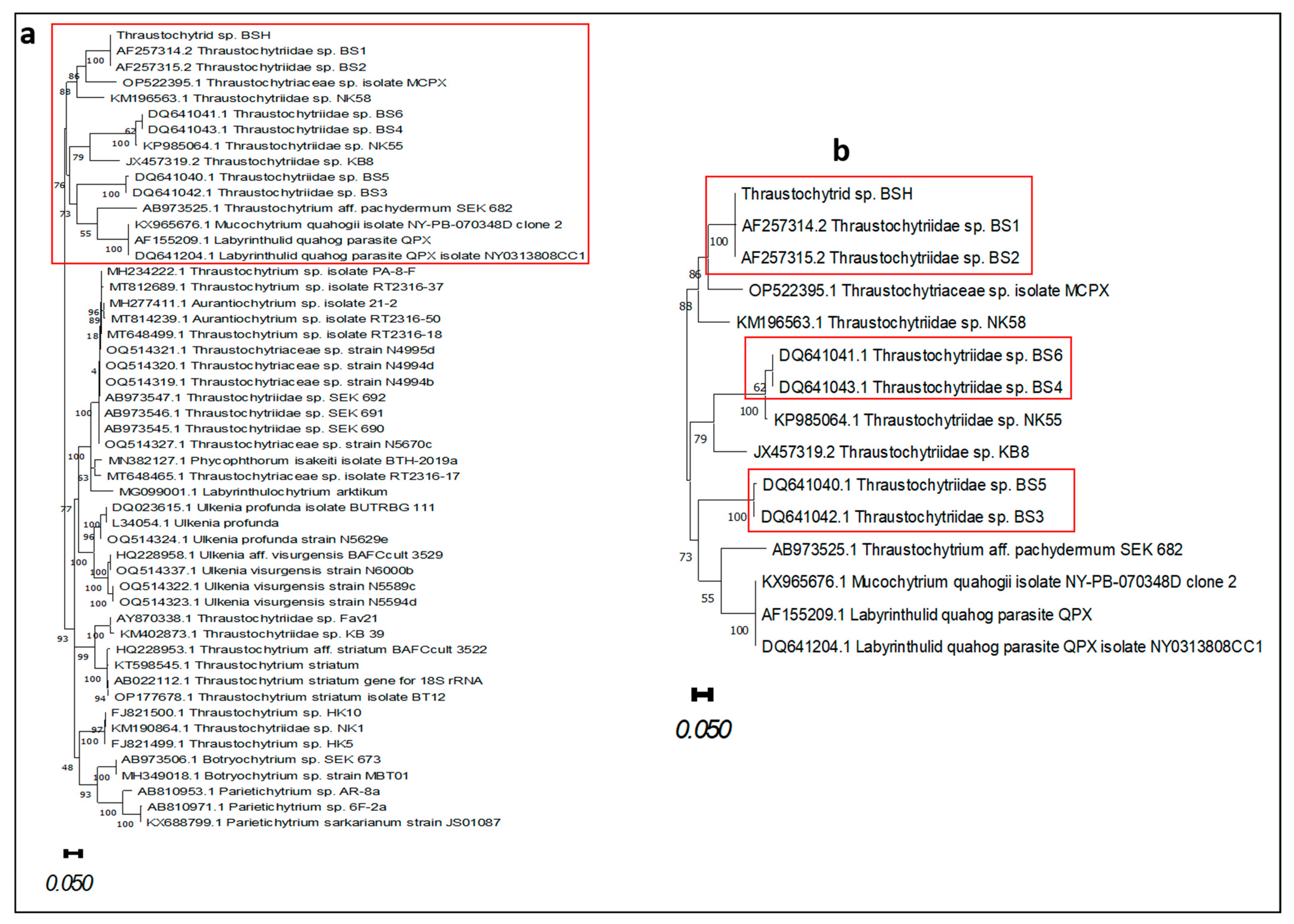
Cell cultures at day 2 (24 days post B. schlosseri cells isolation) were centrifuged at 1000× g for 10 min at room temperature and DNA was extracted using Quick Extract buffer (QE—DNA Extraction Solution; QE09050; LGC Biosearch Technologies, Shanghai, China) according to the manufacturer’s instructions. DNA samples were subjected to a polymerase chain reaction (PCR) protocol optimized for the recovery of the full length of the thraustochytrid 18s rDNA, using four sets of primers (A1, A2, A3, F and R) as described [6]. PCR amplifications were carried out using a ProFlex PCR system (applied biosystems) with the following cycling program: denaturation for 5 min at 95 °C, 35 amplification cycles of 45 sec at 94 °C, 1 min at 55 °C (for A1, A3 primers) or 62 °C (for A2, F and R primers) and 1.5 min at 72 °C, with a 7 min extended elongation step. PCR products were visualized on 1.5% agarose gel. PCR products were sequenced using the above PCR primers at Eurofins Genomics, Germany. Forward and reverse sequences of each PCR product were aligned to each other and assembled using BioEdit (Version 7.2.5 ) [41]. The assembled sequence was compared to NCBI database using BLASTN program. Related sequences were downloaded and aligned using ClustalW application embedded in BioEdit. The phylogenetic tree was inferred by using the maximum likelihood method and the Hasegawa–Kishino–Yano model [42] in MEGA11 [43]. The tree with the highest log likelihood (−14,044.76) was chosen. The tree was drawn to scale with branch lengths measured in the number of substitutions per site. Estimates of evolutionary divergence between fifteen 18S rDNA sequences was conducted using the maximum composite likelihood model [44] in MEGA11.

3. Results

3.1. Emerged Thraustochytrid Cells in B. schlosseri Primary Cultures

As of day 0, the developing thraustochytrid cultures were made of thraustochytrid vegetative cells as previously described [12], including mononucleated cells, multinucleated cells and sporangia. In a previous study [40], we used confocal microscopy to elucidate the natural auto-fluorescence properties of two thraustochytrid cell types (mononucleated and sporangia cells), that were further validated here. To complete the auto-fluorescence properties of the vegetable cells, we added here the confocal microscopy auto-fluorescence results of multinucleated cells (Figure S1). The fluorescence intensity of the multinucleated cells (n = 5) showed values of 17.4 ± 1.4, 21 ± 1.1, 35.4 ± 2.1 and 35.8 ± 2.1 relative fluorescence units (RFU) for blue, green, red and far-red channels, respectively (Figure S1a–d).
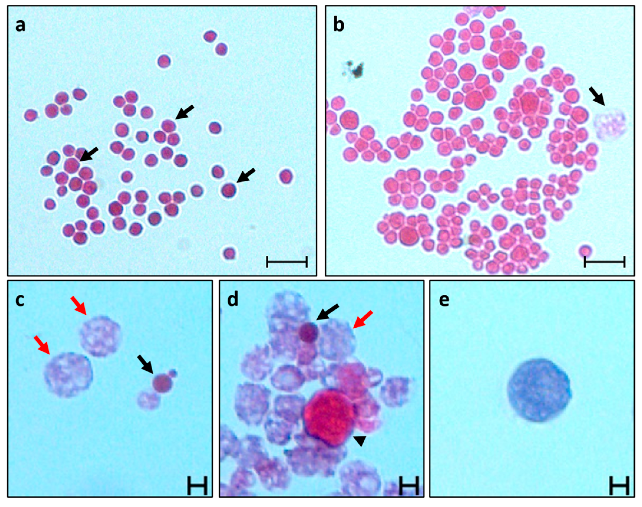
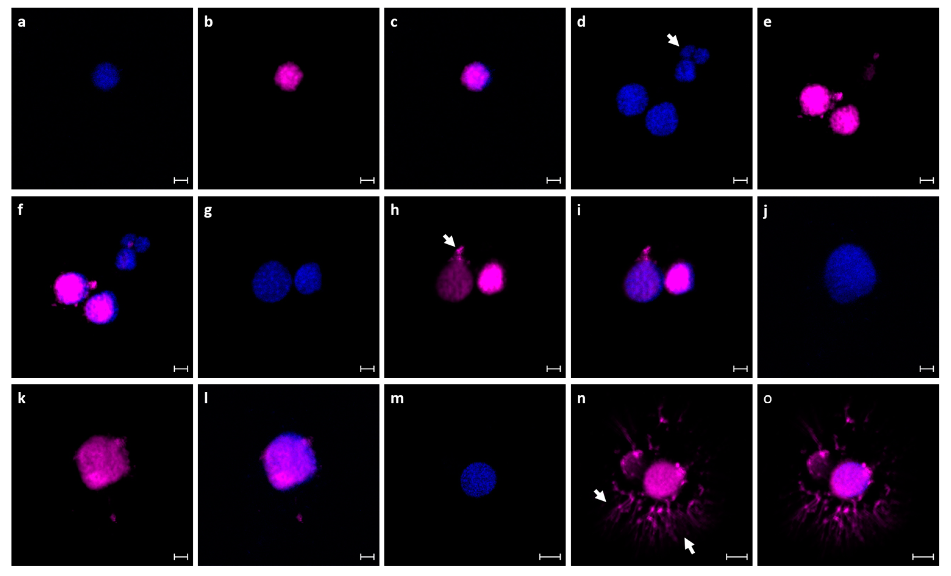
Thraustochytrid cells were subsequently characterized using histological (H&E), nuclear (Hoechst) and membrane (DiD) staining (Figure 2 and Figure 3). The mononucleated cells (4–10 µm) appeared in the cultures as cell clusters (up to 300 µm, composed of at least three cells/cluster) or single cells (Figure 2a). They are spherical and smooth, containing a small, hardly recognizable nucleus under bright field (1–2 µm; Qarri et al. [40]). H&E staining marked the cell cytoplasm as pink, and the membranes surrounding the cell were stained with a thin blue line (Figure 2a,b). They contain a circular nucleus (4–10 µm) positioned in the middle of the cell as validated via confocal microscopy (Figure 3a–f). The multinucleated cells (10–50 µm) are brown, spherical cells with a grainy texture and a barely recognizable nucleus under bright field. Stained (H&E) cells appeared blue (basophilic) with patterns of blue and light pink in the inner cell (Figure 2b–d), containing circular nuclei (1–2 µm), each surrounded by a membrane, and all together occupying the inner cell content (Figure 3d–i). Confocal microscopy revealed nets of cellular secretions on substrates and outside of the cells (Figure 3g–i). The sporangia cells (10–200 µm) are dark brown, spherical (some elongated in shape; Figure 3j) and condensed cells with a grainy texture and barely recognizable nucleus. H&E staining showed two types of stained cells, basophilic and the eosinophilic cells (Figure 2d,e). The cytoplasm of the eosinophilic sporangia was marked as pink, and the membranes surrounding the cells were stained with a thin blue line (Figure 2d), whereas the cell cytoplasm of the basophilic sporangia was marked as condense blue and the membranes surrounding the cells were stained with a thin blue line (Figure 2e). Confocal microscopy images of sporangia cells further revealed nets of cellular secretions on substrates and outside of the cells (Figure 3m–o), which were not seen in H&E staining.

3.2. Thraustochytrid Primary Culture Characteristics

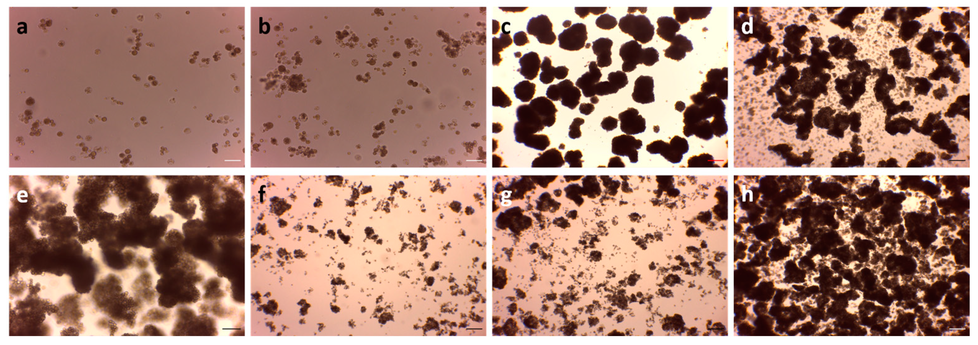
All primary cultures obtained from the two B. schlosseri colonies (n = 13) were made of suspensions of the three vegetable thraustochytrid cell types (mononucleated cells, multinucleated cells and sporangia; Figure 4), appearing as mixtures of single cells (from each type) or aggregates (10–500 µm). At day 0 of thraustochytrid primary cultures (22 days post B. schlosseri blood cells isolation) the plates were composed mainly of aggregates while single cells were barely seen. To facilitate cell counting and ensure the uniform distribution of cells across the different experimental vessels, we used a pipette to disperse the primary cultures until each culture consisted of only individual cells from the three cell types. All thraustochytrid primary cultures were pooled together, mixed and cells were inoculated into the twelve culturing conditions, six times per condition (Figure 1). Remarkably, within 24 h period, the primary cell cultures reverted back to the same aggregate profiles observed on day 0 (Figure 4a,b), with small modifications. For example, while similar size/shape aggregates were observed in the static 35 mm Petri dishes (same conditions as in day 0; Figure 4c), under the agitation condition all six 35 mm Petri dishes showed smaller-size aggregates (10–150 µm instead of 10–500 µm), and in addition, we observed single cells of mononucleated cells, multinucleated cells and sporangia (Figure 4f). In the T25 flasks, thraustochytrid cells grew as aggregates in both conditions (10–150 µm per cluster) with few single cells in the static condition, and many single cells in the agitation condition, that contained aggregates as well (Figure 4d,g). In T75 flasks, thraustochytrid cells grew as aggregates (50–500 µm per cluster) or as single cells in both conditions. The aggregates were composed of all three cell types (Figure 4e,h).

3.3. Thraustochytrid Culture Growth under Various Maintenance Conditions

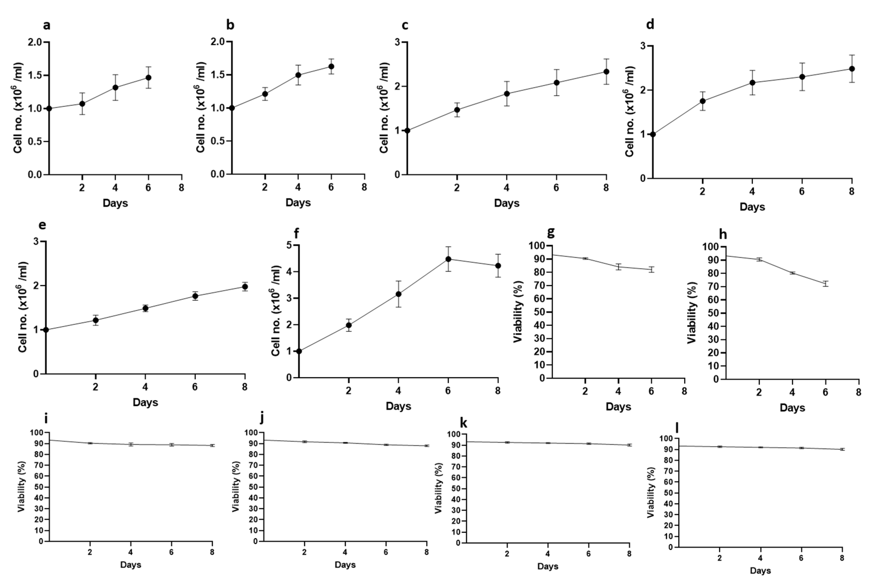
Cell viability at day 0, in all three-vessel types and in all replicates in the two experiments was 93.2%. Cells grew in fast modes. In the last day of the first set of experiments (day 12; 0.5 × 10⁶ cells/vessel) the average cell numbers in the static 35 mm Petri dishes averaged 0.96 × 10⁶ ± 0.02 cells/mL−1 (Figure 5a) and 83.63% ± 0.97 viability (Figure 5g), while under the agitating condition, the average cell numbers reached 1.41 × 10⁶ ± 0.12 cells/mL−1 (Figure 5b) with 81.95% ± 1.97 viability (Figure 5h). In the static T25 flasks, cell numbers reached 2.35 × 10⁶ ± 0.12 cells/mL−1 (Figure 5c) with 87.5% ± 0.85 viability (Figure 5i), and under the agitation condition, they reached 2.15 × 10⁶ ± 0.13 cells/mL−1 (Figure 5d) with 87.4% ± 1.11 viability (Figure 5j). In the static T75 flasks, cell numbers reached 2.22 × 10⁶ ± 0.13 cells/mL−1 (Figure 5e) and 87.2% ± 1.3 viability (Figure 5k), compared to 3.2 × 10⁶ ± 0.32 cells/mL−1 (Figure 5f) with 86.1% ± 0.6 viability under agitation condition (Figure 5l). Thraustochytrid cell growth rates in the present experiment showed an increase in cell numbers for all treatments under the two physical conditions and the two inoculation sizes. Fold increase values for inoculation 0.5 × 10⁶ cells/dish were, for the static condition 1.92 ± 0.33, 4.69 ± 1.3 and 4.4 ± 1.2 in 35 mm Petri dishes, T25 flasks and T75 flasks, respectively, and for the agitation conditions valued 2.82 ± 0.64, 4.3 ± 1.17 and 6.4 ± 1.91 for the 35 mm Petri dishes, T25 flasks and T75 flasks, respectively.
The second set of experiments was performed on thraustochytrid cultures with the inoculum of 1 × 10⁶ cells/vessel and held for up to 8 days. The average cell numbers in the last day of the static 35 mm Petri dishes (day 6) was 1.46 × 10⁶ ± 0.16 cells/mL−1 (Figure 6a) with 82.1% ± 2.1 viability (Figure 6g), while under agitating conditions, the average cell numbers reached 1.63 × 10⁶ ± 0.11 cells/mL−1 (Figure 6b) with 72.2% ± 2 viability (Figure 6h). The T25 and T75 flasks were held for up to 8 days. In the static T25 flasks (day 8), cell numbers reached 2.34 × 10⁶ ± 0.3 cells/mL−1 (Figure 6c) with 88.3% ± 0.94 viability (Figure 6i), and under agitation conditions, they reached 2.5 × 10⁶ ± 0.3 cells/mL−1 (Figure 6d) with 87.95% ± 0.63 viability (Figure 6j). In the static T75 flasks (day 8), cell numbers reached 1.97 × 10⁶ ± 0.1 cells/mL−1 (Figure 6e) and 90.14% ± 0.89 viability (Figure 6k), compared to 4.23 × 10⁶ ± 0.43 cells/mL−1 (Figure 6f) with 90.01% ± 1.2 viability under agitation conditions (Figure 6l). The increase values for inoculation 1 × 10⁶ cells/dish were, for the static condition 1.45 ± 0.33, 2.3 ± 0.9 and 1.97 ± 0.7 in 35 mm Petri dishes, T25 flasks and T75 flasks, respectively, and the agitation conditions were valued 1.62 ± 0.4, 2.5 ± 1.05 and 4.2 ± 2.3 for the 35 mm Petri dishes, T25 flasks and T75 flasks, respectively.

3.4. Molecular Identification

The 18s rDNA PCR amplifications of thraustochytrid cells were performed using four sets of primers revealing the existence of the undescribed strain Thraustochytriidae sp. BS1 and BS2 (GenBank accession number AF257314.2 and AF257315.2, respectively; Mo et al. [6]). Using BLASTN and MAGE 11 analysis tools, sequence comparison (Figure 7a,b) of the present isolated strain (termed thraustochytrid sp. BSH; H for Helgoland, Germany) from the German B. schlosseri colonies, revealed high similarity to Thraustochytriidae sp. BS2 and BS1 at 99.81% and 99.53%, respectively, which were isolated from B. schlosseri colonies collected in Monterey, California (USA), and maintained for generations under laboratory conditions in Haifa, Israel, supplied with Mediterranean seawater [6]. Lower similarities (82% and 77%, Table S1) were revealed for Thraustochytriidae isolates BS3 and BS6, respectively, also isolated from different Israeli B. schlosseri colonies [22].

4. Discussion

The world’s oceans and seas host, in diverse marine habitats and within a variety of marine taxa [17,24], more than one hundred known thraustochytrid strains, a common group of marine eukaryotic protists [9], recorded from the tropical to Antarctic zones and in depths down to 2000 m [14,15]. Thraustochytrids further emerged in primary cell cultures made from marine invertebrates [12,20,22,24], and have garnered significant attention in the fields of industry, transformative medicine, and science. Of prime interest are the valuable bioactive compounds, including omega-3 polyunsaturated fatty acid (ω-3 PUFA), docosahexaenoic acid (DHA), squalene and carotenoids. Additionally, these microorganisms are being explored as potential biofuel sources, highlighting the importance of isolation and cultivation of new strains [9,45,46,47,48]. Some thraustochytrid strains can be successfully cultivated to yield significant biomass with a rich lipid content, particularly high levels of PUFA, further considered promising candidates for the commercial production of omega-3 and DHA, for human health and aquaculture [25,26,29]. The thorough screening of cultures from diverse marine habitats has resulted in the discovery of strains capable of producing a minimum of 50% of their biomass in the form of lipids, with DHA comprising at least 25% of the total fatty acids [25,26]. It is also evident that the cell yields and the PUFA production of certain thraustochytrid strains can be altered through the manipulation of physical culture parameters [25].
By employing marine invertebrates cell culture medium for thraustochytrids isolation [12], this study assessed growth potentialities of a new isolated thraustochytrid strain (thraustochytrid sp. BSH) originated from B. schlosseri colonies from the intertidal zone at Helgoland Island, Germany. These cells were cultivated under two physical conditions (static versus agitation) with two inoculation sizes (0.5 and 1 × 10⁶ cells/dish). The sequence comparison of this strain revealed high similarities to Thraustochytriidae sp. BS2 and BS1 (99.81 and 99.53, respectively) isolated from B. schlosseri colonies from the Monterey, California, that were reared with Mediterranean seawater [6], while showing lower similarities to other strains (82–77%; BS3 to BS6, respectively) isolated from Israeli B. schlosseri colonies [22]. The phylogenetic analysis further revealed that thraustochytrid strains BS1 and BS2 might be considered to be the same species even though morphological characteristics indicated that BS1 and BS2 differed from BSH (Pers. Observ.). While it is premature to make definitive conclusions regarding the species status within this group of organisms (there is currently insufficient scientific evidence to support the separation of distinct species), it is interesting that the Israeli strains BS1 and BS2 (eastern Mediterranean) and the German BSH strain (North Sea), all isolated from the same host tunicate species, reveal high 18S sequence similarities.
In a morphological comparison between the isolated thraustochytrid cells from two different growth conditions (both reared under static condition in 35 mm Petri dishes; this study and Qarri et al. [12]), those originating from the Israeli growth system, where the B. schlosseri host was cultivated with seawater, and the present study, where the host was grown with artificial seawater, the results revealed that the cell types emerged remained consistent. These included mononucleated cells, multinucleated cells and sporangia. However, there was a notable difference in the nature of the cultures from Helgoland, as they primarily consisted of aggregated cells, with single cells being rare or scarcely observed. For the improved identification of the thraustochytrid cells, the present study adds, using confocal microscopy, the differential auto-fluorescence properties of multinucleated cells. This complements the auto-fluorescence characterization of a previous study [40], where the auto-fluorescence properties of mononucleated and sporangia cells were elucidated.
Regarding the extensive screening of thraustochytrid strains for the purpose of generating substantial biomass for biotechnological applications [25,26,29], the present research highlights the potential of thraustochytrid cell growth, demonstrating a significant increase in cell numbers across all treatments, under two distinct physical conditions and with two different inoculation sizes. As examples, the increase in cell numbers for the 0.5 × 10⁶ cells/dish inoculation in T75 flasks varies between 4.4- and 6.4-fold under static and agitation conditions, respectively, and revealed 2.0 and 4.2-fold increase for the 1 × 10⁶ cells/dish inoculation under static and agitation conditions, respectively. The results of the present study also reflect improved increase values for thraustochytrid growth when compared to the 1.45-fold increase per week of our former work, performed under static conditions [12]. In summary, to maximize growth potential in isolated thraustochytrid strains from Helgoland, cultivation under agitation in T75 flasks proves highly productive. However, it is worth noting that generating large biomasses using smaller culture units like T75 flasks may not be economically viable for biotechnological purposes. The findings of the present study demonstrate the feasibility of the mass cultivation of strain BSH isolated from the Helgoland, Germany, Botryllus colonies. The growth potential of the current strain serves as additional evidence that newly isolated thraustochytrid strains can indeed be cultivated for industrial purposes.

5. Conclusions

This study reveals experiments employed on a recently discovered thraustochytrid strain obtained from Helgoland, Germany (referred to as thraustochytrid sp. BSH, isolated from colonies of the colonial tunicate Botryllus schlosseri). By using varying conditions of static versus agitated setups, as well as two different inoculation sizes and three different vessel volumes, this study has successfully demonstrated the route for the cultivation scaling-up of these protist cells. This approach is amenable for future industrial applications (including the extraction of omega-3 oils as fish replacement, their uses as aquafeeds and more), further highlighting that thraustochytrid biomasses are subjected to varying culture conditions. Notably, the newly isolated thraustochytrid strain isolated from German colonies shares a significant genetic resemblance with thraustochytrid strains isolated from Botryllus colonies raised in Israel but originally collected from marinas in California, the USA. In the light of potential industrial applications, this finding further underscores the needs to investigate the relationship between animal hosts and symbiotic thraustochytrids in order to gain deeper insights into the biology of these protists and to unveil the yet undiscovered biodiversity (including the wide spectrum of bioactive compounds they hold) within this intriguing group of microorganisms.

Supplementary Materials

The following supporting information can be downloaded at: https://www.mdpi.com/article/10.3390/fishes9010022/s1. Figure S1: Confocal autofluorescence images of a multinucleated cell at various wavelengths; Table S1: Estimates of Evolutionary Divergence between Sequences.

Author Contributions

Conceptualization, A.Q. and B.R.; methodology, A.Q.; software, A.Q. and J.D.; validation, A.Q.; formal analysis, A.Q. and J.D.; investigation, A.Q.; resources, A.Q.; data curation, A.Q. and A.S.; writing—original draft preparation, A.Q. and B.R.; writing—review and editing, A.Q. and B.R.; visualization, A.Q. and B.R.; supervision, B.R.; project administration, A.Q.; funding acquisition, Y.R. and B.R. All authors have read and agreed to the published version of the manuscript.

Funding

This research was funded by the Human Frontier Science Program Career Development Award (CDA00017/2016), the German Research Foundation (RI 2787/1-1 AOBJ: 628819), the Fritz–Thyssen–Stiftung (2016-01277), the Else-Kröner-Fresenius-Stiftung (2016_A21) and the European Research Council Consolidator Grant (ERC-CoG 819933) (to Y.R.). B.R. was supported by an STSM grant from the COST action CA18238—European transdisciplinary networking platform for marine biotechnology.

Institutional Review Board Statement

Thraustochytrids were isolated from colonies of the tunicate Botryllus schlosseri. This species is considered an invasive species: an invertebrate for which no ethical considerations are required.

Data Availability Statement

The data created or analyzed in this study are included in the figures/tables and supplementary material. Additional data are available on request from the corresponding author.

Conflicts of Interest

The authors declare no conflicts of interest.

References

  1. Ellenbogen, B.B.; Aaronson, S.; Goldstein, S.; Belsky, M. Polyunsaturated fatty acids of aquatic fungi: Possible phylogenetic significance. Comp. Biochem. Physiol. 1969, 29, 805–811. [Google Scholar] [CrossRef]
  2. Cavalier-Smith, T.; Allsopp, M.T.E.P.; Chao, E.E. Thraustochytrids are chromists, not fungi: 18s rRNA signatures of Heterokonta. Philos. Trans. R. Soc. Lond. B 1994, 346, 387–397. [Google Scholar] [CrossRef]
  3. Honda, D.; Yokochi, T.; Nakahara, T.; Raghukumar, S.; Nakagiri, A.; Schaumann, K.; Takanori, H. Molecular phylogeny of labyrinthulids and thraustochytrids based on the sequencing of 18s ribosomal RNA gene. J. Eukaryot. Microbiol. 1999, 46, 637–647. [Google Scholar] [CrossRef] [PubMed]
  4. Tsui, C.K.M.; Marshall, W.; Yokoyama, R.; Honda, D.; Lippmeier, J.C.; Craven, K.D.; Peterson, P.D.; Berbee, M.L. Labyrinthulomycetes phylogeny and its implications for the evolutionary loss of chloroplasts and gain of ectoplasmic gliding. Mol. Phylogenetics Evol. 2009, 50, 129–140. [Google Scholar] [CrossRef] [PubMed]
  5. Mo, C.; Rinkevich, B. A simple, reliable and fast protocol for thraustochytrids DNA extraction. Mar. Biotechnol. 2001, 3, 100–102. [Google Scholar] [CrossRef] [PubMed]
  6. Mo, C.; Douek, J.; Rinkevich, B. Development of a PCR strategy for thraustochytrids identification based on 18S-rDNA sequence. Mar. Biol. 2002, 140, 883–889. [Google Scholar] [CrossRef]
  7. Marchan, L.F.; Chang, K.J.L.; Nichols, P.D.; Mitchell, W.J.; Polglase, J.L.; Gutierrez, T. Taxonomy, ecology and biotechnological applications of thraustochytrids: A review. Biotechnol. Adv. 2018, 36, 26–46. [Google Scholar] [CrossRef] [PubMed]
  8. Strassert, J.F.; Jamy, M.; Mylnikov, A.P.; Tikhonenkov, D.V.; Burki, F. New phylogenomic analysis of the enigmatic phylum Telonemia further resolves the eukaryote tree of life. Mol. Biol. Evol. 2019, 36, 757–765. [Google Scholar] [CrossRef]
  9. Lyu, L.; Wang, Q.; Wang, G. Cultivation and diversity analysis of novel marine thraustochytrids. Mar. Life Sci. Technol. 2021, 3, 263–275. [Google Scholar] [CrossRef]
  10. Chang, K.J.L.; Nichols, C.M.; Blackburn, S.I.; Dunstan, G.A.; Koutoulis, A.; Nichols, P.D. Comparison of thraustochytrids Aurantiochytrium sp., Schizochytrium sp., Thraustochytrium sp., and Ulkenia sp. for production of biodiesel, long-chain omega-3 oils, and exopolysaccharide. Mar. Biotechnol. 2014, 16, 396–411. [Google Scholar] [CrossRef]
  11. Liu, Y.; Singh, P.; Liang, Y.; Li, J.; Xie, N.; Song, Z.; Daroch, M.; Leng, K.; Johnson, Z.I.; Wang, G. Abundance and molecular diversity of thraustochytrids in coastal waters of southern China. FEMS Microbiol. Ecol. 2017, 93, fix070. [Google Scholar] [CrossRef] [PubMed]
  12. Qarri, A.; Rinkevich, Y.; Rinkevich, B. Employing marine invertebrate cell culture media for isolation and cultivation of thraustochytrids. Bot. Mar. 2021, 64, 447–454. [Google Scholar] [CrossRef]
  13. Bai, M.; Sen, B.; Wen, S.; Ye, H.; He, Y.; Zhang, X.; Wang, G. Culturable diversity of Thraustochytrids from coastal waters of Qingdao and their fatty acids. Mar. Drugs 2022, 20, 229. [Google Scholar] [CrossRef]
  14. Raghukumar, S. Ecology of the marine protists, the Labyrinthulomycetes (Thraustochytrids and Labyrinthulids). Eur. J. Protistol. 2002, 38, 127–145. [Google Scholar] [CrossRef]
  15. Raghukumar, S.; Damare, V.S. Increasing evidence for the important role of labyrinthulomycetes in marine ecosystems. Bot. Mar. 2011, 54, 3–11. [Google Scholar] [CrossRef]
  16. Naganuma, T.; Takasugi, H.; Kimura, H. Abundance of thraustochytrids in coastal plankton. Mar. Ecol. Prog. Ser. 1998, 162, 105–110. [Google Scholar] [CrossRef]
  17. Porter, D. Phylum Labryinthulomycota. In Handbood of Protoctista, 3nd ed.; Margulis, L., Corliss, J.O., Melkonian, M., Chapman, D.J., Eds.; Jones and Bartlett: Boston, MA, USA, 1990; pp. 388–398. [Google Scholar]
  18. Mass, P.A.Y.; Kleinschuster, S.J.; Dykstra, M.J.; Smolowitz, R.; Parent, J. Molecular characterization of QPX (quahog parasite unknown), a pathogen of Mercenaria mercenaria. J. Shellfish. Res. 1999, 18, 561–567. [Google Scholar]
  19. Polglase, J.L. A preliminary report on the thraustochytrid(s) and labyrinthulid(s) associated with a pathological condition in the lesser octopus Eledone cirrhosa. Bot. Mar. 1980, 23, 699–706. [Google Scholar] [CrossRef]
  20. Grasela, J.J.; Pomponi, S.A.; Rinkevich, B.; Grima, J. Efforts to develop a cultured sponge cell line: Revisiting an intractable problem. Vitr. Cell. Dev. Biol. -Anim. 2012, 48, 12–20. [Google Scholar] [CrossRef]
  21. Rinkevich, B.; Frank, U.; Gateño, D.; Rabinowitz, C. The establishment of various cell lines from colonial marine invertebrates. In Use of Aquatic Invertebrates as Tools for Monitoring of Environmental Hazards; Mueller, W.E.G., Ed.; Gustav Fischer Verlag: Stuttgart, Germany, 1994; pp. 253–263. [Google Scholar]
  22. Rabinowitz, C.; Douek, J.; Weisz, R.; Shabtay, A.; Rinkevich, B. Isolation and characterization of four novel thraustochytrid strains from a colonial tunicate. IJMS 2006, 35, 341–350. [Google Scholar]
  23. Garcia-Vedrenne, A.E.; Groner, M.; Page-Karjian, A.; Siegmund, G.F.; Singhal, S.; Sziklay, J.; Roberts, S. Development of genomic resources for a thraustochytrid pathogen and investigation of temperature influences on gene expression. PLoS ONE 2013, 8, e74196. [Google Scholar] [CrossRef]
  24. Rinkevich, B. Cell cultures from marine invertebrates: Obstacles, new approaches and recent improvements. J. Biotechnol. 1999, 70, 133–153. [Google Scholar] [CrossRef]
  25. Lewis, T.E.; Nichols, P.D.; McMeekin, T.A. The biotechnological potential of thraustochytrids. Mar. Biotechnol. 1999, 1, 580–587. [Google Scholar] [CrossRef]
  26. Raghukumar, S. Thraustochytrid marine protists: Production of PUFAs and other emerging technologies. Mar. Biotechnol. 2008, 10, 631–640. [Google Scholar] [CrossRef]
  27. Gupta, A.; Barrow, C.J.; Puri, M. Omega-3 biotechnology: Thraustochytrids as a novel source of omega-3 oils. Biotechnol. Adv. 2012, 30, 1733–1745. [Google Scholar] [CrossRef]
  28. Rotter, A.; Barbier, M.; Bertoni, F.; Bones, A.M.; Cancela, M.L.; Carlsson, J.; Carvalho, M.F.; Cegłowska, M.; Chirivella-Martorell, J.; Conk Dalay, M.; et al. A new network for the advancement of marine biotechnology in Europe and beyond. Front. Mar. Sci. 2020, 7, 278. [Google Scholar] [CrossRef]
  29. de La Broise, D.; Ventura, M.; Chauchat, L.; Guerreiro, M.; Michez, T.; Vinet, T.; Gautron, N.; Le Grand, F.; Bideau, A.; Goïc, N.L.; et al. Scale-up to pilot of a non-axenic culture of thraustochytrids using digestate from methanization as nitrogen source. Mar. Drugs 2022, 20, 499. [Google Scholar] [CrossRef] [PubMed]
  30. Singh, P.; Liu, Y.; Li, L.; Wang, G. Ecological dynamics and biotechnological implications of Thraustochytrids from marine habitats. Appl. Microbiol. Biotechnol. 2014, 98, 5789–5805. [Google Scholar] [CrossRef]
  31. Aasen, I.M.; Ertesvåg, H.; Heggeset, T.M.B.; Liu, B.; Brautaset, T.; Vadstein, O.; Ellingsen, T.E. Thraustochytrids as production organisms for docosahexaenoic acid (DHA), squalene, and carotenoids. App. Microbiol. Biotechnol. 2016, 100, 4309–4321. [Google Scholar] [CrossRef]
  32. Hong, W.K.; Heo, S.Y.; Park, H.M.; Kim, C.H.; Sohn, J.H.; Kondo, A.; Seo, J.W. Characterization of a squalene synthase from the thraustochytrid microalga Aurantiochytrium sp. krs101. J. Microbiol. Biotechnol. 2013, 23, 759–765. [Google Scholar] [CrossRef]
  33. Barta, D.G.; Coman, V.; Vodnar, D.C. Microalgae as sources of omega-3 polyunsaturated fatty acids: Biotechnological aspects. Algal Res. 2021, 58, 102410. [Google Scholar] [CrossRef]
  34. Pradeepkiran, J.A. Aquaculture role in global food security with nutritional value: A review. Transl. Anim. Sci. 2019, 3, 903–910. [Google Scholar] [CrossRef] [PubMed]
  35. Fukada, H.; Renato, K.; Junpei, S.; Haruka, M.; Toshiro, M. Effects of complete replacement of fish oil with plant oil mixtures and algal meal on growth performance and fatty acid composition in juvenile yellowtail Seriola quinqueradiata. Fish. Sci. 2020, 86, 107–118. [Google Scholar] [CrossRef]
  36. Lee Chang, K.J.; Parrish, C.C.; Simon, C.J.; Revill, A.T.; Nichols, P.D. Feeding Whole Thraustochytrid Biomass to Cultured Atlantic Salmon (Salmo salar) Fingerlings: Culture Performance and Fatty Acid Incorporation. J. Mar. Sci. Eng. 2020, 8, 207. [Google Scholar] [CrossRef]
  37. Davies, S.J.; Roderick, E.; Brudenell-Bruce, T.; Bavington, C.D.; Hartnett, F.; Hyland, J.; de Souza Valente, C.; Wan, A.H.L. Delivering a nutritionally enhanced tilapia fillet using a pre-harvest phase omega-3 thraustochytrids protist enriched diet. Eur. J. Lipid Sci. Technol. 2022, 124, 2100153. [Google Scholar] [CrossRef]
  38. Turchini, G.M.; Bente, E.T.; Wing-Keong, N. Fish oil replacement in finfish nutrition. Rev. Aquac. 2009, 1, 10–57. [Google Scholar] [CrossRef]
  39. Miller, M.R.; Nichols, P.D.; Carter, C.G. Replacement of fish oil with thraustochytrid Schizochytrium sp. L. oil in Atlantic salmon parr (Salmo salar L.) diets. Comp. Biochem. Physiol. Part A 2007, 148, 382–392. [Google Scholar] [CrossRef]
  40. Qarri, A.; Kültz, D.; Gardell, A.M.; Rinkevich, B.; Rinkevich, Y. Improved Media Formulations for Primary Cell Cultures Derived from a Colonial Urochordate. Cells 2023, 12, 1709. [Google Scholar] [CrossRef]
  41. Hall, T.A. BioEdit: A user-friendly biological sequence alignment editor and analysis program for Windows 95/98/NT. Nucleic Acids Symp. Ser. 1999, 41, 95–98. [Google Scholar]
  42. Hasegawa, M.; Kishino, H.; Yano, T. Dating the human-ape split by a molecular clock of mitochondrial DNA. J. Mol. Evol. 1985, 22, 160–174. [Google Scholar] [CrossRef]
  43. Tamura, K.; Stecher, G.; Kumar, S. MEGA 11: Molecular Evolutionary Genetics Analysis Version 11. Mol. Biol. Evol. 2021, 38, 3022–3327. [Google Scholar] [CrossRef] [PubMed]
  44. Tamura, K.; Nei, M.; Kumar, S. Prospects for inferring very large phylogenies by using the neighbor-joining method. Proc. Natl. Acad. Sci. USA 2004, 101, 11030–11035. [Google Scholar] [CrossRef]
  45. Burja, A.M.; Radianingtyas, H.; Windust, A.; Barrow, C.J. Isolation and characterization of polyunsaturated fatty acid producing Thraustochytrium species: Screening of strains and optimization of omega-3 production. Appl. Microbiol. Biotechnol. 2006, 72, 1161–1169. [Google Scholar] [CrossRef] [PubMed]
  46. Morabito, C.; Bournaud, C.; Maës, C.; Schuler, M.; Aiese Cigliano, R.; Dellero, Y.; Maréchal, E.; Amato, A.; Rébeillé, F. The lipid metabolism in thraustochytrids. Prog. Lipid Res. 2019, 76, 101007. [Google Scholar] [CrossRef]
  47. Nham Tran, T.L.; Miranda, A.F.; Gupta, A.; Puri, M.; Ball, A.S.; Adhikari, B.; Mouradov, A. The nutritional and pharmacological potential of new Australian thraustochytrids isolated from mangrove sediments. Mar. Drugs 2020, 18, 151. [Google Scholar] [CrossRef]
  48. FioRito, R.; Leander, C.; Leander, B. Characterization of three novel species of Labyrinthulomycota isolated from ochre sea stars (Pisaster ochraceus). Mar. Biol. 2016, 163, 170. [Google Scholar] [CrossRef]
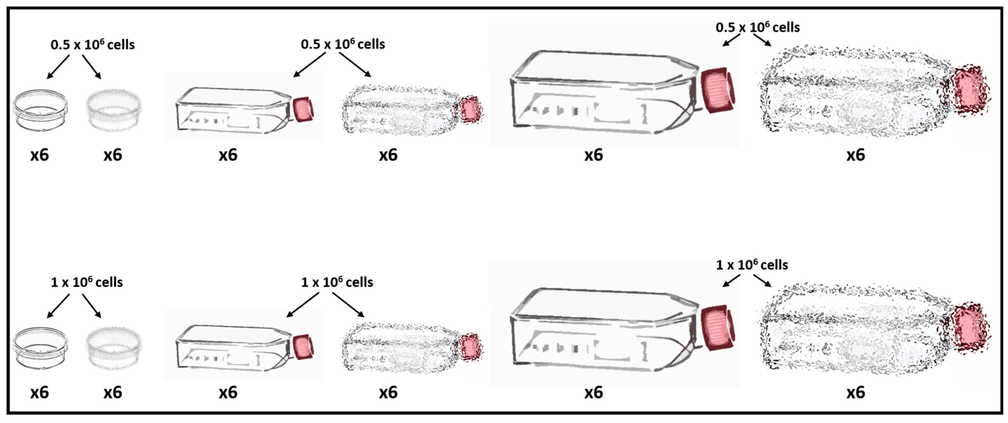
Figure 1. Experimental design for the various thraustochytrid cultures using: three vessel types (35 mm Petri dishes, T25 flasks and T75 flasks), two inoculum types (0.5 and 1 × 10⁶ cells/vessel) and two condition states: static and shaking. “Blurry vessels” represent the shaking condition. Each of the 12 culturing types was repeated ×6.
Fishes 09 00022 g001
Figure 2. H&E staining for thraustochytrid cell types in primary cultures: (a) mononucleated cells appearance as single cells (arrows); (b) aggregates of mononucleated cells and a multinucleated cell (an arrow). (ce) High magnifications of mononucleated cells (a black arrow) and multinucleated cells (red arrows) (c); mononucleated cells (a black arrow), multinucleated cells (a red arrow) and sporangium (a black arrowhead) (d); basophilic sporangium (e). Scale bars: (a) 10 µm, (b) 20 µm, (ce) 5 µm.
Fishes 09 00022 g002
Figure 3. Confocal images of thraustochytrid cell types stained with Hoechst and DiD. (ac) Mononucleated cell stained with Hoechst (a) and DiD (b); (c) a merged image. (df) Mononucleated (white arrow) and multinucleated cells stained with Hoechst (d) and DiD (e); (f) a merged image. (gi) Multi-nucleated cells stained with Hoechst (g) and DiD (h) depicting cell secretion (a white arrow); (i) a merged image. (jl) A single sporangium cell stained with Hoechst (j) and DiD (k); (l) a merged image. (mo) A single sporangium cell stained with Hoechst (m) and DiD (n), depicting cell secretion (white arrows); (o) a merged image. Scale bars: (al) 10 µm, (mo) 20 µm.
Fishes 09 00022 g003
Figure 4. Thraustochytrid primary cultures in the three vessel types at 24 h and in the last day of the experiments. (a,b) Inoculum of 0.5 and 1 × 10⁶ cells/vessel, respectively, under static conditions, 24 h post inoculation. (ce) Thraustochytrid cell cultures growth at day 12 in static condition, after inoculation of 0.5 × 10⁶ cells/vessel. (c) The 35 mm Petri dishes. (d) T25 flasks. (e) T75 flasks. (fh) Thraustochytrid cell cultures at days 6 and 8 under agitating condition after inoculation of 0.5 × 10⁶ cells/vessel. (f) Thraustochytrid cells in 35 mm Petri dishes at day 6. (g) Thraustochytrid cells in T25 flask at day 8. (h) Thraustochytrid cells in T75 flask at day 8. Scale bars = 100 µm.
Fishes 09 00022 g004
Figure 5. Cell numbers (106 cells/mL) and cell viability (%) of thraustochytrid cultures for 0.5 × 10⁶ cells/mL inoculation in the three different vessel sizes and under static/agitation conditions. Cell numbers of 35 mm Petri dishes in static (a) and agitation (b) conditions, T25 flasks in static (c) and agitation (d) conditions and T75 flasks in static (e) and agitation (f) conditions. Cell viabilities of 35 mm Petri dishes in static (g) and agitation (h) conditions, T25 flasks in static (i) and agitation (j) conditions and T75 flasks in static (k) and agitation (l) conditions. Time points represent averages ± STD of six vessels for each treatment.
Fishes 09 00022 g005
Figure 6. Cell numbers (106 cells/mL) and cell viability (%) of thraustochytrid cultures for 1 × 10⁶ cells/mL inoculation in the three different vessel sizes and under static/agitation conditions. Cell numbers of 35 mm Petri dishes in static (a) and agitation (b) conditions, T25 flasks in static (c) and agitation (d) conditions and T75 flasks in static (e) and agitation (f) conditions. Cell viabilities of 35 mm Petri dishes in static (g) and agitation (h) conditions, T25 flasks in static (i) and agitation (j) conditions and T75 flasks in static (k) and agitation (l) conditions. Time points represent averages ± STD of six vessels for each treatment.
Fishes 09 00022 g006
Figure 7. Evolutionary analysis by the Maximum Likelihood method. (a) Maximum likelihood tree inferred by using Hasegawa–Kishino–Yano model for 51 18S rDNA sequences. (b) Subtree including Thraustochytriidae sp. BS isolates, obtained from B. schlosseri (in red boxes). Numbers below branches indicate the accumulated tree percentages for the clustered taxa. Scale bars indicate branch lengths measured in number of substitutions per site.
Fishes 09 00022 g007
Disclaimer/Publisher’s Note: The statements, opinions and data contained in all publications are solely those of the individual author(s) and contributor(s) and not of MDPI and/or the editor(s). MDPI and/or the editor(s) disclaim responsibility for any injury to people or property resulting from any ideas, methods, instructions or products referred to in the content.