
Bio-SELEX: A Strategy for Biomarkers Isolation Directly from Biological Samples


Abstract
:1. Introduction
2. Experimental Design
2.1. Materials
- TRI Reagent (93289, Sigma-Aldrich, Darmstadt, Germany);
- BCA Protein Assay Kit (ab102536, Abcam, Cambridge, UK);
- 2X PCR Hot Start Master Mix with dye (G900-dye, ABM, Richmond, Canada);
- GeneJET Gel Extraction Kit (K0691, Thermo Scientific™, Waltham, MA, USA);
- Dynabeads™ M-270 Streptavidin (65306, Invitrogen™, Waltham, MA, USA);
- 2x Laemmli Sample Buffer (1610737, Bio-Rad, Hercules, CA, USA);
- InstantBlue® Coomassie Protein Stain (ab119211, Abcam, Cambridge, UK).
2.2. Equipment
- S1000 Thermal Cycler (BIORAD, Hercules, CA, USA);
- NanoDrop™ 2000/2000c Spectrophotometers (ND2000, Thermo Scientific™, Waltham, MA, USA);
- MagneSphere 12-Tube Magnetic Separation Stand (Z5343, Promega, Madison, WIS, USA);
- Mini-PROTEAN® Tetra Vertical Electrophoresis Cell, 4-gel, for 1.5 mm thick handcast gels (1658006FC, Hercules, CA, USA).
3. Procedure
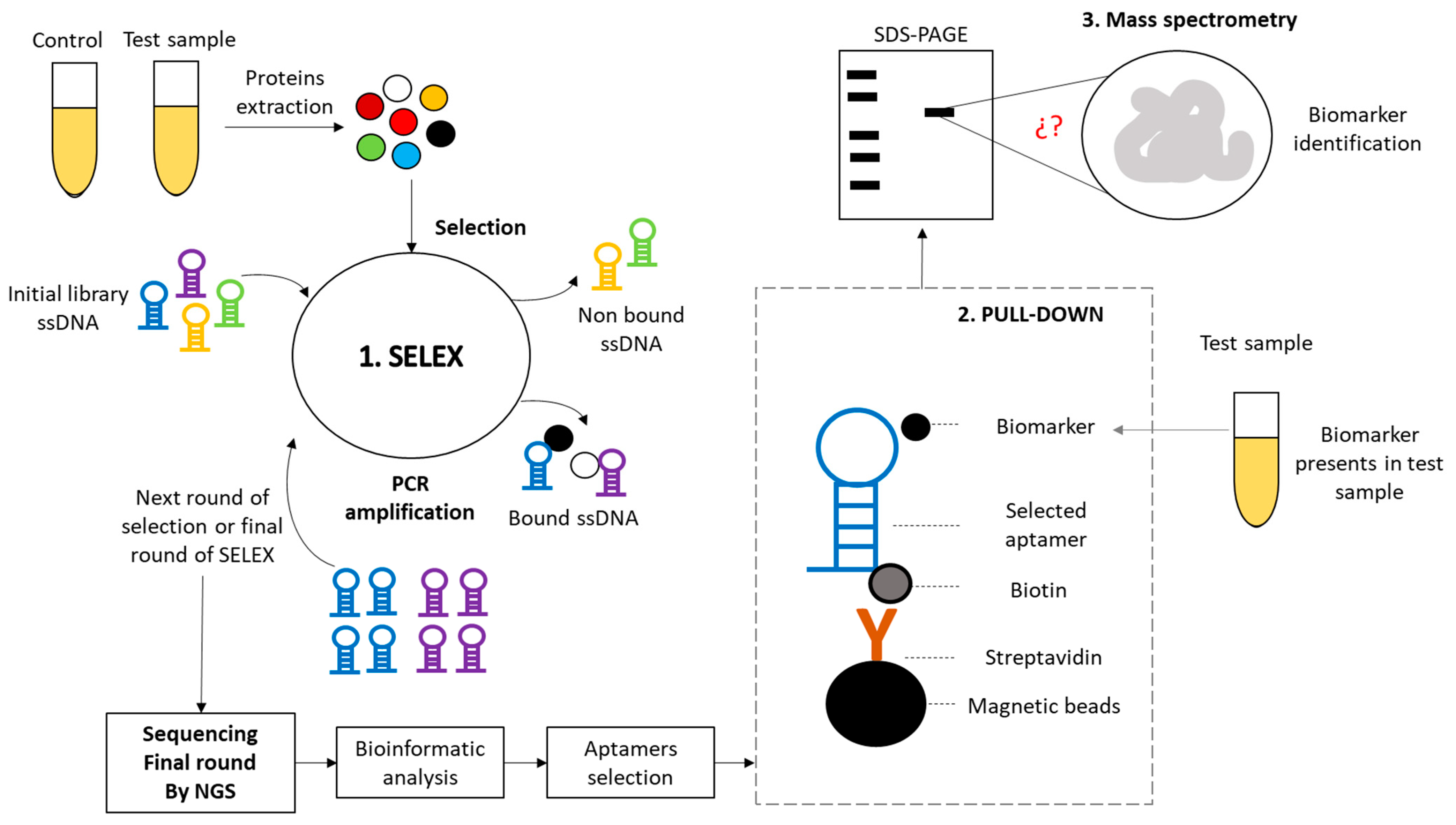
3.1. SELEX Strategy
- Purify PCR amplicons using any of the commercially available kits (we use GeneJET Gel Extraction Kit).
- Heat 200 pmol of amplified and purified library in 500 µL of Binding buffer 1X at 95 °C by 5–10 min, and let it cool slowly to promote 3D structure formation.
- Fix 100 ng of the extracted proteins in 96-well dishes or polypropylene tubes using 1X carbonate-bicarbonate buffer and incubate at 4 °C overnight.
- Wash 3–4 times using PBS 1X buffer.
- Bring the library from step 3 into contact with the fixed proteins from step 5 and allow interaction for 1 h at temperatures between 18 and 24 °C while agitating at 100 rpm.
- Wash 3–4 times using Binding buffer 1X.
- Recover protein-bound sequences by adding 100 µL of hot nuclease-free H2O (95 °C) by shaking at 100 rpm for 5 min at temperatures between 18 and 24 °C.
- Use these 100 µL as a template to be amplified via PCR using 2–5 µL in each reaction under the previously described conditions.
- Repeat the SELEX strategy for 10–20 rounds.
- Send the final round of the SELEX strategy for identification through NGS.
- Analyze the sequencing data and choose the aptamers based on their representativeness (readings and readings per million) for synthesis coupled to biotin.
3.2. Pull-Down
- Add 50 µL (0.5 mg) of magnetic beads coupled to streptavidin in 1.5 mL Eppendorf tube.
- Place the tube in a magnetic rack for 2–5 min until the beads are on one side of the tube. Remove the supernatant carefully and discard it.
- Add 1 mL binding buffer 1X, vortex, separate magnetic beads into a magnetic rack for 2–5 min, remove the supernatant carefully, and discard it.
- Dilute the biotinylated aptamer (100 ng) in 300 µL of binding buffer 1X.
- Add the 300 µL of biotinylated aptamer to the tubes with magnetic beads coupled to streptavidin that were previously washed.
- Allow interaction between 18 and 24 °C for 1–2 h at 100 rpm or overnight at 4 °C.
- Separate aptamer–biotin/streptavidin–magnetic beads complexes into magnetic rack for 2–5 min, remove the supernatant carefully, and discard it.
- Wash three times with binding buffer 1X, separate complexes into magnetic rack for 2–5 min, remove the supernatant carefully and discard it.
- Add 1 mL of test sample (pure or diluted depending on biological sample).
 CRITICAL STEP: During the Pull-Down biomarker separation process, negative controls must be performed in parallel to validate the experiment. It is suggested to at least use the following:
- Allow interaction between 18 and 24 °C for 1–2 h at 100 rpm or overnight at 4 °C.
- Separate aptamer–biotin/streptavidin–magnetic beads/biomarkers complexes into magnetic rack for 2–5 min, remove the supernatant carefully, and discard it.
- Wash three times with binding buffer 1X, separate complexes into magnetic rack for 2–5 min, remove the supernatant carefully, and discard it.
- Resuspend the complexes obtained in 50 µL of 2x Laemmli Sample Buffer.
- Resolve the complexes obtained for the test group and control groups by electrophoresis in an SDS-PAGE polyacrylamide gel using BIORAD instruments.
- Stain the proteins present in the gel using Coomassie or silver staining (we use Instablue Coomassie protein stain).
CRITICAL STEP: Verify on the manufacturer’s data sheet that the stain used is compatible with mass spectrometry (MS).
- Identify differentially expressed protein bands in the study group.
- Remove the identified bands using a sterile lancet and place them in a 1.5 mL Eppendorf tube with 100–200 µL of sterile water.
- Send the bands for protein identification through MS.
3.3. LC-MS/MS
3.4. Data Interpretation
4. Expected Results
- At the end of the SELEX strategy and sequencing the final library, it is expected that a list of aptamers ordered according to their representativeness will be obtained (reads, reads per million), with potential for the recognition of biomarkers. Table 3 shows some problems and solutions that can be presented in the SELEX strategy.
- At the end of the Bio-SELEX strategy, in its three stages, it is expected to obtain a list of biomarkers with the potential for the diagnosis, treatment, follow-up, or understanding of the disease to be studied. Table 4 shows some problems and solutions that can be presented in the Bio-SELEX strategy.
5. Reagents Setup
- Binding buffer 1X (PBS containing 2.5 mM of MgCl2, Tween20 to 0.02%, and 1 mM of heparin in miliQ water, pH 7.4).
- Carbonate/bicarbonate buffer 1X (12.5 mM Sodium bicarbonate, 87.5 mM Sodium carbonate, pH 9.6).
- PBS 1X buffer (137 mM NaCl, 2.7 mM KCl, 10 mM Na2HPO4, and 1.8 mM KH2PO4, pH 7.4).
Author Contributions
Funding
Institutional Review Board Statement
Informed Consent Statement
Data Availability Statement
Conflicts of Interest
References
- Tuerk, C.; Gold, L. Systematic evolution of ligands by exponential enrichment: RNA ligands to bacteriophage T4 DNA polymerase. Science 1990, 249, 505–510. [Google Scholar] [CrossRef] [PubMed]
- Ellington, A.D.; Szostak, J.W. In vitro selection of RNA molecules that bind specific ligands. Nature 1990, 346, 818–822. [Google Scholar] [CrossRef] [PubMed]
- Ellington, A.D.; Szostak, J.W. Selection in vitro of single-stranded DNA molecules that fold into specific ligand-binding structures. Nature 1992, 355, 850–852. [Google Scholar] [CrossRef] [PubMed]
- Jenison, R.D.; Gill, S.C.; Pardi, A.; Polisky, B. High-resolution molecular discrimination by RNA. Science 1994, 263, 1425–1429. [Google Scholar] [CrossRef] [PubMed]
- Mendonsa, S.D.; Bowser, M.T. In vitro evolution of functional DNA using capillary electrophoresis. J. Am. Chem. Soc. 2004, 126, 20–21. [Google Scholar] [CrossRef] [PubMed]
- Lou, X.; Qian, J.; Xiao, Y.; Viel, L.; Gerdon, A.E.; Lagally, E.T.; Atzberger, P.; Tarasow, T.M.; Heeger, A.J.; Soh, H.T. Micromagnetic selection of aptamers in microfluidic channels. Proc. Natl. Acad. Sci. USA 2009, 106, 2989–2994. [Google Scholar] [CrossRef] [PubMed]
- Daniels, D.A.; Chen, H.; Hicke, B.J.; Swiderek, K.M.; Gold, L. A tenascin-C aptamer identified by tumor cell SELEX: Systematic evolution of ligands by exponential enrichment. Proc. Natl. Acad. Sci. USA 2003, 100, 15416–15421. [Google Scholar] [CrossRef] [PubMed]
- Zhao, Y.; He, L.; Huang, B.; Zhang, W.; Hu, A.; Li, B.; Liao, S.; Wang, N. Identification of a novel DNA aptamer that selectively targets lung cancer serum. RSC Adv. 2021, 11, 33759–33769. [Google Scholar] [CrossRef] [PubMed]
- Esposito, C.L.; Van Roosbroeck, K.; Santamaria, G.; Rotoli, D.; Sandomenico, A.; Wierda, W.G.; Ferrajoli, A.; Ruvo, M.; Calin, G.A.; Catuogno, S.; et al. Selection of a Nuclease-Resistant RNA Aptamer Targeting CD19. Cancers 2021, 13, 5220. [Google Scholar] [CrossRef] [PubMed]
- Scoville, D.J.; Uhm, T.K.; Shallcross, J.A.; Whelan, R.J. Selection of DNA Aptamers for Ovarian Cancer Biomarker CA125 Using One-Pot SELEX and High-Throughput Sequencing. J. Nucleic Acids 2017, 2017, 9879135. [Google Scholar] [CrossRef] [PubMed]
- Eaton, R.M.; Shallcross, J.A.; Mael, L.E.; Mears, K.S.; Minkoff, L.; Scoville, D.J.; Whelan, R.J. Selection of DNA aptamers for ovarian cancer biomarker HE4 using CE-SELEX and high-throughput sequencing. Anal. Bioanal. Chem. 2015, 407, 6965–6973. [Google Scholar] [CrossRef] [PubMed]
- Nagarkatti, R.; de Araujo, F.F.; Gupta, C.; Debrabant, A. Aptamer based, non-PCR, non-serological detection of Chagas disease biomarkers in Trypanosoma cruzi infected mice. PLoS Neglected Trop. Dis. 2014, 8, e2650. [Google Scholar] [CrossRef] [PubMed]
- Morales-Velásquez, M.; Barón-Vera, J.P.; Pulgarín-Osorio, M.I.; Sánchez-Jiménez, M.M.; Ospina-Villa, J.D. Identification of the ATPase alpha subunit of Trypanosoma cruzi as a potential biomarker for the diagnosis of Chagas. Biomarkers 2023, 1–9. [Google Scholar] [CrossRef] [PubMed]
- Wang, T.; Yin, W.; AlShamaileh, H.; Zhang, Y.; Tran, P.H.L.; Nguyen, T.N.G.; Li, Y.; Chen, K.; Sun, M.; Hou, Y.; et al. A Detailed Protein-SELEX Protocol Allowing Visual Assessments of Individual Steps for a High Success Rate. Hum. Gene Ther. Methods 2019, 30, 1–16. [Google Scholar] [CrossRef] [PubMed]
- Martin, M. Cutadapt removes adapter sequences from high-throughput sequencing reads. EMBnet J. 2011, 17, 10–12. [Google Scholar] [CrossRef]
- Andrews, S. FastQC: A Quality Control Tool for High Throughput Sequence Data. Babraham Bioinformatics, Babraham Institute: Cambridge, UK, 2010. Available online: https://www.bioinformatics.babraham.ac.uk/projects/fastqc/ (accessed on 1 October 2023).
- Alam, K.K.; Chang, J.L.; Burke, D.H. FASTAptamer: A Bioinformatic Toolkit for High-throughput Sequence Analysis of Combinatorial Selections. Mol. Ther. Nucleic Acids. 2015, 4, e230. [Google Scholar] [CrossRef] [PubMed]
- Arthur Louche, Suzana Pinto Salcedo, Sarah Bigot. Protein-Protein Interactions: Pull-Down Assays.Protein-Protein Interactions: Pull-Down Assays. 2017, pp. 247–255. Available online: https://link.springer.com/protocol/10.1007/978-1-4939-7033-9_20 (accessed on 1 October 2023).

| Composition | 25 µL | 50 µL | Final Conc. |
|---|---|---|---|
| Nuclease-free water | to 25 µL | to 50 µL | |
| 10 µM Forward Primer | 95 | 15–30 s | 0.2 μM (0.05~1 µM) |
| 10 µM Reverse Primer | 45–68 | 15–60 s | 0.2 μM (0.05~1 µM) |
| Template library | 68 | 1 kb/min | <1 µg/50 µL |
| Hot Start Taq 2x PCR Master Mix with Dye | 12.5 µL | 25 µL | 1X |
| Steps | Temperature (°C) | Time | No. of Cycles |
|---|---|---|---|
| Initial denaturalization | 95 | 5 min | 1 |
| Denaturation | 95 | 15–30 s | 10–30 |
| Annealing | 45–68 | 15–60 s | |
| Extension | 68 | 1 kb/min | |
| Final extension | 68 | 5 min | |
| Cooling | 4 | ∞ | 1 |
| Problematic Result | Some Possible Causes | Some Potential Solutions |
|---|---|---|
| Too many or smear bands after negative or positive SELEX rounds. |
|
|
| No band after negative or positive SELEX rounds. |
|
|
| Problematic Result | Some Possible Causes | Some Potential Solutions |
|---|---|---|
| Too many bands after Pull-Down and SDS-PAGE. |
|
|
| No bands observed after Pull-Down and SDS-PAGE. |
|
|
Disclaimer/Publisher’s Note: The statements, opinions and data contained in all publications are solely those of the individual author(s) and contributor(s) and not of MDPI and/or the editor(s). MDPI and/or the editor(s) disclaim responsibility for any injury to people or property resulting from any ideas, methods, instructions or products referred to in the content. |
© 2023 by the authors. Licensee MDPI, Basel, Switzerland. This article is an open access article distributed under the terms and conditions of the Creative Commons Attribution (CC BY) license (https://creativecommons.org/licenses/by/4.0/).
Share and Cite
Ospina-Villa, J.D.; Restrepo-Cano, V.; Sánchez-Jiménez, M.M. Bio-SELEX: A Strategy for Biomarkers Isolation Directly from Biological Samples. Methods Protoc. 2023, 6, 109. https://doi.org/10.3390/mps6060109
Ospina-Villa JD, Restrepo-Cano V, Sánchez-Jiménez MM. Bio-SELEX: A Strategy for Biomarkers Isolation Directly from Biological Samples. Methods and Protocols. 2023; 6(6):109. https://doi.org/10.3390/mps6060109
Chicago/Turabian StyleOspina-Villa, Juan David, Valentina Restrepo-Cano, and Miryan Margot Sánchez-Jiménez. 2023. "Bio-SELEX: A Strategy for Biomarkers Isolation Directly from Biological Samples" Methods and Protocols 6, no. 6: 109. https://doi.org/10.3390/mps6060109
APA StyleOspina-Villa, J. D., Restrepo-Cano, V., & Sánchez-Jiménez, M. M. (2023). Bio-SELEX: A Strategy for Biomarkers Isolation Directly from Biological Samples. Methods and Protocols, 6(6), 109. https://doi.org/10.3390/mps6060109