Next Issue
Volume 10, November
Previous Issue
Volume 10, September


Tomography, Volume 10, Issue 10 (October 2024) – 12 articles

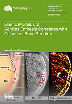
Cover Story (view full-size image): This study investigates the relationship between the mechanical properties of the Achilles tendon enthesis and bone microstructure in the calcaneal crescent. Nineteen human calcaneal-enthesis sections were examined with micro-CT imaging and mechanical indentation tests. The results showed significant correlations between the enthesis elastic modulus and trabecular bone thickness in specific regions, particularly the distal entheseal and proximal plantar regions. These findings suggest that regional differences in load transfer influence the microstructural adaptations in bone. Understanding these relationships could have implications for the management of enthesopathies and other tendon–bone interface pathologies. View this paper
  • Issues are regarded as officially published after their release is announced to the table of contents alert mailing list.
  • You may sign up for e-mail alerts to receive table of contents of newly released issues.
  • PDF is the official format for papers published in both, html and pdf forms. To view the papers in pdf format, click on the "PDF Full-text" link, and use the free Adobe Reader to open them.
Order results
Result details
Section
Select all
Export citation of selected articles as:
12 pages, 450 KiB  
Article
Left Ventricular Diastolic Dysfunction with Elevated Filling Pressures Is Associated with Embolic Stroke of Undetermined Source and Atrial Fibrillation
by Zubair Bashir, Liqi Shu, Yuqian Guo, Edward W. Chen, Shuyuan Wang, Eric D. Goldstein, Maheen Rana, Narendra Kala, Xing Dai, Daniel Mandel, Shadi Yaghi, Phinnara Has, Mingxing Xie, Tao Wang, James Simmons, Christopher Song and Philip Haines
Tomography 2024, 10(10), 1694-1705; https://doi.org/10.3390/tomography10100124 - 14 Oct 2024
Cited by 2 | Viewed by 1708
Abstract
Background/Objectives: Left ventricular diastolic dysfunction (LVDD) and elevated left ventricular filling pressure (LVFP) are strong predictors of clinical outcomes across various populations. However, their diagnostic utility in embolic stroke of undetermined source (ESUS) remains unclear. We hypothesized that LVDD with elevated LVFP [...] Read more.
Background/Objectives: Left ventricular diastolic dysfunction (LVDD) and elevated left ventricular filling pressure (LVFP) are strong predictors of clinical outcomes across various populations. However, their diagnostic utility in embolic stroke of undetermined source (ESUS) remains unclear. We hypothesized that LVDD with elevated LVFP (based on echocardiography) was more likely to be prevalent in ESUS compared to non-cardioembolic stroke (NCE) and to be associated with atrial fibrillation (AF) on follow-up monitoring. Methods: This is a single-center retrospective study that included adult patients with a diagnosis of acute ischemic stroke between January 2016 and June 2017. LV function was assessed by inpatient transthoracic echocardiogram (TTE), and stroke etiology was adjudicated by the neurologist per the consensus criteria. Patients with cardioembolic stroke and those with indeterminate diastolic function on TTE were excluded. Baseline patient characteristics and clinical variables were compared among patients with and without LVDD and elevated LVFP. Multivariable regression models were used to assess the associations between diastolic dysfunction, ESUS, and AF detection in ESUS patients. Results: We identified 509 patients with ESUS and NCE stroke who had reported diastolic function. The mean age was 64.19 years, 45.19% were female, and 146 had LVDD with available LVFP data. LVDD was not associated with ESUS (adjusted OR: 1.43, 95% CI: 0.90–2.27, p = 0.130) or atrial fibrillation (AF) detection on cardiac monitoring (adjusted OR: 1.88, 95% CI: 0.75–4.72, p = 0.179). However, LVDD with elevated LVFP was borderline associated with ESUS (adjusted OR: 2.17, 95% CI: 0.99–4.77, p = 0.054) and significantly associated with AF detection (adjusted OR: 3.59, 95% CI: 1.07–12.06, p = 0.038). Conclusions: Our data suggest that LVDD with elevated LVFP is borderline associated with ESUS and significantly associated with AF detection on follow-up cardiac monitoring. Therefore, the presence of LVDD with an increased probability of elevated LVFP may help identify a subset of stroke patients more likely to have ESUS, potentially due to atrial cardiopathy with underlying occult AF. Further studies are needed to confirm our findings and to evaluate the safety and efficacy of anticoagulation in patients with ESUS and LVDD with elevated LVFP. Full article
(This article belongs to the Section Cardiovascular Imaging)
Show Figures

Figure 1

18 pages, 3821 KiB  
Article
A Hybrid CNN-Transformer Model for Predicting N Staging and Survival in Non-Small Cell Lung Cancer Patients Based on CT-Scan
by Lingfei Wang, Chenghao Zhang and Jin Li
Tomography 2024, 10(10), 1676-1693; https://doi.org/10.3390/tomography10100123 - 10 Oct 2024
Cited by 3 | Viewed by 4316
Abstract
Accurate assessment of N staging in patients with non-small cell lung cancer (NSCLC) is critical for the development of effective treatment plans, the optimization of therapeutic strategies, and the enhancement of patient survival rates. This study proposes a hybrid model based on 3D [...] Read more.
Accurate assessment of N staging in patients with non-small cell lung cancer (NSCLC) is critical for the development of effective treatment plans, the optimization of therapeutic strategies, and the enhancement of patient survival rates. This study proposes a hybrid model based on 3D convolutional neural networks (CNNs) and transformers for predicting the N-staging and survival rates of NSCLC patients within the NSCLC radiogenomics and Nsclc-radiomics datasets. The model achieved accuracies of 0.805, 0.828, and 0.819 for the training, validation, and testing sets, respectively. By leveraging the strengths of CNNs in local feature extraction and the superior performance of transformers in global information modeling, the model significantly enhances predictive accuracy and efficacy. A comparative analysis with traditional CNN and transformer architectures demonstrates that the CNN-transformer hybrid model outperforms N-staging predictions. Furthermore, this study extracts the one-year survival rate as a feature and employs the Lasso–Cox model for survival predictions at various time intervals (1, 3, 5, and 7 years), with all survival prediction p-values being less than 0.05, illustrating the time-dependent nature of survival analysis. The application of time-dependent ROC curves further validates the model’s accuracy and reliability for survival predictions. Overall, this research provides innovative methodologies and new insights for the early diagnosis and prognostic evaluation of NSCLC. Full article
Show Figures

Figure 1

11 pages, 2284 KiB  
Article
The Correlation between the Elastic Modulus of the Achilles Tendon Enthesis and Bone Microstructure in the Calcaneal Crescent
by Kenichiro Doi, Dina Moazamian, Behnam Namiranian, Sheronda Statum, Amir Masoud Afsahi, Takuaki Yamamoto, Karen Y. Cheng, Christine B. Chung and Saeed Jerban
Tomography 2024, 10(10), 1665-1675; https://doi.org/10.3390/tomography10100122 - 10 Oct 2024
Viewed by 1426
Abstract
Background: The calcaneal enthesis, an osseous footprint where the Achilles tendon seamlessly integrates with the bone, represents a complex interface crucial for effective force transmission. Bone adapts to mechanical stress and remodels based on the applied internal and external forces. This study explores [...] Read more.
Background: The calcaneal enthesis, an osseous footprint where the Achilles tendon seamlessly integrates with the bone, represents a complex interface crucial for effective force transmission. Bone adapts to mechanical stress and remodels based on the applied internal and external forces. This study explores the relationship between the elasticity of the Achilles tendon enthesis and the bone microstructure in the calcaneal crescent. Methods: In total, 19 calcaneal-enthesis sections, harvested from 10 fresh-frozen human cadaveric foot-ankle specimens (73.8 ± 6.0 years old, seven female), were used in this study. Indentation tests were performed at the enthesis region, and Hayes’ elastic modulus was calculated for each specimen. Micro-CT scanning was performed at 50-micron voxel size to assess trabecular bone microstructure within six regions of interest (ROIs) and the cortical bone thickness along the calcaneal crescent. Results: Significant Spearman correlations were observed between the enthesis elastic modulus and trabecular bone thickness in the distal entheseal (ROI 3) and proximal plantar (ROI 4) regions (R = 0.786 and 0.518, respectively). Conclusion: This study highlights the potential impacts of Achilles tendon enthesis on calcaneal bone microstructure, which was pronounced in the distal calcaneal enthesis, suggesting regional differences in load transfer mechanism that require further investigation. Full article
Show Figures

Graphical abstract

20 pages, 3823 KiB  
Article
Pulmonary Fissure Segmentation in CT Images Using Image Filtering and Machine Learning
by Mikhail Fufin, Vladimir Makarov, Vadim I. Alfimov, Vladislav V. Ananev and Anna Ananeva
Tomography 2024, 10(10), 1645-1664; https://doi.org/10.3390/tomography10100121 - 9 Oct 2024
Cited by 1 | Viewed by 1511
Abstract
Background: Both lung lobe segmentation and lung fissure segmentation are useful in the clinical diagnosis and evaluation of lung disease. It is often of clinical interest to quantify each lobe separately because many diseases are associated with specific lobes. Fissure segmentation is important [...] Read more.
Background: Both lung lobe segmentation and lung fissure segmentation are useful in the clinical diagnosis and evaluation of lung disease. It is often of clinical interest to quantify each lobe separately because many diseases are associated with specific lobes. Fissure segmentation is important for a significant proportion of lung lobe segmentation methods, as well as for assessing fissure completeness, since there is an increasing requirement for the quantification of fissure integrity. Methods: We propose a method for the fully automatic segmentation of pulmonary fissures on lung computed tomography (CT) based on U-Net and PAN models using a Derivative of Stick (DoS) filter for data preprocessing. Model ensembling is also used to improve prediction accuracy. Results: Our method achieved an F1 score of 0.916 for right-lung fissures and 0.933 for left-lung fissures, which are significantly higher than the standalone DoS results (0.724 and 0.666, respectively). We also performed lung lobe segmentation using fissure segmentation. The lobe segmentation algorithm shows results close to those of state-of-the-art methods, with an average Dice score of 0.989. Conclusions: The proposed method segments pulmonary fissures efficiently and have low memory requirements, which makes it suitable for further research in this field involving rapid experimentation. Full article
(This article belongs to the Topic AI in Medical Imaging and Image Processing)
Show Figures

Figure 1

23 pages, 966 KiB  
Article
Super Learner Algorithm for Carotid Artery Disease Diagnosis: A Machine Learning Approach Leveraging Craniocervical CT Angiography
by Halil İbrahim Özdemir, Kazım Gökhan Atman, Hüseyin Şirin, Abdullah Engin Çalık, Ibrahim Senturk, Metin Bilge, İsmail Oran, Duygu Bilge and Celal Çınar
Tomography 2024, 10(10), 1622-1644; https://doi.org/10.3390/tomography10100120 - 9 Oct 2024
Viewed by 1526
Abstract
This study introduces a machine learning (ML) approach to diagnosing carotid artery diseases, including stenosis, aneurysm, and dissection, by leveraging craniocervical computed tomography angiography (CTA) data. A meticulously curated, balanced dataset of 122 patient cases was used, ensuring reproducibility and data quality, and [...] Read more.
This study introduces a machine learning (ML) approach to diagnosing carotid artery diseases, including stenosis, aneurysm, and dissection, by leveraging craniocervical computed tomography angiography (CTA) data. A meticulously curated, balanced dataset of 122 patient cases was used, ensuring reproducibility and data quality, and this is publicly accessible at (insert dataset location). The proposed method integrates a super learner model which combines adaptive boosting, gradient boosting, and random forests algorithms, achieving an accuracy of 90%. To enhance model robustness and generalization, techniques such as k-fold cross-validation, bootstrapping, data augmentation, and the synthetic minority oversampling technique (SMOTE) were applied, expanding the dataset to 1000 instances and significantly improving performance for minority classes like aneurysm and dissection. The results highlight the pivotal role of blood vessel structural analysis in diagnosing carotid artery diseases and demonstrate the superior performance of the super learner model in comparison with state-of-the-art (SOTA) methods in terms of both accuracy and robustness. This manuscript outlines the methodology, compares the results with state-of-the-art approaches, and provides insights for future research directions in applying machine learning to medical diagnostics. Full article
Show Figures

Figure 1

14 pages, 2219 KiB  
Article
Optimal DaTQUANT Thresholds for Diagnostic Accuracy of Dementia with Lewy Bodies (DLB) and Parkinson’s Disease (PD)
by Phillip H. Kuo, Patrick Cella, Ying-Hui Chou, Alexander Arkhipenko and Julia M. Fisher
Tomography 2024, 10(10), 1608-1621; https://doi.org/10.3390/tomography10100119 - 9 Oct 2024
Viewed by 2205
Abstract
Background: Quantitative thresholds are helpful to define an abnormal DaT SPECT in patients with suspected nigrostriatal degenerative diseases (NSDD). The optimal DaTQUANT threshold for diagnostic accuracy of DaT SPECT across combined movement and cognitive disorder populations has been previously described. Methods: We [...] Read more.
Background: Quantitative thresholds are helpful to define an abnormal DaT SPECT in patients with suspected nigrostriatal degenerative diseases (NSDD). The optimal DaTQUANT threshold for diagnostic accuracy of DaT SPECT across combined movement and cognitive disorder populations has been previously described. Methods: We established optimal DaTQUANT thresholds that enhance the discrimination between dementia with Lewy bodies (DLB) and non-DLB dementia types, as well as between Parkinsonian syndromes (PS) and conditions not characterized by nigrostriatal degeneration (non-PS). Results: Data from a total of 303 patients were used in this retrospective analysis. Posterior putamen of the more affected hemisphere (MAH) was shown to be an accurate single-variable predictor for both DLB and PS and was comparable to the most accurate multi-variable models. Conclusions: Automated quantification with DaTQUANT can accurately aid in the differentiation of DLB from non-DLB dementias and PS from non-PS. Optimal thresholds for assisting a diagnosis of DLB are striatal binding ratio (SBR) ≤ 0.65, z-score ≤ −2.36, and a percent deviation ≤ −0.54 for the posterior putamen of the MAH. Optimal posterior putamen thresholds for assisting a diagnosis of PS are SBR ≤ 0.92, z-score ≤ −1.53, and a percent deviation ≤ −0.33, which are similar to our previously reported posterior putamen threshold values using a blended patient pool from multiple study populations. Full article
(This article belongs to the Section Neuroimaging)
Show Figures

Graphical abstract

3 pages, 154 KiB  
Editorial
Feature Reviews for Tomography 2023
by Yashbir Singh and Emilio Quaia
Tomography 2024, 10(10), 1605-1607; https://doi.org/10.3390/tomography10100118 - 9 Oct 2024
Viewed by 992
Abstract
In an era of rapid technological progress, this Special Issue aims to provide a comprehensive overview of the state-of-the-art in tomographic imaging [...] Full article
(This article belongs to the Special Issue Feature Reviews for Tomography 2023–2024)
14 pages, 3459 KiB  
Article
Diagnostic Value of Contrast-Enhanced Dual-Energy Computed Tomography in the Pancreatic Parenchymal and Delayed Phases for Pancreatic Cancer
by Yusuke Kurita, Daisuke Utsunomiya, Kensuke Kubota, Shingo Koyama, Sho Hasegawa, Kunihiro Hosono, Kuniyasu Irie, Yuichi Suzuki, Shin Maeda, Noritoshi Kobayashi, Yasushi Ichikawa, Itaru Endo and Atsushi Nakajima
Tomography 2024, 10(10), 1591-1604; https://doi.org/10.3390/tomography10100117 - 7 Oct 2024
Viewed by 1797
Abstract
Background/Objectives: The usefulness of dual-energy computed tomography (DECT) for low absorption in the parenchymal phase and contrast effects in the delayed phase for pancreatic cancer is not clear. Therefore, the diagnostic capability of low-KeV images obtained using DECT for pancreatic cancer in [...] Read more.
Background/Objectives: The usefulness of dual-energy computed tomography (DECT) for low absorption in the parenchymal phase and contrast effects in the delayed phase for pancreatic cancer is not clear. Therefore, the diagnostic capability of low-KeV images obtained using DECT for pancreatic cancer in the pancreatic parenchymal and delayed phases was evaluated quantitatively and qualitatively. Methods: Twenty-five patients with pancreatic cancer who underwent contrast-enhanced DECT were included. A total of 50 and 70 KeV CT images, classified as low-keV and conventional CT-equivalent images, were produced, respectively. The tumor-to-pancreas contrast (Hounsfield units [HU]) in the pancreatic parenchymal and delayed phases was calculated by subtracting the CT value of the pancreatic tumor from that of normal parenchyma. Results: The median tumor-to-pancreas contrast on 50 KeV CT in the pancreatic parenchymal phase (133 HU) was higher than that on conventional CT (68 HU) (p < 0.001). The median tumor-to-pancreas contrast in the delayed phase was −28 HU for 50 KeV CT and −9 HU for conventional CT (p = 0.545). For tumors < 20 mm, the tumor-to-pancreas contrast of 50 KeV CT (−39 HU) had a significantly clearer contrast effect than that of conventional CT (−16.5 HU), even in the delayed phase (p = 0.034). Conclusions: These 50 KeV CT images may clarify the low-absorption areas of pancreatic cancer in the pancreatic parenchymal phase. A good contrast effect was observed in small pancreatic cancers on 50 KeV delayed-phase images, suggesting that DECT is useful for the visualization of early pancreatic cancer with a small tumor diameter. Full article
(This article belongs to the Section Abdominal Imaging)
Show Figures

Figure 1

14 pages, 828 KiB  
Article
Lightweight MRI Brain Tumor Segmentation Enhanced by Hierarchical Feature Fusion
by Lei Zhang, Rong Zhang, Zhongjie Zhu, Pei Li, Yongqiang Bai and Ming Wang
Tomography 2024, 10(10), 1577-1590; https://doi.org/10.3390/tomography10100116 - 1 Oct 2024
Cited by 2 | Viewed by 1696
Abstract
Background: Existing methods for MRI brain tumor segmentation often suffer from excessive model parameters and suboptimal performance in delineating tumor boundaries. Methods: For this issue, a lightweight MRI brain tumor segmentation method, enhanced by hierarchical feature fusion (EHFF), is proposed. This method reduces [...] Read more.
Background: Existing methods for MRI brain tumor segmentation often suffer from excessive model parameters and suboptimal performance in delineating tumor boundaries. Methods: For this issue, a lightweight MRI brain tumor segmentation method, enhanced by hierarchical feature fusion (EHFF), is proposed. This method reduces model parameters while improving segmentation performance by integrating hierarchical features. Initially, a fine-grained feature adjustment network is crafted and guided by global contextual information, leading to the establishment of an adaptive feature learning (AFL) module. This module captures the global features of MRI brain tumor images through macro perception and micro focus, adjusting spatial granularity to enhance feature details and reduce computational complexity. Subsequently, a hierarchical feature weighting (HFW) module is constructed. This module extracts multi-scale refined features through multi-level weighting, enhancing the detailed features of spatial positions and alleviating the lack of attention to local position details in macro perception. Finally, a hierarchical feature retention (HFR) module is designed as a supplementary decoder. This module retains, up-samples, and fuses feature maps from each layer, thereby achieving better detail preservation and reconstruction. Results: Experimental results on the BraTS 2021 dataset demonstrate that the proposed method surpasses existing methods. Dice similarity coefficients (DSC) for the three semantic categories ET, TC, and WT are 88.57%, 91.53%, and 93.09%, respectively. Full article
(This article belongs to the Topic AI in Medical Imaging and Image Processing)
Show Figures

Figure 1

13 pages, 4825 KiB  
Article
Identifying Brain Network Structure for an fMRI Effective Connectivity Study Using the Least Absolute Shrinkage and Selection Operator (LASSO) Method
by Xingfeng Li and Yuan Zhang
Tomography 2024, 10(10), 1564-1576; https://doi.org/10.3390/tomography10100115 - 30 Sep 2024
Cited by 1 | Viewed by 1407
Abstract
Background: Studying causality relationships between different brain regions using the fMRI method has attracted great attention. To investigate causality relationships between different brain regions, we need to identify both the brain network structure and the influence magnitude. Most current methods concentrate on magnitude [...] Read more.
Background: Studying causality relationships between different brain regions using the fMRI method has attracted great attention. To investigate causality relationships between different brain regions, we need to identify both the brain network structure and the influence magnitude. Most current methods concentrate on magnitude estimation, but not on identifying the connection or structure of the network. To address this problem, we proposed a nonlinear system identification method, in which a polynomial kernel was adopted to approximate the relation between the system inputs and outputs. However, this method has an overfitting problem for modelling the input–output relation if we apply the method to model the brain network directly. Methods: To overcome this limitation, this study applied the least absolute shrinkage and selection operator (LASSO) model selection method to identify both brain region networks and the connection strength (system coefficients). From these coefficients, the causality influence is derived from the identified structure. The method was verified based on the human visual cortex with phase-encoded designs. The functional data were pre-processed with motion correction. The visual cortex brain regions were defined based on a retinotopic mapping method. An eight-connection visual system network was adopted to validate the method. The proposed method was able to identify both the connected visual networks and associated coefficients from the LASSO model selection. Results: The result showed that this method can be applied to identify both network structures and associated causalities between different brain regions. Conclusions: System identification with LASSO model selection algorithm is a powerful approach for fMRI effective connectivity study. Full article
(This article belongs to the Special Issue New Insights into Functional Magnetic Resonance Imaging (fMRI))
Show Figures

Figure 1

17 pages, 1974 KiB  
Review
Nailfold Video-Capillaroscopy in Sarcoidosis: New Perspectives and Challenges
by Maria Chianese, Gianluca Screm, Paola Confalonieri, Francesco Salton, Liliana Trotta, Beatrice Da Re, Antonio Romallo, Alessandra Galantino, Mario D’Oria, Michael Hughes, Giulia Bandini, Marco Confalonieri, Elisa Baratella, Lucrezia Mondini and Barbara Ruaro
Tomography 2024, 10(10), 1547-1563; https://doi.org/10.3390/tomography10100114 - 25 Sep 2024
Cited by 3 | Viewed by 1683
Abstract
Introduction: Nailfold video-capillaroscopy (NVC) is a non-invasive cost-effective technique involving the microscopic examination of small blood vessels of the distal nailfold with a magnification device. It provides valuable information regarding the microcirculation including anomalies such as tortuous or dilated capillaries, hemorrhages, and avascular [...] Read more.
Introduction: Nailfold video-capillaroscopy (NVC) is a non-invasive cost-effective technique involving the microscopic examination of small blood vessels of the distal nailfold with a magnification device. It provides valuable information regarding the microcirculation including anomalies such as tortuous or dilated capillaries, hemorrhages, and avascular areas, which can characterize connective tissue diseases. The utility of NVC in the diagnosis and monitoring of systemic sclerosis (SSc) has been investigated in numerous studies allowing the distinction of the specific microvascular pattern of scleroderma from different conditions other than scleroderma (non-scleroderma pattern). Sarcoidosis (SA) is a systemic inflammatory disease that can affect various organs, including the lungs, skin, and lymph nodes. The purpose of our review was to evaluate the current state of the art in the use of NVC in the diagnosis of SA, to understand the indications for its use and any consequent advantages in the management of the disease in different settings in terms of benefits for patients. Materials and Methods: We searched for the key terms “sarcoidosis” and “video-capillaroscopy” in a computerized search of Pub-Med, extending the search back in time without setting limits. We provided a critical overview of the literature, based on a precise evaluation. After our analysis, we examined the six yielded works looking for answers to our questions. Results: Few studies have evaluated that microcirculation is often compromised in SA, with alterations in blood flow and consequent tissue damage. Discussion: Basing on highlighted findings, NVC appears to be a useful tool in the initial evaluation of sarcoidosis patients. Furthermore, capillaroscopy is useful in the evaluation of the coexistence of sarcoidosis and scleroderma spectrum disorder or overlap syndromes. Conclusions: In conclusions, no specific pattern has been described for sarcoidosis, and further re-search is needed to fully understand the implications of nailfold capillaroscopy find-ings in this disease and to establish standardized guidelines for its use in clinical practice. Full article
Show Figures

Figure 1

13 pages, 1341 KiB  
Article
Comparison of Traumatic Brain Injury in Adult Patients with and without Facial Fractures
by Iulia Tatiana Lupascu, Sorin Hostiuc, Costin Aurelian Minoiu, Mihaela Hostiuc and Bogdan Valeriu Popa
Tomography 2024, 10(10), 1534-1546; https://doi.org/10.3390/tomography10100113 - 24 Sep 2024
Cited by 1 | Viewed by 2183
Abstract
Objectives: Facial fractures and associated traumatic brain injuries represent a worldwide public health concern. Therefore, we aimed to determine the pattern of brain injury accompanying facial fractures by comparing adult patients with and without facial fractures in terms of demographic, clinical, and imaging [...] Read more.
Objectives: Facial fractures and associated traumatic brain injuries represent a worldwide public health concern. Therefore, we aimed to determine the pattern of brain injury accompanying facial fractures by comparing adult patients with and without facial fractures in terms of demographic, clinical, and imaging features. Methods: This single-center, retrospective study included 492 polytrauma patients presenting at our emergency department from January 2019 to July 2023, which were divided in two groups: with facial fractures (FF) and without facial fractures (non-FF). The following data were collected: age, sex, mechanism of trauma (road traffic accident, fall, and other causes), Glasgow Coma Scale (GCS), the evolution of the patient (admitted to a medical ward or intensive care unit, neurosurgery performed, death), and imaging features of the injury. Data were analyzed using descriptive tests, Chi-square tests, and regression analyses. A p-value less than 0.05 was considered statistically significant. Results: In the FF group, there were 79% (n = 102) men and 21% (n = 27) women, with a mean age of 45 ± 17 years, while in the non-FF group, there were 70% (n = 253) men and 30% (n = 110) women, with a mean age 46 ± 17 years. There was a significant association between brain injuries and facial fractures (p < 0.001, AOR 1.7). The most frequent facial fracture affected the zygoma bone in 28.1% (n = 67) cases. The most frequent brain injury associated with FF was subdural hematoma 23.4% (n = 44), and in the non-FF group, the most common head injury was intraparenchymal hematoma 29% (n = 73); Conclusions: Both groups shared similarities regarding gender, age, cause of traumatic event, and outcome but had significant differences in association with brain injuries, ICU admission, and clinical status. Full article
(This article belongs to the Section Neuroimaging)
Show Figures

Figure 1

Previous Issue
Next Issue
Back to TopTop